Back to Journals » Drug Design, Development and Therapy » Volume 15
In vitro Preparation and Evaluation of Sustained-Release Microcapsules of Salvianolic Acid
Authors Wang YH, Qu T, Wang XL, Yang RP
Received 26 November 2020
Accepted for publication 19 March 2021
Published 21 April 2021 Volume 2021:15 Pages 1623—1631
DOI https://doi.org/10.2147/DDDT.S294314
Checked for plagiarism Yes
Review by Single anonymous peer review
Peer reviewer comments 3
Editor who approved publication: Dr Qiongyu Guo
This paper has been retracted.
Yun-Hong Wang, 1 Tong Qu, 2 Xue-Lian Wang, 3 Rong-Ping Yang 3
1Chongqing Academy of Chinese Materia Medica, Chongqing, 400065, People’s Republic of China; 2Shaanxi University of Chinese Medicine, Xianyang, 712046, People’s Republic of China; 3Department of Pharmacy College, Southwest University, Chongqing, 400715, People’s Republic of China
Correspondence: Rong-Ping Yang
Department of Pharmacy College, Southwest University, No. 2 of Tiansheng Street, Beibei District, Chongqing, 400715, People’s Republic of China
Tel +86 023-68251225
Fax +86 023-68251225
Email [email protected]
Objective: This study aims to investigate the preparation of sustained-release microcapsules of salvianolic acid.
Methods: The stability of salvianolic acid microcapsules was improved, and the time of action was prolonged in the present study. This was prepared using the spray-drying method, with chitosan as the carrier. In the preparation process, the prescription and process were optimized by L9 (34) using an orthogonal design, with yield and drug loading as indexes, in order to obtain optimum conditions.
Results: The optimal process and prescription for the preparation of salvianolic acid microcapsules were found to be as follows: mass concentration of chitosan, 1.5%; mass ratio of salvianolic acid to chitosan, 1:3; inlet air temperature, 190°C; and peristaltic pump speed, 300 mL·h-1. The surface of the microcapsules was round, the drug loading was 25.99% ± 2.14%, the yield was 51.88% ± 2.84%, the entrapment efficiency was 86.21% ± 2.89%, and the average particle size was 105.6 ± 2.56 nm. The microcapsules in vitro had certain sustained release characteristics. The internally fitted first-order release model equation was ln(1-Q) = − 0.236 t + 4.591 7, r = 0.920. In addition, the results of differential scanning calorimetry show that the properties of salvianolic acid were not changed by the microcapsules.
Conclusion: Sustained-release microcapsules of salvianolic acid can be successfully prepared by adopting marine polysaccharide as a carrier.
Keywords: salvianolic acid sustained-release microcapsule, marine polysaccharides, chitosan, Salvia miltiorrhiza, salvianolic acid B
Introduction
The dry root and rhizome1–3 of the plant Salvia miltiorrhiza Bge (of the Labiatae family) are often used to treat cardiovascular and cerebrovascular diseases.2,4,5 Modern pharmacological research has revealed that salvianolic acid B is one of the effective components of S. miltiorrhiza. This is a tetrameric caffeic acid compound, which is condensed by two-molecule tanshinol and one–molecule prolithospermic acid. However, its chemical properties are very unstable and easily degraded.6 Microcapsules are tiny capsules that encapsulate solid or liquid drugs, along with excipients that can improve the stability of the drugs and mask their unpleasant smells.7 As stable carriers, microcapsules are widely used in the preparation of chemical drugs; however, they are rarely used in traditional Chinese medicine. The purpose of this study was to apply microencapsulation technology to certain main components of traditional Chinese medicine, so as to realize their potential as patent medicines. To this end, in the present study, salvianolic acid B was prepared into microcapsules using marine polysaccharide as a carrier.
Methods
Materials and Instruments
All materials used—salvianolic acid B control (National Institute for the Control of Pharmaceutical and Biological Products, Batch No. 110817–200605), salvianolic acid (salvianolic acid B: 58%, Xi’an Honson Biotechnology Co., Ltd., Batch no. 090927), chitosan (degree of deacetylation ≥ 95%, viscosity < 60 cp, Shandong AK Biotech Co., Ltd., Batch no. 090926), hydrochloric acid (Chongqing Chuandong Chemical [Group] Co., Ltd., Chemical Reagent Factory), and other reagents—were analytically pure.
The instruments used were as follows: Agilent 1200SL RRLC (Agilent, USA), SD-1500 small spray dryer (Shanghai Wodi Technology Co., Ltd.), H-7500 transmission electron microscope (Japan Hitachi), S-3000N scanning electron microscope (Japan Hitachi), Zetasizer Nano ZS90 nanometer particle size and zeta potential analyzer (Malvern, UK), Spectrum One infrared spectrometer (Perkin-Elmer, USA), STA-449C differential scanning calorimeter (Netzsch, Germany), AW-200 electronic balance (Shimadzu, Japan), and ZRS-8G intelligent dissolution tester (Tianjin University Wireless Power Plant).
Preparation of Salvianolic Acid Microcapsules
Chitosan was precisely weighed and dissolved with 0.1 moL·L−1 of hydrochloric acid to prepare solutions with different mass concentrations, and these were placed in standby. The salvianolic acid was dissolved into the chitosan solution and prepared into the solution, and sprayed according to a certain proportion. This was evenly stirred for spray drying after standing and removing the bubbles.
Evaluation of Drug Loading
Approximately 60 mg of microcapsule powder was precisely weighed and placed in a 25-mL flask. Hydrochloric acid was added for dissolution and constant volume. After ultrasound, the liquid was shaken, and 1.0 mL was transferred into a flask and diluted to scale with 0.1 mol·L−1 of hydrochloric acid solution. Then, peak area A was determined. The drug mass concentration was calculated according to the standard curve equation, and drug loading was calculated according to the following formula: drug loading = drug content in microcapsules/total mass of microspheres × 100%.
Determination of Yield
The microcapsules in the receiving flask were collected using a spray dryer and weighed. The percentage of feed volume of the microcapsules, ie, the yield, was calculated. The calculation formula was yield = obtained microcapsule quality/total feed volume × 100%.
Preparation Method
Specificity test. Controls were prepared as follows: salvianolic acid B controls of 0.00771 g were accurately weighed, dissolved with 75% ethanol, and diluted to 25 mL to obtain the final product. For the salvianolic acid extract solution, salvianolic acid extract of 0.02065 g was accurately weighed; again, this was dissolved with 75% ethanol and diluted to 25 mL to obtain the final product. For the blank solution, 0.02589 g of chitosan was precisely weighed and dissolved in water, and diluted to 25 mL to obtain the final product. The sample solution was prepared by precisely weighing 0.06081 g of microcapsules, which were dispersed with a small amount of absolute ethyl alcohol; 0.2 moL/L of hydrochloric acid was added to dissolve and dilute this to 25 mL.
Sample assay methods. From each of the above-mentioned solutions, 20 μL was injected into the liquid chromatograph for determination.
Results
Sample Specificity Chromatogram
As stated above, 20 μL of the sample solution was injected into the liquid chromatograph. The results, presented in Figure 1, reveal that chitosan was completely separated from the main drug and had no interference with the determination of the drug. Furthermore, this was strongly specific.
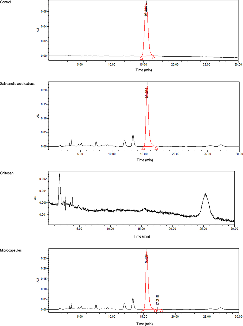 |
Figure 1 Specificity of the chromatogram. |
Establishment of Standard Curves
Next, 1.59 mg of salvianolic acid B control was accurately weighed and dissolved with 75% methanol solution to a constant volume of 25 mL, in order to prepare a solution of 63.60 μg·mL−1. Each 0.25, 0.50, 0.75, 1.00, 1.50, and 2.00 mL of solution was taken into a 10-mL volumetric flask, and 75% methanol solution was used to maintain a constant volume. The above series of standard solutions was filtered via a 0.45 μm millipore filter. This was tested based on chromatographic conditions in Sample Specificity Chromatogram. The sample injection was 20 μL. Peak area A was measured as the longitudinal coordinate, while linear regression was conducted for the concentration according to the regression equation: A = 1.765 6X-0.230 6, r2 = 0.999.8. The results show that salvianolic acid B has a good linear relationship between 1.59 and 12.72 μg·mL−1.
Precision Test and Recovery Test
The prepared mass concentrations were 1.59, 6.36, and 12.72 μg·mL−1. The salvianolic acid B control solution was measured three times a day. The relative standard deviation (RSD) was calculated as 1.73%, 2.16%, and 0.93%, respectively, within the day. Measurements were taken at day 1, 2, and 3, and the calculated RSD was 0.93%, 1.45%, and 1.96%, respectively, within the day.
Quantities of 1.12, 2.23, and 3.03 mg of salvianolic acid B controls were accurately weighed, and blank microcapsules were added according to the prescription dosage. These quantities were dissolved with 0.1 mol·L−1 of hydrochloric acid to prepare mass concentrations of 1.59, 6.36, and 12.72 μg·mL−1, respectively, in order to test peak area A. The results indicate that the average recovery rate was 99.62%, 102.36%, and 99.31%, respectively.
Single-Factor Test for the Selection of the Spray Drying Process
A series of microcapsules was prepared according to the method in Preparation of Salvianolic Acid Microcapsules, based on drug-to-chitosan ratios of 1:1, 1:2, 1:3, and 1:4. The results reveal that when the ratio of drug to chitosan was 1:4, the drug loading and yield of the microcapsules were low, because the ratio of drug to chitosan was too small. When the ratio of drug to chitosan was 1:1, the salvianolic acid extract could not be completely dissolved in the chitosan. Therefore, the drug-to-chitosan ratio should be between 1:2 and 1:3 (Table 1).
 |
Table 1 Drug Loading Caused by Different Ratio of Drug to Chitosan |
A series of microcapsules was prepared using chitosan mass concentrations of 0.5%, 0.8%, 1.0%, and 1.5%, according to the method in Preparation of Salvianolic Acid Microcapsules. The experimental results show that the mass concentration of chitosan should be controlled within the range of 0.8% to 1.5%. This is because when the viscosity of the chitosan solution is too high, the nozzle can easily become blocked, preventing the spray process from being carried out normally; however, when the concentration is too low (0.5% or less), the microcapsule powder is mainly deposited in the shunt flask, resulting in a lower yield (Table 2).
 |
Table 2 Drug Loading Caused by Different Concentration of Chitosan |
According to the preparation method in Preparation of Salvianolic Acid Microcapsules, a series of microcapsules was prepared with peristaltic pump velocities of 300, 400, 500, 600, and 700 mL·h−1. During the experiment, it was found that when the speed of the peristaltic pump was 700 mL·h−1, obvious water droplets formed in the spray dryer, indicating that the spray droplets were not completely dry, and the yield was low. When the speed decreased to 600 mL·h−1, the powder was still slightly wet. As 300 mL·h−1 is the minimum rotation speed of the instrument, the acceptable range of the peristaltic pump speed was thus determined to be between 300 and 500 mL·h−1.
According to the preparation method in Preparation of Salvianolic Acid Microcapsules, a series of microcapsules was prepared at inlet air temperatures of 150°C, 160°C, 170°C, 180°C, 190°C, and 200°C. In the course of the experiment, it was found that when the inlet air temperature was below 170°C, the droplets could not be dried, indicating that the inlet air temperature cannot be lower than 170°C.
According to the results of single-factor investigation, the main influencing factors are as follows: the drug-to-chitosan ratio (A), the mass concentration of chitosan (B), the speed of the peristaltic pump (C), and the air inlet temperature (D). In order to optimize the prescription and process for preparing microcapsules, an orthogonal test was carried out. The trial protocol is presented in Table 3. Drug loading and yield were taken as the comprehensive evaluation indexes. The weight coefficients were all 0.5. The results are presented in Tables 3 and 4.
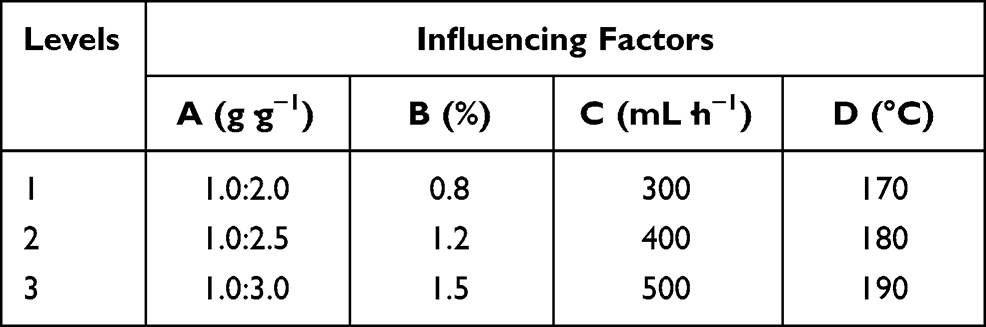 |
Table 3 The Influencing Factors and Levels |
 |
Table 4 The Results of the Orthogonal Test (n=3) |
According to the range analysis, the order of each influencing factor for the salvianolic acid microcapsules was as follows: C > D > B > A. The final optimal process and prescription for the preparation of salvianolic acid microcapsules are A3B3C1D3, ie, salvianolic acid-to–chitosan ratio = 1:3, mass concentration of chitosan = 1.5%, peristaltic pump speed = 300 mL· h−1, and inlet air temperature = 190°C.
Proof Test
The optimal process and prescription conditions were obtained on the basis of the orthogonal test results. Three batches of salvianolic acid microcapsules were prepared. The results, presented in Table 5, show that the process has good reproducibility. Although the results for the microcapsules prepared according to the optimal process and prescription conditions (A3B3C1D3) were close to the results for orthogonal test 6 (A2B3C1D2), in view of the higher proportion of the capsule, and the notion that the higher the rate of encapsulation, the higher the inlet temperature, and the less likely the nozzle is to block, the optimal process and prescription conditions A3B3C1D3 were finally selected for the preparation of the microcapsules.
 |
Table 5 The Results of Confirmatory Experiment |
Quality Evaluation
Morphology of the Microcapsules
The microcapsule sample is a slightly yellow powder, with no bad smell, and good dispersibility and stability. The surface morphology of the microcapsules was observed by scanning electron microscope (SEM) (Figure 2). The results indicate that the microcapsule was round, with a smooth and clean surface, and did not adhere.
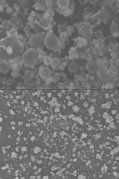 |
Figure 2 Microcapsule scanning electron microscope. |
Particle Diameter
An appropriate number of salvianolic acid microcapsules were taken and diluted with water. The particle size and zeta potential were determined using a Zetasizer laser particle analyzer, as illustrated in Figure 3. As shown in the figure, the average particle size of the microcapsules was 105.63 ± 2.56 nm, the polydispersity index was 0.411 ± 0.023, and the zeta potential was 47.71 ± 1.36 mv. The results indicate that the particle size of the microcapsule was small and the system was stable, but the particle size distribution was not sufficiently uniform; this needs to be improved.
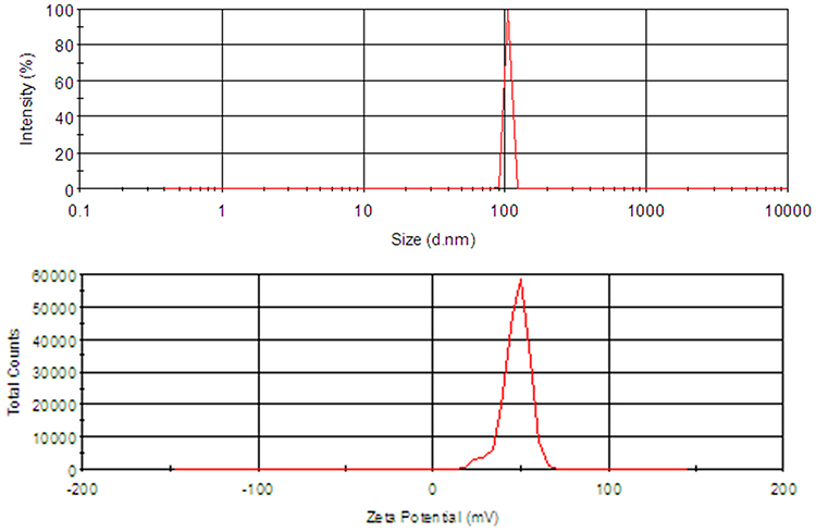 |
Figure 3 Particle size distribution map and potential diagram. |
Differential Scanning Calorimetry (DSC)
The salvianolic acid microcapsules, salvianolic acid extract, chitosan, and physical mixture were determined by DSC. The weight of the sample was approximately 6 mg. The test temperature was 0°C to 450°C, and the temperature rose by 10°C min−1 (the results are shown in Figure 4). These results show that salvianolic acid and salvianolic acid embedded in microspheres have similar absorption peaks, of 250.562°C and 241.53°C, respectively; there was no significant difference between the two groups. The results indicate that the microcapsules did not change the properties of salvianolic acid.
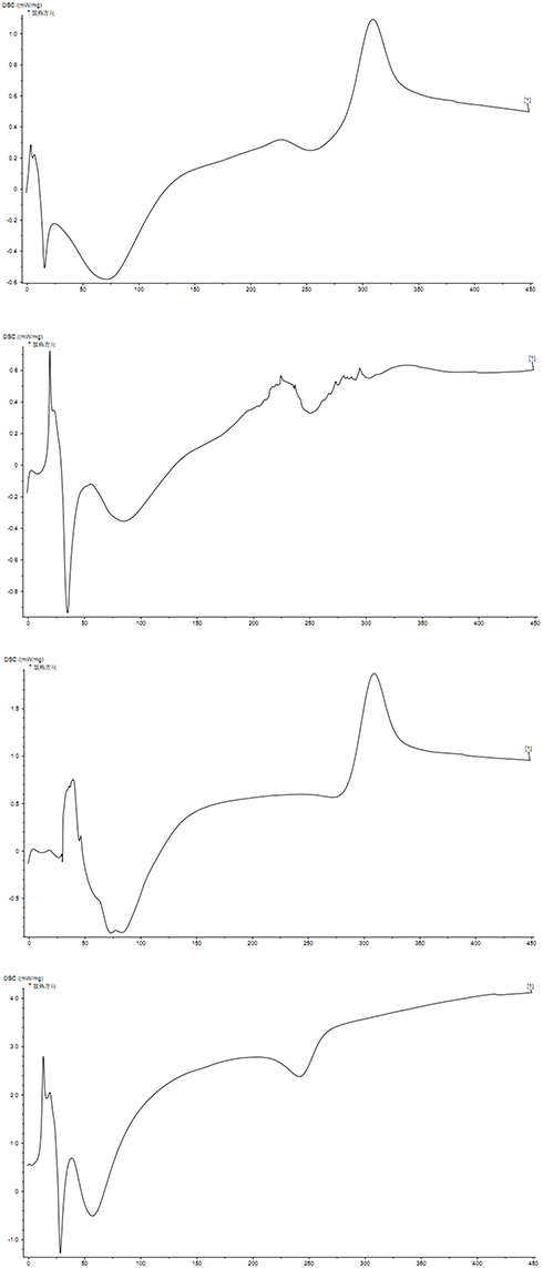 |
Figure 4 Differential thermal scanning diagram. (A) Physical mixture of salvianolic acid extract and chitosan; (B) salvianolic acid extract; (C) chitosan; (D) microcapsule. |
Preliminary Study on the Release Properties of Microcapsules in vitro
Methodological Study on Dissolution in vitro
The release rate was determined using the XD release method in Appendix II of the Chinese Pharmacopoeia 2015. In the pilot experiment, the optimal release medium was selected. Water, 0.01 mol·L−1 hydrochloric acid, phosphate buffer solution pH 6.8, phosphate buffer solution pH 7.4, and phosphate buffer solution pH 4.5 were each used to determine the cumulative release degree. The presence of white turbidity was observed in the dissolution cup for the microcapsules in water, phosphate buffer solution pH 6.8, and phosphate buffer pH 7.4, possibly due to the insolubility of the chitosan.8–10 In addition, the microcapsule release was too fast in 0.01 moL·L−1 hydrochloric acid, and the salvianolic acid was unstable under these conditions.11–14 Therefore, the optimal release medium for the microcapsules was identified as phosphate buffer pH 4.5.
Preparation of Release Medium
For the acetic acid–sodium acetate buffer solution (pH 4.5), 18 g of sodium acetate was taken, 98 mL of acetic acid was added, and this was diluted with water to 1,000 mL to obtain the final product.
Preparation of a Standard Curve
Next, 2.25 mg of salvianolic acid B control was accurately weighed and dissolved with 75% methanol solution to a constant volume of 25 mL, in order to prepare a solution of 63.60 μg·mL−1. Each 0.1, 0.2, 0.4 0.8, 1.60, and 3.2 mL of solution was taken into a 10-mL volumetric flask, and a 75% methanol solution was used to maintain a constant volume. The above series of standard solutions was filtered using a 0.45-μm Millipore filter. This was tested based on chromatographic conditions in Sample Specificity Chromatogram. The sample injection was 20 μL. Peak area A was measured as the longitudinal coordinate, and linear regression was conducted for the concentration according to the regression equation: A = 2.157 0 X-0.185 4, r2 = 0.999.5. The results reveal that salvianolic acid B has a good linear relationship between 1.02 and 32.64 μg·mL−1.
Determination of Sample Release in vitro
According to the Second Method “Device” of General Rule 0931 of the Pharmacopoeia of the People’s Republic of China 2015, 900 mL of each of the above five release media was used as the release medium, at a temperature of 37.0°C ± 0.5°C and a rotating speed of 100 r·min−1. Each 20 mg of prepared salvianolic acid powder and microcapsule was precisely weighed and placed in a large cup. The sampling times were 30, 60, 120, 180, and 240 minutes, and the sampling quantity was 5 mL. The initial filter was discarded, and the sample was injected (the same amount of isothermal medium was added at the same time). The release degree was calculated according to the standard curve. The cumulative release curve was plotted (Figure 5), and drug release curve fitting was carried out. The results, presented in Table 6, reveal that the first-order drug release model can explain the drug release behavior better than the zero-order drug release model and the Higuchi equation. In the release medium, the cumulative release rate of salvianolic acid powder was 100% within 30 minutes, whereas in the salvianolic acid microcapsules prepared by spray drying it was 74.62% ± 3.15%, indicating that the microcapsules had a certain sustained release effect.
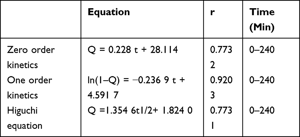 |
Table 6 The Results of the Drug Release Curve Fitting (n=3) |
 |
Figure 5 Release curve (n=3). |
Discussion
In the present study, salvianolic acid microcapsules were prepared by spray drying; this is convenient to operate, easy to control, uses a non–organic solvent, and is suitable for industrial production. In addition, the microcapsules have a round form and good fluidity. The amount of drug loading can reach up to 25.99%, laying a foundation for the development of microcapsules of the solid preparation of salvianolic acid.
The results of the drug release in vitro reveal that the release amount reached 45.3% at the beginning of 30 minutes. Furthermore, there was a sudden release effect, which may be mainly due to the rapid dissolution of microcrystals attached to the surface of the microcapsule. Additionally, there may be micropores on the surface of the microcapsule, through which the release medium can quickly enter the microcapsule and dissolve the drug to form a higher or even saturated concentration. According to the concentration difference, the drug molecules were rapidly diffused from the micropores to create a sudden release. The release was relatively slow within 30 to 240 minutes due to the water absorption and swelling of chitosan, and the micropore diameter was reduced or even disappeared.15–17 When the micropore channel was closed, subsequent drug release could only take place slowly by diffusion through the skeleton, or through the degradation of chitosan.18–20 After 240 minutes, the cumulative release rate decreased, possibly due to the instability and accelerated degradation of salvianolic acid. However, release conditions in vitro are quite different from those in vivo; further in vivo experiments are therefore necessary.
Limitations remain in the present study. First, this study concerned an in vitro experiment. Due to the great difference between in vitro and in vivo conditions, the findings need to be further confirmed through in vivo experiments. Second, although it is known that salvianolic acid B is one of the effective components of S. miltiorrhiza, its specific mechanism remains unclear, and should be further investigated.
Conclusions
The results of this study show that sustained-release microcapsules of salvianolic acid can be successfully prepared using marine polysaccharide as the carrier. However, due to the great difference between in vitro and in vivo conditions, this needs to be further confirmed through in vivo experiments.
Disclosure
The authors declare no conflict of interest in this work.
References
1. Wang L, Ma R, Liu C, et al. Salvia miltiorrhiza: a potential red light to the development of cardiovascular diseases. Curr Pharm Des. 2017;23:1077–1097. doi:10.2174/1381612822666161010105242
2. Yin ZK, Feng ZM, Jiang JS, Zhang X, Zhang PC, Yang YN. Two new tanshinone derivatives from the rhizomes of Salvia miltiorrhiza and their antiviral activities. J Asian Nat Prod Res. 2019;1:1–6.
3. Liang WY, Chen WJ, Yang GH, et al. [Research progress on salvianolic acids of Salvia miltiorrhiza]. Zhongguo Zhong Yao Za Zhi. 2016;41:806–812. Chinese. doi:10.4268/cjcmm20160508
4. Chen W, Chen G. Danshen (Salvia miltiorrhiza Bunge): a prospective healing sage for cardiovascular diseases. Curr Pharm Des. 2017;23:5125–5135. doi:10.2174/1381612823666170822101112
5. Chen F, Li L, Tian DD. Salvia miltiorrhiza roots against cardiovascular disease: consideration of herb-drug interactions. Biomed Res Int. 2017;2017:9868694.
6. Zhou X, Cheung CM, Yang JM, Or PM, Lee WY, Yeung JH. Danshen (Salvia miltiorrhiza) water extract inhibits paracetamol-induced toxicity in primary rat hepatocytes via reducing CYP2E1 activity and oxidative stress. J Pharm Pharmacol. 2015;67:980–989. doi:10.1111/jphp.12381
7. Zhou X, Kang X, Tang L, et al. [Pharmaceutical research of Ligusticum chuanxiong oil microcapsule analgesic gel patch by D-optimal mixture design]. Chin Tradit Herb Drugs. 2019;50:5455–5461. Chinese.
8. Younes I, Rinaudo M. Chitin and chitosan preparation from marine sources. Structure, properties and applications. Mar Drugs. 2015;13:1133–1174. doi:10.3390/md13031133
9. Sanina N. Vaccine adjuvants derived from marine organisms. Biomolecules. 2019;9(8):
10. Garg U, Chauhan S, Nagaich U, Jain N. Current advances in chitosan nanoparticles based drug delivery and targeting. Adv Pharm Bull. 2019;9:195–204. doi:10.15171/apb.2019.023
11. Lu ZZ, Zhu JB, Liu TC. [Influence of pH and additives on stability of salvianolic acid B]. J Dalian Polytech U. 2008;27:209–211. Chinese.
12. Ren ZH, Su HX, Bai YL. [Study on the integration technique for extracting liposoluble and water-soluble components of salvia miltiorrniza]. Chin J Inform Tradit Chin Med. 2009;16:54–56. Chinese.
13. Zhang WX, Xuan L, Ni J. [Stability of salvianolic acid B in water solution]. J Beijing U Tradit Chin Med. 2009;32:856–858. Chinese.
14. Wang B, Zhu WJ, Zeng XL, Fan MW. [Stability of salvianolic acid B in Danshen freeze-dried product injection]. Chin Tradit Pat Med. 2007;29:1479–1482. Chinese.
15. Li S, Hou XP. [Studies on the formation mechanism of alginate-chitosan microcapsule and its drug-loading and release properties on macromolecular drug]. Acta Pharmaceut Sin. 2003;38:380–383. Chinese.
16. Nguyen NT, Nguyen LV, Tran NM, et al. The effect of oxidation degree and volume ratio of components on properties and applications of in situ cross-linking hydrogels based on chitosan and hyaluronic acid. Mater Sci Eng C Mater Biol Appl. 2019;103:109670. doi:10.1016/j.msec.2019.04.049
17. Schmitz C, Auza LG, Koberidze D, Rasche S, Fischer R, Bortesi L. Conversion of chitin to defined chitosan oligomers: current status and future prospects. Mar Drugs. 2019;17(8):
18. El Knidri H, Dahmani J, Addaou A, Laajeb A, Lahsini A. Rapid and efficient extraction of chitin and chitosan for scale-up production: effect of process parameters on deacetylation degree and molecular weight. Int J Biol Macromol. 2019;139:1092–1102. doi:10.1016/j.ijbiomac.2019.08.079
19. Wang S, Lu Y, Ouyang XK, et al. Fabrication of chitosan-based MCS/ZnO@Alg gel microspheres for efficient adsorption of As(V). Int J Biol Macromol. 2019;139:886–895. doi:10.1016/j.ijbiomac.2019.08.070
20. Luo Z, Chen H, Wu S, Yang C, Cheng J. Enhanced removal of bisphenol A from aqueous solution by aluminum-based MOF/sodium alginate-chitosan composite beads. Chemosphere. 2019;237:124493. doi:10.1016/j.chemosphere.2019.124493
 © 2021 The Author(s). This work is published and licensed by Dove Medical Press Limited. The
full terms of this license are available at https://www.dovepress.com/terms
and incorporate the Creative Commons Attribution
- Non Commercial (unported, 3.0) License.
By accessing the work you hereby accept the Terms. Non-commercial uses of the work are permitted
without any further permission from Dove Medical Press Limited, provided the work is properly
attributed. For permission for commercial use of this work, please see paragraphs 4.2 and 5 of our Terms.
© 2021 The Author(s). This work is published and licensed by Dove Medical Press Limited. The
full terms of this license are available at https://www.dovepress.com/terms
and incorporate the Creative Commons Attribution
- Non Commercial (unported, 3.0) License.
By accessing the work you hereby accept the Terms. Non-commercial uses of the work are permitted
without any further permission from Dove Medical Press Limited, provided the work is properly
attributed. For permission for commercial use of this work, please see paragraphs 4.2 and 5 of our Terms.