")
Back to Journals » Clinical, Cosmetic and Investigational Dermatology » Volume 14
In vitro and in vivo Evaluation on the Safety and Efficacy of a Brand-New Intracutaneous Filler with α1-R-Collagen
Authors Rinaldi F, Pinto D , Trink A, Giuliani G, Sparavigna A
Received 14 January 2021
Accepted for publication 29 April 2021
Published 12 May 2021 Volume 2021:14 Pages 501—512
DOI https://doi.org/10.2147/CCID.S295618
Checked for plagiarism Yes
Review by Single anonymous peer review
Peer reviewer comments 5
Editor who approved publication: Dr Jeffrey Weinberg
Fabio Rinaldi,1,2 Daniela Pinto,1,2 Anna Trink,2 Giammaria Giuliani,1 Adele Sparavigna3
1Giuliani SpA, Research and Development Department, Milan, Italy; 2IHRF, International Hair Research Foundation, Milan, Italy; 3Derming, Clinical Research, and Bioengineering Institute, Monza, Italy
Correspondence: Fabio Rinaldi
Giuliani SpA, Research and Development Department, Palagi, 2, Milan, 20129, Italy
Tel +39-02-76006089
Fax +39 0220541
Email [email protected]
Abstract: Nowadays, the most advanced skin anti-aging treatments are addressed to restore the extracellular matrix (ECM) homeostasis. ECM is considered the main player not only as physical support of the tegument but also at the biochemical level, thanks to its capacity to exchange nutrients, water, cellular mediators, and growth factors within and between cells. This study aimed to evaluate the in vitro and in vivo efficacy and aesthetic performance of a brand-new intracutaneous filler. The latter is based on novel concepts: besides filling it exerts a homeostatic balance of nutrients able to delay the skin aging process by sustaining physiological rejuvenation of the tissue and in the surrounding injection/implantation area. The brand-new intracutaneous filler was tested for in vitro capacity to stimulate extracellular matrix components production. Therefore, a single session for injection of the product under study was performed by a specialized dermatologist, using the bolus technique on the zygomatic protuberance of 20 healthy female subjects with midface volume loss, caused by aging. Results confirmed the important and long-term volumizing, anti-wrinkle, the hydrating activity of this product after one single injection session. The biological outcomes also support product effectiveness in skin structure restoration.
Keywords: fillers, hyaluronic acid, α 1-R-collagen, carboxymethylcellulose, dipeptides, amino acids
Introduction
Skin aging is a complex and unavoidable biological phenomenon that starts early in the third decade of life with a high degree of heterogeneity.1 Aging processes are related to genetic background, personal lifestyle, and environmental conditions, and as a consequence, aging phenomenon, and skin aging, in particular, is extremely variable among subjects.2–4 Skin aging is a dynamic process that determines structural alterations of both soft and bone tissues.5 The silhouette of the face and body deals with atrophic processes of various layers composing it: from the skin to the subcutaneous adipose tissue including loss of muscle mass and reabsorption of the bone, that result in the appearance of wrinkles, hypotonicity, lipoatrophy, sarcopenia, and volume displacement. Furthermore, to counteract atrophy, a permanent muscle tone is established neutralizing the incorrect relaxation of the skin by compensating mechanisms.6
Recent studies7 highlighted as a common trait an intranet structure that connects the whole organism: the extracellular matrix (ECM) that fills the space between cells. Research is currently focusing on ECM composition and signaling.8 Recently, results in this field have contributed to developing effective strategies for the prevention and treatment of degenerative diseases and signs of cutaneous aging.9
The product under study contains 2% hyaluronic acid (HA) (200–400 kDa), the primary component of the ECM of human connective tissues, and the most used filler and rejuvenating agent in anti-age treatments and injectables.10–12 Due to its macromolecular size, marked hygroscopicity, and viscoelasticity, HA can modulate tissue hydration, osmotic balance, and the physical properties of ECM, structuring a hydrated and stable extracellular space where cells, collagen, elastin fibers, and other ECM components are maintained.13 The cleavage of high molecular weight chains14–16 into shorter fragments by enzymatic activity not only allows the reuse of the fragments for its de-novo synthesis but generates 200 Dalton fragments that interact directly with a specific receptor on cell membranes (Intercellular Adhesion Molecule 1, ICAM 1) consequently promoting the synthesis of new ECM by fibroblasts of the connective tissue. However, this molecule, through the interaction with reactive oxygen species (ROS) (including superoxide, hydrogen peroxide, nitric oxide, peroxynitrite) generates a strong quenching effect, acting as a free radical scavenger. The product contains also carboxymethylcellulose (CMC), providing the right viscosity and amount of coordinated water to guarantee a stable filler effect and skin hydration. CMC is FDA-approved, water-soluble, cellulose-derived polysaccharide that is available in high-purity forms and has been used in several biomedical applications due to its biocompatibility and low cost. Thanks to the presence of CMC the product becomes a hydrogel injectable in an uncrosslinked form with advantages in terms of handling and administration, compared to existing commercial fillers that need crosslinking before injection.17–19
The main component of the product, in terms of activity and innovation, is a non-triple (unfolded) helical collagen alpha chain produced in transgenic silkworms. Today there are no fillers containing collagen even equine on the market. The recombinant collagen contained in the product under study possesses a safer profile to compare to equine collagen. The recombinant collagen contained in the product under study possesses specific characteristics: it cannot generate a homotrimer with a triple helical structure, since the polypeptide chain is homologous to human collagen alpha 1 chain does not contain the C-telopeptide and C-propeptide, which are known to promote the triple helix formation. Furthermore, it is not post-translationally modified by prolyl hydroxylases and cannot twist since the presence of hydroxyprolines is a prerequisite for forming the stable collagen triple helix. This molecule is characterized by a lower hydrophobicity compared to human native collagen indicating that the molecule can promote a larger number of H-bonds contributing to stabilize the HA secondary structure and can behave as a physiological water donor thus favoring the hydration of the surrounding environment, in which HA and CMS are placed. Water is an essential element for fibroblasts and the maintenance of ECM structure. Unfolded R polypeptide collagen 1 alpha at millimolar concentration generates local gradients in the matrix that accelerate water transfer in the dermis. The polypeptide interacts with HA and CMC that also mediate the interchange of water nutrients and metabolites between the blood, the lymph, the interstitial cell network, and the epidermal layers. This allows the interstitial transfer of biological fluids that, unlike diffusion, can speed the dispersion of water and large solutes within the matrix. Under physiological conditions, the proportion of unfolded R polypeptide collagen alpha 1 is crucial since it has local, microscopic functional properties that contribute to the steady-state renewal and drainage of interstitial matrix fluids. Consistent with the above-described concepts, the concentration was kept in the millimolar range to provide support to hyaluronic acid in a 1:100 ratio and it exposes hydrophilic sites to interact with HA and coordinate water.
Finally, also biologically active amino acids (AA) (L- isomers of Alanine, Valine, Glycine, Proline, OH-Proline, Lysine, and dipeptides (AlaPro, ProGly, GlyPro) were included in the formulation. Advantageously, these AA supports the surrounding environment contributing to the maintenance of the cellular tropism and exerting antioxidant effect.20 The AA and dipeptide composition was specifically selected and each concentration was specifically designed to maintain the dermal homeostatic balance, providing an overall healthy appearance to the skin.21,22
The proposed above composition combines the mechanical function of a dermal filling with homeostatic balance and signaling of nutrients promoting the delay of skin aging by sustaining the physiological cell activity of tissues in and around the injection/implantation area.23 This originates from the idea that intracutaneous injection of Hyaluronic Acid (HA), carboxymethylcellulose (CM), and a combination of the micromolar concentration of dermal compounds like a recombinant polypeptide of collagen α1-chain (r-collagen), selected dipeptides, and amino acids could provide an effective and prolonged anti-aging activity of the skin.
Materials and Methods
Chemicals
A sterile, resorbable filler (Monodermà Fillagen®, Giuliani SpA) registered as Medical Device under the notification number 1,988,658 was used for in vivo assay.
Gly-pro, Pro-gly, Ala-pro dipeptides, HA, and R polypeptide collagen alpha 1 (r-collagen) from Innate s.r.l. (Novi Ligure, Italy) were also tested in vitro on 3D Phenion® full-thickness skin models were purchased from Henkel (Phenion® FT; Düsseldorf, Germany)
In vitro Efficacy of the Main Components of the Filler
Gly-pro (0.001%), Pro-gly (0.001%), Ala-pro (0.001%) dipeptides as a mixture (MIX aa), HA (2%) and R-collagen (0.022%) were analyzed as single component or in combination (Mix aa+ HA; Mix aa + R-collagen; Mix-aa+ HA+ R-collagen; HA+ R-collagen) for their effect on production and homeostasis of Collagen IV (COLIV), Aquaporin 3 (AQP3), and CD44, a cell-surface glycoprotein which acts as the principal cell surface receptor for HA in the ECM.
The Phenion® Full-Thickness Skin Model, produced by Henkel (Düsseldorf, Germany, diameter 1.3 cm) was used for this study. The model includes epidermal keratinocytes and dermal fibroblasts from biopsy material from healthy donors), forming a multilayered skin equivalent that resembles human skin multilayered structure and tissue functionality. Under this experimental model, the products under examination can be tested at the same concentration and mode as in vivo. Upon arrival, the FT models were transferred in small Petri dishes (3.5 cm in diameter) filled with 4 mL pre-warmed air-liquid-interface (ALI) medium, provided by the manufacturer. The medium was refreshed one time after an initial overnight equilibration period. Tissues were subjected to 24h treatment with the above-cited compounds after the overnight equilibration at 37°C and 5% CO2.
Twenty-four hours after treatment RNA for qRT-PCR analysis was extracted. Tri Reagent (Sigma Aldrich, Milan, Italy) methods as described by Chomczynski and Mackey24 were used and the cDNA was then synthesized from 2μg RNA template in a 20μL reaction volume, using the PrimeScript RT-PCR Kit (Takara, Japan). The cDNA was amplified and detected by the Stratagene Mx3000P Real-Time PCR System (Agilent Technologies Italia S.p.A., Milan, Italy). The amplification of cDNA was conducted using the following TaqMan gene expression assays: HS00266237 (Collagen IV, COLIV), HS00185020_M1 (aquaporin 3, AQP3), HS01075864 (Cluster differentiation 44, CD44), and Hs99999905_m1 (human glyceraldehyde-3-phosphate dehydrogenase, GAPDH). GAPDH was used as housekeeping gene. PCR amplifications were carried out in a 20µL of total volume. The mixture of reaction contained 10µL of 2X Premix Ex Taq (Takara, Japan), 1µL of 20× TaqMan gene expression assay, 0.4 µL of RoX Reference Dye II (Takara, Japan), 4.6 µL of water, and 4µL of DNA. PCR conditions were the following: 95°C for 30 sec followed by 40 cycles of 95°C for 5 sec, 60°C for 20 sec. PCR reactions were performed in duplicate using an MX3000p PCR machine (Stratagene, La Jolla, CA). Δ cycle threshold25 was used for the calculation of the relative abundance in the expression of each gene.
In vivo Assay
A spontaneous, pre-marketing, open, clinical trial, that foresaw one micro-injection session of the studied medical device was performed from July 2019 to October 2019, 31th. The study was conducted on 20 female volunteers, aged 40–66 years (mean = 52), with midface volume laxity, whose written informed consent had been obtained. Four visits were performed during the trial: a basal visit, two intermediate visits (7 and 30 days after the injection procedure), and a final visit (90 days after the injection procedure). The intracutaneous injection with the product under study was performed during the basal visit, after the evaluations, in the malar area (zygomatic protuberance), by bolus technique using a needle (30G). The amount of tested product to be injected was 1mL (Figure 1).
 |
Figure 1 Schematic figure illustrating the injection area. |
The primary endpoint of the study was to evaluate, clinically and by non-invasive instrumental evaluations, the safety and the aesthetic performance on the face of a new intracutaneous filler, based on the concept of homeostatic balance and thorough micro-dosage of nutrients that delay the skin aging process.
Aesthetic results were established through the use of qualitative clinical evaluations of cheek ptosis, nasolabial folds, and marionette lines according to the Facial Volume Loss Scale (Table 1) and Wrinkle Severity Rating Scale (Table 2) and quantitative assessments (superficial and deep skin hydration, wrinkles profilometry, face volume image analysis), supported by photographic documentation.26,27
 |
Table 1 Facial Volume Loss Scale (FVLS) |
 |
Table 2 Wrinkle Severity Rating Scale (WSRS) |
Clinical evaluations were carried out, at each time point, mono-laterally on the right or left face side according to the randomization list.
Treatment aesthetic performance and duration (Table 3) was also defined using the Clinical Observer Global Assessment (COGA) and the Patient Global Assessment (PtGA) as well as the self-assessment questionnaire filled in at T90 by all treated subjects, who completed the study as protocol directed.
 |
Table 3 Variation of Face Volume |
Secondary endpoints were aesthetic performance duration, variations in skin parameters during follow-up, and investigator’s and subjects’ satisfaction efficacy degree.
Treatment efficacy was evaluated also by the volunteers’ judgment through the compilation of a self-assessment questionnaire.
Safety and tolerability of the study treatment were assessed by investigating local and expected and unexpected adverse events, as a consequence of the product injection; all the volunteers were followed up for 6 months to evaluate the possible delayed onset of side effects.
The study protocol and related documentation were previously submitted to an Independent Ethic Committee at Derming S.r.l. (Milan, Italy)., obtaining the I.E.C. approval. The study has been registered on clinicaltrials.gov under clinical trial registration (CTR) number NCT04239768 on January 27, 2020.
Clinical Instrumental Evaluations
At each study time, all included volunteer’s 3D pictures of the face were taken by 3D Camera VECTRA H1 (Canfield) handheld imaging system. 3D pictures taken at two different times were merged and automatically compared by Vectra analysis module (VAM). When the merge resulted not applicable/evaluable, the data of the respective subjects were excluded from the evaluation.
To assure comparable images, the pictures will be taken with standardized methods, concerning especially: distance from the subject and intensity of the illumination source.
The subject during the photo execution had to keep still, with open eyes and relaxed facial muscles.
A picture of nasolabial folds and marionette lines was also taken by Primos compact portable device (GFMesstechnik)28 which elaborates 3D representations of skin wrinkles and measures skin principal profilometric parameters, according to DIN EN ISO 4228. Moreover, the software compares directly the different images obtained at the times foreseen by the protocol.
Precise recording optics ensures a wide spectrum of measuring possibilities with ranges up to micrometers.
The following profilometric parameters were measured: Ra (average roughness of the analyzed profile); Rt (wrinkles total height); Rv (wrinkles maximum depth).
Superficial skin hydration was measured by the instrument Corneometer CM825 (Courage – Khazaka, Köln, Germany). The measure of the skin capacitance properties is an indirect expression of its hydration level. To reduce the variability of measurement methods, for each volunteer, three measures on the same skin area were executed: the adjusted mean was considered as the real measure value.
Deep skin hydration was measured by the MoistureMeterD (Delfin Technologies, Kuopio - Finland), which measures non-invasively the dielectric constant of the skin and subcutaneous fat. For the present study 0.5 mm and 1.5 mm, probes were used.
Statistical Analysis
Data processing took into consideration the comparison of each time versus basal conditions (T0) and was performed in terms of descriptive and inferential analysis as follows:
- In vitro assay: non-parametric test (Student’s t-test) followed by Welch’s correction using the statistical software, GraphPad 7.0 (GraphPad Software, La Jolla California USA, www.graphpad.com).
- Clinical data: non-parametric test (Friedman test), followed in case of statistically significant results by Holm-Sidak Adjusted Wilcoxon signed-rank test.
- Instrumental data: non-parametric test (Friedman test) or parametric test (ANOVA test for repeated measures), followed in case of the statistically significant result by Holm-Sidak Adjusted tests.
Moreover, the long-lasting activity of the study treatment was expressed by comparing the image analysis results on face volume obtained at T7 to the ones obtained at T30 and T90 using the statistical tests explained above for the instrumental data analysis.
Results
Biological Evaluation
The results of the quantitative real-time PCR analysis for the FT model treated with the filler main compounds are shown in Figure 2. In particular, RQ values for active compounds treated samples were normalized for FT samples treated with medium alone (negative control). Data analysis demonstrated a significant synergic up-regulation of COLIV (RQ> 5), AQP3 ((RQ=2) and CD44 (RQ> 3.5) 24 h after treatment with Mix-aa+ HA+ R-collagen (Figure 1).
 |
Figure 2 Gene expression analyses (qRT-PCR) in the treated FT-skin model were expressed as relative quantification (RQ) with respect to control. Statistical differences between mean values were determined with Student’s t-test followed by Welch’s correction. The symbols indicate a significant difference (P < 0.05) to the control (* for COLIV, # for AQP3, $ for CD44). */ # /$ (P < 0.05); **/ ##/ $$ (P < 0.01); ***/ $$$ (P < 0.01). |
Clinical Evaluation
No drop-out or important event occurred during the study period.
Tolerance
Some subjects complained about the appearance of light bruises and/or swelling on the injection points, that disappeared/reabsorbed within 5–15 days; these reactions are expected events of the injection procedure, not related to the tested product. Finally, the product tolerance was judged to be good-excellent in 100% of subjects.
Clinical Evaluations
The study treatment determined starting from T7:
- A very significant improvement of cheek volume corresponding to a reduction of the FVLS mean value of 21.2% at T7 and 24.2% at T30 and T90 and a decrease of the clinical score (CS) of at least 1 grade (according to FVLS photographic scale) respectively in 70%, 80% and 75% of included subjects (Table 1).
- A statistically important reduction of wrinkles severity of 18.2% at T7 and 21.2% at T30 and T90, with a reduction of the CS of at least 1 grade (according to WSRS reference photographic scale) respectively on 65%, 70%, and 65% of volunteers (Table 2).
Instrumental Evaluations
3D pictures were taken at each study time by Vectra H1. An average increase of volume versus T0 of 1.036 ccs at T7, 0.964 ccs at T30, and 0.917 ccs was reported. In particular, at T7 75% of subjects (Table 3) presented a marked- very marked improvement of face volume (35% of subjects with a cheek volume increase > 1 cc and 40% of subjects between 0.5 ccs and 1 cc); although at T30 and T90 a trend and progressive reduction of cheek volume mean value was highlighted (−7% Δ T30-T0 vs Δ T7-T0 and −11.5% Δ T90-T0 vs Δ T7-T0), the differences versus T7 were not statistically significant; moreover, the high volumizing performance of the tested treatment was detectable in a more marked percentage of treated cases (90% at T30 and 85% at T90). These results confirm the long-lasting and persistent re-volumizing activity of the tested product.
Image analysis of the midface (Figure 3), nasolabial folds (Figure 4), and marionette lines (Figure 5) confirmed the clinical assessment results; starting from T7, an important “anti-wrinkle” activity of the injective treatment was shown.
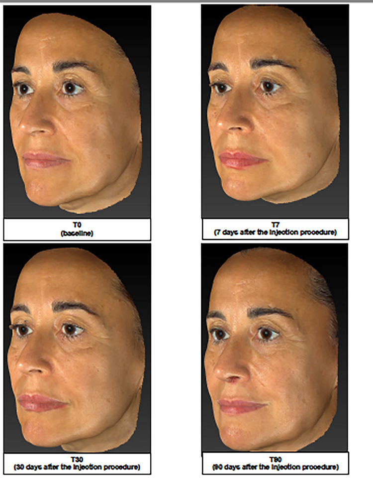 |
Figure 3 Representative image analysis of improvement of midface volume before treatment (T0), 7 days (T7), 30 days (T30) and 90 days after injection of the brand-new intracutaneous filler. The subject’s midface was evaluated by mean of Vectra H1. |
 |
Figure 4 Representative image analysis of improvement of nasolabial folds before treatment (T0), 7 days (T7), 30 days (T30) and 90 days after injection of the brand-new intracutaneous filler. The subject’s midface was evaluated by mean of Vectra H1. |
 |
Figure 5 Representative image analysis of improvement marionette lines before treatment (T0), 7 days (T7), 30 days (T30) and 90 days after injection of the brand-new intracutaneous filler. The subject’s midface was evaluated by mean of Vectra H1. |
For the nasolabial folds, a clinically and statistically significant reduction of all profilometric parameters versus baseline was found (Table 4), an index that the nasolabial folds were generally less visible and deep. The reduction reached its maximum at T30 (−27.5% Ra; −25.5% Rt; −25.1% Rv).
 |
Table 4 Profilometry of Nasolabial Folds |
For the marionette lines, the reduction percentage of Ra, Rt, and Rv parameters were generally less marked, but still clinically/statistically significant vs T0 (Table 5) and maximum values of reduction were reached at T30: −25.2% Ra; −21% Rt; −22.9% Rv).
 |
Table 5 Profilometry of Marionette Lines |
It is important to notice that the anti-wrinkle effect on the nasolabial folds and marionette lines was more marked 30 days after the injection procedure; this result highlights an important activity of bio-stimulation mainly attributable to HA.
As far as skin hydration is concerned, obtained results (Table 6) highlighted the important superficial and deep moisturizing efficacy of the tested treatment started at T7 with the clinically/statistically significant increase of the superficial (Figure 2) and deep skin layers hydration at 0.5 mm of depth. Although no significant variation of deep hydration at 1.5 mm of depth was found at any study time, it is important to notice at T30 a trend increase of the measured parameter.
 |
Table 6 Superficial and Deep Hydration Variations |
Investigator’s and Subjects’ Satisfaction Degree
Regarding the GOGA investigator’s assessment, the percentage of subjects with a moderate-very marked improvement was high (≥70%) for a long time, up to T90 (Table 7), while the PtGA assessment highlighted, especially at T90, a slight reduction of the subject’s satisfaction degree.
 |
Table 7 Investigator’s and Subjects’ Satisfaction Degree |
Discussion and Conclusions
Obtained in vitro and in vivo results confirm the anti-aging performance and the lasting effect of the tested product on midface defects due to the aging mechanism.
In vitro results confirmed the ability of the main components of the studied medical device to stimulate the production of several components of the extracellular matrix such as COLIV and also AQP3.
COLIV, together with COLI, COLIII, and COLVII, is the most frequent collagen type present in the skin, mainly in the basement membrane that anchors the epidermis to the dermis. The up-regulation found for COLIV (Figure 1) suggests the activation of the cutaneous basement membrane, responsible for the optimal skin architecture. This boosting effect on COLIV leads to the regulation of the remodeling process globally.
ECM degrades with age losing HA,8 the main extracellular matrix component in the skin. HA can exist both as freely available but also bound to cells or proteins containing a hyaluronic acid-binding domain29 such as CD44.30 Like HA itself, also CD44 decreased with age. In the present work, we showed that the main components of the studied medical device were able not only to stimulate ECM components but also to induce the main HA receptor CD44 and this contributes to HA homeostasis.
Therefore, the up-regulation of AQP3, one of the most abundant skin aquaglyceroporin suggests a positive effect on skin elasticity, tone, and hydration.
These positive effects were confirmed by the clinical evaluation. The product thanks to its formulation based on recombinant collagen and low molecular weight hyaluronic acid (HA) determined at all considered evaluation times: a statistically significant improvement of FVLS score and face cheek volume (bio-revolumetric effect); a statistically significant reduction of WSRS score (filling efficacy); a clinically/statistically significant reduction of all profilometric parameters index of an anti-wrinkles efficacy (nasolabial folds and marionette lines appeared generally less visible); a clinically/statistically significant improvement of superficial and deep (at 0.5 mm of depth) skin hydration (moisturizing activity).
As confirmed also by the profilometry and 3D-face volume image analysis results, the aesthetic performance of the tested product resulted generally more marked at T30, a sign of significant activity on dermal connective tissue and on epidermis able to provide new tonicity and consistency to the skin (bio-revitalization effect).
Previous studies with different formulations containing natural HA alone, HA with amino acids, or equine collagen18 had demonstrated the performance in terms of both clinical (volume loss, wrinkles) and instrumental evaluations (volume and profilometry measurements, hydration) on facial aging. Although it is not an endpoint of this study to compare these results with those of other trials, it can be noticed that in other cited studies the injection sessions were more than one (from two to four). In the case of Monodermà Fillagen®, a much more prominent effect after only one injection session was observed.
Probably, this result is due to the special composition of the product which exerts an immediate volumizing effect (presence of carboxymethylcellulose) and a more pronounced remodeling (reduction of nasolabial folds and marionette lines). Also, the improvement of superficial and deep hydration of the skin was very pronounced. The product under study could even improve its efficacy by establishing a treatment protocol based on two or three injection sessions and a very long duration of the effects. Specific trials are required to assess the efficacy and duration of the results after repeated injections.
Data Sharing Statement
The authors intend to share individual deidentified participant data upon request.
Ethics
This study was conducted in compliance with the ethical principles originating in or derived from the Declaration of Helsinki and in compliance with Good Clinical Practice Guidelines. All subjects provided signed informed consent.
Acknowledgments
This study was supported by Giuliani SpA.
Disclosure
R.F., A.T. and A.S. served as consultants for Giuliani S.p.A. P.D. is employed by Giuliani S.p.A. G.G. is in the board of director of Giuliani S.p.A. A.S. reports grants from Derming srl, during the conduct of the study.
References
1. Baumann L. Skin ageing and its treatment. J Pathol. 2007;211:241–251.
2. Naylor EC, Watson REB, Sherrat MJ. Molecular aspects of skin ageing. Maturitas. 2011;69(3):249–256. doi:10.1016/j.maturitas.2011.04.011
3. Krutmann J, Bouloc A, Sore G, et al. The skin aging exposome. J Dermatol Sci. 2017;85(3):152–161. doi:10.1016/j.jdermsci.2016.09.015
4. Glogau RG. Aestethic and anatomic analysis of the aging skin. Semin Cutan Med Surg. 1996;5:134–138. doi:10.1016/S1085-5629(96)80003-4
5. Michaud T, Gassia V, Belhaouari L. Facial dynamics and emotional expressions in facial aging treatments. J Cosmet Dermatol. 2015;14:9–21. doi:10.1111/jocd.12128
6. Sparavigna A. How does face age? A retrospective observational study and meta-analysis. J Plast Pathol Dermatol. 2019;1:1–8.
7. Richard OH, Naba A. Overview of the matrisome—an inventory of extracellular matrix constituents and functions. Cold Spring Harb Perspect Biol. 2012;4:1.
8. Sparavigna A. Role of the extracellular matrix in skin aging and dedicated treatment. State of the art. Plast Aesthet Res. 2020;7:14.
9. Limbert G, Masen MA, Pond D, et al. Biotribology of the ageing skin—why we should care. Biotribology. 2019;17:75–90. doi:10.1016/j.biotri.2019.03.001
10. Sparavigna A, Tenconi B, Giori AM, et al. Evaluation of the efficacy of a new hyaluronic acid gel on dynamic and static wrinkles in volunteers with moderate aging/photoaging. Clin Cosmet Investig Dermatol. 2019;12:81–90. doi:10.2147/CCID.S191935
11. Sparavigna A, Orlandini A. Efficacy and tolerance of an injectable medical device containing hyaluronic acid and amino acids: a monocentric six-month open label evaluation. J Clin Trials. 2017;7:4–12. doi:10.4172/2167-0870.1000316
12. Sparavigna A, Tenconi B. Efficacy and tolerance of an injectable medical device containing stable hybrid cooperative complexes of high- and low-molecular-weight hyaluronic acid: a monocentric 16 weeks open-label evaluation. Clin Cosmet Investig Dermatol. 2016;9:297–305. doi:10.2147/CCID.S114450
13. WO2006067608A1 - Aqueous formulations based on sodium hyaluronate for parenteral use. Patent.
14. Winterbourn CC. Reconciling the chemistry and biology of reactive oxygen species. Nat Chem Biol. 2008;4:278–286. doi:10.1038/nchembio.85
15. Kohen R, Nyska A. Invited review: oxidation of biological systems: oxidative stress phenomena, antioxidants, redox reactions, and methods for their quantification. Toxicol Pathol. 2002;30(6):620–650. doi:10.1080/01926230290166724
16. Kammeyer A, Luiten RM. Oxidation events and skin aging. Ageing Res Rev. 2015;21:16–29. doi:10.1016/j.arr.2015.01.001
17. Klein AW, Elson ML. The history of substances for soft tissue augmentation. Dermatol Surg. 2000;26(12):1096–1105. doi:10.1046/j.1524-4725.2000.00512.x
18. Sparavigna A, Tateo A, Inselvini E, Tocchio M, Orlandini MC, Botali G. Anti-age activity and tolerance evaluation of collagen micro-injection treatment associated to topical application of a cosmetic formulation (investigator-initiated multicentre trial). J Clin Exp Dermatol Res. 2017;8(03):13. doi:10.4172/2155-9554.1000391
19. Zielke H, Wölber L, Wiest L, et al. Risk profiles of different injectable fillers: results from the Injectable Filler Safety Study (IFS Study). Dermatol Surg. 2008;34(3):326–335. doi:10.1111/j.1524-4725.2007.34066.x
20. Kawashim K, Itoh H, Miyoshi M, et al. Antioxidant of branched-chain amino acid derivatives. Chem Pharm Bull. 1979;27(8):1912–1916. doi:10.1248/cpb.27.1912
21. Schoffbnibls E. Amino acid metabolism and cell volume regulation. In: Bolis L, Schmidt-Nielsen K, Maddrell SHP, editors. Comparative Physiology, Locomotion, Respiration, Transport and Blood. Amsterdam: North-Holland; 1973.
22. Miller MS, Lay WK, Elcock AH. Osmotic pressure simulations of amino acids and peptides highlight potential routes to protein force field parameterization. J Phys Chem B. 2016;120(33):8217–8229. doi:10.1021/acs.jpcb.6b01902
23. Bhattacharjee O, Ayyangar U, Kurbet AS, et al. Unraveling the ECM-immune cell crosstalk in skin diseases. Front Cell Dev Biol. 2019;7:68.
24. Chomczynski P, Mackey K. Modification of the TRI reagent procedure for isolation of RNA from polysaccharide- and proteoglycan-rich sources. Biotechniques. 1995;19:942–945.
25. Vigetti D, Viola M, Karousou E, et al. Hyaluronan-CD44-ERK1/2 regulate human aortic smooth muscle cell motility during aging. J Biol Chem. 2008;283:4448–4458. doi:10.1074/jbc.M709051200
26. Day DJ, Littler CM, Swift RW, et al. The wrinkle severity rating scale: a validation study. Am J Clin Dermatol. 2004;5(1):49–52. doi:10.2165/00128071-200405010-00007
27. Lemperle G, Holmes RE, Cohen SR, et al. A classification of facial wrinkles. Plast Reconstr Surg. 2001;108(6):1735–1750. doi:10.1097/00006534-200111000-00049
28. Jaspers S, Bretschneider T, Maerkeer U, et al. Optical topometry with Primos: a powerful tool to prove the efficacy on skin care products in in-vivo studies.
29. Kogan G, Šoltés L, Stern R, et al. Hyaluronic acid: its function and degradation in vivo systems. Stud Nat Prod Chem. 2008;34:789–882.
30. Peach RJ, Hollenbaugh D, Stamenkovic I, et al. Identification of hyaluronic acid binding sites in the extracellular domain of CD44. J Cell Biol. 1993;122(1):257–264. doi:10.1083/jcb.122.1.257
 © 2021 The Author(s). This work is published and licensed by Dove Medical Press Limited. The full terms of this license are available at https://www.dovepress.com/terms.php and incorporate the Creative Commons Attribution - Non Commercial (unported, v3.0) License.
By accessing the work you hereby accept the Terms. Non-commercial uses of the work are permitted without any further permission from Dove Medical Press Limited, provided the work is properly attributed. For permission for commercial use of this work, please see paragraphs 4.2 and 5 of our Terms.
© 2021 The Author(s). This work is published and licensed by Dove Medical Press Limited. The full terms of this license are available at https://www.dovepress.com/terms.php and incorporate the Creative Commons Attribution - Non Commercial (unported, v3.0) License.
By accessing the work you hereby accept the Terms. Non-commercial uses of the work are permitted without any further permission from Dove Medical Press Limited, provided the work is properly attributed. For permission for commercial use of this work, please see paragraphs 4.2 and 5 of our Terms.