
Back to Journals » International Journal of Nanomedicine » Volume 20
Implication of Bimodal Magnetic Resonance and Fluorescence Imaging Probes in Advanced Healthcare: Enhancing Disease Diagnosis and Targeted Therapy
Authors Gong T, Ullah Z, Roy S  , Cheng C , Li M, Cheng J, Zhang Y, Guo B , Qiu Y
Received 23 February 2025
Accepted for publication 16 July 2025
Published 29 July 2025 Volume 2025:20 Pages 9473—9503
DOI https://doi.org/10.2147/IJN.S524454
Checked for plagiarism Yes
Review by Single anonymous peer review
Peer reviewer comments 5
Editor who approved publication: Professor Farooq A. Shiekh
Tingting Gong,1,* Zia Ullah,2,* Shubham Roy,2 Caixue Cheng,1 Menglong Li,1 Jing Cheng,3 Yinghe Zhang,2 Bing Guo,2 Yingwei Qiu1
1Department of Radiology, Shenzhen Nanshan People’s Hospital (NSPH), Shenzhen, Guangdong, People’s Republic of China; 2School of Science, Shenzhen Key Laboratory of Flexible Printed Electronics Technology, Shenzhen Key Laboratory of Advanced Functional Carbon Materials Research and Comprehensive Application, Harbin Institute of Technology, Shenzhen, Guangdong, People’s Republic of China; 3Education Center and Experiments and Innovations, Harbin Institute of Technology, Shenzhen, Guangdong, People’s Republic of China
*These authors contributed equally to this work
Correspondence: Bing Guo, Email [email protected] Yingwei Qiu, Email [email protected]
Abstract: Molecular imaging probes hold great promise in disease diagnostics and their therapeutic interventions. However, a single imaging modality sometimes lacks efficiency in all kinds of diseases. Conditions such as cancer, cardiovascular disorders, and neurodegenerative diseases critically require multimodal imaging to characterize complex biological environments accurately. Efficient targeting further enhances probe performance, improving diagnostic precision. This study explores the design rationale and application of bimodal probes designed for Magnetic Resonance (MR) and fluorescence (FL) imaging for their complementary strengths. MRI enables deep tissue visualization, while fluorescence offers high sensitivity and cellular-level resolution. Meticulous attention has been devoted to presenting methodologies aimed at improving the targeting efficacy of these probes. This involves the methodology to enhance targeting efficacy, including ligand-based strategies, nanoparticle functionalization, and molecularly imprinted polymers. The probes combine fluorescence for precise cellular imaging with MRI for in-depth tissue visualization, providing synergistic benefits that elevate their diagnostic potential. Moreover, we offer recent developments in Machine Learning and Artificial Intelligence-based computational approaches for image analysis, enabling more precise diagnosis across a range of diseases that may propel their diagnostic abilities for better therapeutic outcomes. Through systematic analysis and in vitro and in vivo evaluations, we demonstrate the ability of the probes to achieve superior spatial and temporal resolution, facilitating the accurate delineation of biological targets. The integration of these bimodal probes holds great promise for advancing our understanding of complex biological processes, enabling more precise diagnostics, and paving the way for targeted therapeutic interventions.
Keywords: magnetic resonance imaging, fluorescence imaging, bimodal imaging, molecular imaging probes, targeting
Introduction
Imaging modalities have become an integral part of the medical realm because of the need for proper diagnostics before any therapeutic intervention is required for bodily dysfunctions. The clinically available imaging modalities employed for diagnostics include X-ray imaging, positron emission tomography (PET), computed tomography (CT), ultrasound imaging (USI), and magnetic resonance imaging (MRI).1 The above-mentioned imaging modalities are commonly used in clinical setups to diagnose the abnormalities present in the body. Imaging modalities such as X-ray, PET, CT, ultrasound (USI), and MRI are critical instruments in modern medical diagnosis, with each having unique advantages and limits. X-rays and CT scans employ ionizing radiation to produce quick and detailed images, mostly of bones and internal structures, but they pose radiation concerns. PET gives essential functional information, particularly in cancer and neurological illnesses, but at a high radiation dose and cost. Ultrasound is safe, portable, and real-time, making it excellent for soft tissue and prenatal imaging, albeit image quality and operator expertise are limited. MRI provides improved soft tissue contrast without radiation, but it is costly, time-consuming, and not appropriate for individuals who have specific implants. Despite their vital responsibilities, these devices have varying levels of safety, accessibility, and diagnostic precision. However, these imaging techniques have several limitations, like the presence of ionizing radiation, which is harmful to the human body, long acquisition time, complex operating systems, expensive apparatus, and harmful side effects to the patients.2 Among all the available modalities, MRI is preferred over all the other owing to its high spatiotemporal resolution, satisfactory tissue penetration depth, and non-ionizing radiation. MRI offers unique advantages such as good soft tissue resolution, multi-sequence, multi-parameter and multi-directional imaging, which provides precise acquisition of the entire lesion size and thickness, internal structural features, peripheral structural infiltration, degree of local infiltration, degenerative changes such as cyst formation or hemorrhage, including assessment of tumor extension, lymph node involvement and distant metastases during progression, all without usage of hazardous ionizing radiation.3 Additional MRI sequences for functional imaging include diffusion-weighted magnetic resonance imaging (DWI), apparent diffusion coefficient (ADC), functional magnetic resonance imaging (fMRI), etc, which help to reveal the state of the tumor tissue.4 Beside the offered advantages, MRI also have some limitations like long acquisition time, inability in real-time visualization of bodily dysfunctions, very expensive machinery, and toxicity caused by contrast agents (Gd causing nephrogenic systemic fibrosis and Mn causing neurodegenerative disorder “manganism” with symptoms resembling Parkinson’s disease) injected during the imaging process. Therefore, there is an utmost need for an imaging modality capable of real-time visualization of defective body tissues, short acquisition time, and optimal biocompatibility for effective diagnostics.5 To address the above-mentioned concerns, fluorescence (FL) imaging has captured significant attention in the medical realm due to its higher sensitivity for early detection and real-time visualization of abnormalities at the cellular level than conventional imaging modalities.6 There have been many research attempts to apply visible FL imaging to real-time intraoperative surgical navigation in the visible range, but its low spatial resolution and insufficient depth of penetration into tissues have limited its clinical translation.7 Researchers have engineered fluorescent agents capable of near-infrared (NIR) FL imaging to address the shortcomings, such as lower penetration depth, autofluorescence, tissue scattering, lower signal-to-background ratio (SBR), and limited spatial resolution. Moreover, studies have demonstrated that NIR neural probes enabled µm-scale neural visualization to the maximum reported tissue depth up to ~2-3 mm by using the first near-infrared (NIR-I) window (700–900 nm) and the second near-infrared (NIR-II 1000–1700 nm).8 The NIR-II region has better diagnostic ability than the NIR-I region due to its low absorption by the biological tissues and good bio-permeability, resulting in higher SBR, significant tissue penetration, and excellent resolution, enabling real-time imaging of tissues at mm depths and µm resolution with enhanced contrast.9,10 Therefore, NIR FL imaging is the best available imaging modality for real-time intraoperative visualization of the abnormalities in the patient’s body and efficient early detection of bodily complications.11,12 However, besides all the benefits of NIR FL imaging, the penetration depth and contrast resolution are still limited for deep-seated abnormalities of the patient’s body due to energy dissipation and light scattering encountered during the imaging process. Given the limitations of conventional single-modality imaging techniques currently used in clinical practice, such as limited depth penetration, insufficient resolution, or low sensitivity, there is an urgent need to adopt more advanced approaches. Bimodal imaging, particularly the combination of MR and FL modalities, offers unparalleled advantages by bridging the gap between deep tissue visualization and high-resolution cellular imaging. This integrated approach enables more accurate disease characterization, earlier detection, and more precise monitoring of therapeutic responses. As diseases like cancer, cardiovascular, and neurodegenerative disorders demand increasingly sophisticated diagnostic tools, the adoption of bimodal imaging represents a critical step forward in improving clinical outcomes and personalizing patient care. Therefore, there should be an imaging modality that is equally non-invasive as NIR FL imaging but has better SBR and greater penetration depth than NIR FL imaging.
Hence, the combination of the two imaging modalities is a great way to complement each other’s strengths and significantly expand the range of clinical applications.13 MRI provides non-invasive, deep tissue penetration with excellent anatomical and soft tissue contrast which allows whole-organ or whole-body imaging. However, it typically lacks molecular sensitivity. On the other hand, fluorescence imaging offers high sensitivity and resolution at the cellular and molecular level, making it ideal for visualizing specific biomarkers or guiding interventions at microscopic scales, though its tissue penetration is limited. By combining these two modalities in a single bimodal probe, it becomes possible to achieve both broad anatomical context and precise molecular-level detail, providing synergistic diagnostic benefits.
Currently, researchers have engineered materials capable of MR and NIR FL bimodal imaging to investigate tumors,14 inflammation, brain degeneration,15 and other diseases, as well as in the fields of photodynamic16,17 chemotherapeutic, immunotherapy, and photothermal treatment of tumors.18,19 However, simply combining the two imaging modalities also does not achieve the desired results; factors contributing to this include low uptake of the probes at the lesion site, low ability of the target cells to internalize the probe, and insufficient concentration of the contrast agent within the target site to achieve a satisfactory imaging or therapeutic outcome. Additionally, the lack of specificity in vivo distribution of the probes and the excessive uptake of normal tissues may affect the biosafety as well as the diagnostic accuracy. Thus, enhancing the targeting and biocompatibility of the bimodal probes is a matter of concern to achieve the desired results with minimal side effects to the surrounding bodily tissues.
Thus, scientists have designed targeted drug delivery systems (TDDS) for selective delivery of the drug to the target tissue or organ through carriers after administration through a certain route for efficient targeting.20 There are two main mechanisms of targeted delivery in organisms: passive targeting and active targeting. (i) Tumor sites have special lymphatic drainage and abundant neovascularization, and the enhanced permeability and retention (EPR) effect. By taking advantage of these features, passive aggregation and signal amplification of the nanoprobe to image the lesion have been achieved.21,22 Particle size is the main factor affecting the EPR effect, with larger nanoparticles having longer retention times, as opposed to smaller particle sizes having deeper tissue penetration. However, specific surface area and bioactivity can be increased with decreasing particle size, leading to an increased risk of toxicity.23,24 (ii) Active targeting mechanism is the delivery of drugs to specific tissues or cells through biomimetics or carrier structure modification while ensuring safety and no damage to normal tissues or cells. Specifically, through ligand-receptor interactions, thereby directing drug delivery in vivo as well as specific recognition and uptake by target cells.25,26 Surface modification of ligands on nanomaterials facilitates surface functionalization, which further expands their biological applications, and the ligands used for designing assemblies can be surfactants, sugars, peptides, transferrin, copper transporters, folic acid, and membrane coatings. Therefore, the use of such TDDS for targeted delivery of MR and NIR fluorescent bimodal imaging probes to the required site for efficient diagnostics is the main concern nowadays.27,28
This review sets itself apart from previous research by providing a broader, more application-oriented perspective. Unlike others, which focus primarily on cancer or single-modality applications, it covers various disease types, such as cancer, cardiovascular, and neurodegenerative, highlighting the importance of multimodal imaging. It goes beyond traditional ligand-based targeting by including molecularly imprinted polymers (MIPs) to improve specificity. Furthermore, it incorporates AI and ML not only for picture analysis but also for probe design and optimization, which is rarely described in previous literature. The study also balances in vitro and in vivo validation, considering long-term biocompatibility, and addresses clinical translation problems using regulatory and standardization insights, areas that are generally underexplored in such reviews.
In this work, we are going to summarize the designing strategies of targeted molecular bimodal imaging probes for exceptional MR/FL controlled imaging efficacies, improved disease diagnosis and real-time monitoring, targeted imaging-mediated therapeutic capabilities, and the new-generation computation-based regimens for accurate diagnostic interventions. While demonstrating their targeting strategies, the ligand-based, nanoparticle-based, and polymer-based functionalization methods to capture the overall developments in this domain have also been explored. Therefore, the use of biomimetic MR/FL bimodal imaging probes with combined strengths of targeting, MRI, and FL imaging can demonstrate promising results with effective targeting, real-time visualization, deeper penetration, biocompatibility, and higher SBR for optimal diagnostics. Herein, the idea of deploying Machine Learning and AI-based technologies for result analysis and diagnostic interventions for better healthcare, and to explore the functionalities of these probes, has also been discussed. Finally, future opportunities and challenges in this exciting research area have been summarized, which could generate interest among researchers and scientists to pursue their future research.
Strategies to Enhance Target Specificity for Bimodal Imaging Probes
Disease targeting is the most crucial thing in imaging probes for enhanced specificity and accuracy in medical diagnostics. By selectively binding to disease-associated biomarkers or tissues, the targeted probes have improved the contrast and SBR, enabling precise localization at the pathological sites, minimizing false positives, and reducing the risk of misdiagnosis. Additionally, disease targeting enhanced the probe’s efficiency in tracking and real-time monitoring of disease progression, facilitating early detection and intervention for proper and timely disease management.29 Overall, incorporating disease-targeting strategies in imaging probes ensured heightened sensitivity and reliability, contributing to the advancement of non-invasive, real-time diagnostics in medical applications (Scheme 1). Herein, we demonstrated some rationale to enhance the targeting efficacy of MR/FL imaging probes for disease diagnostics.
 |
Scheme 1 Schematic illustration of enhanced target specificity strategies for bimodal imaging probes. |
Ligand-Based Approaches
In molecular imaging, the ligand-based targeting approach plays a pivotal role by utilizing specific moieties while designing an imaging probe, known as ligands, to selectively bind to molecular targets associated with the diseased part of the body. These ligands, often designed to recognize unique biomarkers or receptors overexpressed in pathological conditions, serve as the targeting part of the imaging probes. Through this approach, molecular interactions are harnessed for enhanced sensitivity and specificity, enabling precise and real-time visualization of diseased tissues.30 Ligand-based targeting in molecular imaging facilitates early disease diagnosis, monitoring therapeutic responses, and guiding personalized treatment strategies. This strategy holds promise in advancing diagnostics and therapeutics, offering a tailored and efficient means to investigate and combat serious medical conditions. There are several strategies to design molecular ligands discussed in this section for targeted biomedical imaging to investigate the disease deeply and plan to treatment accordingly.
Contrast agents circulate in vivo and are readily taken up by macrophages and removed by the reticuloendothelial system (RES), which shortens circulation time and reduces their biological effects on target tissues. Although contrast agents allow passive targeting through poor lymphatic drainage and EPR effects at the tumor site, they are less effective.31 Hence, modification of the particle surface with targeting ligands, which guide specific targeting to the diseased site in a receptor-mediated manner, is necessary to increase the accumulation of imaging probes at the tumor site. Low molecular weight targeting ligands such as folic acid (FA), hyaluronic acid (HA), lactobionic acid (LA), and chloramphenicol are employed to modify the probe surface and enable targeting by increasing penetration at the disease site.32 FA attached to the surface of the particles specifically recognizes tumor cells or normal cells through the interaction of FA with its receptor. Its presence on the cell membranes of a wide range of cancer cells includes tumors of the female reproductive system such as ovarian and uterine tumors as well as brain, breast, and intestinal tumors. In contrast, normal healthy tissues contain extremely low levels of the folate receptor, a dramatic difference that makes tumor-targeted diagnostics possible.33 For example, Au/Gd@BSA nanoparticles were prepared by a simple hydrothermal method, which effectively improved the absorption and targeting of the probe by modifying it with FA molecule. The nanoparticles possessed the ability of T1-weighted MRI and NIR FL with high fluorescence QY and meanwhile, possessed a good biosafety enabling the nanoparticles to be excreted with the excrement after imaging without influencing the normal physiological metabolism of the mice, thereby successfully realizing the safe in vivo MR/FL bimodal targeted imaging of breast cancer.34 Additionally, a naturally occurring polymer known as HA, involved in a variety of physiological processes, is a water-soluble glycosaminoglycan with D-glucuronic acid and N-acetyl-D-glucosamine repeating units have excellent targeting potential to the diseased microenvironment. It has high affinity for overexpressed CD 44 receptor in a variety of tumors.35 Exploiting this feature, HA nowadays acting as a potential targeting ligand for enhanced imaging and therapeutics. For instance, Li et al have grown MnO2 in situ on the surface of GSH-responsive Ag&Mn:ZnInS QDs@SiO2–MnO2-HA probe coated with a layer of HA on the surface of the quantum dots in order to improve its stability and target recognition ability. It has been successfully applied to in vivo MR/FL bimodal cell imaging, cancer cell and normal cell identification, and lysosomal localization (Figure 1).36
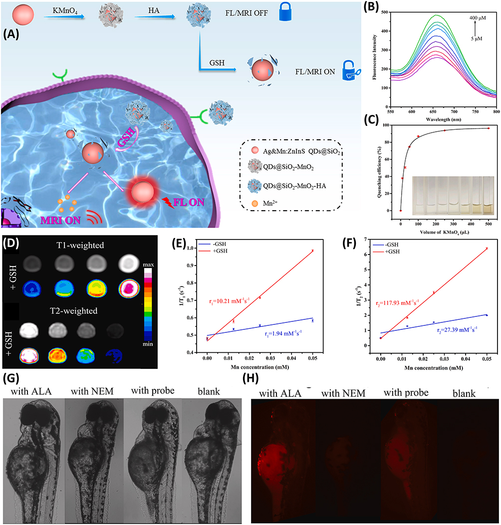 |
Figure 1 (A) Graphical representation of GSH-responsive MR/FL bimodal probes for targeted diagnosis. (B) FL spectra of the HA functionalized bimodal probe for different GSH concentrations. (C) The FL quenching efficiency and corresponding color changes of the bimodal probe solution upon addition of different concentration of KMnO4. (D) T1 and T2-weighted MR images at different concentrations of HA functionalized bimodal probe in the presence of 5 mM GSH. (E and F) Relaxivity plots of T1 and T2-weighted MRI in the presence and absence of GSH at different Mn concentrations. (G and H) FL images of zebrafish only; zebrafish incubated with 0.5 mg mL−1 of HA functionalized bimodal probe for 1 h; zebrafish pretreated with 500 μM of NEM for 20 min and followed by incubation with 0.5 mg mL−1 of HA functionalized bimodal probe for 1 h; zebrafish pretreated with 500 μM ALA for 2 h and followed by incubation with 0.5 mg mL−1 of HA functionalized bimodal probe for 1 h. Reproduced with permission from Anal Chim Acta, volume: 1221, Liu Y, Xue J, Wang W et al. Multifunctional probe based on modified Ag&Mn:ZnInS QDs for dual-mode fluorescence and magnetic resonance imaging of intracellular glutathione. 340172. Copyright (2022), with permission from Elsevier.36 |
According to the studies, such chemical ligand can also be applied for targeted delivery of therapeutic drugs and prodrugs for different diseases. Thus, besides bimodal targeted imaging, scientists may focus on targeted delivery of drugs by using these chemical ligands for a sustainable therapeutic regime. However, these ligands can only attach the receptors overexpressed on surface of the tumor cells or diseased site.37 For effective targeting, the ligands should reach the diseased cite via blood circulation. Because of their small size, these ligands can easily be excreted through the blood before reaching the infection site. Therefore, there is a need of targeting agents having affinity with the blood and should not be excreted from the blood before reaching the infection site.
Similarly, Peptides and antibodies have been exploited for various ligand modification strategies.38 Within the context of these ligands, the RGD and GSH peptides have been attractive as they mediate effective targeting of tumor microvasculature and cancer cells such as glioblastoma through the binding of αvβ3 integrins overexpressed on the cell surface. Once the contrast agent reaches the target area through systemic circulation, tumor-specific targeting is performed by a receptor-mediated approach and the particle surface is modified with targeting ligands to augment the uptake and accumulation within the tumor site of the probe.39,40 Taking advantage of such feature, Wang et al covalently attached the scavenger receptor (SRAI)-specific peptide PP1 to Gd-AuNCs, and the negatively charged carboxyl groups of Gd3+ and AuNCs self-assembled into mono-dispersed spherical particles (GNCNs) by electrostatic interaction, which enhanced the photoluminescence ability of AuNCs. In vitro cell binding studies showed that the NCs could selectively adhere to activated macrophages in an SR-AI-dependent mode. Due to the favorable targeting efficacy of PP1-Au@GSH@Gd NCs, in vivo MR/FL imaging in an established ApoE-/- mouse model showed robust and long-lasting enhancement of plaque contrast (Figure 2).41
 |
Figure 2 (A) Graphical representation of GSH functionalized MR/FL bimodal imaging probes for precise atherosclerosis diagnosis. (B) T1-weighted MRI and mapping of GSH functionalized bimodal probes and Magnevist. (C) Longitudinal relaxivity of GSH functionalized bimodal probe at various Gd3+ concentrations. (D) Enhancement in the T1 signals with GSH functionalization of bimodal probe. (E) FL imaging at different concentrations of GSH functionalized bimodal probe. (F) Increase in the average FL intensities at different concentrations of GSH functionalized bimodal probe by using Caliper IVIS Lumina II system. (G) In vivo T1-weighted MRI and mapping of GSH functionalized bimodal probe at different time frames. (H) Ex vivo FL images of aorta at 24 h post injection of GSH functionalized bimodal probe (white arrows indicate the maximum intensity at the aorta plaque site). (I) Histology and immunofluorescent staining demonstrating the targeting ability of the GSH functionalized bimodal probes with efficient macrophage penetration within aortic vulnerable plaques (*p < 0.05). Reproduced with permission from Nanomedicine, volume: 19, Wang J, Wu M, Chang J et al. Scavenger receptor-AI-targeted ultrasmall gold nanoclusters facilitate in vivo MR and ex vivo fluorescence dual-modality visualization of vulnerable atherosclerotic plaques. 81–94. Copyright (2019), with permission from Elsevier.41 |
Moreover, multifunctional nanospheres were developed to functionalize Ce 6-loaded lipid/PEG-coated ultralarge-pore mesoporous silica nanoparticles (UMSN) (Ce 6-LUMSN) with an acidity-triggered rational membrane (ATRM) peptide. Under weakly acidic conditions, ATRM has been inserted into the lipid bilayer as a transmembrane α-helix, with a membrane insertion of peptide with a pKa of 6.5, which made the ATRM ideally suited for targeting malignant cells in the weakly acidic (pH = 6.5–6.8) microenvironment of solid tumors. ALUMSNs displayed a high potential for efficient internalization into tumor cells through multiple pathways. On one hand, in the case of membrane translocation, ALUMSNs would enter directly into the cytoplasmic lysate; on the other hand, after grid protein-mediated endocytosis uptake, acidification of the mature endocytosis compartment would drive endosomal membrane insertion and disruption by ATRM, leading to cytoplasmic release of ALUMSNs. Further MR, thermography, and FL imaging for accurate early tumor diagnosis and monitoring of ALUMSNs promote the anti-tumor effects of NIR laser-induced PDT and PTT without detectable systemic toxicity.42 In another study, a convenient all-in-one therapeutic nanoplatform (FTY 720@AM/T7-TL) was designed for the treatment of hepatocellular carcinoma (HCC). The multifunctional platform consisted of gold manganese dioxide nanoparticles (AM), tetrastyrene (T), fingolimod (FTY 720), hybrid liposomes (L), and a T7 peptide (T7). Doping of the T7 tumor-targeting peptide further contributed to the nanoplatform to provide an effective way for the accurate diagnosis of tumors by bimodal imaging.43
Furthermore, due to specific structural characteristics and pathophysiological progression, atheromatous plaques carry a risk of sudden rupture, causing acute cardiovascular events as a major factor in patient death. Consequently, early identification of atheromatous plaques with MR/FL bimodal probes for timely intervention could be an effective approach. For example, a highly sensitive nanoprobe (mZGS-OPN) based on PLNPs was constructed for in vivo imaging of early atheromatous plaques after surface functionalization with maleimide-polyethylene glycol activated ester (MALPEG 5000-NHS) and anti-osteoclastogenic protein (OPN) antibody to construct a foam-cell-specific targeting nanoprobe (mZGS-OPN). OPN is a glycosylated phosphoprotein that is highly expressed on the surface of foam cells but hardly expressed in normal arterial walls, which is an effective goal for improving targeting. With the prolongation of co-incubation time with foam cells, the FL intensity was gradually enhanced and peaked at the 8 h time point, indicating that mZGS-OPN could effectively target foam cells. In vitro experiments showed its good targeting of foam cells. In vivo experiments showed that mZGS-OPN achieved high SBR imaging of plaques and was able to detect the earliest atheromatous plaques within 2 weeks.44 Because of deep penetration and higher spatiotemporal resolution, MRI is highly sensitive to plaque components of necrotic cores, calcifications, and hemorrhages that ensemble FL and MR imaging to improve early diagnostic accuracy effectively. In one study, the excellent targeting ability of model foamy macrophages obtained by concatenating a foamy macrophage-specific osteobridging protein (OPN) antibody with highly luminescent core@shell NaGdF4:Yb, Er@NaGdF4 nanoparticles enabled successful in vivo imaging of fragile and stable atherosclerotic plaques by both FL and MR imaging, with OPN in atherosclerotic plaques was upregulated and promoted macrophage adhesion, migration and activation as well as leukocyte recruitment and increased viability. Per-OPN demonstrated excellent macrophage targeting ability and binding affinity strongly correlated with OPN expression.45 In addition, expression of adhesion molecule-1 (VCAM-1) occurs early in the formation of atherosclerosis, leading to an increase in adhesion between monocytes and activated endothelial cells, with monocytes expressing more inflammatory cytokines in order to further stimulate the endothelial cells to express more VCAM-1, a positive feedback process serving as a useful imaging biomarker for early atherosclerosis. This study synthesized a liposome-encapsulated contrast agent, USPIO (Fe3O4), in combination with a low-dose therapeutic drug, Rap, which was then linked to a plaque-targeting VHPKQHR (VHP) peptide and fluorescent reagents for diagnosis and early treatment of atherosclerotic lesions. The results of the MR and FL imaging demonstrated that Rap/Fe3O4 @ VHP enables early and effective diagnosis of atherosclerosis.46,47 However, the use of peptide ligands as a targeting agent could increase the blood circulation time of the imaging probe to a toxic level by reacting with the blood proteins and could cause serious complications. Therefore, the use of more target-oriented biological ligands should be designed and applied to increase the targeting efficiency of the imaging probe to reduce the toxicity and unwanted side effects.
Moreover, aptamers, concise single-stranded DNA and RNA sequences, exhibit a high specific affinity for their target molecules. The in vitro selection of these oligonucleotides, capable of binding to specific target molecules, was independently reported in a brief time frame in 1990 by three distinct groups of scientists: Tuerk and Gold, Ellington and Szostak, and Robertson and Joyce. FL-based aptasensing technology has already been explored by numerous groups in a detailed manner. Zhang et al extensively discussed multiple FL response systems. In addition, they conducted a review on the detection of aflatoxins using fluorescent aptasensors, addressing challenges associated with detection methodologies.48 In a separate study, He et al demonstrated progress in amplifying current signals within aptamer-based electrochemical biosensors.49 On the other hand, aptasensor targeted MRI agents has also been demonstrated by different groups over the past few decades. For instance, Yigit et al functionalized iron oxide NPs with aptamers for MRI guided thrombin detection. The aptamer lowers the detection limit to 25 nM for thrombin in human serum.50 Such a low detection limit makes aptamers useful candidates for biomedical applications. As mentioned earlier, the use of aptamers in FL or MRI based probes have been explored quite deeply. However, there is a room to search typical aptamer conjugated MR/FL bimodal imaging probes for biomedical research having ability to demonstrate exceptional targeting for precise diagnosis of various diseases.
Zhao et al developed an activable MR/FL bimodal probe based on aptamer modified MnO2 nanosheets. When target cells are not present, the nanoprobe remains inactive, with no activation of FL signaling or MRI contrast. However, in the presence of target cells, the aptamers binding to their targets weaken the adsorption of aptamers on the MnO2 nanosheets. This results in partial FL recovery, illuminating the target cells and facilitating the endocytosis of nanoprobes into them. Following endocytosis, the reduction of MnO2 nanosheets by GSH activates FL signals and produces abundant Mn2+ ions, making the nanoprobe suitable for MRI.51 Several other groups have recently shown some extraordinary data on different cancer models such as ovarian and breast cancer.52 In a study, gadolinium-doped CDs were successfully developed by solvothermal method, and AS1411 aptamer was grafted onto the surface of Gd-CD in order to improve the targeting ability. The synthesized AS1411-Gd-CD had highly efficient red fluorescence emission with a QY of 5.6%, and the MR signal intensity of the AS1411-Gd-CD group was almost twice as high as that of the Gd-CD group after co-cultivation with 4T1 cells, demonstrating the specific targeting property of AS1411-Gd-CD, for use in tumor-targeted FL imaging and T1-weighted MRI. AS1411-Gd-CD additionally has a high photothermal conversion rate, giving good PTT therapeutic results guided by MR/FL imaging. H&E tissue staining images showed that laser-irradiated Gd-CD induced tumor tissue damage, and that apoptosis of tumor cells was more severe in tumor tissues irradiated with AS1411-Gd-CD plus NIR laser. The results of biosafety performance testing also confirmed that H & E staining of major organ sections revealed no significant tissue damage and no tissue inflammation or lesions (Figure 3).53 The AS1411 aptamer is a guanine-rich oligonucleotide that specifically binds nucleolin, a hormone that is abnormally upregulated both intracellularly as well as on the cell surface in many types of tumor cells. In another study, MR/FL bimodal imaging of renal cell carcinoma using the AS1411 aptamer in combination with a CLT 1 cyclic peptide was achieved, resulting in a more accurate depiction of tumor boundaries.54 One of the prime important characteristics of aptamer-based targeting is their extremely low limits of detection (LOD), which make these probes more precise (sometimes in the nM range). Moreover, aptamer does not increase the size of the sample significantly making it potential for crossing various pathological barriers (such as blood-brain barrier). Aptamer is typically a chain of several nucleotides. Thus, they are very flexible to alter and modify according to the domain of application and the use of aptamers or nucleotide-based ligands can bring a scientific revolution for efficient targeting of different harmful diseases in near future. However, aptamers and DNA or RNA based ligands are very sensitive and it is very difficult to engineer and attach them as ligand on the probe according to the desired target disease. The technological advancements could make the functionalization possible as desired.
 |
Figure 3 (A) Graphical representation of preparation of aptamer functionalized MR/Fl bimodal probe for efficient targeting and photothermal therapy of tumor. (B) Excitation and emission wavelength of aptamer functionalized MR/FL bimodal probe. (C) FL spectra of the MR/FL bimodal probe by exciting the probe at 470 nm to 550 nm. (D) T1-weighted MR images of aptamer functionalized MR/FL bimodal probe at different Gd concentrations. (E) Longitudinal relaxivity of aptamer functionalized MR/FL bimodal probe at different Gd concentrations. (F) In vivo FL imaging of tumor-bearing mice after i.v. injection of MR/FL bimodal probe while, the red circles are indicating the tumor selectivity of the probe. (G) FL signal intensities at different time points after i.v. injection of MR/FL probe with and without aptamer functionalization demonstrating the targeting ability of the probe. (H) T1-weighted MR imaging of mice model before and after injecting the MR/Fl probe with and without aptamer functionalization demonstrating the targeting ability of the probe. (I) T1-weighted MRI signals of the tumors at different time points after injecting the MR/FL probe with and without aptamer functionalization while, the red circles are indicating the targeting ability of the probe. Reproduced with permission from Chemical Engineering Journal. Volume: 440, Jiao M, Wang Y, Wang W et al. Gadolinium doped red-emissive carbon dots as targeted theranostic agents for fluorescence and MR imaging guided cancer phototherapy. 135965. Copyright (2022), with permission from Elsevier.53 |
Surface Engineering of Bimodal Nanoprobes for Targeting
Nanoparticle functionalization involves modifying the surface of nanoparticles with specific molecules to impart desired properties. In biomedical imaging, functionalization enhances biocompatibility, stability, and targeting capabilities of nanoparticles. Ligands or biomolecules can be attached to enable specific interactions with biological targets, aiding in precise imaging of tissues or cells. Functionalization also allows the incorporation of imaging agents for contrast enhancement and therapeutic payloads for targeted drug delivery. This tailored approach improves the efficacy and selectivity of biomedical imaging agents, making them vital tools for diagnostics, monitoring, and personalized medicine in the field of healthcare.
Surface Engineering for Target Recognition
The continuous progress of research related to nanomaterials has demonstrated excellent qualities in all aspects in vivo. Surface modification of inorganic nanoparticles is very crucial for subsequent biomedical applications. Improving targeting properties through surface modification, as well as improving biocompatibility and chemical stability in living organisms, could also laterally ensure the targeting function; modified nanoparticles are more susceptible to enter the disease site.55,56 Previous studies have proposed various approaches, such as water-soluble polymer substrates, non-mesoporous silica shells, amphiphilic ligands, and hydrophilic inorganic materials. A Comparative Analysis of Surface Engineering Strategies in MR/FL Bimodal Nanoprobes in different disease models has been studied in a tabular form (Table 1). These molecules allow easy bonding to the surface of nanoparticles either by direct adhesion or by doping/encapsulation in transparent micelles (lipids or polymers) or silica shells at the core of the nanoparticles. Covalent bonding of polymers to inorganic nanoparticles is usually carried out using two main methods: (i) “grafting to” and (ii) “grafting from”. The “grafting to” method allows functionalization with low-density polymers on the surface of nanoparticles through chemical reactions. In contrast, the “grafting from” method uses an initiator surface on which a high-density polymer layer is grafted, resulting in a high graft density nanocomposite.57,58
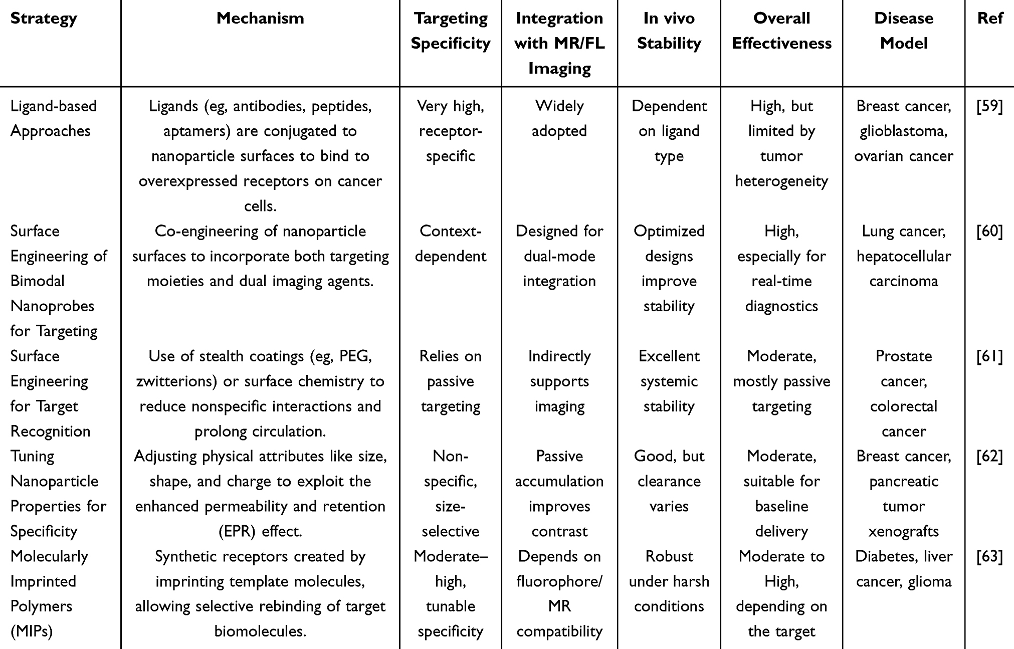 |
Table 1 Comparative Analysis of Surface Engineering Strategies in MR/FL Bimodal Nanoprobes in Different Disease Models |
Additionally, surface modification of nanomaterials by targeted ligands enables increased cellular uptake by specific cancer cells thereby enabling targeted applications in MR imaging. To establish a successful nanoparticle-mediated targeted drug delivery system, the first consideration should be the particle size-selective synthesis, followed by their interaction, stability and aggregation effects under in vivo conditions.64 The probes with biomimetic modification by biological membranes are also possible, and the encapsulation of cancer cell membranes on the surface of nanoconjugates enables these probes to target the tumor site uniformly in vivo, displaying contrast-enhanced imaging signals. By loading GOD and Ag2S quantum dots on the surface of MnO2 nanosheets (AM-GOD), further encapsulated by 4T1 cell membranes (AMG@CM), where NIR-II FL imaging, MRI, and PA signals were focused on the tumor site, suggesting that AMG@CM has excellent tumor-targeting ability, with tumor cell membranes encapsulation able to effectively evade immune clearance and target tumor sites in homozygous 4T1 homozygous mice with little to no inflammatory response. GSH-induced Mn+2 release serves for application in TME-responsive tumor visualization. Due to higher GSH concentrations and more acidic nature of TME, GSH is an exemplary triggering generator of TME-responsive tumor therapies. AMG@CM exists in a similar way to AM “off-on” process, where GSH induces MnO2 decomposition for FL recovery and greatly reduces the phototoxicity of NIR-II FL as it circulates in vivo. Such probes, which feature an activation mode or an “on” mode, enable specific activation of diagnostic and therapeutic capabilities in diseased areas, while remaining silent in normal tissues. In contrast to “always-on” nanomedicine, “on” therapeutic diagnostics systems tend to exploit intrinsic features of pathological stimuli (eg, pH, redox reactions, enzyme content, hypoxia) to achieve the “off” to “on” mode of nanomedicine. Activated imaging probes prevent most nanomedicines from remaining in an “always-on” mode, whether from reaching the target tissue or not, thus avoiding the influence of background signals generated by normal tissues, thereby increasing the sensitivity and specificity of the imaging.65,66
For example, Dai et al engineered MnO2-CQ4T-GOx based homology-activated ultrasensitive MR/FL imaging guided nanomedicines for effective “knock-on” dynamic therapy (CDT and PDT) in rheumatoid arthritis (RA). The NIR-II FL signal of the probe was quenched because of the aggregation-induced quenching effect of CQ4T-BSA. The probe rapidly accumulated in inflamed joints through a combination of the inherent high affinity of BSA and ELVIS (extravasation through leaky vasculature and subsequent inflammatory cell-mediated sequestration) effect when injected. Taking advantage of the unique RA-microenvironment characteristics, an ultrasensitive MnO2-Mn2+ reaction system was successfully constructed. Such a single-homology response system has greatly facilitated the realization of precision theranostics based on the following pathways: 1) The RA-microenvironment could rapidly trigger the degradation of the probe, releasing Mn2+ ions, GOx, and CQ4T-BSA for achieving a fast and precise “turn on” MR/FL bimodal imaging. 2) The degradation of probe not only facilitates the simultaneous restoration of NIR-II FL and activation of MR imaging but also effectively guides a GOx-mediated “knock-on” dynamic therapy (Figure 4).66
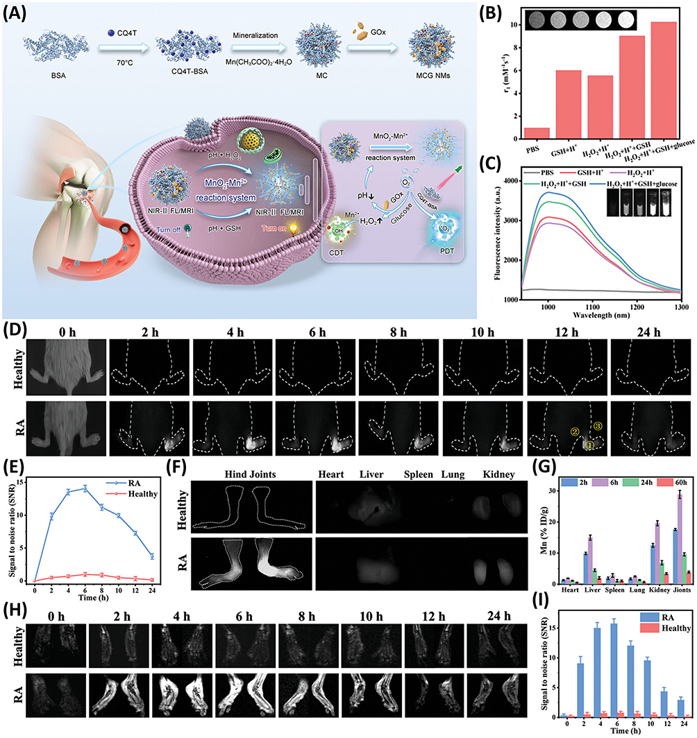 |
Figure 4 (A) Graphical representation of designing the MR/FL bimodal probe and its diagnosing and “knock-on” dynamic therapeutic ability for rheumatoid arthritis. (B and C) T1-weighted MR images of the probe at different concentration of Mn+2 and NIR-II FL imaging of the bimodal probe in the presence of different reagents. (D) In vivo NIR-II FL imaging of diseased and healthy mice at different time frames after 200 µg mL–1 i.v. injection of the probe (rheumatoid arthritis infected joints are indicated as 1, 2, and 3). (E) SBR changes determined between diseased and healthy mice by the targeted NIR-II FL imaging. (F) Ex vivo NIR-II FL images of dissected legs and body organs in diseased and healthy mice after injecting the probe. (G) Demonstration of biodistribution of Mn in major body organs and hind joints of mice measured by ICP-MS at different time frames. (H) In vivo T1-weighted MR images of diseased and healthy mice at different time frames after injecting the probe. (I) SBR changes determined between diseased and healthy mice by the targeted MR imaging. Reproduced with permission from Dai R, Zhao M, Zheng X et al. Homology-Activated Ultrasensitive Nanomedicines for Precise NIR-II FL/MRI Imaging-Guided “Knock-On” Dynamic Therapy in Rheumatoid Arthritis. Adv Healthc Mater. 2024; 13: 2303892. © 2024 Wiley-VCH GmbH.66 |
Hence, NPs could be a promising alternative of FL/MR bimodal probes for targeted imaging. However, the coating of the nanoparticles with organic ligands, cellular membranes or organic polymers could not serve as the ultimate approach for targeted imaging. Therefore, modifying the size and morphology of the nanoparticles could be another impressive strategy to engineer efficient targeted imaging probes. The surface to volume ratio (ie, aspect ratio) is an important parameter to determine the encapsulation of the payload (which might be the MR/FL agent). By tuning the aspect ratio, the loading capacity can be enhanced significantly. Thus, it can be said that the tunability of surface properties significantly depends on the NPs structure, which can be controlled quite easily by altering the reaction parameters.67–69
Tuning Nanoparticle Properties for Attaining Specificity
The physicochemical properties of nanoparticles depend strongly on size and shape. It has been shown that hepatic uptake begins to dominate the clearance pathway at 8 nm, the reticuloendothelial excretory pathway becomes the main clearance pathway at 10–12 nm, and particles ranging in size from 60 nm to 150 nm rapidly appear in the liver and spleen.70 The larger the particle size the longer retention time of the probe in the targeted tissue. The magnetic moment of the particles increases 1000-fold and 10-fold with increasing particle size, so the larger the particle, the higher the magnetic moment, and the easier it is to aggregate in a specific region. However, oversized particles may bring about biotoxicity.71 By simply combining different imaging agents and then surface modification, the resulting composite particles are usually >50 nm, which are susceptible to larger absorption by the reticuloendothelial system (RES). Ding et al directly coated the QDs with paramagnetic Gd chelates, by self-assembly or covalent combination to achieve highly fluorescent and magnetic QDs without greatly enhancing the particle size. The small size of the particles facilitates the rapid excretion of intravenously administered particle probes, which greatly reduces the possible side effects of QDs in vivo, and results in further evidence that they can target subcutaneous and intraperitoneal tumors well in vivo.72
Another important aspect of using NPs is their morphology controllability. In reality, different morphologies have different advantages in pharmacokinetics and pharmacodynamics (PK/PD). For instance, rod-shaped particles penetrate more into the cells than spherical one of the same sizes.73 Even morphology tunability can effectively improve the excretion of the samples from the body making them less toxic. Besides, such tunability in other ways can also alter the band structure of the NPs, which in turn affect the FL and MR properties of the sample.74,75 Such enhancement in FL/MR properties can affect the overall MR/FL imaging capabilities of the probe quite significantly. However, the stability of nanoparticles in aqueous solution is one of the major issues for their biomedical applications, as particles with hydrophobic surfaces tend to gradually aggregate in vivo and get immediately cleared by the immune system. Therefore, there is a need to engineer the nanoparticles having excellent water stability to avoid early excretion from the body to increase their bioavailability for efficient targeting and better diagnosis.
Molecularly Imprinted Polymers for Targeting
The polymer coating of the nanoparticles is a promising strategy to improve the stability and targeting properties with respect to the in vivo aggregation.76,77 Polymer-based MR/FL bimodal contrast agents have been widely reported to improve lesion targeting of small-molecule contrast agents. Additionally, the ability to increase water solubility and reduce biotoxicity has offered great potential in targeted MR/FL imaging. MRI/FL bimodal contrast agents have been prepared by efficient synthetic methods such as reverse addition-fracture chain transfer (RAFT) polymerization and click chemistry, which are extensively applied for multifunctional modification of polymers to achieve improved probe targeting performance due to their mild reaction conditions and extremely high reaction efficiency.78,79 Moreover, amphiphilic surface-modified nanomaterials are proven to exhibit superior targeted ligand binding function than polyethylene glycol-modified nanomaterials, which resists the adsorption of non-specific proteins or the adhesion of blood cells, improves biocompatibility, enhances the blood circulation time. Widely utilized amphiphiles include small-molecule amphiphiles, large-molecule amphiphiles, and positive-negative charge mixtures, which allow modification to the surface of nanoplatforms via ATPR or self-assembly after covalent bonding, with the benefits of amphiphilic modification, targeted ligand binding, in response to the tumor microenvironment.80–82 In a study, polymeric micelles of Fe3O4 coated with magnetic nanoparticles as core and polyethylene glycol (PEG) as shell were synthesized by self-assembly of amphiphilic poly(HFMA-co-VBK)-g-PEG copolymer with oleic acid-stabilized Fe3O4 nanoparticles. The multifunctional micelles possessed good MR properties and specific FL characteristics, showing enhanced contrast between liver and spleen, and the superior multifunctional properties suggest its potential clinical applications as a nanocarrier in MR/FL imaging.83
Therefore, molecularly imprinted polymers (MIPs) have found compelling applications in biomedical imaging, offering a versatile platform for selective and sensitive detection of specific molecular targets.84,85 In the realm of diagnostics, MIPs can be tailored to mimic the binding sites of biomolecules, such as proteins or nucleic acids, facilitating their recognition and capture. In cancer imaging, MIPs can be engineered to recognize cancer-specific biomarkers, enabling early detection and accurate localization of tumors. This not only enhances diagnostic precision but also aids in monitoring therapeutic responses. Moreover, MIPs can be incorporated into contrast agents, improving the visibility of specific tissues or pathological lesions in imaging modalities. For example, Qin et al designed novel multifunctional metal organic frameworks (MOFs) by integrating Si–Gd nanoparticles, the Ce6 photosensitizer, DOX, ZIF-8, HOOC-PDMAEMA-SH, and PEG–FA into single nanoplatform. The multifunctional MOF achieved a pH-sensitive DOX release with precise control, due to the targeted PEG–FA and the pH-responsive polymer HOOC-PDMAEMA-SH that swells in acidic tumor environment, which are modified on the surface of the nanoparticles. It was observed that MCF-7 cells with highly expressed folate receptors were able to phagocytose more MOFs than A549 cells with lowly expressed folate receptors. The multifunctional MOF exhibited excellent drug delivery capacity and effectively avoided side effects caused by drug leakage under physiological environmental conditions. The MOF nanocomposites were successfully accumulated at the tumor sites and exhibit both MR/FL bimodal imaging (Figure 5).86
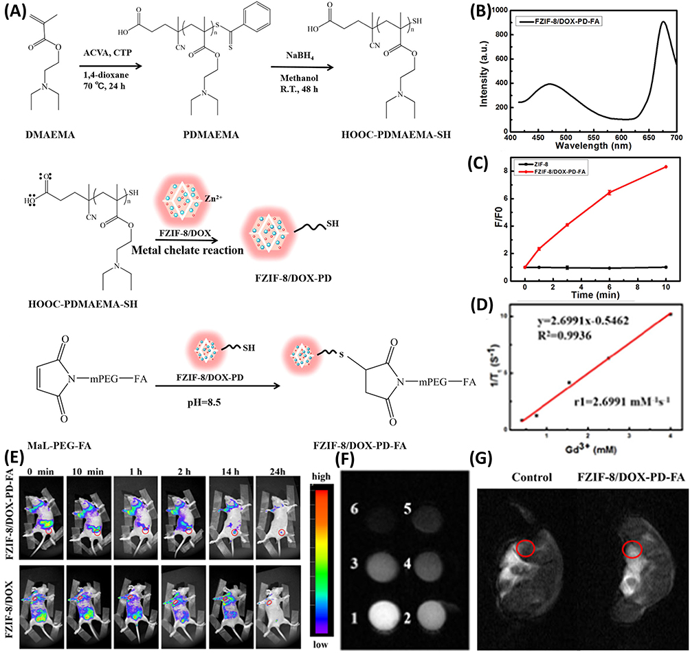 |
Figure 5 (A) Representation of synthetic scheme of multifunctional MOFs. (B) Fluorescence spectra of the multifunctional MOFs (λex = 408 nm). (C) Comparison of ROS generation ability of simple and multifunctional MOFs. (D) T1 relaxivity of multifunctional MOFs for MRI. (E) In vivo NIR FL imaging tumor-bearing mice at different time frames after injection of multifunctional MOFs. (F) T1-weighted MR images of multifunctional MOFs with different Gd concentrations. (G) In vivo MR images of mice before and after injection of multifunctional MOFs while, tumor sites are demonstrated in red circles. Reproduced with permission from Qin YT, Peng H, He XW et al. PH-Responsive Polymer-Stabilized ZIF-8 Nanocomposites for Fluorescence and Magnetic Resonance Dual-Modal Imaging-Guided Chemo-/Photodynamic Combinational Cancer Therapy. ACS Appl Mater Interfaces. 2019; 11: 34,268–34281. Copyright (2019) American Chemical Society Publications.86 |
Additionally, MIPs contribute to drug delivery systems by acting as carriers for imaging agents or therapeutic compounds. Their controlled release properties, combined with selective targeting, enhance drug delivery precision while minimizing off-target effects.87,88 Overall, the integration of MIPs in biomedical imaging showcases their potential to revolutionize diagnostics, imaging, and targeted therapy, promising advancements in personalized medicine and improved patient outcomes.
Benefits of Using Targeted Bimodal MR/FL Imaging Probes
Bimodal targeted MR/FL probes hold several advantages such as precise diagnostic capability, theranostic interventions, high SBR, and most importantly the diverse applicability of the probes in different disease models. Herein, we discuss the targeted bimodal MR/FL probes in the light of their advantages and offer rationality to convey our thoughts for advanced imaging probes for the future biomedical applications.
Improved Disease Diagnosis and Real-Time Monitoring
The bimodal MR/FL imaging possesses several advantages such as high sensitivity, enhanced resolution, strong penetration, and also offers important real-time monitoring for both oncologic and non-oncologic diseases.89 Especially for tumors with heterogeneous and highly aggressive growth, preoperative failure to accurately locate the extent of the tumor will make it difficult to resect, and excessive resection may cause irreversible injuries by damaging the surrounding structures of the tumor. Consequently, to define the extent and margin of tumors and to accurately outline the morphology of the tumor and precisely locate the lesion area before surgery is a powerful approach for precise resection of tumors and reduction of recurrence.89,90 Herein, bimodal probes have been applied for different carcinogenic diseases such as pancreatic cancer, breast cancer, gliomas, and other diseases91,92 (Table 2).
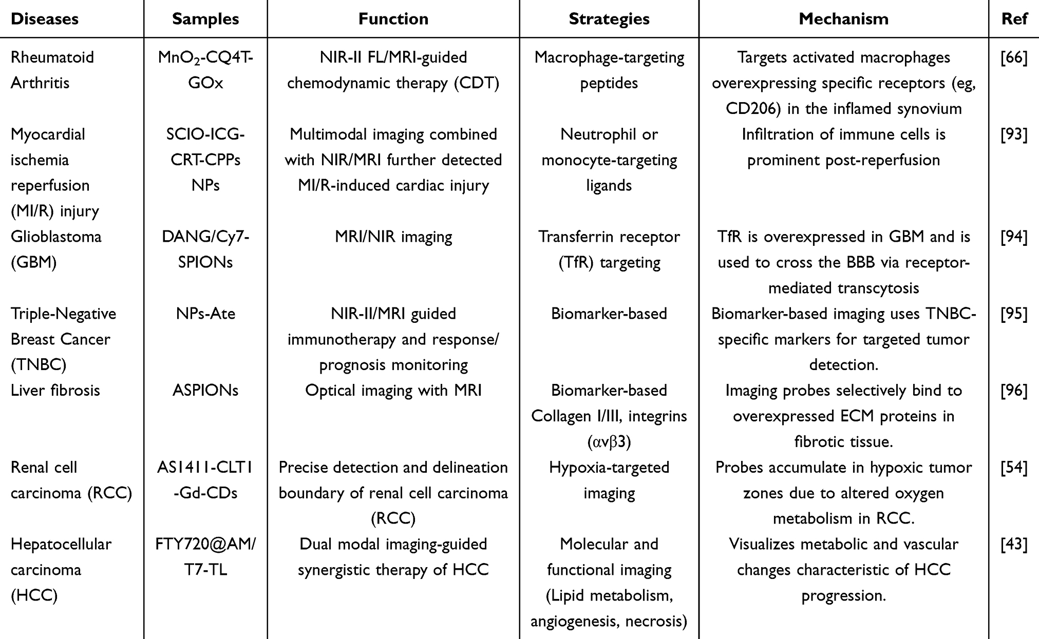 |
Table 2 Applications of Bimodal Probes in Different Disease Models |
SPION was investigated in a study as a core nanoprobe platform, integrating it with the NIR dye Cyanine 7 (Cy 7) to construct MR/NIR bimodal nanoprobes. THR peptide (Ac-THRPPMWSPVWP-COOH, TfR ligand) promotes receptor-mediated endocytosis, contributing to the blood brain barriers (BBB) penetration and specificity of nanoprobe for GBM imaging, which not only prolongs circulation time in vivo, but also exhibits less biotoxicity, showing great value in preoperative diagnosis and intraoperative localization of gliomas.97 In this study SPION (ASPION) stabilized with alginate had a hydrodynamic diameter of 40 nm, and at room temperature ASPION demonstrated very excellent proton relaxation rate ratios. To introduce a bimodal imaging capability, ASPIONS were coupled with the NIR dye ATTO 700. After i.v. administration in hepatocellular carcinoma cells and in an animal model of hepatic fibrosis the very good cytocompatibility of ASPION validated its suitability for use in living systems, and histological analyses further confirmed liver-targeting efficacy for magnetic therapy and optical imaging while demonstrating its short-term safety in vivo.96
A study reported a dual-targeting probe with the main components of superparamagnetic cubic iron oxide nanoparticles (SCIO NPs), indocyanine green (ICG), tfr1-targeting peptide (CRT), and cell-penetrating peptides (CPPs) for noninvasive quantitative assessment of the extent of myocardial ischemia/reperfusion injury (MI/r)-induced heart injury in vivo, CCI NPs imaging probes with high MI/r detection sensitivity, excellent targeting capability and high reliability, and integrated MR/FL with more complete MI/r characterization and higher detection accuracy, identifying MI/r-induced cardiac injury ≥ 48 h earlier than echocardiography, thus compensating for the difficulty in detecting MI/r injury during cardiac remodeling. In addition, the CCI NPs probe enhanced the MPI signal and MPI specificity of MI/r-induced cardiac injury, allowing a longer observation time window to continuously assess MI/r-induced cardiac injury.93
Moreover, up-regulation of β-galactosidase is commonly associated with cancer development and cellular senescence; therefore the in vivo detection and localization of β-galactosidase activity promises to be an invaluable tool in the diagnosis and therapeutic monitoring of cancer. A study here demonstrates that LnL 5 Gal allows successful imaging β-galactosidase activity, making the detection of enzyme activity possible in complementary imaging modalities including NIR luminescence, T1 and CEST MRI. The LnL 5/LnL 5 NH2 complex has good luminescence, CEST and relaxation properties.98 Similarly, in another study, a bimodal probe Gal-Cy-Gd-1, with integrated MR/FL, was proposed to be capable of sensing and labelling β-gal. Gal-Cy-Gd-1 showed high sensitivity and selectivity for β-gal, accompanied by turned-on FL signals and enhanced longitudinal relaxation. In vivo evaluation of GalCy-Gd-1 for bimodal β-gal imaging in ovarian cancer model mice demonstrated promising results, facilitating NIR FL-guided real-time surgical resection of in situ ovarian cancer.99 Here design of a NIR probe, MitoSQ-DOPA, utilizing a lipophilic cationic triphenyl scale (TPP+) moiety, which is tethered at the axis to target live cell mitochondria, was undertaken for the targeting of magnetic NPs delivered within the mitochondria coupled with T2-weighted MR imaging, thus enabling the selective targeting and imaging of live cell mitochondria.100 Another study developed and characterized a new nanoparticle, NPs-Ate, consisting of Atezolizumab, ICG and GD-DTPA, which constitutes an innovative bimodal NIR-II/MRI imaging platform that offers the opportunity to optimize and monitor the outcome and improve the efficacy of ICT treatments in the clinical setting.95
Pancreatic cancer is often difficult to detect due to the lack of obvious symptoms in the early stage, and when clinically diagnosed, it is often already in advanced stage, with a very short survival period, thus the use of bimodal molecular probes for early and accurate diagnosis of the patients has an important value for the subsequent early treatment and prognosis of the patients.101,102 Wang et al developed a novel selectin/integrin-targeted bispecific molecular probe (Gd-Cy7-PTP/RGD) conjugated complex for MR/FL imaging of pancreatic cancer in vivo, successfully realizing MR/FL-guided intraoperative pancreatic cancer surgical margin delineation under bimodal imaging guidance.40 In another study, Xie et al utilized indocyanine (Cy7) molecules and peptide (ANG or DANG)-modified superparamagnetic iron oxide nanoparticles (SPION), an MR/NIR FL bimodal nanoprobe modified with a trans-isomer peptide, to have precise preoperative imaging and intraoperative real-time imaging of glioblastoma in vivo (Figure 6).94 These studies suggest that these bimodal probes can be used in clinics for imaging-guided surgery and guided therapeutic applications.
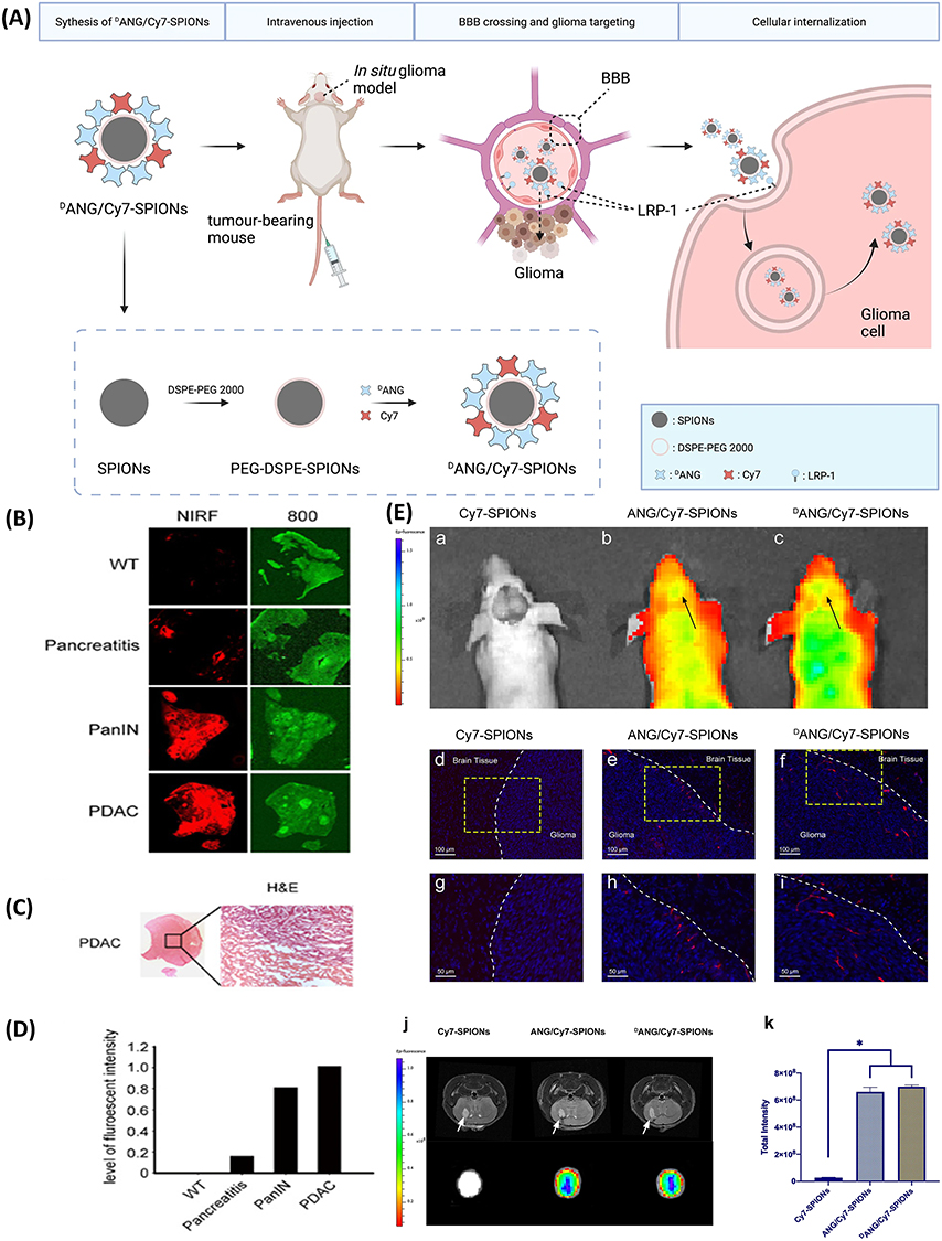 |
Figure 6 (A) Graphical representation of the fabrication and features of DANG/Cy7-SPIONs, including their strategy to cross the BBB and target glioma cells. (B) Near-infrared fluorescence (NIRF) imaging was performed on pancreatic tissues from Ptf1a^Cre/+; LSL-Kras^G12D/+ mice with mPanIN lesions or PDAC. Wild-type and caerulein-induced pancreatitis mice served as controls. Tissues were collected 24 h after i.v. injection of a cathepsin-activated NIRF probe. Cryosections were scanned at 680 nm (red signal) and 800 nm (green, morphology). (C) Histology of the same PDAC section revealed necrotic and viable tumor areas aligning with the NIRF signal (left: 2×; right: 200×). (D) Fluorescence intensity from the scans in (B) was quantified using Odyssey software. (E) a, b, c. In vivo pictures of tumor-bearing mice injected with different samples. d, e, f. Pictures of glioma-containing brain slices from mice injected with different samples which were taken with an automated quantitative pathology imaging system. g–i. The lower images showed a 2x magnification of the areas in the yellow boxes of the images above. The borderline of the glioma is highlighted by the white dotted line. The DAPI signal in blue indicates cells, and the Cy7 signal in red indicates probes. j. Ex vivo NIR fluorescence images of three probes in tumor-bearing brains after i.v. injection for 24 h. The upper images demonstrate the MRI of the tumor-bearing mice, and the lower images demonstrate the fluorescence signals. (white arrows are showing the tumor site). k. The total intensity of the three probes in tumor-bearing brains (*p < 0.05). Reproduced from Xie R, Wu Z, Zeng F et al. Retro-enantio isomer of angiopep-2 assists nanoprobes across the blood-brain barrier for targeted magnetic resonance/fluorescence imaging of glioblastoma. Signal Transduction and Targeted Therapy. 2021; 6: 1–13. Creative Commons.94 |
In addition to oncological diseases, bimodal imaging finds application in cell tracking as well. By conjugating paramagnetic chelates to fluorophores, which are highly water-soluble and cell-permeable as well as excellent cell labeling agents, bimodal imaging allows the labeling of cells of different origins, including tissue origin (cervical, mammary, and cutaneous), species origin (human and mouse), cell size (12–20 mm in diameter), and cell growth rate (20–38 h doubling time).103 Furthermore, caspase-3 and caspase-7 are the executors that carry out extensive protein hydrolysis, ultimately leading to apoptosis, which has been detected by bi-modal probes in vivo by detecting elevated caspase-3/7 activity.13 To investigate the possibility of multimodal imaging, the surface of the probe was functionalized with fluorescein isothiocyanate (FITC) for lymph node imaging.104
Enhanced Signal-to-Background Ratio (SBR)
A bimodal contrast agent requires a high SBR in order to achieve good imaging contrast. Current studies have demonstrated that the SBR of MRI can be enhanced by increasing the contact between the imaging agent and water molecules, adjusting the size of the contrast agent, and introducing other paramagnetic ions.105–107 MRI contrast agents currently in clinical practice are mainly hydrophilic small-molecule gadolinium-based chelates. Gd3+ provides a high paramagnetic relaxation rate due to the seven unpaired 4f electrons, which makes Gd3+-based probes ideal paramagnetic relaxants.108,109 The MR signal enhancement of paramagnetic Gd3+ is primarily achieved by increasing the longitudinal relaxation rate of the adjacent water protons, and sufficient contact or cooperation between the Gd3+ contrast agent and the water protons and it is important for polymer-based MRI contrast agents. Its contrast capability is computed by the relaxation rate (r1), which is certainly correlated with the rotatory correlation time (τR) of the imaging agent and the diffusion correlation time (τD) of the water molecules nearby the contrast agent.110,111 However, some of the surface modification methods reported so far for the preparation of multifunctional nanoplatforms severely limit the exposure of Gd3+ ions, which inevitably causes low SBR in MRI. The solutions include constructing geometrically confined structures and reducing nanoparticle size to increase the exposure of Gd3+ ions. Simply increasing the dose of paramagnetic ions to obtain high contrast imaging results in the retention of metal ions in the body and the risk of biotoxicity, which in turn leads to the introduction of other paramagnetic ions, such as Mn2+ or Fe3+, combined with a variety of ions to achieve high contrast at low concentrations (Figure 7A).16
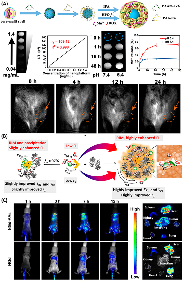 |
Figure 7 (A) Synthesis of Mn-based nanoplatform, T1-weight MR images of different concentration nanoplatform with Relaxation rates of the nanoplatform at different concentrations using a 1.2T MR scanner, and T1-weight MR images of the nanoplatform against different pH conditions over time. Release profiles determined by ICP and T1-weight MR images of tumor-bearing mice at different time points after i.v. injection of the nanoplatform (the sample accumulation in the subcutaneous tumor has been indicated by Orange-colored circles and arrows). Reproduced with permission from Chen H, Wu F, Xie X et al. Hybrid Nanoplatform: Enabling a Precise Antitumor Strategy via Dual-Modal Imaging-Guided Photodynamic/Chemo-/Immunosynergistic Therapy. ACS Nano. 2021; 15: 20,643–20655. Copyright (2021) American Chemical Society Publications.16 (B) Mechanism of simultaneous enhancement of FL and relaxivity. τR1, τR2, and τR3 refer to rotational correlation time of NGd molecules, NGd-AAs, and NGd aggregates in BSA aqueous solution, respectively while, τD1, τD2, and τD3 are the diffusion correlation time of water molecules around them, respectively. (C) FLI in vivo after i.v. injection of NGd-AAs or NGd at different time intervals (white dotted circles refer to the tumors) and ex vivo FL images of main body organs and tumors from mice at different time points post-injection. Reproduced with permission from Wang L, Wan Q, Zhang R et al. Synergistic Enhancement of Fluorescence and Magnetic Resonance Signals Assisted by Albumin Aggregate for Dual-Modal Imaging. ACS Nano. 2021; 15: 9924–9934. Copyright (2021) American Chemical Society Publications.112 |
In addition, MR contrast agents for signal amplification are used in combination with more sensitive imaging modalities, including FL imaging. Different from paramagnetic contrast agents, most of the small molecule fluorescent probes in service today are hydrophobic compounds with aggregation-induced quenching (ACQ) effects in aqueous solution, which usually affects their imaging performance.113,114 Aggregation-induced emission (AIE) with a limitation of intramolecular movement (LIM) working phenomenon is an effective approach to address this issue. Before activation, these AIE bioprobes remain in molecularly dispersed state in water. In this state, due to the rotation of the benzene ring, the “propeller-shaped” AIEgen enables access to the non-radiative decay pathway, efficiently dissipating excited state energy and quenching FL. Upon activation, the bioprobes aggregate through π-π stacking that restricts intramolecular movement, reducing the nonradiative decay rate and enhancing FL.13,115,116 In one study, an NGd amphiphilic MR/FL bimodal probe that binds tightly to bovine serum albumin (BSA) was designed by combining an AIE motif with Gd-DOTA. NGd albumin aggregates (NGd-AAs) were then constructed by dissolution and glutaraldehyde cross-linking to BSA for loading NGd.117 The geometrically confined structure of these nanoaggregates eases the entry and exit of water molecules at a slow diffusion rate, ie, prolongs the τD of the water molecules, which would further result in the improvement of the relaxation. Meanwhile, the larger size and longer τR of nanoaggregates compared to individual molecules will greatly augment the relaxation of MRI. Assembling the Gd chelate with the AIE portion (AIE-Gd) to form an amphiphilic bimodal probe will not only intensify the FL intensity, but the relaxation rate will also be raised and extended τR (Figure 7B and C).112
Besides, NIR-based fluorescent probes have also been an effective strategy to enhance FL intensity, tissue absorption and minimizing autofluorescence in the NIR optical window (>700 nm). NIR light penetrates into deep tissues without being absorbed by the blood. Thus, provides higher SBR during signal acquisition. Herein, lanthanide-based upconversion and downconversion nanoparticles (UCNPs and DCNPs) show unique NIR to NIR upconversion and downconversion luminescence (UCL), which provides high SBR and penetrate longer in the small animal imaging.118–120 The incorporation of paramagnetic ions (eg, Gd3+ and Mn2+) into such lanthanide-based nanoparticles provides magnetic properties that make these probes usable for MR/FL bimodal imaging.121–123 Indocyanine green (ICG) and methylene blue are approved by the US Food and Drug Administration (FDA) as NIR fluorescent dyes for use in a variety of surgical applications in addition to other NIR fluorescent dyes such as Cy7, Cy5.5, and others. These NIR fluorescent dyes in combination with MR contrast agents can achieve high SBR for MR/FL bimodal imaging.124,125
Apart from these, semiconductive quantum dots (QDs) are well exploited for bimodal imaging applications with MRI active nanoparticles. For instance, Fe3O4 NPs has been conjugated with CdS or CdTe QDs and acted as bimodal imaging agents to enable high sensitivity and enhanced resolution of FL imaging, as well as non-invasive and high spatial resolution in MRI. The nanohybrids were prepared by a facile ligand-exchange method, which easily adjusted with the ratio of SPIO/QDs with good water solubility and small stability, results showed that the sample exhibited good magnetic properties as well as promising luminescence intensity. This method provides more in-depth biological information about the target site.126 High FL quantum yield (QY) by encapsulating prefabricated magnetic nanoparticles and QDs into inert matrices such as silica or polymers to gain high FL QY can also be achieved by several other groups. Besides, organic dyes-magnetic probes, SPIO-rare earths, gadolinium reagents attached to QDs or dyes, Gd3+ reagents, and CdSe-containing molecular probes have also shown promise in this domain.127–129
It is noted that a higher amount of contrast does not always mean a higher dose of the imaging probes, which may perturb the overall metabolism of the body and can induce severe toxicity. In contrast, a high-resolution image can be produced by tuning the SBR of the imaging probe or improving the FL QY of the sample. On the other hand, scientists are working on various Gd-alternatives to achieve better MRI contrast. Using Fe/Mn could aid such issues. Therefore, more feasible strategies are needed for the upcoming days to address the public health issues.130
Targeted Imaging-Mediated Efficient Therapeutics
Imaging-guided precision and personalized therapies are the mainstream strategies with important clinical translational significance. The construction of an integrated MR/FL imaging-guided diagnosis and treatment platform can also help to improve therapeutic efficacy while reducing therapeutic side effects.131 Researchers have designed a homologous activated ultrasensitive nanomedicine, MnO2-CQ4T-GOx (MCG NMs), which takes advantage of the fact that the rapid proliferation of inflammatory cells in the synovial tissues of RA creates a unique microenvironment in which the nanomedicine allows for the passive targeting and accumulation of the nanomedicine at the inflamed joint via ELVIS effect. In vivo imaging showed that MCG NMs effectively “turn on” NIR-II FL and MRI bimodal imaging at the site of joint inflammation at 6 hours post-injection.132 Under the guidance of bimodal imaging, MCG NMs + laser irradiation can achieve GOx-mediated “Knock-On” dynamic treatment.66 For, photodynamic therapy (PDT) uses photosensitizers (PS) to convert oxygen in cancer cells into cytotoxic reactive oxygen species (ROS) when irradiated with specific light, and then eliminates cancer cells through apoptosis or necrosis, which has the benefits of being highly selective to the tumor site and non-invasive to normal tissues, etc.39 However, many obstacles still exist in PDT that need to be solved, for instance, the therapeutic efficacy of PDT is restricted by the low penetration depth of tissues as well as the inappropriate timing and location of triggering.133 Chen and coworkers designed a photo-switchable lanthanide-doped nanoparticle-based real-time NIR-IIb imaging and image-guided PDT (980 nm) with high SBR, which is further loaded with paramagnetic Mn2+ ions to enhance T1 MRI signal in the tumor microenvironment. In addition to direct killing of tumor cells by bimodal imaging-guided PDT/chemotherapy, this theranostic nanoplatform can inhibit distant tumors and lung metastasis by immunogenic cell death (ICD), which has great clinical application prospects.16 Moreover, a carbon-dotted (Gd/Ru-CD) nanoprobe was fabricated by a one-step microwave-assisted synthesis technique, which demonstrated excellent performance in FL and MR bimodal tumor imaging both in vitro and in vivo. Furthermore, the study highlights the efficacy of imaging-guided PDT, which showed remarkable anticancer potential in 4T1 hormonal mice. Gd/Ru-CD is also well biocompatible, as the probe also exits the body efficiently after imaging, avoiding the accumulation of the protein in vivo and the potential long-term toxicity of the proctor.134
Many features exist within the complex tumor microenvironment, including the presence of reduced GSH, limited H2O2, hypoxia, and weak acidity; thus, different tumor therapeutic strategies involve modulation and combinations based on these features.135,136 Numerous studies have been carried out to combine chemotherapeutic agents, targeted agents and PDT, guided by MR/FL bimodal imaging, to form an integrated platform for diagnosis and treatment, frequently used chemotherapeutic agents in research include doxorubicin (DOX), paclitaxel (PTX), and so on.65 Apart from PDT, photothermal therapy (PTT) is based on nanostructured samples such as gold nanoparticles, carbon nanotubes, and graphene, which intensely absorb NIR light and locally generate cytotoxic heat to eliminate the cancer cells.137,138 On one hand, previous studies demonstrated that the coupling of PTT with chemotherapy enhances the therapeutic effect because chemotherapeutic probes exhibit increased cytotoxicity at higher temperatures. On the other hand, it reduces the dosage of chemotherapeutic agents required, thereby reducing systemic side effects.139–141
For instance, Wang et al reported a novel highly unified hybrid nanoparticle that combines magnetic iron oxide nanocrystals and fluorescent carbon dots (CD) combine combined into a single carbon-based nanosystem in which Fe3O4 nanocrystals are aggregated in the core and CDs and drugs are loaded in porous carbon (C) shells. Materials with high yields of MR/FL properties in this design can be synthesized in a single step to achieve a combined bimodal imaging diagnostic, NIR PTT, or chemotherapy as an effective strategy. The intrinsic and simultaneous growth of the carbon dots into the Fe3O4 nanocrystals is quite a new approach and not only could aid in bimodal diagnostic applications but also help in NIR light mediated therapeutic applications (Figure 8).142 CDs are carbon nanoparticles less than 10 nm in size that combine the fluorescent properties with the magnetic properties of metals for MR/FL imaging. By combining different surface-specific ligands that enable recognition of receptors on tumor cells, binding of different targeted therapeutic agents, and doping with various metals such as Gadolinium-doped Carbon Quantum Dots (Gd-CD), Manganese-doped Carbon Quantum Dots (Mn-CD), and Iron-doped Carbon Quantum Dots (Fe-CD), it is possible to carry out more accurate tumor diagnostics and a variety of different therapeutic modalities such as chemotherapy, PDT, PTT, and CDT.143
 |
Figure 8 (A) Hybrid Nanoplatform for Targeted Cancer Therapy via Dual-Modal Imaging-Guided Photodynamic, Chemotherapy, and Immunotherapy Synergy. Chen H, Wu F, Xie X et al. Hybrid Nanoplatform: Enabling a Precise Antitumor Strategy via Dual-Modal Imaging-Guided Photodynamic/Chemo-/Immunosynergistic Therapy. ACS Nano. 2021;15(12):20,643–20655. Copyright (2021) American Chemical Society Publications.16 (B) Graphical representation of MR/Fl bimodal imaging guided probe for photothermal therapy. (C) FL spectra of bimodal probe at different excitation wavelengths. (D) The hysteresis loop of the bimodal probe measured at room temperature while, the inset is demonstrating the respective expanded plots between −40 and 40 Oe. (E) T2 relaxivity of the bimodal probe at different concentration of Fe. (F) T2-weighted MR images of the bimodal probe at different concentrations of Fe. (G) The photothermal curves of water, 100 mg L−1 of bimodal probe in water, and only C-CDs in water under NIR illumination for 5 min. (H) Release profiles of the drug from the bimodal probe in PBS at 37 °C, illuminated with/without 1.5 W cm−2 NIR for 5 min at different time frames. Used with permission of Royal Society of Chemistry, from Wang H, Shen J, Li Y et al. Magnetic iron oxide-fluorescent carbon dots integrated nanoparticles for dual-modal imaging, near-infrared light-responsive drug carrier and photothermal therapy. Biomater Sci. 2014; 2: 915–923. Permission conveyed through Copyright Clearance Centre, Inc..142 |
Although, the efficient targeting and MR/FL bimodal imaging has achieved deeper penetration, higher SBR, enhanced resolution, and optimal biocompatibility to generate clear images and real-time monitoring of the diseased body parts. However, the result interpretation and disease diagnosis are still human dependent, which could sometimes lead to false positives or misleading information due to human errors that could affect the overall therapeutic regimen. Therefore, there should be a universal diagnostic tool having ability to deeply analyze the produced results and then diagnose the respective disease to avoid false positives for better healthcare of the patients.
Computational Approach-Based Image Analysis for Accurate Diagnostics
In the era of precision medicine, computational approaches have transformed medical image analysis by enhancing diagnostic accuracy and efficiency. Utilizing AI, machine learning, and deep learning algorithms, these methods enable automated detection, segmentation, and classification of complex patterns, reducing subjectivity and improving reproducibility. They are crucial for early disease detection, monitoring progression, and supporting personalized treatments. As imaging data becomes increasingly complex, computational analysis is emerging as a vital tool in clinical decision-making and data-driven diagnostics. In this section, we will discuss the computational approach-based image analysis for accurate diagnostics.
Early and Accurate Disease Detection
Computational approaches for image analysis have significantly advanced early and accurate disease detection by automating the recognition and interpretation of complex patterns in medical images. This is especially crucial in detecting life-threatening conditions such as brain tumors at early stages. For instance, Chato and Latifi presented a machine learning- based approach, which introduces an automated approach for predicting the survival rates of glioma brain tumor patients through the classification of their MRI images using machine learning techniques. The utilized dataset, BraTS 2017, comprises 163 samples, each including four sequences of MRI brain images, the age of the patient, and overall survival time in days. The dataset is categorized into three survivor classes: short-term, mid-term, and long-term. To enhance prediction accuracy, diverse features were extracted and trained using various machine learning methods.144 In another study, Shen et al demonstrated the real-time glioma diagnosis using fluorescence imaging. In their study, they used deep convolutional neural networks (CNNs) in conjunction with NIR-II imaging, referred to as FL-CNN, for the automatic real-time diagnosis of glioma in situ during patient surgery. The gold standard for evaluation was based on pathological examination results. The study suggests that deep CNNs are better when it comes to decoding the fluorescent signals in glioma detection (Figure 9).145
 |
Figure 9 (A) a. The no. of cases per class with pathological diagnosis results serving as the Au standard. Pathological results for tumor cases included grade of tumor and Ki-67. b. The tumors were surgically removed with the guidance of NIR-II imaging. Photos of specimens obtained with white light and FL were input into CNNs for distinguishing between tumor and non-tumor tissues. Subsequently, photos of tumors were input into the CNNs for the classification of tumor grade and Ki-67 level. The diagnostic process by the models took less than 1 sec on the CPU and could be even quicker on the GPU, making the time negligible. (B) Diagnostic ability of the CNNs and neurosurgeons. a. Receiver operating characteristic curves were designed for the CNNs and neurosurgeons on FL and white light photos for the tumor tissue versus non-tumor classification. The receiver operating characteristic curves demonstrate the performance gained by the CNNs, while crosses indicate each neurosurgeon performance. Results for FL and white are shown in different colors, respectively. b. Selectivity of the CNNs and individual neurosurgeons were plotted, using pathological diagnostic results as the Au standard, and relative on FL and white light. (C) Gradient-weighted Class Activation Maps (GCAMs) visually represent the learned character demonstration for the categorization of tumor tissue versus non-tumor. FL/white light photos of tumor and non-tumor tissues were sampled randomly. The targeted layer of GCAM was the last state of the CNNs, and target types guided backpropagation to produce Class Activation Maps (***p < 0.001, ****p < 0.0001). Reproduced from Shen B, Zhang Z, Shi X et al. Real-time intraoperative glioma diagnosis using fluorescence imaging and deep convolutional neural networks. Eur J Nucl Med Mol Imaging. 2021; 48: 3482–3492. Creative Commons.145 |
Integration of Artificial Intelligence, Machine Learning, and Deep Learning
Modern computational imaging heavily relies on artificial intelligence (AI), particularly its subfields—machine learning (ML) and deep learning (DL). While machine learning generates logical solutions from structured datasets, deep learning mimics the human brain through neural networks to execute complex tasks. Despite their functional similarities, they differ in how they process data and learn patterns. Both approaches are now extensively integrated into MR (Magnetic Resonance) and FL (Fluorescence) imaging systems, enhancing image analysis capabilities. In clinical practice, AI has become indispensable for automating workflows, improving diagnostic consistency, and extracting meaningful insights from high-dimensional imaging data. As these systems continue to evolve, AI is also being considered in the development of novel imaging agents. Although there is currently no direct evidence of AI being used to design new fluorophores or contrast agents, researchers are exploring the potential of combining AI with emerging technologies like quantum computing to create contrast agents with improved targeting efficiency, biocompatibility, and resolution.
Quantitative Image Analysis
Quantitative image analysis is another key strength of computational approaches, enabling precise measurements and tracking of disease progression. Recent reviews suggest that AI can greatly enhance the image resolution by exploiting several machine learning and deep learning algorithms, which benefits in analyzing the images and offers better diagnosis. AI-driven techniques can enhance image resolution and quality even when using lower-cost imaging agents or machines, making advanced diagnostics more accessible. Computational methods also support objective data extraction, such as lesion volume, intensity profiles, and morphological changes over time, which are critical for treatment planning and monitoring response. Beyond AI, traditional mathematical techniques like Contrast Limited Adaptive Histogram Equalization (CLAHE), Active Contour Models (Snake), filtering algorithms, and feature extraction tools continue to play a role in biomedical image analysis. However, these conventional methods have limited scalability and adaptability compared to modern AI-driven tools. Scientists are actively refining these algorithms and integrating them with AI models to improve the understanding and interpretation of MR and FL images, making them more reliable and clinically relevant. But there is no such proof of designing new fluorophores and contrast agents by using machine learning and deep learning algorithms. Scientists are continuously searching for such improved AI technologies equipped with quantum computing to solve this puzzle in near future.
A table (Table 3) outlines essential features of each system, including the nanomaterials used, integrated therapeutic strategies (eg, photodynamic therapy, chemotherapy, immunotherapy), diagnostic modalities (eg, MRI, NIR-II fluorescence), and targeted biomedical applications. This comparison highlights the versatility of these nanoplatforms in enabling precise, image-guided treatments and real-time diagnostics, while also reflecting emerging trends and innovations in the field of nanotheranostics.
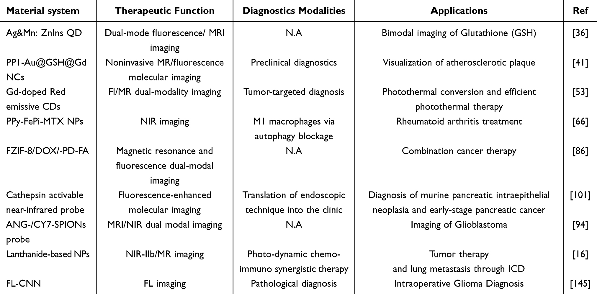 |
Table 3 A Summary Table of Referenced Studies (as Shown in the Figures), Highlighting Material Systems, Therapeutic Functions, Diagnostic Modalities, and Biomedical Applications |
Challenges and the Future Prospective
Despite remarkable advancements, several persistent challenges remain to hinder the clinical translation of magnetic resonance/fluorescence (MR/FL) bimodal nanoprobes, especially those designed for targeted diagnostics. One of the key limitations is targeting efficiency, often compromised by tumor heterogeneity and the complexity of the tumor microenvironment. Most current ligand-conjugated nanoprobes are tailored to a single cancer type or biomarker, restricting their broader application across diverse malignancies. To overcome this, future strategies must focus on engineering dual- or multi-targeting nanoplatforms capable of recognizing multiple biomarkers simultaneously. Such designs could significantly improve tumor localization and retention, particularly in heterogeneous or treatment-resistant cancer types.
Signal stability and intensity in complex in vivo environments represent another critical hurdle. Enzymatic degradation, nonspecific uptake by the reticuloendothelial system, and rapid systemic clearance can substantially reduce the imaging signal. Addressing these issues requires the development of robust, biocompatible nanostructures, such as core–shell architectures and stealth coatings (eg, PEGylation) that resist degradation and prolong circulation time. Additionally, incorporating stimuli-responsive elements, such as pH, enzyme, redox, or temperature-sensitive moieties, could enable on-demand signal activation and real-time monitoring of therapeutic responses. Biocompatibility remains a central concern for clinical translation. MR/FL nanoprobes must be non-toxic, non-immunogenic, and efficiently eliminated from the body to avoid long-term accumulation. This necessitates surface modifications using biodegradable or naturally derived materials and rigorous preclinical testing to ensure safety without sacrificing diagnostic performance. To further enhance diagnostic utility, integration of MR/FL imaging with other emerging modalities, such as photoacoustic imaging, positron emission tomography (PET), or Raman spectroscopy, can provide complementary information, improving spatial resolution, sensitivity, and molecular specificity. Such multimodal platforms could substantially expand diagnostic capabilities and support more comprehensive patient evaluations.
On the computational front, artificial intelligence (AI) and quantum computing are poised to revolutionize the design, optimization, and clinical application of MR/FL nanoprobes. AI can aid in probe screening, predict biodistribution, analyze complex imaging datasets, and enhance diagnostic accuracy. Quantum computing may offer solutions for rapid modeling of nanostructure behavior and optimizing probe–target interactions. However, these technologies face several barriers to clinical integration, including the need for large, high-quality annotated datasets, concerns over model transparency and interpretability, and the absence of standardized regulatory guidelines. Clinical deployment also demands robust, multi-center validation and seamless integration into existing workflows. Therefore, future efforts should prioritize the development of interpretable, reproducible, and clinically validated AI frameworks to build trust and facilitate adoption among healthcare providers. AI can play a central role in the development of next-generation MRI/NIR-II probes by accelerating material discovery and probe design through predictive modeling and high-throughput screening. Machine learning algorithms can analyze structure–activity relationships to identify optimal nanomaterial compositions, surface modifications, and targeting ligands. AI can also simulate in vivo behavior,such as circulation time, clearance, and tissue accumulation,allowing for more efficient preclinical optimization. In the diagnostic phase, AI enables precise interpretation of MRI/NIR-II imaging by integrating multimodal data, enhancing lesion detection, and distinguishing subtle signal variations that may be overlooked by human observers. This results in improved sensitivity, specificity, and overall diagnostic accuracy, enabling earlier and more personalized intervention. Finally, addressing current limitations, including low targeting efficiency, signal instability, and limited biocompatibility, through advanced material design, multi-targeting strategies, stimuli-responsive elements, and computational innovations is essential for the successful implementation of MR/FL bimodal imaging nanoprobes. When empowered by AI-driven design and supported by next-generation imaging modalities, these platforms promise to enable real-time, personalized diagnostics and targeted therapeutic interventions in translational medicine.
Conclusion
This work critically reviews relevant research on the improvement of targeting performance in MRI and FL imaging, emphasizing its clinical value through the enhancement of targeting properties in multimodal molecular imaging probes. The integration of imaging contrast agents and multiple targeting ligands into nanomaterials, guided by principles of active and passive targeting, enables the delivery of more comprehensive information for disease diagnosis. Bimodal MR/FL nanoprobes with specific targeting capabilities have demonstrated promising potential in enhancing imaging quality, photoconductive therapy, and drug delivery efficiency. These developments hold significant promise for accurate tumor localization and image-guided surgical treatments. Such advances signal a strong potential for these nanoprobes to play an increasingly important role in disease diagnosis and therapeutic applications, ultimately contributing to translational medicine and precision healthcare.
Acknowledgment
The authors thank for the support from the “Chunhui Plan” cooperative scientific research project of the Ministry of Education, China (HZKY20220312), the Guangdong Basic and Applied Basic Research Foundation (2021A1515110086), the General Project of Guangdong Natural Science Foundation (2022A1515011781), the Science and Technology Innovation Commission of Shenzhen (RCB20200714114910141, JCYJ20210324132816039) and Shenzhen Key Laboratory of Advanced Functional Carbon Materials Research and Comprehensive Application (ZDSYS20220527171407017).
Data Sharing Statement
No data has been used in this manuscript.
Disclosure
The authors declare no conflicts of interest in this work.
References
1. Lu Z-R, Laney V, Li Y. Targeted contrast agents for magnetic resonance molecular imaging of cancer. Acc Chem Res. 2022;55(19):2833–2847. doi:10.1021/acs.accounts.2c00346
2. Keall PJ, Brighi C, Glide-Hurst C, et al. Integrated MRI-guided radiotherapy — opportunities and challenges. Nat Rev Clin Oncol. 2022;19(7):458–470. doi:10.1038/s41571-022-00631-3
3. Lin H, Tang X, Li A, Gao J. Activatable 19F MRI nanoprobes for visualization of biological targets in living subjects. Adv Mater. 2021;33(50):2005657. doi:10.1002/ADMA.202005657
4. Fu C, Yu Y, Xu X, et al. Functional polymers as metal-free magnetic resonance imaging contrast agents. Prog Polym Sci. 2020;108:101286. doi:10.1016/J.PROGPOLYMSCI.2020.101286
5. Barrett T, de Rooij M, Giganti F, Allen C, Barentsz JO, Padhani AR. Quality checkpoints in the MRI-directed prostate cancer diagnostic pathway. Nat Rev Urol. 2022;20(1):9–22. doi:10.1038/s41585-022-00648-4
6. Wang S, Ren WX, Hou JT, et al. Fluorescence imaging of pathophysiological microenvironments. Chem Soc Rev. 2021;50(16):8887–8902. doi:10.1039/D1CS00083G
7. Refaat A, Yap ML, Pietersz G, et al. In vivo fluorescence imaging: success in preclinical imaging paves the way for clinical applications. J Nanobiotechnol. 2022;20(1):1–22. doi:10.1186/S12951-022-01648-7
8. Wang F, Ren F, Ma Z, et al. In vivo non-invasive confocal fluorescence imaging beyond 1700 nm using superconducting nanowire single-photon detectors. Nature Nanotechnol. 2022;17(6):653–660. doi:10.1038/s41565-022-01130-3
9. Wang LG, Barth CW, Kitts CH, et al. Near-infrared nerve-binding fluorophores for buried nerve tissue imaging. Sci Transl Med. 2020;12(542). doi:10.1126/scitranslmed.aay0712
10. Ullah Z, Roy S, Muhammad S, et al. Fluorescence imaging-guided surgery: current status and future directions. Biomater Sci. 2024;12(15):3765–3804. doi:10.1039/D4BM00410H
11. Schraivogel D, Kuhn TM, Rauscher B, et al. High-speed fluorescence image-enabled cell sorting. Science. 2022;375(6578):315–320. doi:10.1126/science.abj3013
12. Ullah Z, Roy S, Gu J, Ko Soe S, Jin J, Guo B. NIR-II fluorescent probes for fluorescence-imaging-guided tumor surgery. Biosensors. 2024;14(6):282. doi:10.3390/BIOS14060282
13. Li H, Parigi G, Luchinat C, Meade TJ. Bimodal fluorescence-magnetic resonance contrast agent for apoptosis imaging. J Am Chem Soc. 2019;141(15):6224–6233. doi:10.1021/JACS.8B13376
14. Huang Y, Guo X, Wu Y, et al. Nanotechnology’s frontier in combatting infectious and inflammatory diseases: prevention and treatment. Signal Transduction Targeted Ther. 2024;9(1):1–50. doi:10.1038/s41392-024-01745-z
15. Lauwerends LJ, van Driel PBAA, Baatenburg de Jong RJ, et al. Real-time fluorescence imaging in intraoperative decision making for cancer surgery. Lancet Oncol. 2021;22(5):e186–e195. doi:10.1016/S1470-2045(20)30600-8
16. Chen H, Wu F, Xie X, et al. Hybrid nanoplatform: enabling a precise antitumor strategy via dual-modal imaging-guided photodynamic/chemo-/immunosynergistic therapy. ACS Nano. 2021;15(12):20643–20655. doi:10.1021/ACSNANO.1C09635
17. Getachew G, Korupalli C, Rasal AS, Dirersa WB, Fahmi MZ, Chang JY. Highly luminescent, stable, and red-emitting CsMgxPb1–xI3 quantum dots for dual-modal imaging-guided photodynamic therapy and photocatalytic activity. ACS Publ. 2022;14(1):278–296. doi:10.1021/acsami.1c19644
18. Zhang L, Liu Y, Huang H, et al. Multifunctional nanotheranostics for near infrared optical imaging-guided treatment of brain tumors. Adv Drug Deliv Rev. 2022;190:114536. doi:10.1016/J.ADDR.2022.114536
19. Cheng G, Zong W, Guo H, et al. Programmed size-changeable nanotheranostic agents for enhanced imaging-guided chemo/photodynamic combination therapy and fast elimination. Adv Mater. 2021;33(21):2100398. doi:10.1002/ADMA.202100398
20. Wu D, Chen Q, Chen X, Han F, Chen Z, Wang Y. The blood–brain barrier: structure, regulation and drug delivery. Signal Transduction Targeted Ther. 2023;8(1):1–27. doi:10.1038/s41392-023-01481-w
21. Jiang G, Wei C, Chen Y, et al. Targeted drug delivery system inspired by macropinocytosis. J Control Release. 2023;359:302–314. doi:10.1016/J.JCONREL.2023.06.011
22. Manzari MT, Shamay Y, Kiguchi H, Rosen N, Scaltriti M, Heller DA. Targeted drug delivery strategies for precision medicines. Nature Rev Mater. 2021;6(4):351–370. doi:10.1038/s41578-020-00269-6
23. Yi Z, Luo Z, Qin X, Chen Q, Liu X. Lanthanide-activated nanoparticles: a toolbox for bioimaging, therapeutics, and neuromodulation. Acc Chem Res. 2020;53(11):2692–2704. doi:10.1021/ACS.ACCOUNTS.0C00513
24. Park H, Otte A, Park K. Evolution of drug delivery systems: from 1950 to 2020 and beyond. J Control Release. 2022;342:53–65. doi:10.1016/J.JCONREL.2021.12.030
25. Tian T, Zhang T, Shi S, Gao Y, Cai X, Lin Y. A dynamic DNA tetrahedron framework for active targeting. Nature Protocols. 2023;18(4):1028–1055. doi:10.1038/s41596-022-00791-7
26. Izci M, Maksoudian C, Manshian BB, Soenen SJ. The use of alternative strategies for enhanced nanoparticle delivery to solid tumors. Chem Rev. 2021;121(3):1746–1803. doi:10.1021/acs.chemrev.0c00779
27. Gao C, Wang Y, Ye Z, Lin Z, Ma X, He Q. Biomedical micro-/nanomotors: from overcoming biological barriers to in vivo imaging. Adv Mater. 2021;33(6):2000512. doi:10.1002/ADMA.202000512
28. Ouyang J, Xie A, Zhou J, et al. Minimally invasive nanomedicine: nanotechnology in photo-/ultrasound-/radiation-/magnetism-mediated therapy and imaging. Chem Soc Rev. 2022;51(12):4996–5041. doi:10.1039/D1CS01148K
29. Li H, Kim H, Zhang C, et al. Mitochondria-targeted smart AIEgens: imaging and therapeutics. Coord Chem Rev. 2022;473:214818. doi:10.1016/J.CCR.2022.214818
30. Zi Y, Yang K, He J, Wu Z, Liu J, Zhang W. Strategies to enhance drug delivery to solid tumors by harnessing the EPR effects and alternative targeting mechanisms. Adv Drug Deliv Rev. 2022;188:114449. doi:10.1016/J.ADDR.2022.114449
31. Pang X, He X, Qiu Z, et al. Targeting integrin pathways: mechanisms and advances in therapy. Signal Transduction Targeted Ther. 2023;8(1):1–42. doi:10.1038/s41392-022-01259-6
32. Chen L, Hong W, Ren W, Xu T, Qian Z, He Z. Recent progress in targeted delivery vectors based on biomimetic nanoparticles. Signal Transduction Targeted Ther. 2021;6(1):1–25. doi:10.1038/s41392-021-00631-2
33. Zhu X, Tang R, Wang S, et al. Protein@Inorganic nanodumpling system for high-loading protein delivery with activatable fluorescence and magnetic resonance bimodal imaging capabilities. ACS Nano. 2020;14(2):2172–2182. doi:10.1021/ACSNANO.9B09024
34. Dong X, Ye J, Wang Y, et al. Ultra-small and metabolizable near-infrared Au/Gd nanoclusters for targeted FL/MRI imaging and cancer theranostics. Biosensors. 2022;12(8):558. doi:10.3390/BIOS12080558/S1
35. Liang Y, Wang Y, Wang L, et al. Self-crosslinkable chitosan-hyaluronic acid dialdehyde nanoparticles for CD44-targeted siRNA delivery to treat bladder cancer. Bioact Mater. 2021;6(2):433–446. doi:10.1016/J.BIOACTMAT.2020.08.019
36. Liu Y, Jiang X, Wang W, et al. Multifunctional probe based on modified Ag&Mn:ZnInS QDs for dual-mode fluorescence and magnetic resonance imaging of intracellular glutathione. Anal Chim Acta. 2022;1221:340172. doi:10.1016/J.ACA.2022.340172
37. Kara G, Calin GA, Ozpolat B. RNAi-based therapeutics and tumor targeted delivery in cancer. Adv Drug Deliv Rev. 2022;182:114113. doi:10.1016/J.ADDR.2022.114113
38. Li K, Ding D, Prashant C, et al. Gadolinium-functionalized aggregation-induced emission dots as dual-modality probes for cancer metastasis study. Adv Healthc Mater. 2013;2(12):1600–1605. doi:10.1002/ADHM.201300135
39. Hua Y, Wang Y, Kang X, et al. A multifunctional AIE gold cluster-based theranostic system: tumor-targeted imaging and Fenton reaction-assisted enhanced radiotherapy. J Nanobiotechnol. 2021;19(1):1–14. doi:10.1186/S12951-021-01191-X
40. Wang Q, Yan H, Jin Y, et al. A novel plectin/integrin-targeted bispecific molecular probe for magnetic resonance/near-infrared imaging of pancreatic cancer. Biomaterials. 2018;183:173–184. doi:10.1016/J.BIOMATERIALS.2018.08.048
41. Wang J, Wu M, Chang J, et al. Scavenger receptor-AI-targeted ultrasmall gold nanoclusters facilitate in vivo MR and ex vivo fluorescence dual-modality visualization of vulnerable atherosclerotic plaques. Nanomedicine. 2019;19:81–94. doi:10.1016/J.NANO.2019.04.003
42. Palanikumar L, Kalmouni M, Houhou T, et al. pH-responsive upconversion mesoporous silica nanospheres for combined multimodal diagnostic imaging and targeted photodynamic and photothermal cancer therapy. ACS Nano. 2023;17(19):18979–18999. doi:10.1021/acsnano.3c04564
43. Chen T, Tam N, Mao Y, et al. A multi-hit therapeutic nanoplatform for hepatocellular carcinoma: dual stimuli-responsive drug release, dual-modal imaging, and in situ oxygen supply to enhance synergistic therapy. Mater Today Bio. 2022;16:100338. doi:10.1016/J.MTBIO.2022.100338
44. Luo X, Shi J, Wang R, et al. Near-infrared persistent luminescence nanoprobe for early detection of atherosclerotic plaque. ACS Nano. 2024;18(8):6500–6512. doi:10.1021/acsnano.3c12136
45. Qiao R, Qiao H, Zhang Y, et al. Molecular imaging of vulnerable atherosclerotic plaques in vivo with osteopontin-specific upconversion nanoprobes. ACS Nano. 2017;11(2):1816–1825. doi:10.1021/acsnano.6b07842
46. Huang C, Huang W, Zhang L, et al. Targeting peptide, fluorescent reagent modified magnetic liposomes coated with rapamycin target early atherosclerotic plaque and therapy. Pharmaceutics. 2022;14(5):1083. doi:10.3390/pharmaceutics14051083
47. Zhou T, Wu B, Xing D. Bio-modified Fe3O4 core/Au shell nanoparticles for targeting and multimodal imaging of cancer cells. J Mater Chem. 2011;22(2):470–477. doi:10.1039/C1JM13692E
48. Zhang M, Guo X. Emerging strategies in fluorescent aptasensor toward food hazard aflatoxins detection. Trends Food Sci Technol. 2022;129:621–633. doi:10.1016/J.TIFS.2022.11.013
49. He L, Huang R, Xiao P, et al. Current signal amplification strategies in aptamer-based electrochemical biosensor: a review. Chin Chem Lett. 2021;32(5):1593–1602. doi:10.1016/J.CCLET.2020.12.054
50. Yigit MV, Mazumdar D, Lu Y. MRI detection of thrombin with aptamer functionalized superparamagnetic iron oxide nanoparticles. Bioconjug Chem. 2008;19(2):412–417. doi:10.1021/BC7003928
51. Zhao Z, Fan H, Zhou G, et al. Activatable fluorescence/MRI bimodal platform for tumor cell imaging via MnO2 nanosheet-aptamer nanoprobe. J Am Chem Soc. 2014;136(32):11220–11223. doi:10.1021/JA5029364
52. Asgari M, Khanahmad H, Motaghi H, Farzadniya A, Mehrgardi MA, Shokrani P. Aptamer modified nanoprobe for multimodal fluorescence/magnetic resonance imaging of human ovarian cancer cells. Appl Phys A. 2021;127(1):1–9. doi:10.1007/s00339-020-04171-4
53. Jiao M, Wang Y, Wang W, et al. Gadolinium doped red-emissive carbon dots as targeted theranostic agents for fluorescence and MR imaging guided cancer phototherapy. Chem Eng J. 2022;440:135965. doi:10.1016/J.CEJ.2022.135965
54. Yu N, Huang T, Duan T, et al. Accurate detection and delineation boundary of renal cell carcinoma based on dual-targeted magnetic-fluorescent carbon dots. Chem Eng J. 2022;440:135801. doi:10.1016/J.CEJ.2022.135801
55. Lee JE, Lee N, Kim H, et al. Uniform mesoporous dye-doped silica nanoparticles decorated with multiple magnetite nanocrystals for simultaneous enhanced magnetic resonance imaging, fluorescence imaging, and drug delivery. J Am Chem Soc. 2010;132(2):552–557. doi:10.1021/JA905793Q
56. Xie J, Lee S, Chen X. Nanoparticle-based theranostic agents. Adv Drug Deliv Rev. 2010;62(11):1064–1079. doi:10.1016/J.ADDR.2010.07.009
57. Zdyrko B, Luzinov I. Polymer brushes by the “grafting to” method. Macromol Rapid Commun. 2011;32(12):859–869. doi:10.1002/MARC.201100162
58. Enomoto K, Takahashi S, Maekawa Y. A novel characterization method for graft-polymer structures chemically attached on thermally stable polymer films. Macromol Chem Phys. 2012;213(1):72–78. doi:10.1002/MACP.201100341
59. Gao G, Yang F, Zhou F, et al. Bioinspired self-healing human–machine interactive touch pad with pressure-sensitive adhesiveness on targeted substrates. Adv Mater. 2020;32(50):2004290. doi:10.1002/ADMA.202004290
60. Xie M, Zhang B, Jin Z, Li P, Yu G. Atomically reconstructed palladium metallene by intercalation-induced lattice expansion and amorphization for highly efficient electrocatalysis. ACS Nano. 2022;16(9):13715–13727. doi:10.1021/ACSNANO.2C05190
61. Sun Z, Zhou X, Luo W, et al. Interfacial engineering of magnetic particles with porous shells: towards magnetic core – porous shell microparticles. Nano Today. 2016;11(4):464–482. doi:10.1016/J.NANTOD.2016.07.003
62. Blanco E, Shen H, Ferrari M. Principles of nanoparticle design for overcoming biological barriers to drug delivery. Nature Biotechnol. 2015;33(9):941–951. doi:10.1038/nbt.3330
63. Hoshino A, Ariyoshi M, Okawa Y, et al. Inhibition of p53 preserves Parkin-mediated mitophagy and pancreatic β-cell function in diabetes. Proc Natl Acad Sci U S A. 2014;111(8):3116–3121. doi:10.1073/pnas.1318951111
64. Li S, Li L, Lin X, Chen C, Luo C, Huang Y. Targeted inhibition of tumor inflammation and tumor-platelet crosstalk by nanoparticle-mediated drug delivery mitigates cancer metastasis. ACS Nano. 2022;16(1):50–67. doi:10.1021/ACSNANO.1C06022
65. Li H, Liu Y, Huang B, et al. Highly efficient GSH-responsive “Off-On” NIR-II fluorescent Fenton nanocatalyst for multimodal imaging-guided photothermal/chemodynamic synergistic cancer therapy. Anal Chem. 2022;94(29):10470–10478. doi:10.1021/ACS.ANALCHEM.2C01738
66. Dai R, Zhao M, Zheng X, et al. Homology-activated ultrasensitive nanomedicines for precise NIR-II FL/MRI imaging-guided “Knock-On” dynamic therapy in rheumatoid arthritis. Adv Healthc Mater. 2024;13(11):2303892. doi:10.1002/ADHM.202303892
67. Cao YC. Synthesis of square gadolinium-oxide nanoplates. J Am Chem Soc. 2004;126(24):7456–7457. doi:10.1021/JA0481676
68. Si R, Zhang YW, You LP, Yan CH. Rare-earth oxide nanopolyhedra, nanoplates, and nanodisks. Angewandte Chemie - Int Ed. 2005;44(21):3256–3260. doi:10.1002/ANIE.200462573
69. Hu H, Liu S, Li D, et al. The synthesis of lanthanide-doped GdVO4 ultrathin nanosheets with great optical and paramagnetic properties for FRET biodetection and in vivo MR imaging. J Mater Chem B. 2014;2(25):3998–4007. doi:10.1039/C4TB00144C
70. Aaron AJ, Bumb A, Brechbiel MW. Macromolecules, dendrimers, and nanomaterials in magnetic resonance imaging: the interplay between size, function, and pharmacokinetics. Chem Rev. 2010;110(5):2921–2959. doi:10.1021/CR900232T
71. Xu M, Qi Y, Liu G, Song Y, Jiang X, Du B. Size-dependent in vivo transport of nanoparticles: implications for delivery, targeting, and clearance. ACS Nano. 2023;17(21):20825–20849. doi:10.1021/ACSNANO.3C05853
72. Ding K, Jing L, Liu C, Hou Y, Gao M. Magnetically engineered Cd-free quantum dots as dual-modality probes for fluorescence/magnetic resonance imaging of tumors. Biomaterials. 2014;35(5):1608–1617. doi:10.1016/J.BIOMATERIALS.2013.10.078
73. Bardhan S, Pal K, Roy S, et al. Nanoparticle size-dependent antibacterial activities in natural minerals. J Nanosci Nanotechnol. 2019;19(11):7112–7122. doi:10.1166/JNN.2019.16658
74. Tong S, Quinto CA, Zhang L, Mohindra P, Bao G. Size-dependent heating of magnetic iron oxide nanoparticles. ACS Nano. 2017;11(7):6808–6816. doi:10.1021/ACSNANO.7B01762
75. Bardhan S, Roy S, Chanda DK, et al. Nitrogenous carbon dot decorated natural microcline: an ameliorative dual fluorometric probe for Fe3+ and Cr6+ detection. Dalton Trans. 2020;49(30):10554–10566. doi:10.1039/D0DT02166K
76. Xie J, Liu G, Eden HS, Ai H, Chen X. Surface-engineered magnetic nanoparticle platforms for cancer imaging and therapy. Acc Chem Res. 2011;44(10):883–892. doi:10.1021/AR200044B
77. Gao J, Gu H. research BXA of chemical, 2009 undefined. Multifunctional magnetic nanoparticles: design, synthesis, and biomedical applications. ACS Publ. 2009;42(8):1097–1107. doi:10.1021/ar9000026
78. Li Q, Zhang L, Bai L, et al. Multistimuli-responsive hybrid nanoparticles with magnetic core and thermoresponsive fluorescence-labeled shell via surface-initiated RAFT polymerization. Soft Matter. 2011;7(15):6958–6966. doi:10.1039/C1SM05211J
79. Wen X, Wang Y, Zhang F, et al. In vivo monitoring of neural stem cells after transplantation in acute cerebral infarction with dual-modal MR imaging and optical imaging. Biomaterials. 2014;35(16):4627–4635. doi:10.1016/J.BIOMATERIALS.2014.02.042
80. Mi L, Jiang S. Integrated antimicrobial and nonfouling zwitterionic polymers. Angewandte Chemie Int Ed. 2014;53(7):1746–1754. doi:10.1002/ANIE.201304060
81. Vaisocherová H, Yang W, Zhang Z, et al. Ultralow fouling and functionalizable surface chemistry based on a zwitterionic polymer enabling sensitive and specific protein detection in undiluted blood plasma. Anal Chem. 2008;80(20):7894–7901. doi:10.1021/AC8015888
82. Liu X, Jin Q, Ji Y, Ji J. Minimizing nonspecific phagocytic uptake of biocompatible gold nanoparticles with mixed charged zwitterionic surface modification. J Mater Chem. 2012;22(5):1916–1927. doi:10.1039/C1JM14178C
83. Yan K, Li H, Li P, et al. Self-assembled magnetic fluorescent polymeric micelles for magnetic resonance and optical imaging. Biomaterials. 2014;35(1):344–355. doi:10.1016/J.BIOMATERIALS.2013.09.035
84. Haupt K, Medina Rangel PX, Bui BTS. Molecularly imprinted polymers: antibody mimics for bioimaging and therapy. Chem Rev. 2020;120(17):9554–9582. doi:10.1021/ACS.CHEMREV.0C00428
85. Gui R, Jin H, Guo H, Wang Z. Recent advances and future prospects in molecularly imprinted polymers-based electrochemical biosensors. Biosens Bioelectron. 2018;100:56–70. doi:10.1016/J.BIOS.2017.08.058
86. Qin YT, Peng H, He XW, Li WY, Zhang YK. PH-responsive polymer-stabilized ZIF-8 nanocomposites for fluorescence and magnetic resonance dual-modal imaging-guided chemo-/photodynamic combinational cancer therapy. ACS Appl Mater Interfaces. 2019;11(37):34268–34281. doi:10.1021/ACSAMI.9B12641/SUPPL_FILE/AM9B12641_SI_001.PDF
87. Pilvenyte G, Ratautaite V, Boguzaite R, et al. Molecularly imprinted polymers for the recognition of biomarkers of certain neurodegenerative diseases. J Pharm Biomed Anal. 2023;228:115343. doi:10.1016/J.JPBA.2023.115343
88. Pan J, Chen W, Ma Y, Pan G. Molecularly imprinted polymers as receptor mimics for selective cell recognition. Chem Soc Rev. 2018;47(15):5574–5587. doi:10.1039/C7CS00854F
89. She ZP, Wang WX, Jiang WL, et al. Accurate fluorescence diagnosis of cancer based on sequential detection of hydrogen sulfide and pH. Anal Chem. 2021;93(34):11826–11835. doi:10.1021/ACS.ANALCHEM.1C02449
90. Hou M, Lu X, Zhang Z, et al. Conjugated polymer containing organic radical for optical/MR dual-modality bioimaging. ACS Appl Mater Interfaces. 2017;9(51):44316–44323. doi:10.1021/ACSAMI.7B15052
91. Chen H, Wang Y, Wang T, et al. Application prospective of nanoprobes with MRI and FI dual-modality imaging on breast cancer stem cells in tumor. J Nanobiotechnol. 2016;14(1):1–10. doi:10.1186/S12951-016-0195-8/FIGURES/8
92. Zheng S, Zhang M, Bai H, et al. Preparation of AS1411 aptamer modified Mn-MoS2 QDs for targeted MR imaging and fluorescence labelling of renal cell carcinoma [Corrigendum]. Int J Nanomed. 2023;18:505–507. doi:10.2147/IJN.S405918
93. Yang W, Wang Y, Fu C, et al. Quantitative visualization of myocardial ischemia-reperfusion-induced cardiac lesions via ferroptosis magnetic particle imaging. Theranostics. 2024;14(3):1081. doi:10.7150/THNO.89190
94. Xie R, Wu Z, Zeng F, et al. Retro-enantio isomer of angiopep-2 assists nanoprobes across the blood-brain barrier for targeted magnetic resonance/fluorescence imaging of glioblastoma. Signal Transduction Targeted Ther. 2021;6(1):1–13. doi:10.1038/s41392-021-00724-y
95. Liu WL, Zhang YQ, Luo XJ, et al. Novel dual-mode NIR-II/MRI nanoprobe targeting PD-L1 accurately evaluates the efficacy of immunotherapy for triple-negative breast cancer. Int J Nanomed. 2023;18:5141–5157. doi:10.2147/IJN.S417944
96. Nazeer SS, Saraswathy A, Nimi N, et al. Near infrared-emitting multimodal nanosystem for in vitro magnetic hyperthermia of hepatocellular carcinoma and dual imaging of in vivo liver fibrosis. Sci Rep. 2023;13(1):1–12. doi:10.1038/s41598-023-40143-3
97. Hao J, Cai H, Gu L, et al. A transferrin receptor targeting dual-modal MR/NIR fluorescent imaging probe for glioblastoma diagnosis. Regen Biomater. 2024:11. doi:10.1093/RB/RBAE015.
98. Jouclas R, Laine S, V ES, et al. Lanthanide-based probes for imaging detection of enzyme activities by NIR luminescence, T1- and ParaCEST MRI. Angew Chem Int Ed. 2024;63(16):e202317728. doi:10.1002/ANIE.202317728
99. Yu Q, Zhang L, Jiang M, et al. An NIR fluorescence turn-on and mrl bimodal probe for concurrent real-time in vivo sensing and labeling of β-Galactosidase. Angew Chem. 2023;135(46):e202313137. doi:10.1002/ANGE.202313137
100. Das RS, Maiti D, Kar S, et al. Design of water-soluble rotaxane-capped superparamagnetic, ultrasmall Fe3O4 nanoparticles for targeted NIR fluorescence imaging in combination with magnetic resonance imaging. J Am Chem Soc. 2023;145(37):20451–20461. doi:10.1021/JACS.3C06232/SUPPL_FILE/JA3C06232_SI_004.PDF
101. Eser S, Messer M, Eser P, et al. In vivo diagnosis of murine pancreatic intraepithelial neoplasia and early-stage pancreatic cancer by molecular imaging. Proc Natl Acad Sci U S A. 2011;108(24):9945–9950. doi:10.1073/PNAS.1100890108
102. Zhu L, Staley C, Kooby D, El-Rays B, Mao H, Yang L. Current status of biomarker and targeted nanoparticle development: the precision oncology approach for pancreatic cancer therapy. Cancer Lett. 2017;388:139–148. doi:10.1016/J.CANLET.2016.11.030
103. Harrison VSR, Carney CE, Macrenaris KW, Meade TJ. A multimeric MR-optical contrast agent for multimodal imaging. Chem Commun. 2014;50(78):11469–11471. doi:10.1039/C4CC05651E
104. Yang KM, Il CH, Choi HJ, Piao Y. Synthesis of water well-dispersed PEGylated iron oxide nanoparticles for MR/optical lymph node imaging. J Mater Chem B. 2014;2(21):3355–3364. doi:10.1039/C4TB00084F
105. Caravan P, Ellison JJ, Mcmurry TJ, Lauffer RB. Gadolinium(III) Chelates as MRI contrast agents: structure, dynamics, and applications. Chem Rev. 1999;99(9):2293–352. doi:10.1021/cr980440x
106. Subasinghe SAAS, Romero J, Ward CL, et al. Magnetic resonance thermometry using a GdIII-based contrast agent. Chem Commun. 2021;57(14):1770–1773. doi:10.1039/D0CC06400A
107. Ni K, Zhao Z, Zhang Z, et al. Geometrically confined ultrasmall gadolinium oxide nanoparticles boost the T1 contrast ability. Nanoscale. 2016;8(6):3768–3774. doi:10.1039/C5NR08402D
108. Ananta JS, Godin B, Sethi R, et al. Geometrical confinement of gadolinium-based contrast agents in nanoporous particles enhances T1 contrast. Nature Nanotechnol. 2010;5(11):815–821. doi:10.1038/nnano.2010.203
109. Gizzatov A, Stigliano C, Ananta JS, et al. Geometrical confinement of Gd(DOTA) molecules within mesoporous silicon nanoconstructs for MR imaging of cancer. Cancer Lett. 2014;352(1):97–101. doi:10.1016/J.CANLET.2014.06.001
110. Hu J, Liu S. Recent advances on stimuli-responsive macromolecular magnetic resonance imaging (MRI) contrast agents. Sci China Chem. 2018;61(9):1110–1122. doi:10.1007/S11426-018-9291-9
111. Wang L, Lin H, Ma L, et al. Albumin-based nanoparticles loaded with hydrophobic gadolinium chelates as T1–T2 dual-mode contrast agents for accurate liver tumor imaging. Nanoscale. 2017;9(13):4516–4523. doi:10.1039/C7NR01134B
112. Wang L, Wan Q, Zhang R, et al. Synergistic enhancement of fluorescence and magnetic resonance signals assisted by albumin aggregate for dual-modal imaging. ACS Nano. 2021;15(6):9924–9934. doi:10.1021/ACSNANO.1C01251/SUPPL_FILE/NN1C01251_SI_001.PDF
113. Mei J, Leung NLC, Kwok RTK, Lam JWY, Tang BZ. Aggregation-induced emission: together we shine, united we soar! Chem Rev. 2015;115(21):11718–11940. doi:10.1021/ACS.CHEMREV.5B00263
114. Huang Y, Xing J, Gong Q, et al. Reducing aggregation caused quenching effect through co-assembly of PAH chromophores and molecular barriers. Nat Commun. 2019;10(1):1–9. doi:10.1038/s41467-018-08092-y
115. Gao H, Kam C, Chou TY, Wu MY, Zhao X, Chen S. A simple yet effective AIE-based fluorescent nano-thermometer for temperature mapping in living cells using fluorescence lifetime imaging microscopy. Nanoscale Horiz. 2020;5(3):488–494. doi:10.1039/C9NH00693A
116. Nie H, Chen B, Zeng J, Xiong Y, Zhao Z, Tang BZ. Excellent n-type light emitters based on AIE-active silole derivatives for efficient simplified organic light-emitting diodes. J Mater Chem C Mater. 2018;6(14):3690–3698. doi:10.1039/C7TC05062C
117. Alam M, Guo Y, Bai Y, Luo S. Post-critical nonlinear vibration of nonlocal strain gradient beam involving surface energy effects. J Sound Vib. 2025;601:118930. doi:10.1016/J.JSV.2025.118930
118. Chen F, Zhang S, Bu W, et al. A “Neck-formation” strategy for an antiquenching magnetic/upconversion fluorescent bimodal cancer probe. Chem a Eur J. 2010;16(37):11254–11260. doi:10.1002/CHEM.201000525
119. Zhou J, Sun Y, Du X, Xiong L, Hu H, Li F. Dual-modality in vivo imaging using rare-earth nanocrystals with near-infrared to near-infrared (NIR-to-NIR) upconversion luminescence and magnetic resonance properties. Biomaterials. 2010;31(12):3287–3295. doi:10.1016/J.BIOMATERIALS.2010.01.040
120. Xia A, Gao Y, Zhou J, et al. Core–shell NaYF4:Yb3+,Tm3+@FexOy nanocrystals for dual-modality T2-enhanced magnetic resonance and NIR-to-NIR upconversion luminescent imaging of small-animal lymphatic node. Biomaterials. 2011;32(29):7200–7208. doi:10.1016/J.BIOMATERIALS.2011.05.094
121. Chen H, Qi B, Moore T, et al. Synthesis of brightly PEGylated luminescent magnetic upconversion nanophosphors for deep tissue and dual MRI imaging. Small. 2014;10(1):160–168. doi:10.1002/SMLL.201300828
122. Hou Y, Qiao R, Fang F, et al. NaGdF4 nanoparticle-based molecular probes for magnetic resonance imaging of intraperitoneal tumor xenografts in vivo. ACS Publ. 2012;7(1):330–338. doi:10.1021/nn304837c
123. Xia A, Chen M, Gao Y, Wu D, Feng W, Li F. Gd3+ complex-modified NaLuF4-based upconversion nanophosphors for trimodality imaging of NIR-to-NIR upconversion luminescence, X-Ray computed tomography and magnetic resonance. Biomaterials. 2012;33(21):5394–5405. doi:10.1016/J.BIOMATERIALS.2012.04.025
124. Yang RQ, Lou KL, Wang PY, et al. Surgical navigation for malignancies guided by near-infrared-Ii fluorescence imaging. Small Methods. 2021;5(3). doi:10.1002/SMTD.202001066
125. Zheng S, Zhang Z, Qu Y, et al. Radiopharmaceuticals and fluorescein sodium mediated triple-modality molecular imaging allows precise image-guided tumor surgery. Adv Sci. 2019;6(13). doi:10.1002/ADVS.201900159
126. Kas R, Sevinc E, Topal U, Acar HY. A universal method for the preparation of magnetic and luminescent hybrid nanoparticles. J Phys Chem C. 2010;114(17):7758–7766. doi:10.1021/JP100312E
127. Jiao Y, Sun Y, Tang X, Ren Q, Yang W. Tumor-targeting multifunctional rattle-type theranostic nanoparticles for MRI/NIRF bimodal imaging and delivery of hydrophobic drugs. Small. 2015;11(16):1962–1974. doi:10.1002/SMLL.201402297
128. Ratnesh RK, Mehata MS. Investigation of biocompatible and protein sensitive highly luminescent quantum dots/nanocrystals of CdSe, CdSe/ZnS and CdSe/CdS. Spectrochim Acta A Mol Biomol Spectrosc. 2017;179:201–210. doi:10.1016/J.SAA.2017.02.028
129. Deng DW, Yu JS, Pan Y. Water-soluble CdSe and CdSe/CdS nanocrystals: a greener synthetic route. J Colloid Interface Sci. 2006;299(1):225–232. doi:10.1016/J.JCIS.2006.01.066
130. Banerjee SS, Khobragade V, Khandare J. Designing multicomponent nanosystems for rapid detection of circulating tumor cells. Methods Mol Biol. 2017;1530:271–281. doi:10.1007/978-1-4939-6646-2_16
131. Kim S, Yang JU, Ahn JH, et al. Porphyrin-based tumor-targeting theranostic agent: gd-TDAP. ACS Med Chem Lett. 2021;12(9):1459–1463. doi:10.1021/ACSMEDCHEMLETT.1C00283
132. Alam M, Mishra SK. Postcritical imperfection sensitivity of functionally graded piezoelectric cylindrical nanoshells using boundary layer solution. J Eng Mech. 2023;149(9):04023055. doi:10.1061/JENMDT.EMENG-6984;PAGE:STRING:ARTICLE/CHAPTER
133. Getachew G, Korupalli C, Rasal AS, Dirersa WB, Fahmi MZ, Luminescent CJYH. Stable, and red-emitting CsMgxPb1–xI3 quantum dots for dual-modal imaging-guided photodynamic therapy and photocatalytic activity. ACS Publ. 2022;14(1):278–296. doi:10.1021/acsami.1c19644
134. Shi Y, Xia Y, Zhou M, et al. Facile synthesis of Gd/Ru-doped fluorescent carbon dots for fluorescent/MR bimodal imaging and tumor therapy. J Nanobiotechnol. 2024;22(1):1–13. doi:10.1186/S12951-024-02360-4/FIGURES/6
135. Brown JM, Wilson WR. Exploiting tumour hypoxia in cancer treatment. Nat Rev Cancer. 2004;4(6):437–447. doi:10.1038/NRC1367
136. Pérez-Herrero E, Fernández-Medarde A. The reversed intra- and extracellular pH in tumors as a unified strategy to chemotherapeutic delivery using targeted nanocarriers. Acta Pharm Sin B. 2021;11(8):2243–2264. doi:10.1016/J.APSB.2021.01.012
137. Liu R, Wu D, Liu S, Koynov K, Knoll W, Li Q. An aqueous route to multicolor photoluminescent carbon dots using silica spheres as carriers. Angewandte Chemie Int Ed. 2009;48(25):4598–4601. doi:10.1002/ANIE.200900652
138. Cao L, Wang X, Meziani MJ, et al. Carbon dots for multiphoton bioimaging. J Am Chem Soc. 2007;129(37):11318–11319. doi:10.1021/JA073527L
139. Fang Y, Guo S, Li D, et al. Easy synthesis and imaging applications of cross-linked green fluorescent hollow carbon nanoparticles. ACS Nano. 2012;6(1):400–409. doi:10.1021/NN2046373
140. Zhu S, Meng Q, Wang L, et al. Highly photoluminescent carbon dots for multicolor patterning, sensors, and bioimaging. Angewandte Chemie Int Ed. 2013;52(14):3953–3957. doi:10.1002/ANIE.201300519
141. Luo PG, Sahu S, Yang ST, et al. Carbon “quantum” dots for optical bioimaging. J Mater Chem B. 2013;1(16):2116–2127. doi:10.1039/C3TB00018D
142. Wang H, Shen J, Li Y, et al. Magnetic iron oxide–fluorescent carbon dots integrated nanoparticles for dual-modal imaging, near-infrared light-responsive drug carrier and photothermal therapy. Biomater Sci. 2014;2(6):915–923. doi:10.1039/C3BM60297D
143. Zhong Y, Chen L, Yu S, Yang Y, Liu X. Advances in magnetic carbon dots: a theranostics platform for fluorescence/magnetic resonance bimodal imaging and therapy for tumors. ACS Biomater Sci Eng. 2023;9(12):6548–6566. doi:10.1021/ACSBIOMATERIALS.3C00988/ASSET/IMAGES/MEDIUM/AB3C00988_0015.GIF
144. Chato L, Latifi S. Machine learning and deep learning techniques to predict overall survival of brain tumor patients using MRI Images. In:
145. Shen B, Zhang Z, Shi X, et al. Real-time intraoperative glioma diagnosis using fluorescence imaging and deep convolutional neural networks. Eur J Nucl Med Mol Imaging. 2021;48(11):3482–3492. doi:10.1007/S00259-021-05326-Y/TABLES/2
 © 2025 The Author(s). This work is published and licensed by Dove Medical Press Limited. The
full terms of this license are available at https://www.dovepress.com/terms
and incorporate the Creative Commons Attribution
- Non Commercial (unported, 4.0) License.
By accessing the work you hereby accept the Terms. Non-commercial uses of the work are permitted
without any further permission from Dove Medical Press Limited, provided the work is properly
attributed. For permission for commercial use of this work, please see paragraphs 4.2 and 5 of our Terms.
© 2025 The Author(s). This work is published and licensed by Dove Medical Press Limited. The
full terms of this license are available at https://www.dovepress.com/terms
and incorporate the Creative Commons Attribution
- Non Commercial (unported, 4.0) License.
By accessing the work you hereby accept the Terms. Non-commercial uses of the work are permitted
without any further permission from Dove Medical Press Limited, provided the work is properly
attributed. For permission for commercial use of this work, please see paragraphs 4.2 and 5 of our Terms.
Recommended articles
Multifunctional Poly(Acrylic Acid)-Coated EuBiGd2O3 Nanocomposite as an Effective Contrast Agent in Spectral Photon Counting CT, MRI, and Fluorescence Imaging

Ibrahim YO, Maalej N, Raja AY, Qurashi A, Rahmani M, Venkatachalam T, Abdullah O, Paterson HJ, Das G, Bradley CC, Nasser RA, Pitsalidis C
International Journal of Nanomedicine 2025, 20:4759-4775
Published Date: 14 April 2025