Back to Journals » Research and Reports in Urology » Volume 13
Implementation of Supine Percutaneous Nephroscopic Surgery to Remove an Upward Migration of Ureteral Catheter in Infancy: A Case Report
Authors Ketsuwan C , Phengsalae Y, Viseshsindh W, Ratanapornsompong W, Kiatprungvech N, Kongchareonsombat W
Received 6 March 2021
Accepted for publication 16 April 2021
Published 5 May 2021 Volume 2021:13 Pages 215—219
DOI https://doi.org/10.2147/RRU.S309894
Checked for plagiarism Yes
Review by Single anonymous peer review
Peer reviewer comments 2
Editor who approved publication: Dr Jan Colli
Chinnakhet Ketsuwan, Yada Phengsalae, Wit Viseshsindh, Wattanachai Ratanapornsompong, Nattaradee Kiatprungvech, Wisoot Kongchareonsombat
Division of Urology, Department of Surgery, Faculty of Medicine Ramathibodi Hospital, Mahidol University, Bangkok, 10400, Thailand
Correspondence: Wisoot Kongchareonsombat Tel +66-2-2011315
Fax +66-2-2794704
Email [email protected]
Background: Double-J stents are favorably utilized after pyeloplasty. In rare situations, the stent may migrate upward. Here, we demonstrate the implementation and result of a supine percutaneous nephroscopic surgery (PNS) to retrieve a proximately migrated ureteral catheter in a pediatric patient.
Patient and Methods: A 1-year-old boy was suffering from an upward migration of a ureteric catheter into the right ureter after an open Anderson-Hynes pyeloplasty. The child was placed in the Galdakao-modified supine Valdivia (GMSV) position and a PNS procedure was performed. The calyceal access was carefully punctured by ultrasonographic guidance. The nephrostomy tract was dilated with a metal dilator using a one-step technique. An exploratory nephroscopy of the renal pelvis was conducted with a 12Fr miniature nephroscope and the migrated ureteral catheter was removed. A hybrid guidewire was retrogradely inserted into the ureteric orifice using a rigid ureteroscope. An antegrade double J stent was inserted in the proper position and a percutaneous nephrostomy was performed.
Results and Conclusion: This is the first report of a successfully removed upwardly migrated ureteral catheter with concurrent insertion of an antegrade double J stent by supine PNS in the GMSV position in an infant. The patient recovered well after surgery with no adverse event, demonstrating that this operation can be carried out safely on pediatric patients.
Keywords: GMSV, percutaneous nephroscopic surgery, migration of ureteral catheter, infant
Introduction and Background
Indwelling double-J ureteral stents are regularly inserted after pyeloplasty for internal urinary diversion. In rural hospitals, the lack of pediatric double-J stents is compensated by adapting a small ureteric catheter for drainage in cases of reconstructive pediatric surgery. In some situations, the catheter migrates upward into the ureter where removal by ureteroscopy may not be feasible, especially in infancy.
Case Presentation
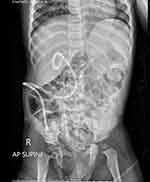
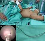
The patient is a one-year-old boy that experienced Anderson-Hynes pyeloplasty at 10 months-old due to right ureteropelvic junction (UPJ) obstruction. A 3Fr ureteric catheter was placed for internal drainage. At the time of cystourethroscopic retrieval two months later, no part of the ureteric catheter could be seen in the bladder. Plain X-ray and computed tomography of the abdomen demonstrated that the distal part of the ureteric catheter had migrated upward into the right ureter, and the tip of the catheter had been placed in the renal pelvis (Figure 1A and B). Percutaneous removal of the ureteric catheter was arranged. We determined to extract the catheter due to the risk of infection and obstruction, and decided to accomplish this procedure by percutaneous nephroscopic surgery (PNS). At the time of surgery, urine culture was negative. Prophylactic antibiotics (ceftriaxone) were administered during the anesthetic induction period and extended until the patient was discharged. The procedure was performed by an experienced urologist. To optimize patient position in association with anesthesia and simultaneous performance of retrograde surgery, the child was placed in the Galdakao-modified supine Valdivia (GMSV) position (Figure 2A), by using a small pillow below the ipsilateral flank to place him in the supine position. Thus, the right flank was elevated 20 degrees, causing the posterior calyx to project more laterally. The right leg was arranged on an extended padded stirrup, with the ankle in axis with the body, and fixed. The left leg was well abducted and flexed. The reference lines marking the anterior limit was the posterior axillary line, the cranial limit was the 12th rib, and the caudal limit was the iliac crest. Ultrasound-guided percutaneous puncture was performed by using 18 G metallic needle. Percutaneous trajectory points were to the lower pole of the right kidney. A Sensor hybrids guidewire (Boston Scientific, Natick, Massachusetts) was inserted into the pelvicalyceal system and the tract was dilated with a metallic one-step dilator. A 15/16F operating sheath was introduced and a percutaneous 12Fr nephroscope MIP-M system (Karl Storz, Germany) was used to explore the renal pelvis. The migrated ureteric catheter was caught and retrieved with a 4Fr rigid forceps (Figure 2B and C).
We attempted to insert a guidewire from the UPJ down to the ureter but failed due to extreme edema of the mucosa. The management was consequently changed to the retrograde route. The bladder was emptied to avoid compression of the ureteric orifice using a 8Fr Foley catheter. Then, a 6.5Fr rigid ureteroscopy was performed throughout the urethra and the bladder was evaluated. Another guidewire was passed under direct vision to the right ureteric orifice. The endourologist who performed the nephroscopy had to perform an antegrade ureteropyelography with a fluoroscopic dye to delineate the collecting system and ureter. When the distal end of the hybrid wire reached the renal pelvis, forceps were used to retrieve and pull the guidewire out of the kidney through the nephroscope sheath. A 4Fr double J stent (Rusch International, Kernen, Germany) was placed over the guidewire. A final fluoroscopic image was stored for correcting the stent position. Rigid ureteroscope was applied thereafter to ensure the proper placement of the distal end of double J stent in the bladder.
Finally, a 10.3F loop nephrostomy catheter was left in the renal pelvis (Figure 3) and a 8Fr Foley catheter was inserted for complete diversion of urine. The duration of the entire operation was less than 40 minutes. The patient made an uneventful postoperative recovery. Minimal gross hematuria was presented, which faded within 24 hours. We obtained KUB films to confirm that the patient retained the proper position of the double J stent and nephrostomy tube. The patient only stayed in hospital for 3 days.
 |
Figure 3 Post-operative plain X-ray with percutaneous nephrostomy and double J stent insertion. |
Discussion
Open pyeloplasty (OP) is an effective first-line treatment for UPJ obstruction with a 93.4% success rate.1 Placement of internal double-J stents is usually necessitated during dismembered type of pyeloplasty to prevent anastomotic obstruction from edema, hemorrhage, or fibrosis.2 Lack of pediatric-sized double-J stents in provincial hospitals often results in a modification using a small ureteric catheter for urinary drainage. There is a risk for an upward migration of the ureteric catheter due to the absence of a coil sitting in the bladder. In general, a stent should be removed or exchanged under general anesthesia 4–8 weeks after pyeloplasty. Stent malfunction occasionally occurs as a result of obstruction, encrustation, stent fracture, ureteral erosion, or migration, and it requires a retrieval procedure. Upward malposition of the stent is typically observed when clinical presentation occurs, unexpected findings are noted during an investigation, or when acute renal failure with urosepsis occurs. Superior migration of the double-J stent is less commonly reported, with an incidence of 2%.3 Aperistaltic segment and the expansion of the renal pelvis in patients with UPJ obstruction may induce the superior migration of the stent. UPJ obstruction associated with vesicoureteral reflux may also promote the malposition of the stent.
The optimal management of an upward ureteral stent migration is ureteroscopic removal. Another approach uses ureteric balloon dilators, a Fogarty catheter, stone forceps, and a basket.4 Shin et al5 demonstrated the use of a snare or basket to retrieve indwelling double-J stents via a percutaneous nephrostomy. In our opinion, direct vision management by using miniature nephroscopy to remove the migrated stent, as present in this report, is safer than using snare or basket via fluoroscopic guidance because blind technique can lacerate the pelvicalyceal system and cause blood clot retention. Furthermore, the percutaneous approach was more suitable for this patient, compared to the retrograde procedure, due to the very small size of the ureter in this infant patient. However, multiple concerns with PNS in pediatric patients should be mentioned. In particular, the use of large equipment in pediatric kidneys may result in renal parenchymal destruction, a decline in glomerular filtration rate, and it may invoke major complications including sepsis, hydrothorax, septicemia, and kidney hemorrhage that may require blood transfusion.6,7 Moreover, the major disadvantage of the prone position in PNS is presumably the complexities and difficulties with respiration management for the anesthesiologist. We decided to perform the procedure in the GMSV position to ensure safe access to the airway. We used a miniature nephroscope, which has the advantage of reducing morbidity, causing less kidney trauma, and lowering the risk of bleeding due to its smaller tract size.8 The use of the GMSV position can also allow the simultaneous performance of cystoscopy or ureteroscopy for an antegrade or retrograde double J stent insertion, as presented in this patient.
To our knowledge, antegrade percutaneous approach for retrieval of ureteral stents has been reported in a very limited number of patients.5,9 However, as far as we know, this is the first case report of the implementation of supine PNS using GMSV position to remove an upward migration of ureteral catheter in infancy. In the future, a large multi-center studies of upward malposition of the stent in pediatric patients are needed to further confirm the benefits of this technique.
Conclusion
This is the first presentation of a pediatric postoperative case of Anderson-Hynes pyeloplasty in which a migrated ureteric catheter was successfully removed from the renal pelvis by supine PNS in the GMSV position, with a concurrent antegrade double-J stent insertion.
Abbreviations
GMSV, Galdakao-modified supine Valdivia; PNS, percutaneous nephroscopic surgery; UPJ, ureteropelvic junction.
Ethics Approval
The present study was approved for publication by the Ethics Committee of the Faculty of Medicine Ramathibodi hospital (ID: COA. MURA2020/1623). The parents of this patient provided informed and signed consent to have the case details and any accompanying images published.
Acknowledgment
There was no research support/fund in this study.
Disclosure
None of the authors have any conflicts of interest to report for this work.
References
1. Klingler HC, Remzi M, Janetschek G, Kratzik C, Marberger MJ. Comparison of open versus laparoscopic pyeloplasty techniques in treatment of uretero-pelvic junction obstruction. Eur Urol. 2003;44(3):340–345. doi:10.1016/S0302-2838(03)00297-5
2. Smith KE, Holmes N, Lieb JI, et al. Stented versus nonstented pediatric pyeloplasty: a modern series and review of the literature. J Urol. 2002;168(3):1127–1130. doi:10.1016/S0022-5347(05)64607-1
3. Breau RH, Norman RW. Optimal prevention and management of proximal ureteral stent migration and remigration. J Urol. 2001;166(3):890–893. doi:10.1016/S0022-5347(05)65858-2
4. Menezes P, Gujral S, Elves A, Timoney A. Ureteroscopic retrieval of proximally displaced ureteric stents using triradiate grasping forceps. Br J Urol. 1998;81(5):758–759. doi:10.1046/j.1464-410x.1998.00777.x
5. Shin JH, Yoon HK, Ko GY, et al. Percutaneous antegrade removal of double J ureteral stents via a 9-F nephrostomy route. J Vasc Interv Radiol. 2007;18(9):1156–1161. doi:10.1016/j.jvir.2007.06.016
6. Ganpule AP, Mishra S, Desai MR. Percutaneous nephrolithotomy for pediatric urolithiasis. Indian J Urol. 2010;26(4):549–554. doi:10.4103/0970-1591.74458
7. Ketsuwan C, Pimpanit N, Phengsalae Y, Leenanupunth C, Kongchareonsombat W, Sangkum P. Peri-operative factors affecting blood transfusion requirements during PCNL: a Retrospective Non-Randomized Study. Res Rep Urol. 2020;12:279–285. doi:10.2147/RRU.S261888
8. Mishra DK, Agrawal MS. Use of a novel flexible mini-nephroscope in minimally invasive percutaneous nephrolithotomy. Urology. 2017;103:59–62. doi:10.1016/j.urology.2017.01.009
9. Liang HL, Yang TL, Huang JS, et al. Antegrade retrieval of ureteral stents through an 8-French percutaneous nephrostomy route. AJR Am J Roentgenol. 2008;191(5):1530–1535. doi:10.2214/AJR.07.3999
 © 2021 The Author(s). This work is published and licensed by Dove Medical Press Limited. The full terms of this license are available at https://www.dovepress.com/terms.php and incorporate the Creative Commons Attribution - Non Commercial (unported, v3.0) License.
By accessing the work you hereby accept the Terms. Non-commercial uses of the work are permitted without any further permission from Dove Medical Press Limited, provided the work is properly attributed. For permission for commercial use of this work, please see paragraphs 4.2 and 5 of our Terms.


