Back to Journals » Vascular Health and Risk Management » Volume 17
Impending Aortic Rupture in a Patient with Syphilitic Aortitis
Authors Cocora M , Nechifor D, Lazar MA, Mornos A
Received 4 November 2020
Accepted for publication 3 May 2021
Published 25 May 2021 Volume 2021:17 Pages 255—258
DOI https://doi.org/10.2147/VHRM.S289455
Checked for plagiarism Yes
Review by Single anonymous peer review
Peer reviewer comments 2
Editor who approved publication: Dr Konstantinos Tziomalos
Mioara Cocora,1 Dan Nechifor,1 Mihai-Andrei Lazar,2 Aniko Mornos2
1Department of Cardiovascular Surgery, Institute of Cardiovascular Diseases, Timisoara, Romania; 2Department of Cardiology, Institute of Cardiovascular Diseases, Timisoara, Romania
Correspondence: Mioara Cocora
Department of Cardiovascular Surgery, Institute of Cardiovascular Diseases, Timisoara, Romania
Tel +40 722 824 703
Fax +40 256 284 463
Email [email protected]
Abstract: We report the case of a 48-year-old man, admitted for atrial fibrillation with rapid heart rate and intense chest pain. A quick evaluation revealed a giant aortic aneurysm with severe aortic regurgitation and pericardial fluid without a trace of aortic dissection. Because of high suspicion of aortic rupture, an emergency surgery was planned, and a Bentall procedure was performed. On examination of the aortic wall revealing vertical wrinkling with a tree bark aspect, suspicion of syphilitic aortitis arose. The diagnosis was confirmed through postoperative serologic testing and histological examination. Histopathologic differential diagnosis, special treatment and follow-up are presented.
Keywords: tertiary syphilis, giant aortic aneurysm, aortic rupture, Bentall procedure
Introduction
Despite the efficient treatment and prophylaxis available, syphilis still exists and persists at endemic levels in low- and middle-income countries. In Europe, it remains at the rate of 2–6 cases in 100.000 inhabitants. 38% of syphilis patients are HIV positive.1
Among patients with primary syphilis 6–10% remain undiagnosed. Untreated but even previously treated syphilis could reach in 20–30 years the tertiary syphilis with neurological and cardiovascular pathology.
The three main cardiac problems caused by syphilis are thoracic aneurysm formation (30–40%), aortic valve disease (30%), coronary arteries ostial stenosis (10–20%), whereas myocardium is rarely affected directly. All of these could be the cause of severely life-threatening events.
Because this pathology occurs in patients with no history of cardiovascular diseases or risk factors, the diagnosis is generally late, leading to irreversible damage such as extended myocardial infarction,2 severe aortic insufficiency with a dilated ventricle, cardiac failure and giant aneurysms that could be sacciform with regional erosion in vertebral bodies, rib, sternum, vena cava, and skin.3,4
Owing to intense scarring of the media layer, aortic dissection is highly unlikely to occur. Fatal outcomes of such aneurysms are already known from autopsic findings.5 Necroptic studies show a mortality rate of 10%, caused by aortic rupture. Some literature reports exist about surgical treatment of aortic aneurysm and parietal hematoma.6 Here we present the case of an impending rupture of a fusiform ascending aortic aneurysm in the pericardial sac successfully treated through Bentall surgery.
Case Report
A 48-year-old Caucasian heterosexual man presented to our emergency department for atrial fibrillation with a rapid heart rate and complaints of severe chest pain. The patient had a known history of smoking and alcoholism. Clinical evaluation revealed peripheral signs of aortic regurgitation (a wide pulse pressure), cardiomegaly, and an early aortic diastolic decrescendo murmur. The blood chemistry, coagulation tests, and hemogram were unremarkable; D-dimer, CK, and troponin levels were within normal range. The viral markers tested negative for HIV 1–2, hepatitis B and C. A 6-lead ECG revealed an atrial fibrillation converted into 4 hours in sinus rhythm with Cordarone. The chest X-ray showed a significant cardiomegaly, dilated ascending aorta, and pulmonary stasis. Transthoracic echocardiography (TTE) revealed an enlarged left ventricle (DTDVS 7.9 cm, VTDVS 340 mL) with severe hypokinesis of the left ventricular wall – FE 35%, severe aortic regurgitation, central jet, and a dilated ascending aorta (7.8 cm) without aortic dissection visualized at the ascending aortic level, medium amount of liquid in the pericardial sac without any sign of cardiac tamponade. Suprasternal incidence measured an aortic arch of 3 cm. We performed an emergency computed tomographic examination, which showed a fusiform ascending aortic aneurysm measuring 78 mm of maximum diameter, extending near the origin of the brachiocephalic artery with normal diameter across the arch, descending and abdominal aorta, without significant atherosclerotic disease of supra-aortic arteries. No aortic hematoma or dissection was visible.
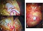
The sinus rhythm was restored under treatment with Cordarone, and the pain subsequently disappeared but re-emerged a few hours later with a rise in the amount of liquid in the pericardial sac. Under the suspicion of aortic rupture, we scheduled an emergency surgery. Extracorporeal circulation was established through axillary arterial cannulation and venous cavo-atrial cannulation. Upon opening the pericardial sac, we discovered a considerable amount of clots and fresh blood, confirming an aortic rupture. The highly dilated ascending aorta presented no dissection. The aortic aneurysm ending at a 2–3 cm distance to the brachiocephalic artery allowed aortic clamping below its origin. After opening the aorta, custodiol cardioplegia was administered into the coronary ostia. A wrinkling wall with tree bark aspect, suggesting an inflammatory aortitis (luetic), was noticeable. Coronary ostia was of normal aspect without stenosis or atherosclerotic deposit, and aortic valve was tricuspid with dilated annulus and major coaptation deficit (Figure 1A-C). The exact location of the aortic tear could not be determined upon examination. The aortic root was replaced with a 25 St. Jude valvular conduit with coronary arteries reimplantation (Bentall surgery). The distal anastomosis of the Dacron prosthesis was realized at ascending aortic level 1 cm below the brachiocephalic artery.
 |
Figure 1 (A-C) Intraoperative view of the ascending aortic aneurysm, wrinkling intima (tree bark aspect). |
The postoperative course was uneventful with a 48-hour stay in the intensive care unit requiring positive inotropic support.
Because of the aspect of the aorta, a high suspicion of luetic aetiology was raised (which was not obvious in that case taking into account the lack of well-identified risk factors). We tested the patient, and the venereal disease research laboratory test (VDRL) was intensely positive, as were the Treponema pallidum hemagglutination test (TPHA) and rapid plasma reagin (RPR).
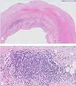
Our suspicion was confirmed through histological examination of the aortic wall, revealing obliterating endarteritis of the vasa vasorum, lymphoplasmacytic infiltrate, mesoaortitis with reduction in the musculo-elastic medial tissue (Figure 2A and B).
The polymerase chain reaction did not reveal traces of living treponemal spirochetes. In this regard, we have also analysed the cerebrospinal liquid, which is normal. The patient received intravenous penicillin for 5 weeks. Early postoperative evolution was favourable, asymptomatic, a control CT–scan one month postoperatively revealed no pathology, VDRL and TPHA tests dropped three months postoperatively.
Discussion
To promote a better understanding of this aortic disease and to enhance patient care, we have attempted to shed light on the aetiology of this aneurysm. In order to identify this aetiology, we have used the Guidelines for Diagnosing Aortic Diseases created by the Standards and Definitions Committee of the Society of Cardiovascular Pathology and the Association for European Cardiovascular Pathology.
Due to the sudden emergence of this aneurysm, it could lead to the hypothesis of a recent aneurysm with rapid growth and expansion evolving to early tendency to rupture, acting as local mass compressing adjacent structures. All of these could be considered characteristics of a mycotic aneurysm. However, the general condition of the patient does not meet the diagnosis criteria.7 The patient does not have a septic condition (bacterial endocarditis or proximal septic outbreaks) and also no predisposing factors such as severe atherosclerosis, immunodeficient patient or the patient being an intravenous drug user. Mycotic aneurysms are typically saccular, multi-lobular or eccentric, with a very low incidence of 0.65%–2% of all aortic aneurysms on the thoracic aorta – more commonly affecting the descending thoracic aorta and the aortic arch.8,9
The overall clinical condition of the patient allowed us to rule out from the beginning the most frequent genetic diseases causing an aneurysm or an ascending aortic dissection – bicuspid aortic valve, Marfan disease, Ehlers-Danlos syndrome type IV, Turner syndrome, arterial tortuosity syndrome, autosomal dominant polycystic kidney disease and autosomal recessive cutis laxa type 1.10
The general clinical examination supports this affirmation, as for the histological examination – typical lesions for this type of disease such as cystic medial degeneration, diffuse medial degeneration, laminar medial necrosis and elastic fibre fragmentation have not been found in the course of the histological examination of the aortic wall.10
But the macroscopic aspect of the aortic specimen – thickening of the wall, cracking, wrinkling of the intima as well as a tree bark appearance are features suggestive of an inflammatory aortic disease.
We therefore think about atherosclerosis and inflammatory atherosclerotic aneurysm or an aortitis. The patient did not have any other sign or risk factor for atherosclerosis. The inflammatory atherosclerotic aneurysm is associated with severe atherosclerosis and is most common in the abdominal aorta. Atherosclerosis is associated with some degree of inflammation but also the presence of fibrosis and medial loss or destruction and plaque disruption. We therefore focus our attention on other types of aortitis. Histological examinations revealed an aortitis with lymphoplasmacytic pattern which is specific for syphilis or IG4-related aortitis. Owing to the state of emergency and without a suggestive anamnesis, a proper serological investigation or coronarography was not possible. After this histo-pathological finding, we performed a proper serological examination, which confirmed the syphilitic aetiology. A special stain for spirochetes could have been obtained but such an investigation was not performed.
Serology and histology helped us bring up the diagnosis of syphilitic aortitis, which was not so obvious in that case taking into account the lack of well identified risk factors. This was certainly a requisite for a secure long-term result owing to the appropriate medical treatment. Close multidisciplinary team follow-up is necessary because of the disease’s evolutionary potential toward general, neurological or coronary complications.
Relevant literature did neither reveal the outcome of surgical interventions in aortic rupture linked to syphilitic aortitis nor regarding the size where aortic surgery is advised, but fatal outcomes of such aneurysms are already known from autopsic findings.5 No relevant research has presented this outcome of reconstructive aortic surgery given this aetiology.6
Conclusion
Syphilis has not yet disappeared. Therefore, when faced with any individuals with high risk for syphilis (transgender, sex workers, homosexuals, intravenous drug users or HIV patients) periodical, cardiological check-ups to discover cardiovascular syphilis are required in order to prevent irreversible or fatal outcomes through early surgery.11
Faced with any morphological lesion suspected for cardiovascular syphilis, serological tests are necessary to confirm the syphilitic aetiology.
Ethics Statement
To ensure that this case report meets national and international guidelines for research on humans, and that it has been conducted in accordance with the principles stated in the Declaration of Helsinki, we have consulted with the ethics committee of IBCV Timisoara, and received approval. Additionally, the consent of the patient and of all participants involved, for this information to be published, has also been granted. These statements and the approval of the ethics committee can be provided upon request.
Disclosure
The authors report no conflicts of interest in this work.
References
1. de Araujo DB, Oliveira DS, Rovere RK, de Oliveira Filho UL. Aortic aneurysm in a patient with syphilis-related spinal pain and paraplegia. Reumatologia. 2017;55:151–153. doi: 10.5114/reum.2017.68916.
2. Tanaka M, Okamoto M, Murayama T A case of acute myocardial infarction due to cardiovascular syphilis with aortic regurgitation and bilateral coronary ostial stenosis. Surg Case Rep 2, 138 (2016). doi: 10.1186/s40792-016-0267-x.
3. Asano M, Hiroshi O, Hanafusa Y, Kazuma H, Tanji M Intramural hematoma and thoracic aortic aneurysm with syphilis. 2007; J Thoracic Cardiovasc Surg, 133 (4), 1085–1086. doi: 10.1016/j.jtcvs.2006.11.042.
4. Hofmann-Wellenhof R, Domej W, Schmid C, Rossmann-Moore D, Kullnig P, Annelli-Monti M Mediastinal mass caused by syphilitic aortitis. Thorax. 1993;48(5):568–569. doi:10.1136/thx.48.5.568
5. Tsokos M Syphilitic aortic aneurysm rupture as cause of sudden death. Forensic Sci Med Pathol 8, 325–326 (2012). doi: 10.1007/s12024-011-9266-1
6. Paulo N, Cascarejo J, Vouga L Syphilitic aneurysm of the ascending aorta. Interact Cardiovasc Thorac Surg. 2012;14(2):223–225. doi:10.1093/icvts/ivr067
7. Sörelius K, Wanhainen A, Wahlgren CM, et al. Nationwide study on treatment of mycotic thoracic aortic aneurysms. Eur J Vasc Endovasc Surg. 2019;57(2):239–246. doi: 10.1016/j.ejvs.2018.08.052.
8. Sörelius K, Mani K, Björck M; European MAA collaborators, et al.. Endovascular treatment of mycotic aortic aneurysms: a European multicenter study. Circulation. 2014;130(24):2136–2142. doi:10.1161/CIRCULATIONAHA.114.009481.
9. Hsu RB, Lin FY Infected aneurysm of the thoracic aorta. J Vasc Surg. 2008;47(2):270–276. doi: 10.1016/j.jvs.2007.10.017.
10. Jain D et al. Causes and histopathology of ascending aortic disease in children and young adults. Cardiovasc Pathol. 2011;20(01):15–25. doi: 10.1016/j.carpath.2009.09.008
11. Graciaa DS, Mosunjac MB, Workowski KA, Kempker RR Asymptomatic cardiovascular syphilis with aortic regurgitation requiring surgical repair in an HIV-infected patient. Open Forum Infect Dis. 2017;4(4):ofx198. doi:10.1093/ofid/ofx198
 © 2021 The Author(s). This work is published and licensed by Dove Medical Press Limited. The full terms of this license are available at https://www.dovepress.com/terms.php and incorporate the Creative Commons Attribution - Non Commercial (unported, v3.0) License.
By accessing the work you hereby accept the Terms. Non-commercial uses of the work are permitted without any further permission from Dove Medical Press Limited, provided the work is properly attributed. For permission for commercial use of this work, please see paragraphs 4.2 and 5 of our Terms.

