")
Back to Journals » Clinical, Cosmetic and Investigational Dermatology » Volume 16
Impact of Leave-on Skin Care Products on the Preservation of Skin Microbiome: An Exploration of Ecobiological Approach
Authors Callejon S, Giraud F, Larue F, Buisson A, Mateos L, Grare L, Guyoux A, Perrier E , Ardiet N, Trompezinski S
Received 22 February 2023
Accepted for publication 25 August 2023
Published 29 September 2023 Volume 2023:16 Pages 2727—2735
DOI https://doi.org/10.2147/CCID.S409583
Checked for plagiarism Yes
Review by Single anonymous peer review
Peer reviewer comments 2
Editor who approved publication: Dr Jeffrey Weinberg
Sylvie Callejon,1,2 Félix Giraud,1,2 Florence Larue,1 Armonie Buisson,1 Léa Mateos,1,2 Laurence Grare,1 Aurélie Guyoux,1 Eric Perrier,2 Nathalie Ardiet,1 Sandra Trompezinski1,2
1NAOS Group, Research and Development Department, Aix-en-Provence, France; 2NAOS Institute of Life Science, Aix-en-Provence, France
Correspondence: Sandra Trompezinski, 355 Rue Pierre Simon Laplace, 13593 Aix-en-Provence Cedex 03, Aix-en-Provence, France, Tel +33 4 42 60 73 65, Email [email protected]
Purpose: Skincare products are used daily to maintain a healthy skin, although their skin microbiome impact is still poorly known. Preserving the natural resources and mechanisms of the skin ecosystem is essential, and a novel approach based on these premises, called ecobiology, has recently emerged in skincare. We evaluated the impact on the skin microbiome of three types of leave-on face skincare products: a hydrophilic solution, a micellar solution, and an oil-in-water emulsion.
Patients and Methods: Samples for microbial profiling were obtained from 20 Caucasian females twenty-four hours and four days following daily application of the skincare products and compared to an untreated area. The bacterial diversity and the abundance of the skin microbiome were analyzed by 16S rRNA gene sequencing using an Illumina MiSeq platform.
Results: Our results confirmed the skin microbiome diversity and the prevalence of Cutibacterium spp. and Staphylococcus spp. at sebaceous sites. The bacterial diversity and abundance were not affected by the products, and no dissimilarities versus the control nor between each product were noted at both times.
Conclusion: These preliminary results demonstrate for the first time that three types of leave-on face skincare products have no impact on the human skin microbiome and can be considered to be “microbiome friendly”.
Keywords: cosmetic product, ecobiology, microbiome friendly
Introduction
The skin is an ecosystem at the interface with the physical environment, and its primary role is to serve as a physical barrier, cooperating with chemical defenses (pH, antimicrobial peptide synthesis, etc.) to protect the body from external aggressions. Recent studies have shown that the outer layer of the skin is intimately linked to its microbial communities, the skin microbiome, effectively representing the first line of skin defense, and disruptions in its balance can result in skin disorders and infections.1 The human skin microbiome comprises fungi, viruses, and bacteria, particularly four major phyla: Actinobacteria, Proteobacteria, Firmicutes, and Bacteroidetes. The microbiome is essential for the skin’s equilibrium. Indeed, the cutaneous microbiome protects the skin from pathogen colonization (by competition and surface occupation, acidic pH, and antimicrobial peptide synthesis), and it interacts with the cutaneous immune system in order to educate it to tolerate its environment.2 Preservation of the skin’s immune system is paramount, and recent evidence has underscored its dependency on the resident microbiota for both host defense and tissue repair.3,4 The skin microbiome composition is highly heterogeneous, as it exhibits intrapersonal variations depending on the location on the body, gender, age, ethnicity, and the environment (climate, stress, diet, lifestyle, drugs, hygiene habits, etc.).5–7 The variability of the microbial composition according to the body sites is influenced by physiological characteristics.1,8 Three typical broad microenvironment types have been identified: sebaceous sites (such as the forehead, the face, and the back), which are mainly composed of Propionibacteriaceae including Cutibacterium spp. and Staphylococcaceae; moist sites (such as the armpits, the inguinal crease, and the popliteal and antecubital fossae), exhibiting a prevalence of Corynebacteriaceae and to a lesser extent Staphylococcaceae; and dry areas (such as the forearm), with a greater prevalence of β-Proteobacteria and Flavobacteria.8 Interpersonal variations of the skin microbiome are also observed for a given site.8
The biodiversity of the skin microbial ecosystem can be directly linked to the skin’s overall health, since skin diseases, such as atopic dermatitis and psoriasis, are often associated with dysbiosis.6 Thus, maintaining the ecological balance of the skin is essential in dermatology, and this can be considered to be the ultimate aim of ecobiology. Ecobiology is an original approach, considering the skin as an ever-evolving ecosystem that hosts human and microbial cells that interact together with their environment and for which the natural resources and mechanisms must be preserved.9,10 This approach applies particularly to skincare products, which play a fundamental role in dermatological treatments for healthy as well as diseased skin.11 In this context, thanks to the creation of a 3D molecular topographical map to visualize both the chemical and the microbial compositions of the skin human surface, Bouslimani et al succeeded in showing that components of skincare products remained on the skin for weeks after the first use, despite regular showering.12,13 These components inevitably have an impact on the microbial ecosystem. Indeed, Bouslimani et al demonstrated an increase in bacterial diversity with deodorant and foot powder use after nine weeks of application, while there was a minor effect of face sunscreens and moisturizers on the forearm.13 Two other studies have reported that deodorants and antiperspirants affect the axillary microbiota by increasing its diversity, and their daily use was shown to shift the microbiome into a stable composition and density by leading to the establishment of a new ecological balance.14,15 Similarly, the use of make-up (foundation and powder) or basic cosmetics (skin softener, lotion, essence, and cream) have been reported to increase the microbial diversity on the forehead16 and on the cheeks,17 respectively. As far as skin cleansers are concerned, bacterial communities appear to reestablish rapidly. Fierer et al showed alteration of microbial communities due to hand washing, but the overall levels of bacterial diversity were unrelated to the time since the last hand washing.18 Moreover, two other studies demonstrated that short-term use of skin cleansers did not significantly affect the cutaneous microbiome in terms of diversity and abundance.19,20 One study has suggested that some cleansers can even increase the bacterial diversity and richness after 2 and 4 weeks.21 Furthermore, Perez et al have suggested that changing of soaps and shampoos may not have substantial impacts on the microbial ecology.22
Therefore, despite the increasing awareness of the impact of cosmetic products on the skin ecosystem,23–26 few studies have been performed to date on leave-on face skincare products applied daily to healthy skin. The aim of this study was to investigate the impact of three types of leave-on face skin care products (a hydrophilic sterile solution, a micellar solution, and an emulsion) on the skin microbiome diversity and abundance.
Materials and Methods
Subject Recruitment and Sample Collection
Twenty Caucasian subjects between 24 to 46 years of age (mean age 34.6 years) with phototype II to IV were recruited at the Cutaneous Investigation and Research Center in Lyon (France) by an advertisement on its website and from its database. Participants with the following characteristics were excluded: (1) pregnant or lactating women, (2) smokers, (3) post-menopausal women, (4) began or had a modified hormonal treatment within three months, (5) had an auto-immune, severe, or progressive disease, (6) had allergy or hypersensitivity to cosmetics, (7) had cutaneous disease on the experimental area, (8) had a sensitive skin, (9) had acne on their back, (10) had diabetes, (11) or had a treatment (antibiotics, anti-inflammatory, anti-acne drugs, etc.) more than five consecutive days within the past four weeks. The subjects were required to avoid excessive sun exposure during the previous month and use of a swimming pool for a period of 14 days before the study. They were asked to use a generic neutral shampoo and soft cleanser and to otherwise avoid any other product application on their back starting 14 days before and during the study All subjects provided written informed consent before participating. All protocols and procedures used in this study were conducted according to the Declaration of Helsinki. The study did not require approval from local ethics committees prior to inclusion of volunteers according to the local legal requirements (Loi Jardé) for non-invasive investigations of cosmetic products on healthy subjects.
Only women were included, in order to rule out gender differences in the ecosystem of the skin microbiome.13,18 Three different cosmetic products were applied once daily on their back over an area of 15 cm2 at 1.5 μL/cm2 by the same investigator from Day 0 to Day 3. Four areas were defined on the back of each subject for application of: (1) a hydrophilic sterile solution (Eau Cellulaire Mist, Institut Esthederm, NAOS), (2) a micellar solution with surfactants and a preservative (Sensibio H20, Laboratoire Bioderma, NAOS), (3) an oil-in-water emulsion with emulsifiers (B23 Crème fondante hydratante, Etat Pur, NAOS) and, (4) a control without any product application. The swab samples were collected three times: just before the use of the products, on Day 1, and on Day 4. The samples were collected using pre-moistened swabs in sterile-filtered and nuclease-free water, BioPerformance Certified (Sigma-Aldrich, St. Quentin Fallavier, France) by the same investigator and following the same swabbing process to prevent possible variation between samples. Each swab head was placed in a Sigma-Transwab® tube containing 1 mL of liquid Amies medium (Medical Wire & Equipment Co., Wiltshire, UK) for 30 min and was then frozen at −18°C until DNA extraction.
Swab DNA Extraction
DNA was extracted from the swabs with an optimized and standardized protocol devised for extraction of bacterial DNA from human samples. An optimized bacterial recovery step from the swabs was added using a NucleoSpin™ Microbial DNA kit (Macherey Nagel, France). After DNA extraction, the DNA concentration was amplified by PCR.
16S rRNA Gene Amplification and Sequencing
Microbial diversity was determined for each sample by targeted amplification of part of the 16S ribosomal gene. A 16S rRNA gene fragment comprising V1 and V3 hypervariable regions was amplified using an optimized and standardized 16S-amplicon-library preparation protocol (Metabiote®, GenoScreen, Lille, France). Briefly, 16S rRNA gene PCR was carried out using the maximal volume of genomic DNA according to the Metabiote® protocol instructions using 192 bar-coded primers (Metabiote® MiSeq Primers, GenoScreen, Lille, France) at final concentrations of 0.2 μM and an annealing temperature of 50°C for 30 cycles. The PCR products were purified with an Agencourt AMPure XP-PCR Purification system (Beckman Coulter, Brea, USA), quantified according to the manufacturer’s protocol, and multiplexed at equal concentrations. Sequencing was performed using a 300-bp paired-end sequencing protocol on an Illumina MiSeq platform (Illumina, San Diego, USA).
Sequence Processing and Analysis
Raw paired-end reads were subjected to the following process: (1) quality filtering with the PRINSEQ-lite PERL script27 by truncation of bases from the 3’ end not with quality < 30 based on the Phred algorithm, (2) paired-end read assembly using fast length adjustment of short reads (FLASH) to improve genome assemblies28 with a minimum length overlap of 30 bases and 97% overlap identity, and (3) search and removal of both forward and reverse primer sequences using CutAdapt, with no mismatches allowed in the primer sequences. Assembled sequences for which perfect forward and reverse primers were not found were eliminated. Sequences were clustered into operational taxonomic units (OTUs) with a 97% identity threshold. Taxonomic assignment was performed using the Ribosomal Database Project (RDP) classifier on the Greengenes database V13_8.29 A total of 323 swabs from volunteers were collected and used for the analysis. Unfortunately, 141 samples could not be analyzed because they presented a profile similar to the background in negative controls corresponding to an insufficient quantity of DNA. Since the taxa Escherichia was identified as background in negative controls, a threshold of 15% of Escherichia reads was applied. The samples with greater relative abundance were discarded from the study, reducing our dataset to 182 samples shared between eight volunteers on Day 1 and nine volunteers on Day 4.
Data and Statistical Analysis
The analysis was performed with R software. The Shannon–Weaver index, the number of observed OTUs, and the Bray–Curtis distance matrix were computed with the Phyloseq package.30 Linear discriminant analysis (LDA) effect size (LEfSe) analysis31 was performed on the galaxy web server. Only genera with an LDA score above 3.0 were included. Statistical significance was determined using the Wilcoxon rank-sum test. A p-value < 0.05 was considered statistically significant.
Results
16S rRNA Gene Amplicon Sequencing
A total of 8,911,437 sequencing reads were generated by sequencing 323 specimen samples from nineteen volunteers (one volunteer dropped out of the study before completion). Following trimming and quality control, 6,797,231 reads of 16S rRNA bacterial sequences were obtained for further analysis. Because biodiversity indexes (the Shannon–Weaver index and the observed OTUs) are impacted by the number of reads,32 samples were normalized at a read depth of 3100 reads per sample.
α-Diversity Analysis Between the Three Products
The number of observed OTUs found in a sample is a standard indicator of the diversity. Application of the hydrophilic solution, the micellar solution, or the emulsion did not significantly change the number of observed OTUs compared to the control on Day 1 or Day 4 (Figure 1A and B). In order to analyze the variations in abundance and the number of observed OTUs, the Shannon–Weaver index was computed for each sample and compared to the controls. Using the Shannon–Weaver index, no significant differences were found between the hydrophilic solution, the micellar solution, and the emulsion compared to their controls (paired Wilcoxon test, p > 0.05) on Day 1 or on Day 4 (Figure 1C and D). However, the Shannon–Weaver index was increased slightly on Day 4 by application of the micellar solution. Since there were not enough subjects in common between Day 1 and Day 4, no statistical comparison was calculated.
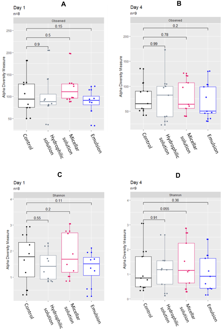 |
Figure 1 Bacterial diversity using observed OTUs and the Shannon–Weaver index of bacterial 16S rRNA gene sequencing reads after 24 hours (Day 1) and 4 days (Day 4) of application of the hydrophilic solution, the micellar solution, or the emulsion compared to the control at each time. (A) Number of observed OTUs on Day 1; (B) number of observed OTUs on Day 4; (C) Shannon–Weaver index on Day 1; (D) Shannon–Weaver index on Day 4. The p-value was calculated with a paired Wilcoxon test. |
Relative Abundance Analysis Between the Three Products
The relative abundance of the fifteen most abundant genera, representing 94%–96% of the total number of reads (Figure 2) and at the phylum level, demonstrated no major alteration of the skin microbiome after use of the products compared to their controls. The LEfSe, analyzing significant changes at the genus level, demonstrated no significant modification for the hydrophilic solution, the micellar solution, or the emulsion on Day 4 (data not shown).
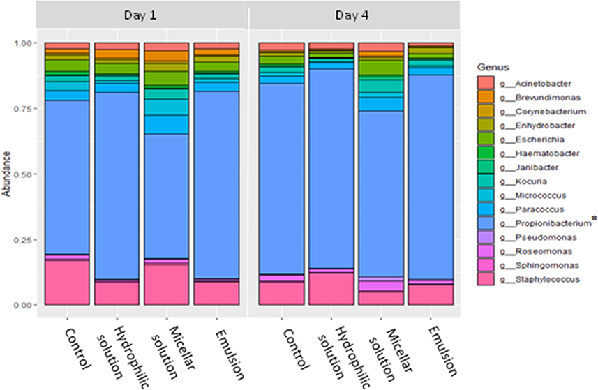 |
Figure 2 Mean relative abundances of the 15 most represented bacterial taxa at the genus level after 24 hours (Day 1) and 4 days (Day 4) of application of the hydrophilic solution, the micellar solution, or the emulsion compared to their controls. *Superseded genus name for Cutibacterium. |
Dissimilarity Analysis Between the Three Products
The dissimilarity between each product and the controls was investigated using the Bray–Curtis distance between each product and their control. The Bray–Curtis distances to the control were then compared between the hydrophilic solution, the micellar solution, and the emulsion. No significant differences were observed on Day 1 (Figure 3A) or Day 4 (Figure 3B). However, the three products displayed low distances overall compared to their controls, suggesting that the hydrophilic solution, the micellar solution, and the emulsion had no impact on the skin microbiome.
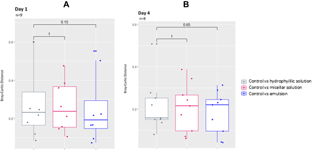 |
Figure 3 Bray–Curtis distances after 24 hours (Day 1) and 4 days (Day 4) of application of the hydrophilic solution, the micellar solution, or the emulsion versus their controls. (A) Bray–Curtis distances on Day 1; (B) Bray–Curtis distances on Day 4. The p-value was calculated with a paired Wilcoxon test. |
Discussion
Firstly, the results of this study confirmed a high degree of skin microbiome diversity, associated with Cutibacterium and Staphylococcus as the most dominant genera, on sebaceous sites such as the back or the face. This is in line with previous observations.3,8 Secondly, the hydrophilic solution, the micellar solution, and the emulsion did not have a negative impact on the bacterial diversity or abundance, and this was observed consistently compared with the control after one application (Day 1) as well as after repeated daily application over a period of four days (Day 4). Thus, the three skincare products preserve the skin microbial ecological balance and can be considered to be “microbiome-friendly” skincare products. Preserving the microbial biodiversity is essential to maintain the natural protection of the skin against pathogens and skin barrier function and to prevent or offset immune system disorders.6 Indeed, it is well established that patients affected by psoriasis or atopic dermatitis have a lower microbial diversity on plaques than on unaffected skin.33,34 Maintaining the relative abundance of the most represented taxa is also a challenge for ecobiological skincare products. In addition to altering the skin physiology and consequently the skin microbiome environment, the application of skincare products can also release new components on the skin, which can be beneficial for underrepresented taxa.17 Thirdly, no dissimilarity was observed between the products and their controls, nor between each product, despite their different overall compositions linked to their functions: the micellar solution, a leave-on cleanser with a surfactant and a preservative; the hydrophilic solution, a sterile sprayable water; and the emulsion, a moisturizing oil-in-water emulsion with emulsifiers.
Regarding the composition of the products, the hydrophilic solution is a filtered sterile formula at pH 6 filled in an airless aerosol devoid of any preservative ingredients. It also exhibits bioelectrical characteristics including physiological osmotic pressure. Therefore, we expected that the hydrophilic solution would have no impact on the skin microbiome when applied on the skin. The micellar solution and the emulsion’s formulae have pH values between 4.7 and 5.5, which simultaneously helps protect the formula from microbial contamination and corresponds to the physiological pH of skin. However, since the formulae of these two products are likely to come into contact with air, suitable cosmetic ingredients are added to avoid potential microbial contamination upon use by the consumer, as required by cosmetic regulations. Moreover, the micellar solution, as a cleanser, also contains a mild surfactant to solubilize dirt and, inevitably, microbes. Yet our results showed no impact on the microbiome profile after application of the micellar solution or the emulsion, thus confirming the importance of the ingredient choice in terms of the number (limited ingredients), concentration (in accordance with efficacy and toxicological requirements), and purity (very high-quality ingredients and reduced levels of potentially harmful trace elements such as heavy metals).
Our study was performed on the back of subjects, as this area provides a larger area for application of the three skincare products, and, as such, there is ample room for a suitable control area. Moreover, sufficient microbial material for DNA amplification can be recovered from the back. In addition, the back is also subject to limited exposure to cosmetic products, which, as previously mentioned, influences the number and the species of bacteria present on the face. For this reason, although the investigated cosmetic products are generally applied on the face, we chose to perform sampling on the back, which presents a microbial composition and physiological characteristics comparable to those of the face.1 Moreover, it was presumed that the back, which is generally less exposed to external factors, may provide a more sensitive site to evaluate the effects of cosmetic products on the skin microbiome. These preliminary results should nonetheless be confirmed by controlled application on the face and also under normal conditions of use, ie, in the context of application twice a day by the consumer.
As far as we know, this study represents the first assessment under controlled conditions of the impact of leave-on skin cleansers on the skin microbiome. Similarly to our results for the micellar solution on Day 1, comparable studies evaluating rinsed skin cleansers have shown that bacterial communities appear to rapidly regain their diversity and abundance following application of a skin product.18–20 Since the micellar solution is left on, like the hydrophilic solution and the emulsion, we also evaluated the skin microbiome on Day 4, and no significant changes in the bacterial diversity or abundance were observed. As far as the hydrophilic solution and the emulsion are concerned, Lee et al demonstrated that the application twice a day on thirty Korean females of basic cosmetics consisting of a skin softer (solubilized type), a lotion (oil in water), an essence (solubilized type), and a cream (oil-in-water emulsion type) for four weeks appeared to increase the microbial diversity on the cheeks.17 Therefore, the use of these basic cosmetic products appeared to positively impact the skin microbiome’s equilibrium. The discordant results observed in our study may be partially explained by the different experimental conditions tested, the quality of the ingredients, and the simplicity of the formula. Lee et al performed their experiments on the cheeks, with a larger sample group and a longer application under conditions of normal use. Moreover, several cosmetic products were applied sequentially. To the best of our knowledge, only one study to date has been performed with a moisturizing emulsion. This analysis demonstrated a moderate increase in bacterial diversity after daily application of the emulsion on the front forearms of twelve subjects over a period of three weeks.13 Another study, evaluating an essence skincare product on the face of twenty-five Korean women over a period of four weeks also found an increase in bacterial diversity after a twice-daily application, but no control condition without application was included.35 The results of these two studies suggest potential improvement of the microbial health of facial skin. In contrast, we observed no changes in the bacterial diversity or abundance after daily application of the emulsion over a period of 4 days compared to the control conditions. These preliminary results should, therefore, be confirmed by assessment of daily application over a longer timeframe (28–30 days) and by increasing the sample size to evaluate temporal diversity.
This study was limited to 16S rRNA sequencing and bacterial identification. For a more in-depth analysis, further investigations should use shotgun sequencing to explore microbiome variations after the application of cosmetic products.
Conclusion
In conclusion, these preliminary results show that a hydrophilic solution, a micellar solution, and an emulsion, all formulated with selected cosmetic ingredients by an ecobiological approach, did not significantly alter the skin microbiome balance over several days of application. This study lays the foundations for a new way of formulating skincare products whereby the unique skin microbiome profile of each individual is determined in order to cater to individual skin needs. Indeed, our ecobiological values have consistently demonstrated that, rather than merely being a barrier against external aggressions, the skin is an interface between two alive worlds.
Acknowledgments
Drafting of the article was assisted by Marlène Chavagnac-Bonneville and Jacopo Novelli (NAOS Group, France).
Funding
This research was funded by NAOS Group (Aix-en-Provence, France).
Disclosure
Sylvie Callejon, Félix Giraud, Florence Larue, Armonie Buisson, Léa Mateos, Laurence Grare, Aurélie Guyoux, Eric Perrier, Nathalie Ardiet, and Sandra Trompezinski are employees of NAOS Group, France.
References
1. Grice EA, Segre JA. The skin microbiome. Nat Rev Microbiol. 2011;9(4):244–253. doi:10.1038/nrmicro2537
2. Chen YE, Tsao H. The skin microbiome: current perspectives and future challenges. J Am Acad Dermatol. 2013;69(1):143–155. doi:10.1016/j.jaad.2013.01.016
3. Fournière M, Latire T, Souak D, Feuilloley MGJ, Bedoux G. Staphylococcus epidermidis and cutibacterium acnes: two major sentinels of skin microbiota and the influence of cosmetics. Microorganisms. 2020;8(11):1–31. doi:10.3390/microorganisms8111752
4. Belkaid Y, Tamoutounour S. The influence of skin microorganisms on cutaneous immunity. Nat Rev Immunol. 2016;16(6):353–366. doi:10.1038/nri.2016.48
5. Schommer NN, Gallo RL. Structure and function of the human skin microbiome. Trends Microbiol. 2013;21(12):660–668. doi:10.1016/j.tim.2013.10.001
6. Baldwin HE, Bhatia ND, Friedman A, Martin R, Seité S. The role of cutaneous microbiota harmony in maintaining a functional skin barrier. J Drugs Dermatol. 2017;16(1):12–18.
7. Dimitriu PA, Iker B, Malik K, Leung H, Mohn WW, Hillebrand GG. New insights into the intrinsic and extrinsic factors that shape the human skin microbiome. MBio. 2019;10(4):e00839–19. doi:10.1128/mBio.00839-19
8. Grice EA, Kong HH, Conlan S, et al. Topographical and temporal diversity of the human skin microbiome. Science. 2009;324(5931):1190–1192. doi:10.1126/science.1171700
9. Dréno B. The microbiome, a new target for ecobiology in dermatology. Eur J Dermatol. 2019;29(S1):15–18.
10. Radman M. Ecobiological approach to research regarding ageing and diseases. Eur J Dermatol. 2019;29(S1):11–14.
11. Polena H, Chavagnac-Bonneville M, Sayag M. Improvement of Quality of Life in Dialysis and Diabetic Patients by Skin Dryness and Pruritus Management with an Ecobiological Dermo-Cosmetic Product. Clin Cosmet Investig Dermatol. 2022;6(15):2143–2152. doi:10.2147/CCID.S375472
12. Bouslimani A, Porto C, Rath CM, et al. Molecular cartography of the human skin surface in 3D. Proc Natl Acad Sci U S A. 2015;112(17):E2120–9. doi:10.1073/pnas.1424409112
13. Bouslimani A, Da Silva R, Kosciolek T, et al. The impact of skin care products on skin chemistry and microbiome dynamics. BMC Biol. 2019;17(1):47. doi:10.1186/s12915-019-0660-6
14. Urban J, Fergus DJ, Savage AM, et al. The effect of habitual and experimental antiperspirant and deodorant product use on the armpit microbiome. PeerJ. 2016;4:e1605. doi:10.7717/peerj.1605
15. Callewaert C, Hutapea P, Van de Wiele T, Boon N. Deodorants and antiperspirants affect the axillary bacterial community. Arch Dermatol Res. 2014;306(8):701–710. doi:10.1007/s00403-014-1487-1
16. Staudinger T, Pipal A, Redl B. Molecular analysis of the prevalent microbiota of human male and female forehead skin compared to forearm skin and the influence of make-up. J Appl Microbiol. 2011;110(6):1382–1389. doi:10.1111/j.1365-2672.2011.04991.x
17. Lee HJ, Jeong SE, Lee S, Kim S, Han H, Jeon CO. Effects of cosmetics on the skin microbiome of facial cheeks with different hydration levels. Microbiologyopen. 2018;7(2):e00557. doi:10.1002/mbo3.557
18. Fierer N, Hamady M, Lauber CL, Knight R. The influence of sex, handedness, and washing on the diversity of hand surface bacteria. Proc Natl Acad Sci U S A. 2008;105(46):17994–17999. doi:10.1073/pnas.0807920105
19. Two AM, Nakatsuji T, Kotol PF, et al. The Cutaneous Microbiome and Aspects of Skin Antimicrobial Defense System Resist Acute Treatment with Topical Skin Cleansers. J Invest Dermatol. 2016;136(10):1950–1954. doi:10.1016/j.jid.2016.06.612
20. Numata S, Akamatsu H, Akaza N, et al. Quantitative effect of face washing on cutaneous resident microbiota in female subjects who wear make-up. J Dermatol. 2012;39(12):1100–1101. doi:10.1111/j.1346-8138.2012.01644.x
21. Wallen-Russell C. The role of every-day cosmetics in altering the skin microbiome: a study using biodiversity. Cosmetics. 2019;6(1):2. doi:10.3390/cosmetics6010002
22. Perez GIP, Gao Z, Jourdain R, et al. Body site is a more determinant factor than human population diversity in the healthy skin microbiome. PLoS One. 2016;11(4):e0151990. doi:10.1371/journal.pone.0151990
23. Holland KT, Bojar RA. Cosmetics: what is their influence on the skin microflora? Am J Clin Dermatol. 2002;3(7):445–449. doi:10.2165/00128071-200203070-00001
24. Beri K. Skin microbiome & host immunity: applications in regenerative cosmetics & transdermal drug delivery. Futur Sci OA. 2018;4(6):FSO302. doi:10.4155/fsoa-2017-0117
25. Sfriso R, Egert M, Gempeler M, Voegeli R, Campiche R. Revealing the secret life of skin with the microbiome you never walk alone. Int J Cosmet Sci. 2022;42(2):116–126. doi:10.1111/ics.12594
26. Carvalho MJ, Oliveira ALS, Pedraosa SS, Pintado M, Pinto-Ribeiro I, Madureira AR. Skin Microbiota and the Cosmetic Industry. Microb Ecol. 2022;86(1):86–96. doi:10.1007/s00248-022-02070-0
27. Schmieder R, Edwards R. Quality control and preprocessing of metagenomic datasets. Bioinformatics. 2011;27(6):863–864. doi:10.1093/bioinformatics/btr026
28. Magoč T, Salzberg SL. FLASH: fast length adjustment of short reads to improve genome assemblies. Bioinformatics. 2011;27(21):2957–2963. doi:10.1093/bioinformatics/btr507
29. DeSantis TZ, Hugenholtz P, Larsen N, et al. Greengenes, a chimera-checked 16S rRNA gene database and workbench compatible with ARB. Appl Environ Microbiol. 2006;72(7):5069–5072. doi:10.1128/AEM.03006-05
30. McMurdie PJ, Holmes S Phyloseq: an R package for reproducible interactive analysis and graphics of microbiome census data. PLoS one. 2013;8(4):290 e61217.
31. Segata N, Izard J, Waldron L, et al. Metagenomic biomarker discovery and explanation. Genome Biol. 2011;12(6). doi:10.1186/gb-2011-12-6-r60
32. Morris EK, Caruso T, Buscot F, et al. Choosing and using diversity indices: insights for ecological applications from the German Biodiversity Exploratories. Ecol Evol. 2014;4(18):3514–3524. doi:10.1002/ece3.1155
33. Tett A, Pasolli E, Farina S, et al. Unexplored diversity and strain-level structure of the skin microbiome associated with psoriasis. npj Biofilms Microbiomes. 2017;3(1):1–12. doi:10.1038/s41522-017-0022-5
34. Byrd AL, Deming C, Cassidy SKB, et al. Staphylococcus aureus and Staphylococcus epidermidis strain diversity underlying pediatric atopic dermatitis. Sci Transl Med. 2017;9(397):eaal4651. doi:10.1126/scitranslmed.aal4651
35. Hwang BK, Lee S, Myoung J, et al. Effect of the skincare product on facial skin microbial structure and biophysical parameters: a pilot study. Microbiologyopen. 2021;10(5):e1236. doi:10.1002/mbo3.1236
 © 2023 The Author(s). This work is published and licensed by Dove Medical Press Limited. The full terms of this license are available at https://www.dovepress.com/terms.php and incorporate the Creative Commons Attribution - Non Commercial (unported, v3.0) License.
By accessing the work you hereby accept the Terms. Non-commercial uses of the work are permitted without any further permission from Dove Medical Press Limited, provided the work is properly attributed. For permission for commercial use of this work, please see paragraphs 4.2 and 5 of our Terms.
© 2023 The Author(s). This work is published and licensed by Dove Medical Press Limited. The full terms of this license are available at https://www.dovepress.com/terms.php and incorporate the Creative Commons Attribution - Non Commercial (unported, v3.0) License.
By accessing the work you hereby accept the Terms. Non-commercial uses of the work are permitted without any further permission from Dove Medical Press Limited, provided the work is properly attributed. For permission for commercial use of this work, please see paragraphs 4.2 and 5 of our Terms.