Back to Journals » International Journal of General Medicine » Volume 15
Identification of the Potential Key Genes and Pathways Involved in Lens Changes of High Myopia
Received 24 December 2021
Accepted for publication 1 March 2022
Published 10 March 2022 Volume 2022:15 Pages 2867—2875
DOI https://doi.org/10.2147/IJGM.S354935
Checked for plagiarism Yes
Review by Single anonymous peer review
Peer reviewer comments 2
Editor who approved publication: Dr Scott Fraser
Weixia Lai,1,2 Xixi Wu,2 Hao Liang1
1Department of Ophthalmology, First Affiliated Hospital of Guangxi Medical University, Nanning, Guangxi Zhuang Autonomous Region, People’s Republic of China; 2Department of Ophthalmology, First Affiliated Hospital of Guangxi Traditional Chinese Medical University, Nanning, Guangxi Zhuang Autonomous Region, People’s Republic of China
Correspondence: Hao Liang, Department of Ophthalmology, First Affiliated Hospital of Guangxi Medical University, 6 Shuangyong Road, Qingxiu District, Nanning, People’s Republic of China, Email [email protected]
Aim: High myopia (HM) is a global problem; however, the molecular pathogenesis of HM underlying lens remains largely unknown. The aims of the present study were to identify the potential key genes and pathways involved in lens changes of HM.
Methods: Gene set enrichment analysis was carried out to identify the HM-specific pathway gene sets. The differentially expressed genes (DEGs) in lens epithelia of HM eyes compared to emmetropic control were screened using limma R package. A DEG-based protein–protein interaction network was constructed and used to identify hub genes and gene cluster analysis. The functional enrichment analysis was performed to reveal the potential biological functions for each gene cluster.
Results: Multiple metabolism-related pathways were significantly enriched in lens epithelia of HM. The expression patterns of DEGs could accurately distinguish HM and emmetropic and CD34, CD40, EGF, IL1A, CD40LG, and CXCL12 maybe the potential key genes involved in HM. Three gene clusters were identified and involved in distinct pathways. MAPK signaling pathway and calcium signaling pathway were considered the key pathways involved in lens changes of HM, due to two gene clusters both involve in these two pathways.
Conclusion: We identified potential key genes in pathological lens growth of HM eyes and proposed that the imbalances of MAPK signaling pathway and calcium signaling pathway may be the two crucial steps of pathological lens growth in HM.
Keywords: high myopia, lens epithelia, hub genes, MAPK signaling pathway, calcium signaling pathway
Introduction
High myopia (HM) is estimated to affect near 10% of the global population in 2050, a leading cause of blindness worldwide.1,2 Various mechanisms in myopia progression was reported in previous studies.3–6 Similar to other complex diseases, myopia is considered result from the interaction of genetic and environmental factors.7 Several biological pathways, including ion transport, neurotransmission, retinoic acid metabolism, eye development and extracellular matrix remodelling, partially explained mechanism of a retina to sclera signalling cascade in myopia.8 In addition, these previous studies on HM mainly focused on the genome (eg genome-wide association studies and single nucleotide polymorphism).9,10 However, the underlying molecular dysregulation of HM in transcriptome remain largely unknown.
Lens is the core refracting medium of human visual system and responsible for the full range of vision. A previous study11 found that patients with HM may have higher rate of lens diseases. Yet despite the lens diameter of HM eyes was considered larger and associated with the TGF-β1-Smad signaling.12
It is not clear whether these lens changes are causal or merely markers for HM. It is urgently needed to propose more potential mechanisms. The development of high-throughput sequencing technology and bioinformatics has greatly accelerated our understanding of human diseases.13–15 Studied of mining and re-analysis of these big-data also saves research expenditure. Interestingly, HM-related high-throughput data and bioinformatics research are few. Herein, we used a HM-related data set to perform bioinformatics analysis to reveal the potential key genes and pathway in lens epithelia of HM.
Methods and Materials
Data Processing
The HM-related data set GSE13670112 was downloaded from Gene Expression Omnibus (https://www.ncbi.nlm.nih.gov/geo/). GSE136701 contains gene expression profiles based on the GPL570 of lens epithelia from three human highly myopic eyes and three emmetropic control eyes and was based on the GPL570 platform. In our present study, the probes corresponding to multiple genes are deleted. If a gene corresponds to multiple probes, the average value of these probes is considered as the expression value of the gene. As these data are publicly available and open-access, ethical approval from the ethics committee of First Affiliated Hospital of Guangxi Traditional Chinese Medical University was not necessary for the present study.
Gene Set Enrichment Analysis (GSEA)
GSEA16,17 was carried out using the GSEA java software to identify the HM-specific Kyoto Encyclopedia of Genes and Genomes (KEGG) pathways. The canonical pathways gene sets derived from the KEGG pathway database were download from the Molecular Signatures Database (version 7.4).17,18 Gene set with the nominal P value < 0.05 was considered significantly enriched in HM.
Gene Differential Expression Analysis and Cluster Analysis
To identify the potential HM-specific genes, we screened the differentially expressed genes (DEGs) in lens epithelia of HM compared to emmetropic control using limma19 package. We considered genes with a |log2 (fold change)|>1 and a P value < 0.05 to be significant. Subsequently, we performed bidirectional hierarchical clustering to the optimal DEGs based on Euclidean distance and displayed the results as a heat map using the pheatmap [https://CRAN.R-project.org/package=pheatmap] package.
Protein–Protein Interaction (PPI) Network Analysis
The PPI networks of the DEGs were constructed in the STRING database (version 11.5)20 and visualized using the Cytoscape software.21 The interactions with medium confidence (combine score > 0.4) were included in our present study. Then, we separately identified the hub genes using three methods (degree,22 Maximum Neighborhood Component (MNC),23 and Maximal Clique Centrality (MCC)24) using the cytoHubba24 plug-in of Cytoscape. Additionally, we used the Cytoscape plug-in ClusterONE25 to predict gene clusters based on a cohesion algorithm and nearest neighbor selection.
Functional Enrichment Analysis
In order to preliminary reveal the biological functions of each gene cluster, we performed functional enrichment analysis using the clusterProfiler26 package. The P adjusted by Benjamini & Hochberg <0.05 was considered significant.
Results
High Metabolism Tendency Found in Lens Epithelia of HM
The GSEA results indicated that multiple metabolism-related pathways were significantly enriched in lens epithelia of HM, including galactose metabolism (Figure 1A), pyruvate metabolism (Figure 1B), insulin signaling pathway (Figure 1C), glycolysis gluconeogenesis (Figure 1D), neurotrophin signaling pathway (Figure 1E), and purine metabolism (Figure 1F).
Multiple Genes Differentially Expressed in HM Compared to Emmetropic
A total of 873 genes were considered as DEGs according to our cutoff criteria, including 426 up-regulated genes and 447 down-regulated genes (Figure 2A). The top 10 (ranked by FC) up-regulated genes in HM were USP9Y, CRYBB1, PALM2, HTATSF1P2, CTXN3, LGSN, TMEM130, SFTA3, CRYBA4, and CRYBA2, while the top 10 (ranked by FC) down-regulated genes in HM were LINC00314, RP1-28C20.1, CNGA4, RP11-327J17.2, CBLN1, LINC00682, LOC102724718, HIST1H2BK, BLACAT1, and LOC100506122. The results of clustering analysis indicated the expression patterns of these DEGs could accurately distinguish HM and emmetropic (Figure 2B).
Identification of Potential Hub Genes Involving in HM from PPI Networks
A PPI networks of the DEGs, including 477 nodes and 1017 edges, were constructed using STRING database (Figure 3). This suggested that HM results from the synergy of multiple dysregulated genes. The ten hub genes identified by degree method were LCK, FGR, SHC1, EGF, CAMK2A, CD40, CD34, IL1A, CD40LG, and CXCL12 (Figure 4A). The ten genes identified by MCC method were IL33, CD34, IFNB1, CCL11, CD40, CXCR2, EGF, IL1A, CD40LG, and CXCL12 (Figure 4B). The ten genes identified by MNC method were VAV1, CD34, SHC1, CCL11, CD40, LCK, EGF, IL1A, CD40LG, and CXCL12 (Figure 4C). Thus, CD34, CD40, EGF, IL1A, CD40LG, and CXCL12, as the overlap hub genes in the three algorithms, maybe the potential key genes involved in HM.
 |
Figure 3 The protein–protein interaction (PPI) networks of the differentially expressed genes. |
Identification of Potential Key Pathways Involving in HM from PPI Networks
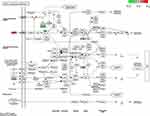
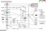
Three gene clusters were identified in the PPI networks of DEGs may possess according to the clusterONE analysis. The three gene clusters involved in distinct GO terms (Figure 4D) and pathways (Figure 4E). The gene cluster 2 were significantly involved in proliferation-related pathways, such as TGF−beta signaling pathway, and signaling pathways regulating pluripotency of stem cells. Genes of cluster 3 were significantly involved in immune-related pathways and NF−kappa B signaling pathway. We also found that gene clusters 1 and 2 are both involved in the MAPK signaling pathway and calcium signaling pathway, which indicated that these two pathways may play a critical role in HM. Subsequently, we separately highlighted the DEGs of clusters 1 and 2 in MAPK signaling pathway (Figure 5) and calcium signaling pathway (Figure 6).
Discussion
HM is one of the blinding ocular diseases with an increasing prevalence worldwide. As the crystalline lens were the core refracting medium of the eye,27 thus it may be a critical step to understand the changes of the crystalline lens to prevent HM. Previous studies suggested no difference in lens thickness28–30 but larger in lens diameter size12 in highly myopic eyes compared to emmetropic eyes. In our present study, various energy metabolic-related gene sets were enriched in the lens epithelia of HM eyes, which indicated more lens content is produced in HM eyes. This may partly explain the reason for the increase in the diameter of the lens. Yet further studies are required to validate this assumption.
In the current management of myopia, one of the reasons why few drugs are recommended31 is that the molecular mechanism of this disease is not well understood. Zhu et al reported that the aberrant TGF-β1 signaling activation by MAF underlies pathological lens growth in HM.12 In our present study, compared to emmetropic eyes, we found multiple genes showed differentially expression patterns in lens epithelium of HM eyes. The potential key genes (CD34, CD40, EGF, IL1A, CD40LG, and CXCL12) were identified using different methods. EGF was considered associated with various ophthalmological diseases.32–34 A previous study showed there is association between IL1A polymorphisms and primary open angle glaucoma.35 CD40 and CD40LG were considered associated with pathogenesis of thyroid eye disease.36 In addition, we also proposed that the imbalances of MAPK signaling pathway and calcium signaling pathway may be the two crucial steps of pathological lens growth in HM. In HM, up-regulated growth factors (EGF and FGF9) and down-regulated RASGRP1, PTPN5, CACNA1B, CACNA2D1, CACNA2D2, and FGFR4 maybe the mainly dysregulated in MAPK signaling pathway. However, this may be a compensatory result, as the energy metabolism-related pathway is enriched in HM compared to emmetropic control. A more active calcium signaling pathway was found in HM, behaving as the up-regulation of EGF, FGF9, SLC8A2, RYR3, and CAMK2A.
Though our present study may provide new insight into the pathological lens in HM, it has several limitations. Firstly, the sample size was small, however, this limitation cannot be addressed for a short time due to the lacking attention in this field. Secondly, this is bioinformatics-based study and further molecular experimental validation for these potential key genes and pathways is required. Thirdly, the key genes were identified based on transcriptomic level and PPI networks, whether these genes mutate or exhibit abnormal epigenetic changes is unknown.
In conclusion, the present study identified potential key genes in pathological lens growth of HM eyes and proposed that the imbalances of MAPK signaling pathway and calcium signaling pathway may be the two crucial steps of pathological lens growth in HM.
Data Sharing Statement
The raw analyses from this study can be obtained from the corresponding author upon reasonable request.
Funding
This work was supported by Guangxi Science and Technology Department (Guike AB18126034), and Guangxi Science and Technology Department (Guike AB20238029).
Disclosure
The authors declare that the research was conducted in the absence of any commercial or financial relationships that could be construed as a potential conflict of interest.
References
1. Holden BA, Fricke TR, Wilson DA, et al. Global prevalence of myopia and high myopia and temporal trends from 2000 through 2050. Ophthalmology. 2016;123(5):1036–1042. doi:10.1016/j.ophtha.2016.01.006
2. Dolgin E. The myopia boom. Nature. 2015;519(7543):276–278. doi:10.1038/519276a
3. Miyake M, Yamashiro K, Tabara Y, et al. Identification of myopia-associated WNT7B polymorphisms provides insights into the mechanism underlying the development of myopia. Nat Commun. 2015;6:6689. doi:10.1038/ncomms7689
4. Wu H, Chen W, Zhao F, et al. Scleral hypoxia is a target for myopia control. Proc Natl Acad Sci U S A. 2018;115(30):E7091–E7100. doi:10.1073/pnas.1721443115
5. Jonas JB, Wang YX, Dong L, Guo Y, Panda-Jonas S. Advances in myopia research anatomical findings in highly myopic eyes. Eye Vis (Lond). 2020;7:45. doi:10.1186/s40662-020-00210-6
6. Jonas JB, Wang YX, Dong L, Panda-Jonas S. High myopia and glaucoma-like optic neuropathy. Asia Pac J Ophthalmol (Phila). 2020;9(3):234–238. doi:10.1097/APO.0000000000000288
7. Baird PN, Saw SM, Lanca C, et al. Myopia. Nat Rev Dis Primers. 2020;6(1):99. doi:10.1038/s41572-020-00231-4
8. Wojciechowski R, Hysi PG. Focusing in on the complex genetics of myopia. PLoS Genet. 2013;9(4):e1003442. doi:10.1371/journal.pgen.1003442
9. Verhoeven VJ, Hysi PG, Wojciechowski R, et al. Genome-wide meta-analyses of multiancestry cohorts identify multiple new susceptibility loci for refractive error and myopia. Nat Genet. 2013;45(3):314–318. doi:10.1038/ng.2554
10. Hysi PG, Choquet H, Khawaja AP, et al. Meta-analysis of 542,934 subjects of European ancestry identifies new genes and mechanisms predisposing to refractive error and myopia. Nat Genet. 2020;52(4):401–407. doi:10.1038/s41588-020-0599-0
11. Pan CW, Cheung CY, Aung T, et al. Differential associations of myopia with major age-related eye diseases: the Singapore Indian Eye Study. Ophthalmology. 2013;120(2):284–291. doi:10.1016/j.ophtha.2012.07.065
12. Zhu X, Du Y, Li D, et al. Aberrant TGF-beta1 signaling activation by MAF underlies pathological lens growth in high myopia. Nat Commun. 2021;12(1):2102. doi:10.1038/s41467-021-22041-2
13. Lin Y, Liang R, Qiu Y, et al. Expression and gene regulation network of RBM8A in hepatocellular carcinoma based on data mining. Aging (Albany NY). 2019;11(2):423–447. doi:10.18632/aging.101749
14. Levy SE, Boone BE. Next-generation sequencing strategies. Cold Spring Harb Perspect Med. 2019;9(7):a025791. doi:10.1101/cshperspect.a025791
15. Prokop JW, May T, Strong K, et al. Genome sequencing in the clinic: the past, present, and future of genomic medicine. Physiol Genomics. 2018;50(8):563–579. doi:10.1152/physiolgenomics.00046.2018
16. Mootha VK, Lindgren CM, Eriksson KF, et al. PGC-1alpha-responsive genes involved in oxidative phosphorylation are coordinately downregulated in human diabetes. Nat Genet. 2003;34(3):267–273. doi:10.1038/ng1180
17. Subramanian A, Tamayo P, Mootha VK, et al. Gene set enrichment analysis: a knowledge-based approach for interpreting genome-wide expression profiles. Proc Natl Acad Sci U S A. 2005;102(43):15545–15550. doi:10.1073/pnas.0506580102
18. Liberzon A, Birger C, Thorvaldsdottir H, Ghandi M, Mesirov JP, Tamayo P. The Molecular Signatures Database (MSigDB) hallmark gene set collection. Cell Syst. 2015;1(6):417–425. doi:10.1016/j.cels.2015.12.004
19. Ritchie ME, Phipson B, Wu D, et al. limma powers differential expression analyses for RNA-sequencing and microarray studies. Nucleic Acids Res. 2015;43(7):e47. doi:10.1093/nar/gkv007
20. Szklarczyk D, Gable AL, Lyon D, et al. STRING v11: protein-protein association networks with increased coverage, supporting functional discovery in genome-wide experimental datasets. Nucleic Acids Res. 2019;47(D1):D607–D613. doi:10.1093/nar/gky1131
21. Shannon P, Markiel A, Ozier O, et al. Cytoscape: a software environment for integrated models of biomolecular interaction networks. Genome Res. 2003;13(11):2498–2504. doi:10.1101/gr.1239303
22. Jeong H, Mason SP, Barabasi AL, Oltvai ZN. Lethality and centrality in protein networks. Nature. 2001;411(6833):41–42. doi:10.1038/35075138
23. Lin CY, Chin CH, Wu HH, Chen SH, Ho CW, Ko MT. Hubba: hub objects analyzer–a framework of interactome hubs identification for network biology. Nucleic Acids Res. 2008;36(WebServer issue):W438–W443. doi:10.1093/nar/gkn257
24. Chin CH, Chen SH, Wu HH, Ho CW, Ko MT, Lin CY. cytoHubba: identifying hub objects and sub-networks from complex interactome. BMC Syst Biol. 2014;8(Suppl 4):S11. doi:10.1186/1752-0509-8-S4-S11
25. Nepusz T, Yu H, Paccanaro A. Detecting overlapping protein complexes in protein-protein interaction networks. Nat Methods. 2012;9(5):471–472. doi:10.1038/nmeth.1938
26. Yu G, Wang LG, Han Y, He QY. clusterProfiler: an R package for comparing biological themes among gene clusters. OMICS. 2012;16(5):284–287. doi:10.1089/omi.2011.0118
27. Alio JL, Schimchak P, Negri HP, Montes-Mico R. Crystalline lens optical dysfunction through aging. Ophthalmology. 2005;112(11):2022–2029. doi:10.1016/j.ophtha.2005.04.034
28. Yong KL, Gong T, Nongpiur ME, et al. Myopia in asian subjects with primary angle closure: implications for glaucoma trends in East Asia. Ophthalmology. 2014;121(8):1566–1571. doi:10.1016/j.ophtha.2014.02.006
29. Xie R, Zhou XT, Lu F, et al. Correlation between myopia and major biometric parameters of the eye: a retrospective clinical study. Optom Vis Sci. 2009;86(5):E503–E508. doi:10.1097/OPX.0b013e31819f9bc5
30. Hashemi H, Khabazkhoob M, Emamian MH, et al. Association between refractive errors and ocular biometry in Iranian adults. J Ophthalmic Vis Res. 2015;10(3):214–220. doi:10.4103/2008-322X.170340
31. Leo SW. Scientific Bureau of World Society of Paediatric O, Strabismus. Current approaches to myopia control. Curr Opin Ophthalmol. 2017;28(3):267–275. doi:10.1097/ICU.0000000000000367
32. Dong F, Call M, Xia Y, Kao WW. Role of EGF receptor signaling on morphogenesis of eyelid and meibomian glands. Exp Eye Res. 2017;163:58–63. doi:10.1016/j.exer.2017.04.006
33. Candar T, Asena L, Alkayid H, Altinors DD. Galectin-3, IL-1A, IL-6, and EGF levels in corneal epithelium of patients with recurrent corneal erosion syndrome. Cornea. 2020;39(11):1354–1358. doi:10.1097/ICO.0000000000002422
34. Kenchegowda S, Bazan NG, Bazan HE. EGF stimulates lipoxin A4 synthesis and modulates repair in corneal epithelial cells through ERK and p38 activation. Invest Ophthalmol Vis Sci. 2011;52(5):2240–2249. doi:10.1167/iovs.10-6199
35. Oliveira MB, de Vasconcellos JPC, Ananina G, Costa VP, de Melo MB. Association between IL1A and IL1B polymorphisms and primary open angle glaucoma in a Brazilian population. Exp Biol Med (Maywood). 2018;243(13):1083–1091. doi:10.1177/1535370218809709
36. Khong JJ, McNab AA, Ebeling PR, Craig JE, Selva D. Pathogenesis of thyroid eye disease: review and update on molecular mechanisms. Br J Ophthalmol. 2016;100(1):142–150. doi:10.1136/bjophthalmol-2015-307399
 © 2022 The Author(s). This work is published and licensed by Dove Medical Press Limited. The full terms of this license are available at https://www.dovepress.com/terms.php and incorporate the Creative Commons Attribution - Non Commercial (unported, v3.0) License.
By accessing the work you hereby accept the Terms. Non-commercial uses of the work are permitted without any further permission from Dove Medical Press Limited, provided the work is properly attributed. For permission for commercial use of this work, please see paragraphs 4.2 and 5 of our Terms.


