Back to Journals » Clinical, Cosmetic and Investigational Dermatology » Volume 19
Hyperbaric Oxygen Exposure During the Holding Phase Modulates Early Apoptotic Signaling in Hair Grafts: A Molecular Pilot Study
Authors Sahan A, Simsek G ![]() , Akyurek ME, Kucun MK
, Akyurek ME, Kucun MK ![]() , Erol SS, Arslan S
, Erol SS, Arslan S
Received 22 February 2026
Accepted for publication 7 May 2026
Published 13 May 2026 Volume 2026:19 604164
DOI https://doi.org/10.2147/CCID.S604164
Checked for plagiarism Yes
Review by Single anonymous peer review
Peer reviewer comments 2
Editor who approved publication: Dr Michela Starace
Ali Sahan,1 Gozde Simsek,2 Murat Eser Akyurek,3 Mustafa Kemal Kucun,4 Selime Semra Erol,5 Serdal Arslan6
1Dermatology, Private Ali Şahan Clinic, Ankara, Türkiye; 2Dermatology Department, University of Health Sciences, Gulhane School of Medicine, Ankara, Türkiye; 3Atlas Biotechnology Laboratory, Ankara, Türkiye; 4Plastic Surgery, Private Avicenna Hospital, İstanbul, Türkiye; 5Department of Medical Biology, Yalova University Faculty of Medicine, Yalova, Türkiye; 6Department of Medical Biology, Mersin University Faculty of Medicine, Mersin, Türkiye
Correspondence: Gozde Simsek, Dermatology Department, University of Health Sciences, Gulhane School of Medicine, Ankara, Türkiye, Email [email protected]
Introduction
During the ex vivo holding phase of hair restoration surgery, follicular units (FUs) experience unavoidable ischemic stress that rapidly activates early intrinsic apoptotic signaling despite preserved structural integrity. Even under standard hypothermic saline storage, oxygen and nutrient deprivation leads to progressive ATP depletion, mitochondrial dysfunction, and reactive oxygen species accumulation — molecular events that prime grafts for apoptotic commitment within the first hour after extraction.1 At the mitochondrial checkpoint, this decision is governed by the balance between pro-apoptotic BAX, which promotes outer membrane permeabilization, and anti-apoptotic BCL-2, which preserves membrane integrity; their ratio therefore serves as an early indicator of cellular apoptotic threshold. This early ischemic vulnerability may be particularly relevant in clinical scenarios where graft survival is known to be more variable, such as extended surgical sessions, revision procedures, and patients with impaired wound healing.1 Hyperbaric oxygen therapy (HBOT), which increases dissolved oxygen tension and mitigates ischemia-related injury in grafted or compromised tissues, also demonstrates a favorable safety profile across non-emergency dermatologic applications.2–5 However, its potential as an ex vivo biological modulator for hair grafts has not previously been investigated.
We conducted a pilot, donor-matched molecular evaluation to determine whether brief HBOT exposure can influence early apoptotic balance in human hair grafts. Six male FUE patients provided paired samples; each donor had 2000–3500 grafts harvested during surgery, of which approximately 500–750 intact follicular units were allocated for the experimental protocol.
Methods
All tissue samples were obtained as surgical by-products of routine FUE procedures performed with written informed patient consent. The study protocol was approved by the Clinical Research Ethics Committee of Mersin University (Date: October 2, 2024; Decision No: 939) and conducted in accordance with the Declaration of Helsinki. Grafts in the HBOT arm were placed in sterile petri dishes containing saline and exposed to hyperbaric oxygen at 1.0 ATA gauge (≈ 2.0 ATA absolute) using 100% oxygen, with the custom chamber maintaining a stable 5–6°C, consistent with standard hypothermic-preservation ranges (4–8°C). Donor-matched control grafts were kept in saline under the same temperature conditions. Preliminary optimization evaluated different pressure settings and storage media; ∼2.0 ATA absolute yielded the most reproducible early anti-apoptotic shifts and was selected for all final experiments. Total RNA was extracted using a RiboEx-based protocol, converted to cDNA with the A.B.T.™ High-Capacity Kit, and quantified using SYBR Green qPCR for intrinsic (BAX, BCL-2, CASP9, CYC) and extrinsic (FasL, CASP3) apoptotic markers. Relative expression was calculated using the ΔΔCt method with paired donor-matched controls, and statistical comparisons were performed using the Wilcoxon signed-rank test with significance set at p < 0.05.
Results
A time-dependent molecular shift was observed. At 30 minutes, HBOT-exposed grafts demonstrated a transient rise in BAX, consistent with the acute stress response after extraction. By 60 minutes, however, BCL-2 expression increased significantly, surpassing BAX levels (shown in Figure 1A). This pattern was more clearly reflected in the BAX/BCL-2 ratio, a recognized indicator of cellular apoptotic threshold: the ratio increased at 30 minutes but declined markedly by 60 minutes, suggesting a shift toward a pro-survival state within the ischemic interval (shown in Figure 1B).
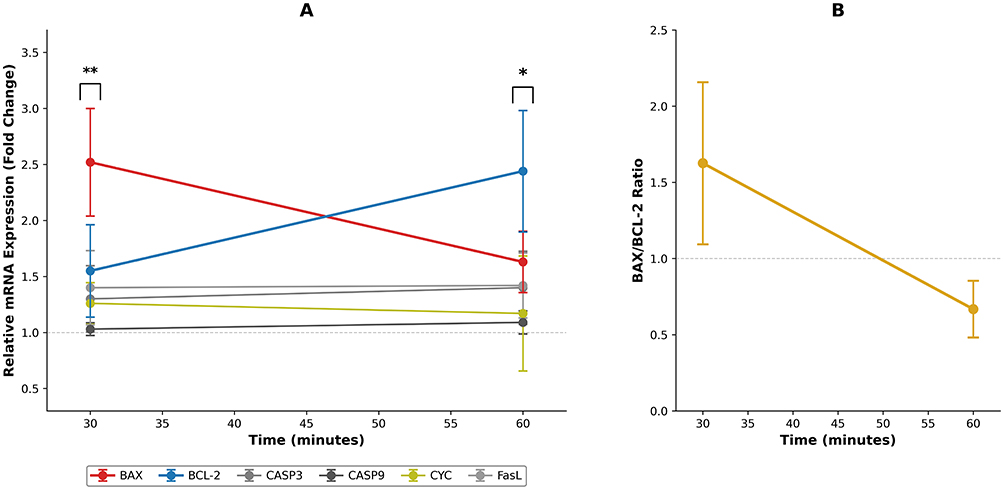 |
Figure 1 Early apoptotic signaling kinetics in human hair follicle grafts following hyperbaric oxygen (HBOT) exposure. (A) Relative mRNA expression of intrinsic (BAX, BCL-2, CASP9, CYC) and extrinsic (CASP3, FasL) apoptotic markers in follicular unit grafts exposed to hyperbaric oxygen at 1.0 ATA gauge (≈ 2.0 ATA absolute) for 30 and 60 minutes. BAX demonstrates a significant transient increase at 30 minutes (**p < 0.01), whereas BCL-2 shows significant upregulation at 60 minutes (*p < 0.05). Error bars represent standard error of the mean (SEM) for BCL-2; SEM values for other genes were not available. (B) BAX/BCL-2 ratio at 30 and 60 minutes, illustrating a time-dependent shift toward a more anti-apoptotic balance. Error bars are not shown because donor-level variance for ratio calculations was unavailable; values represent mean-of-means across donor-matched graft sets (n = 6). |
Notably, CASP3, CASP9, CYC, and FasL showed no significant activation, suggesting that HBOT may have modulated the upstream mitochondrial checkpoint without progression toward irreversible caspase-mediated commitment. This pattern is consistent with HBOT’s previously described cytoprotective effects in other ischemic tissue models and raises the possibility that HBOT may influence early mitochondrial stress integration rather than downstream executioner caspases.2,3
Discussion
Although the sample size was modest and statistical power consequently limited — both understandable constraints in a pilot study - the paired, donor-matched design minimized inter-individual variability, allowing each graft set to serve as its own control. We acknowledge that the study relies on RNA-level data without protein validation or functional viability tests. However, the 30–60-minute ex vivo timeframe is insufficient for meaningful changes in protein abundance, as translation and post-translational processing require hours. In this acute ischemic window, mRNA modulation represents the earliest measurable biological response, making transcriptional profiling the most appropriate method. Therefore, the absence of protein assays reflects the biological constraints of the observation period rather than a methodological gap.
These findings should be interpreted as a preliminary molecular signal rather than evidence of clinical benefit, and whether these transcriptional changes will translate into improved graft outcomes remains to be determined. Nevertheless, the consistent intra-donor pattern — particularly the reproducible reduction in the BAX/BCL-2 ratio — supports the biological plausibility of an early modulatory effect. Confirming whether this molecular shift translates into meaningful outcomes will require larger cohorts, protein-level validation, and longitudinal assessments that track graft behavior through implantation, wound healing, and eventual hair regrowth.
Conclusion
If validated through larger mechanistic and functional, and clinical studies, ex vivo HBOT preconditioning may have the potential to represent a simple, noninvasive, workflow-compatible adjunct that supports enhance early molecular resilience of hair grafts prior to implantation.
AI Statement
Portions of this manuscript (language editing and formatting assistance) were revised using ChatGPT (OpenAI). All scientific content, data interpretation, and conclusions were generated and verified by the authors.
Data Sharing Statement
Available from the corresponding author upon reasonable request.
Ethical Approval
The study was conducted in accordance with the principles of the Declaration of Helsinki. The study protocol was approved by the Clinical Research Ethics Committee of Mersin University (Date: October 2, 2024; Decision No: 939).
Funding
No funding was received for this research.
Disclosure
Dr Ali Sahan reports royalties from patents issued (PCT/TR2023/051865 and PCT/TR2025/050214). The authors report no other conflicts of interest in this work.
References
1. Huang J, Zhou Y, Li H, et al. Preservation solution protects isolated hair micrografts by inhibiting apoptosis of hair bulb. Life Sci. 2025;361:123292. doi:10.1016/j.lfs.2024.123292
2. Zaman T, Canarslan Demir K, Gunduz SH, Gulap Y, Basak AM, Yilmaz KB. Hyperbaric oxygen therapy as an effective adjunctive treatment in the reconstruction of tissue defects with graft in diabetic foot patients: a retrospective cohort study. Int Wound J. 2025;22(6):e70686. doi:10.1111/iwj.70686
3. Mladenov A, Diehl K, Müller O, von Heymann C, Kopp S, Peitsch WK. Outcome of necrotizing fasciitis and Fournier’s gangrene with and without hyperbaric oxygen therapy: a retrospective analysis over 10 years. World J Emerg Surg. 2022;17(1):43. doi:10.1186/s13017-022-00448-6
4. Lee H, Kang S, Paik J, et al. Characteristics of side effects in non-emergency indications using computer-controlled pressurized monoplace hyperbaric chambers: a retrospective multicenter study. J Clin Med. 2024;13(22):6835. doi:10.3390/jcm13226835
5. Parnis J, Magrin AMF, Hassan H. The role, safety, and efficacy of hyperbaric oxygen therapy in aesthetic practice - an evidence-based review. J Cosmet Dermatol. 2024;23(6):1940–3. doi:10.1111/jocd.16228
 © 2026 The Author(s). This work is published and licensed by Dove Medical Press Limited. The
full terms of this license are available at https://www.dovepress.com/terms
and incorporate the Creative Commons Attribution
- Non Commercial (unported, 4.0) License.
By accessing the work you hereby accept the Terms. Non-commercial uses of the work are permitted
without any further permission from Dove Medical Press Limited, provided the work is properly
attributed. For permission for commercial use of this work, please see paragraphs 4.2 and 5 of our Terms.
© 2026 The Author(s). This work is published and licensed by Dove Medical Press Limited. The
full terms of this license are available at https://www.dovepress.com/terms
and incorporate the Creative Commons Attribution
- Non Commercial (unported, 4.0) License.
By accessing the work you hereby accept the Terms. Non-commercial uses of the work are permitted
without any further permission from Dove Medical Press Limited, provided the work is properly
attributed. For permission for commercial use of this work, please see paragraphs 4.2 and 5 of our Terms.
Recommended articles
Caffeic Acid Phenethyl Ester as a DHODH Inhibitor and Its Synergistic Anticancer Properties in Combination with 5-Fluorouracil in a Breast Cancer Cell Line
Amalia E, Diantini A, Endang Prabandari E, Waluyo D, Subarnas A
Journal of Experimental Pharmacology 2022, 14:243-253
Published Date: 23 July 2022
Prussian Blue Nanozyme Potentiates Venetoclax Efficacy in Acute Myeloid Leukemia by Inducing a More Mature Phenotype
Yang F, Wu H, Yao J, Peng X, Yang A, Wen T, Meng J, Liu J, Zhang Y, Wang T, Xu H
International Journal of Nanomedicine 2026, 21:601085
Published Date: 25 May 2026