
Back to Journals » International Journal of Nanomedicine » Volume 21
Hydrogels in Autoimmune Disease: A Comprehensive Review of Current Research and Clinical Potentials
Authors Lu Y, Su H, Fan X, Hu J, Tang X, Hu J  , Zhang L, Ma D
Received 18 December 2025
Accepted for publication 28 June 2026
Published 8 July 2026 Volume 2026:21 590131
DOI https://doi.org/10.2147/IJN.S590131
Checked for plagiarism Yes
Review by Single anonymous peer review
Peer reviewer comments 3
Editor who approved publication: Professor Jie Huang
Yangyang Lu,1,* Haodong Su,1,* Xinying Fan,1 Jie Hu,2 Xiaoyu Tang,1 Jingjin Hu,1 Liyun Zhang,1 Dan Ma1
1Department of Rheumatology, Third Hospital of Shanxi Medical University, Shanxi Bethune Hospital, Taiyuan, Shanxi, People’s Republic of China; 2Stem Cell Translation Laboratory, Shanxi Bethune Hospital, Taiyuan, Shanxi, People’s Republic of China
*These authors contributed equally to this work
Correspondence: Dan Ma, Department of Rheumatology, Third Hospital of Shanxi Medical University, Shanxi Bethune Hospital, No. 99, Longcheng Street, Xiaodian District, Taiyuan, Shanxi, 030032, People’s Republic of China, Email [email protected]
Research Objective: Autoimmune diseases are chronic conditions in which the immune system abnormally attacks the body’s own tissues. Their incidence is on the rise, and the pathogenesis remains incompletely understood. The clinical manifestations are complex and diverse. Despite active treatment, some patients still have poor therapeutic outcomes and rely on immunosuppressants for a long time. Although these drugs can control symptoms, they are associated with increased infection risks and serious side effects such as organ toxicity. Hydrogels are three-dimensional network structures with excellent water absorption properties, good biocompatibility, injectability, and physical and chemical properties. By loading adjuvants, drugs, cells, and other substances, they are applied in the establishment of disease models, drug delivery, bioimaging and biosensors, tissue engineering, and other fields, providing new strategies for studying disease pathogenesis, drug screening, local targeted delivery, and immune regulation. This article aims to systematically review the mechanism of action and research progress of hydrogels in the treatment of autoimmune diseases and assess their potential for clinical translation.
Research Methods: Relevant literature on the medical applications of hydrogels published from January 1, 2004 to October 31, 2025 was retrieved, and the scope was narrowed down to studies on their application in autoimmune diseases for summary and analysis.
Main Results: The main findings are as follows: (1) Through bibliometric analysis, it was found that research on hydrogels in the medical field is increasingly prominent, with studies in autoimmune diseases mainly focusing on drug delivery; (2) By constructing injectable bioadhesive hydrogels, the adhesion of hydrogels to joint tissues is enhanced, prolonging drug retention time and improving treatment efficiency; (3) Specific hydrogel designs can actively regulate immune cell functions - for example, inhibiting inflammation-related signaling pathways to reverse M1 polarization of macrophages and ferritin autophagy/ferroptosis in chondrocytes, maintaining the integrity of cartilage structure, and inducing mitochondrial dysfunction to promote apoptosis of FLS and macrophages and regulate the inflammatory microenvironment; (4) Hydrogel microneedle systems, as transdermal drug delivery platforms, have shown good compliance and efficacy in rheumatoid arthritis.
Summary: Hydrogel technology, through localized, controllable, and intelligent drug delivery, is expected to break through the bottlenecks of traditional autoimmune disease treatment. Current research is gradually evolving from passive carriers to active participants in immune regulation as “intelligent platforms”, and their potential to reshape the inflammatory microenvironment has been verified in animal models. However, issues such as material degradability, long-term biological safety, and consistency in large-scale production still need to be further addressed in preclinical and clinical studies. Future interdisciplinary collaboration and translational medical research are key to promoting the development of this field.
Keywords: hydrogels, medical applications, autoimmune diseases
Introduction
Hydrogels are three-dimensional network structures composed of water and polymer macromolecules, with excellent water absorption properties. They possess good biocompatibility and physical and chemical properties, and can simulate the physical and chemical environment of the extracellular matrix, providing appropriate mechanical support, porosity, and viscoelasticity, promoting cell adhesion, proliferation and differentiation, and can meet specific requirements under different conditions.1,2 In the 1960s, Otto Wichterle and Drahoslav Lim produced a hydrogel that was first successfully applied to contact lenses, and for a long time thereafter was mainly used in ophthalmology and drug delivery through simple chemical crosslinking.3 In the 1970s, inspired by the conversion of chemical energy into mechanical energy, the research focus shifted towards intelligent hydrogels that could respond to environmental changes (such as pH, temperature, electromagnetic field changes, and biomolecular concentration) and undergo volume, structure or phase transitions, achieving “on-demand” drug release or degradation.4 In the 1990s, researchers discovered other physical forces acting on gelation that made it possible to regulate the mechanical, thermal, and degradation properties of hydrogels. With the continuous development of organic chemistry, more complex chemically crosslinked hydrogels have been successfully developed to compensate the poor mechanical properties of physical hydrogels. Hydrogels can be equipped with antigens, adjuvants, or chemical inducers to establish different disease models for studying the pathogenesis or drug screening. By carrying drugs or cells that exert anti-inflammatory, antibacterial, antioxidant, and antitumor functions, making it an ideal platform for drug delivery and tissue repair, applicable in fields such as tissue repair and regeneration, drug delivery, drug carriers, biological imaging, and biological sensors, with remarkable advantages.5,6
Using the Web of Science Core Collection (WoSCC) database, we searched for literature on the medical applications of hydrogels from January, 01, 2004 to October, 31, 2025. The search formula was as follows: (TS=(hydrogel) AND TS=(medicine)) AND LA=(English) 5841 articles were retrieved, and 5491 articles and review articles were obtained after excluding conference papers and abstracts, book chapters, early access literature, editorial materials, retracted literature, and letters (Figure 1a). According to the annual number of published documents (Figure 1b), hydrogels are being applied and researched more and more in the medical field, and their status in the medical field is becoming increasingly prominent. Burst word analysis (Figure 1c) and time diagram (Figure 1d) intuitively showed that hydrogels are hot spots in wound healing, drug delivery, 3D printing, 3D cell culture, and other fields.
 |
Figure 1 Literature on the application of hydrogels in medicine. (a) Data sources and methods; (b) Number of annual publications; (c) Burst word; (d) Time diagram. |
Autoimmune diseases are chronic diseases, such as rheumatoid arthritis (RA), systemic lupus erythematosus (SLE), Sjögren’s disease (SjD), systemic sclerosis (SSc), multiple sclerosis(MS), and diabetes mellitus, caused by abnormal immune system attacks on tissues,and can seriously affect the patient’s quality of life. The incidence of this condition is increasing annually.7–9 Common pathological features include local chronic inflammatory microenvironment, abnormal activation of immune cells, continuous release of pro-inflammatory factors (such as TNF-α and IL-6), and tissue fibrosis. These often present abnormal microenvironmental signals such as low pH, high oxidative stress, and overexpression of reactive oxygen species (ROS). The incidence of these diseases is increasing year by year. The main therapeutic drugs are immunosuppressants, which have limitations such as limited efficacy, severe side effects, poor targeting, low bioavailability, and difficulty in penetrating the fibrotic barrier. Opportunistic infections and tumors may also occur during the treatment process; therefore, precise targeted therapy will be the trend of future treatment, especially biomaterial-based drug delivery systems, providing more opportunities for traditional drug therapy. For example, hydrogels are used to encapsulate drugs or cells so that drugs can be concentrated on the damaged site in a controlled manner to improve function, while reducing the number of doses to increase patient compliance.9
Through searching the literature on the application of hydrogels in autoimmune diseases from January, 01, 2004 to October, 31, 2025, the search formula is as follows: (TS=(hydrogel) AND TS=(autoimmune diseases)) AND LA=(English) 169 articles were retrieved, and a total of 160 articles and review articles were obtained after excluding conference papers, book chapters, and autoimmune diseases (Figure 2a). The annual publication volume map (Figure 2b), burst word analysis (Figure 2c), and time graph (Figure 2d) visually demonstrated the great potential of drug delivery for the treatment of autoimmune diseases. Therefore, herein,we have focused on the application of hydrogels in the treatment of autoimmune diseases and look forward to future development directions and challenges.
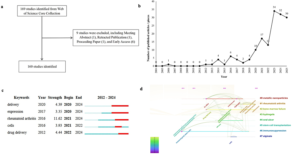 |
Figure 2 Literature on the application of hydrogels in autoimmune diseases. (a) Data sources and methods; (b) Number of annual publications; (c) Burst word; (d) Time diagram. |
Hydrogel Classification
Several types of gels are classified according to different classification methods (Figure 3).2,10 According to different preparation materials, hydrogels are divided into natural hydrogels such as chitosan, hyaluronic acid, alginate acid, synthetic hydrogels such as polyethylene glycol (PEG) and polyn-isopropyl acrylamide (PNIPAm), and hybrid hydrogels.11,12 The former exhibits good biocompatibility and degradability. Compared to the latter, it is safer for biomedical applications, but its mechanical properties and stability are poor. The latter has the advantage of human intervention in synthesis, but must be strictly controlled to avoid possible biological incompatibility and adverse drug reactions. Therefore, several researchers have continued to develop new hydrogels that combine their advantages. Chitosan is one of the most widely used hydrogel materials, with good antibacterial properties, biodegradability, and biocompatibility, and is widely used in drug delivery, tissue engineering, bone regeneration, and as an antimicrobial agents.13,14 Hyaluronic acid (HA), the main components of extracellular matrix (ECM), has good biological compatibility and low immunogenicity, and can regulate the inflammatory response and promote angiogenesis, action, and migration. These properties make HA widely used in medical fields such as anticancer, ophthalmology, and surgery. In addition, HA exhibits high water retention and unique rheological properties. Gels with high viscoelasticity are formed in aqueous solutions and are widely used in the pharmaceutical field.15,16 The alginate structure is widely used in tissue engineering because of its excellent biocompatibility with the natural ECM; however, its high hydrophilicity affects cell adhesion and proliferation, limiting its application in cell delivery.17 PEG has unique advantages in the design and preparation of sustainable and controllable release systems, owing to its unique hydrophilicity, biocompatibility, and biodegradability. However, as a synthetic polymer, further optimization of the effective release of its supported drugs at the target site and reduction in production costs are the next steps to be studied and improved upon. PEG hydrogels are bioinert and do not provide ideal environments for cell survival, adhesion, and growth.18 PNIPAAm has excellent properties such as an adjustable structure, thermal sensitivity, and low toxicity, and is injectable at room temperature. As a commonly used heat-sensitive water-soluble homopolymer, the PNIPAAm hydrogel has been widely used in tissue engineering and supporting cell co-culture; however, its poor biocompatibility and weak mechanical properties limit its wide application.19 Hybrid hydrogels, which combine the advantages of the former two, have better high strength and toughness. For example, alginate/gelatin-based hybrid hydrogels are widely used in cell culture scaffolds and tissue engineering.
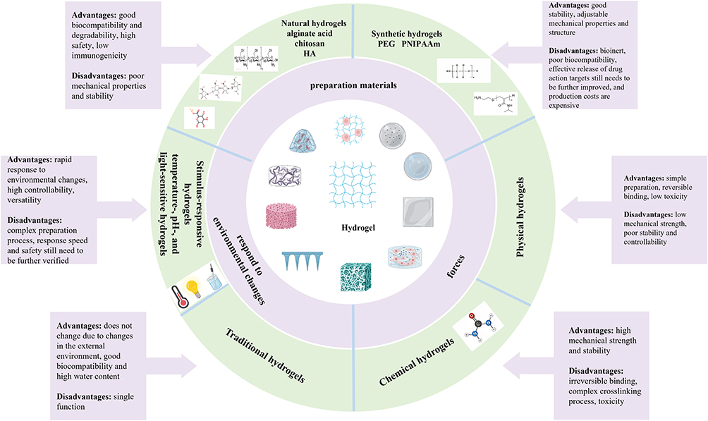 |
Figure 3 Hydrogel classification. |
Hydrogels can be divided into physical and chemical types according to their forces during gelation. The former is formed by physical entanglement, hydrogen bonding, and hydrophobic interactions, whereas the latter is formed by chemical bonding and crosslinking. The combination of the former is temporary, can be changed from the gel state to the solution state by changing temperature, pH and other ways, so it is called “false gel”, the latter through the combination of chemical bonds is irreversible, also known as “true gel”, compared with the former more stable Depending on whether hydrogels respond to environmental changes, they can be divided into traditional hydrogels and stimulus-responsive hydrogels (also known as smart hydrogels).20 Smart hydrogels can be further divided into temperature-, pH-, and light-sensitive hydrogels. Traditional hydrogels do not change owing to changes in the external environment; smart hydrogels respond quickly to changes in the environment, which allows for secondary specific events such as drug release; therefore, they are widely used in the medical field. Temperature-sensitive hydrogels can be classified into those with high critical phase transition temperatures (thermal-expansion temperature-sensitive hydrogels) and those with low critical phase transition temperatures (thermal-shrinkage temperature-sensitive hydrogels). The latter is commonly used in biomedical applications. By changing the composition and structure of hydrogels, the low critical phase transition temperature is adjusted to be close to the human body temperature of 37°C, so that it becomes a solution state at room temperature. It is easy to inject drugs into the human body and form a gel after entering the body for a period of time, which can be used mainly for sustained drug release. pH-sensitive hydrogels induce protonation or ionization at different pH values to change the charge distribution and internal interactions, thereby changing the gel structure. They are primarily used for specific drug administration at different pH levels. When photosensitive hydrogels are exposed to light radiation, the structure of the photosensitive groups is isomerized or dissociated, resulting in a local temperature rise or configuration change, and then a change in the swelling rate of the hydrogels, which is of great significance in drug release and tissue engineering. However, problems such as long crosslinking time of photosensitive hydrogels, light penetration, and light damage to body tissues must be resolved.
Application of Hydrogels
Drug Carrier
Traditional drug delivery methods have problems such as systemic toxicity and repeated administration. Hydrogels as drug carriers can minimize these adverse factors while protecting easily degradable drugs to optimize therapeutic effects. Controlled release methods for hydrogel-loaded drugs include diffusion, swelling, and chemical erosion control. Diffusion control is the most commonly used mechanism of drug release from hydrogels. When the drug diffuses, the hydrogel will swells, and when the drug diffusion rate is higher than the swelling rate of the hydrogel, a controlled swelling release occurs. Chemically controlled release mainly occurs in the delivery carrier where the drug chemically binds to the hydrogel, most commonly through hydrolysis or enzymatic degradation, leading to polymer chain breaks or reversible or irreversible reactions between the polymer network and the releasing drug, resulting in drug release. In this case, the surface or overall erosion of the hydrogel and the equilibrium coefficient of drug binding to the hydrogel controls the drug release rate.21 The advantage of hydrogels is that they can be efficiently loaded with drugs. With the discovery and preparation of stimulus-responsive hydrogels, “intelligent” and “controlled” drug release is also possible. For example, the HA hydrogel has a high affinity for CD44 overexpressed on the surface of tumor cells, which is advantageous as an anticancer drug carrier. It has high adhesion to the oral mucosa and provides theoretical support for the preparation oral medicines. The hydrophilicity of the hydrogel allows it to bypass the host immune response and reduce phagocytic activity, increasing the in-body circulation time of the delivery device for better efficacy. However, hydrophilic polymer cores may not be suitable for hydrophobic drug applications. In addition, many hydrogels have weak tensile strength, which can lead to early release of the drug before it reaches the target site. Therefore, it is necessary to improve the loading rate and the precise release of hydrophobic drugs.
Tissue Engineering and 3D Cell Culture
The purpose of tissue engineering is to repair dysfunctional tissues in the body caused by gene mutations, congenital deformities, aging, disease, or injury, focusing on the joint repair of tissues through cell repair and generation of new tissues, scaffold support and guidance of cells, and biomolecules regulating cell activities.22 The hydrogel can house cells and provide mechanical support, and its mild gelation conditions and in situ polymerization ability allow it to not only encapsulate cells and growth factors but also control the release of growth factors and other agents, which are critical for cell migration, differentiation, angiogenesis, and new tissue generation. Some studies have used the pancreatic ECM and platelet plasma to construct 3D injectable hydrogels for the transplantation into diabetic rats and found that they can enhance the vitality of islet cells and promote angiogenesis, which is a potential tissue engineering method for the treatment of T1MD.23
In vitro cell culture is an integral part of regenerative medicine and preclinical evaluation. Traditional two-dimensional (2D) cell culture has been widely used because of its simplicity, economy, and other advantages; however, 2D scaffolds cannot simulate the actual growth conditions of living cells and may cause changes in metabolism, gene expression patterns, and adhesion inhibition. Therefore, functional three-dimensional (3D) structures are required for cell culture. Hydrogels for cell culture usually need to meet the following conditions: ease of handling under physiological conditions, mechanical properties similar to those of natural tissues, uniformity at the micro and macro levels, compatibility with long-term cell culture, possibility of matching different cell types, and optical transparency that can be used for analysis.21 The hydrogel’s high water content, biocompatibility, and properties similar to those of the ECM matrix make it a potential application for 3D cell culture.
Dressing
Natural skin has defensive properties such as preventing bacterial invasion, facilitating the exchange of substances, and resisting cold or heat damage. Traditional wound dressings promote wound healing by providing growth factors, and there is a mismatch in wound healing stages, leading to passive soft tissue repair. Ideal wound dressings should promote wound healing while functioning as a skin barrier. Hydrogels show broad application prospects in wound dressings because of their good biocompatibility, effective adhesion, antibacterial properties, and high water content (70%–95%). Gao et al constructed an ECM simulating a nanofiber/hydrogel interpenetration network (NFHIN), which balances the wound microenvironment through a three-dimensional nanofiber framework and aerogels, which facilitate cell migration.24 Aerogels can collect wound exudates and transform them into polycationic hydrogels with a contact killing effect, while removeing pathogens and reactive oxygen species. Fibroblast activity significantly enhanced after co-culturing with NFHIN, which promoted wound healing. Chang et al constructed a quaternary ammonium-polyphosphate-chitosan hydrogel.25 The tissue adhesion of the hydrogel seals the blood flow, the activation of coagulation factor V is promoted by polyphosphate to enhance hemostasis, and the antibacterial effect is enhanced by the quaternary ammonium salt. Clinically, it can be used as a dressing for hemostasis and for easy infection repair. A self-healing hydrogel (PAHB/OD hydrogel) combined with CuS nanoparticles made of polyaspartate and glucan loaded with sodium diclofenate can promoted tissue regeneration, inhibited inflammation and promoted wound angiogenesis after being application to the skin of mice.26
Microneedle
Hydrogel microneedles are innovative drug delivery systems that combine hydrogel materials with microneedle technology.27,28 They are usually composed of tens to hundreds of microne-level needle-like structures that can penetrate the skin cuticle and deliver drugs directly to the epidermis or dermis without touching nerves and blood vessels. These procedures are painless, efficient, and controllable. Studies have shown that the use of microneedles to treat of immune-mediated hair loss in mouse models sustains regeneration while reducing the inflammatory response.27 Additionally, the application of TCM-integrated reactive microneedles in SSc effectively improved skin fibrosis in mice.29
Application of Hydrogels in Autoimmune Diseases
In the biomedical field, hydrogels are primarily used in drug delivery systems, tissue-engineered scaffolds, and immunomodulatory platforms. Hydrogels can achieve continuous drug release, improve drug utilization, and reduce adverse effects. In tissue engineering, hydrogels can simulate ECM and provide a suitable microenvironment for cell growth and differentiation. In terms of immune regulation, hydrogels can regulate the behavior of immune cells by regulating their physical and chemical properties, providing new ideas for the treatment of autoimmune diseases (Table 1).
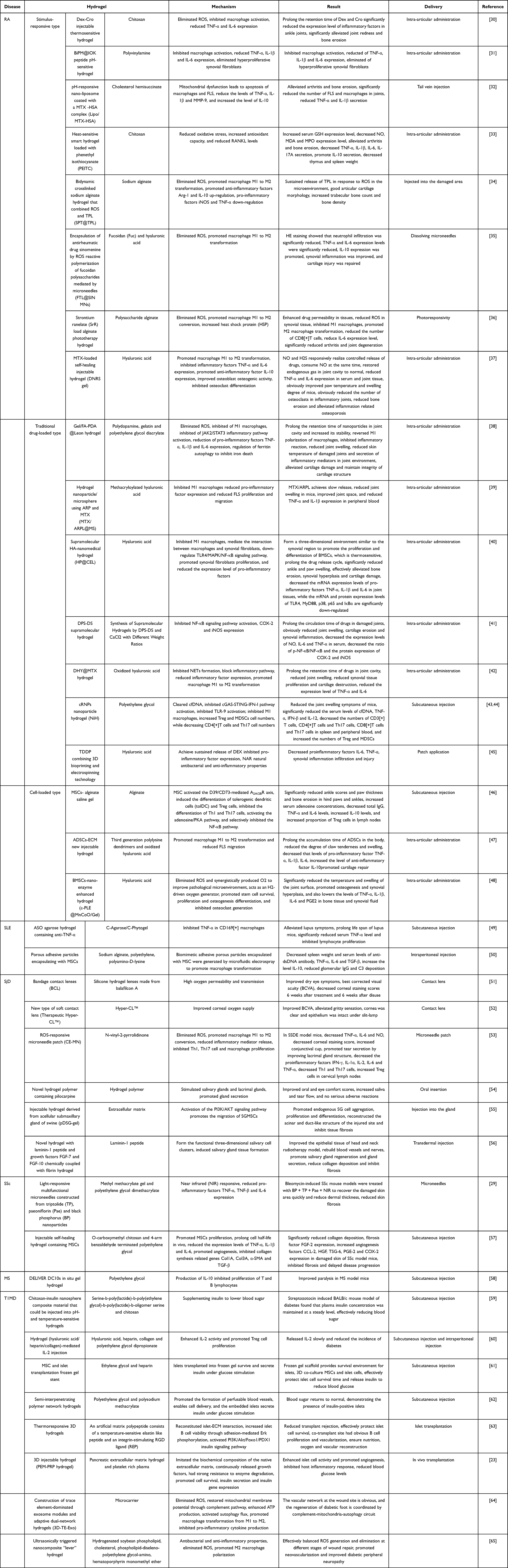 |
Table 1 Application of Hydrogels in Autoimmune Diseases |
Rheumatoid Arthritis
RA is an autoimmune disease, involving symmetrical facet joints of the whole body and is caused by a variety of environmental, genetic, and epigenetic factors.66 The main symptoms are joint swelling and pain, characterized by joint inflammation leading to irreversible joint destruction and deformity, which seriously affects the patients’ quality of life. Commonly used drugs include nonsteroidal anti-inflammatory drugs (NSAIDs), disease-modifying anti-rheumatic drugs (DMARDs), glucocorticoids, and biologics. However, these drugs require high dose and frequent and long-term administration, which may lead to serious side effects and poor patient compliance. Therefore, it is important to avoid these problems while simultaneously improving drug efficacy at the same time.67 By constructing an injectable bioadhesive hydrogel, the adhesion between the hydrogel and the joint tissue was enhanced to extend the drug retention time and improve treatment efficiency.38 A drug delivery system (DDS) carries out passive and active targeting through optimal nano particle size and surface modification, reducing adverse drug reactions while ensuring that the drug concentration at the target site enhances the therapeutic effect.68,69
Macrophages can produce a variety of pro-inflammatory cytokines such as TNF-α, IL-1, IL-6, and prostaglandins, which are intertwined with reactive ROS to cause joint injury; thus, targeting macrophages is a hot spot in RA treatment.30,70 Liu et al developed a heat-sensitive injection hydrogel loaded with dexamethasone (Dex) and crocetin 1 (Cro), which ensured the release of Dex and Cro while extending the retention time.30,71–77 Yang et al encapsulated and loaded leonurine (Leon) folate-functionalized polydopamine (FA-PDA) nanoparticles with anti-inflammatory properties and gradually released FA-PDA@Leon nanoparticles, while Wu et al developed an injectable pH-sensitive IOK peptide hydrogel supported by methotrexate (MTX) and bismuth nanosheets/BiNS/PEI, which could reduce the activity of macrophages and simultaneously eliminate overproliferating synovial fibroblasts (FLS).31,38 Zheng et al also developed a pH-responsive nano-liposome (Lipo/MTX-HSA) coated with a methotrexate (MTX) -human serum albumin (HSA) complex, Haloi et al developed a heat-sensitive smart hydrogel loaded with phenethyl isothiocyanate (PEITC), which has antioxidant and anti-inflammatory activities; Du et al constructed a hydrogel nanoparticle/microspheres (MTX/ARPL@MS) using artemisinin prodrug (ARP) and MTX.32,33,39 After local administration, MTX/ARPL is slowly released and targets both synovial macrophages and fibroblasts. These modified drugs were injected into the joint cavity of arthritis rats, and they could significantly reduce the levels of TNF-α, IL-1β, IL-6, IL-17A and other pro-inflammatory factors, improve joint redness and bone erosion, and reduce the weight of thymus and spleen.30–33,38,39 Increased serum glutathione (GSH) levels and decreased levels of nitric oxide (NO), malondialdehyde (MDA), and myeloperoxidase (MPO), inhibit inflammatory signaling pathways to reverse M1 polarization of macrophages and ferritin autophagy/iron death in chondrocytes, thus maintaining the structural integrity of cartilage.33,38 Additionally, they effectively reduce the number of FLS and macrophages in joints, induce mitochondrial dysfunction, promote FLS and macrophage apoptosis, and regulate the inflammatory microenvironment.32 Wang et al developed a bidynamic crosslinked sodium alginate hydrogel (SPT@TPL) that combined “stimulus response (such as ROS)” and “drug release (such as triptolide (TPL))”, with the ability to respond and regulate ROS levels on demand, and injected hydrogel locally into the damaged femur region of RA model rats.34 It was found that the articular cartilage morphology, trabecular number and bone mineral density increased in SD rats, which may be related to SPT@TPL response and effective clearance of ROS and promotion of macrophage M2 polarization. Wang et al developed a supramolecular HA-nanomedical hydrogel (HP@CEL).40 Through continuous local release of celastrol (CEL), which mediates macrophage-synovial fibroblast crosstalk, the hydrogel was injected into the joint cavity of CIA model mice, and ankle and paw swelling was significantly reduced, and bone density increased. Bone erosion, synovial hyperplasia, and cartilage injury were effectively alleviated. mRNA and protein expressions of TLR4, MyD88, p38, p65 and IκBα in joint tissues were significantly down-regulated, suggesting that HP@CEL can inhibit the TLR4/NF-κB pathway by inhibiting M1 macrophage infiltration and down-regulating pro-inflammatory cytokines. To reduce local inflammation and improve bone regeneration. Song et al studied a dexamethasone sodium phosphate (DPS) -diclofenac sodium (DS) supramolecular hydrogel, which exerts anti-inflammatory effects by down-regulated the levels of NO, TNF-α and IL-6 in RAW 264.7 macrophages activated by lipopolysaccharide (LPS).41 By injecting the DMS-DS hydrogel into the joint cavity of AIA rats, joint swelling, bone and cartilage erosion and synovial inflammation were significantly reduced, serum levels of NO, IL-6 and TNF-α were decreased, p-NF-κB/NF-κB ratio and protein expression of COX-2 and iNOS were decreased. In addition, Liu et al developed an ROS-reactive polymer for the anti-rheumatic drug sinusine (FTL@SIN MNs), which was characterized by the synergistic effect of the algae polysaccharide and showed good mechanical strength and physical stability compared with the microneedles prepared with only hyaluronic acid.35 After local topical application, macrophage inflammation was significantly reduced, the secretion of key pro-inflammatory cytokines was reduced, the polarization of macrophages towards the M2 type was promoted, synovial inflammation improved, and cartilage repair was promoted. This system combines nanoparticles with traditional Chinese medicine to provide a new strategy for the treatment of RA. Weng et al develop alginate (ALG) phototherapy hydrogels supported with strontium ranelate (SrR), combined with photoresponsive molybdenum disulfide nanoflower (MoS2NFs) and photothermally responsive polypyrrole nanoparticles (Ppy NPs).36 Local application of yeast to RA animal models was found to significantly reduce inflammation, increase heat shock protein (HSP), promote macrophage M2 polarization, and alleviate RA-related joint degradation.
Neutrophils are key cellular components infiltrating synovial tissue and rheumatoid nodules in RA patients, and it has recently been found that abnormal neutrophils forming neutrophil extracellular traps (NET) may contribute to the pathogenesis of arthritis.78,79 NET are net-like structures composed of histone proteins, DNA, and antibacterial proteins released by activated neutrophils, which lead to cartilage destruction by enhancing the immunogenicity of the cartilage components. Deoxyribonuclease I (DNase) is a natural endonuclease responsible for the hydrolysis of extracellular DNA outside the cell, which prevents NET formation.80 Wang et al coupled DNase with oxyhyaluronic acid (OHA) to form DHA, which was then crosslinked with carboxymethyl chitosan (CMCS) to form an injectable, degradable, and biocompatible hydrogel (DHY), and then loaded MTX into the posterior articular cavity of the system for injection into CIA mice.42 It was found that after DHY@MTX intervention, the joint symptoms of mice were basically normal, the articular surface was clear, and the levels of TNF-α and IL-6 were decreased, which were significantly better than those in DHY and DNase@HY groups. Jiang H et al utilized the characteristics of traditional Chinese medicine agents to self-assemble Sinomenine-glycyrrhizic acid self-assembly nanohydrogel.81 After intra-articular injection to the rheumatoid arthritis model mice, it not only alleviated joint swelling at a low dose but also reduced the adverse reactions caused by high-dose drugs. In vitro experiments demonstrated that this self-assembled hydrogel could significantly reduce the migration of neutrophils.
Abundant free DNA (cfDNA) in peripheral blood and synovial fluid is involved in the pathogenesis of RA. cfDNA enhances inflammation by activating pattern recognition receptors (PRRS), such as cytoplasmic double-stranded DNA (dsDNA), cyclic guanosine-adenosine phosphate synthase (cGAS) and toll-like receptor 9 (TLR9). cGAS activation can induce the abnormal activation of the interferon gene-stimulating factor (STING) pathway, thus promoting a type I interferon (IFN) response. TLR9 activation can also promote inflammatory responses.43,44 Therefore, clearing cfDNA and inhibiting cGAS are promising therapeutic strategies. Cheng et al loaded cGAS inhibitors (RU) into cationic nanoparticles (cNPs) to make nano-drug cRNPs, and then loaded cRNPs into a hydrogel to form a nanoparticle hydrogel (NiH), which was injected into CIA mice subcutaneously through the tail; they found that the joint swelling of mice was significantly reduced after NiH intervention. Serum cfDNA, TNF-α, IFN-β and IL-12 water were significantly decreased, the number of CD3T cells, CD4T cells, CD8T cells and Th17 cells in spleen and peripheral blood was decreased, and the number of Treg cells was increased, which was significantly better than that in RU-H and cNPs-H groups. These results indicated that NiH can effectively clear cfDNA, inhibit inflammatory responses, promote immune homeostasis, and treat RA.
MSCs are a class of pluripotent stem cells capable of self-renewal, multi-differentiation, and immune regulation. They can inhibit T cells, B cells and NK cells, promote the generation of Treg cells, reduce the production of antibodies, and reduce the secretion of IFN-γ, TNF-a, IL-6, IL-17, IL-22 and other pro-inflammatory factors. They can promote the secretion of PGE2, TGF-β, IL-4, IL-10 and other factors and migrate to the lesion area in response to the inflammatory signal of the injured site, and play the role of tissue repair by secreting extracellular vesicles and growth factors. Shi et al encapsulated MSCs in alginate brine gel, which has the advantages of low toxicity, low cost, good gelling power, and high bioavailability.46,82 It is an ideal scaffold for delivering MSCs, improving the proliferation ability, vitality, and survival time of MSCs in vivo, and enhancing the immune regulation ability of DCs. They injected alginate gel containing MSCs into CIA mice subcutlely and found that RA incidence, ankle joint score and paw thickness, and bone erosion degree of hind paws and ankles were significantly decreased, serum adenosine concentration was increased, total IgG level was significantly decreased, TNF-α and IL-6 levels were basically reduced to normal, and IL-10 levels were increased. The proportion of Tregs cells in the lymph nodes increased, and the effect was comparable to that of the MSCs group, showing superior preventive and therapeutic effects. Zhu et al injected a novel injectable hydrogel loaded with adipose-derived stem cells (ADSCs) in the ECM into the joint cavity of a CIA rat model to prolong the accumulation time of ADSCs in the body and found that the degree of claw tenderness and swelling was significantly reduced, promoting cartilage repair.47 Zhao et al proposed that nanoenzyme-enhanced hydrogels can effectively remove endogenously overexpressed ROS and produce O2.48 They injected a nano-enzyme enhanced hydrogel encapsulated in bone marrow-derived mesenchymal stem cells (BMSCs) (ε-PLE@MnCoO/Gel) into the large and micro pores of the 3D printed titanium alloy prosthesis of the RA rabbit model, and found that joint surface temperature and swelling were significantly reduced, promoting osteogenesis and synovial hyperplasia. In addition, the levels of TNF-α, IL-1β, IL-6 and PGE2 in bone tissue and synovial fluid were decreased.
Endogenous gases (such as NO and H2S) play a key role in the regulation of the RA microenvironment; however, these gases are highly concentration-dependent. Excessive NO and insufficient H2S cause inflammation and activate osteoclasts to dissolve bone tissue, thus delaying bone healing and exacerbating RA.83,84 Therefore, Geng et al developed a self-healing injectable hydrogel (DNRS gel) that can consume NO and release H2S and was injected into CIA rats in the joint cavity after loading MTX.37 They found that paw temperature and swelling of rats were significantly reduced, and bone erosion was alleviated, which may be related to M2 polarization promoting osteogenesis and inhibiting osteoclast differentiation.
In addition to dressing applications, Elshabrawy et al developed a unique double-layer transdermal patch (TDDP), that combined 3D bioprinting and electrospinning technology.45 The first layer was a newly developed hydrogel (HG) containing the joint-maintaining component HA and anti-inflammatory agent dexamethasone (DEX) through 3D printing. The second layer was electrospun polycaprolactone (PCL) nanofibers (NFs) loaded with the natural plant extract naringin (NAR), which has antibacterial and anti-inflammatory effects, and was applied to the ankle joints of RA rats. The levels of IL-6 and TNF-α were significantly reduced, and the inflammatory infiltration and damage of synovium were reduced.
These studies found that, in addition to traditional drugs that can reduce the secretion of inflammatory factors and block the inflammatory pathway, the use of drug delivery to load drugs, DNA, enzymes, cells, and other direct effects on the lesion site, while reducing side effects, ensures that the concentration of drugs at the lesion site enhances efficacy.
Systemic Lupus Erythematosus
SLE is a multisystem chronic autoimmune disease characterized by relapse and remission. Its prevalence is higher in women of reproductive age, with women accounting for 9:1, the exact cause is unknown, but it has been established that environmental and genetic factors interact to trigger an immune response that leads to the overactivation of B cells to produce a large number of pathogenic autoantibodies, resulting in tissue and organ damage, which can affect any part of the body and can lead to death when kidney damage (lupus nephritis) is severe.85 Dysfunction of the peripheral lymphatic organs, such as the spleen and lymph nodes, is associated with SLE.49 Huang et al injected agarose hydrogel containing anti-TNF-A antisense oligonucleotide (ASO) into MRL/Lpr mice subcutaneously, and found that ASO containing anti-TNF-A aggregated in macrophages, significantly reduced the level of TNF-a, inhibited lymphocyte proliferation, and alleviated lupus-like symptoms.49 Nie et al intraperitoneal injected porous adhesive particles encapsulated with MSCs into MRL/Lpr mice, and found that spleen weight and serum levels of anti-DSDNA antibody, TNF-α, IL-6 and TGF-β were significantly reduced, IL-10 levels were increased, and glomerular IgG and C3 deposition were also significantly reduced.50
Sjögren’s Disease
SjD is a chronic autoimmune disease caused by immune disorders characterized by the activation of T and B lymphocytes and the infiltration of exocrine glands and epithelial cells. The main clinical manifestations are dry mouth and eye caused by the destruction of exocrine glands, such as the salivary and lacrimal glands. If SjD-associated dry eye is not treated promptly, it may lead to chronic conjunctivitis, persistent epithelial defects, recurrent infectious keratitis, neovascularization, and perforation.86–88 Currently, the treatment of SjD-related dry eye syndrome mainly depends on the severity of the disease, which can be treated with dexamethasone, serum eye drops (SED), contact lenses, or surgery. The efficacy of SED has been confirmed in most studies. Although hydrogel has been used in contact lenses for a long time, there are few studies on the treatment of SjD with contact lenses.
In a prospective study, Li et al included 37 patients with severe dry eye caused by SjD and randomly divided them into two groups to evaluate the efficacy of bandage contact lenses (BCL) and autologous serum eye drops (AS).51 After 6 weeks of treatment, it was found that the best-corrected visual acuity (BCVA) in the BCL group improved significantly and remained stable after disuse. However, no significant differences were observed in the AS group. The ocular surface disease index was lower in the BCL group than that in the AS group. Corneal staining scores were lower in the BCL group than those in the AS group after 6 weeks of treatment and after 6 weeks of discontinuation. Therapeutic Hyper-CL™ is a new type of soft contact lens that can increase the contact time of eye drops on the corneal surface to improve the bioavailability of active drugs. Romano et al reported a case of new contact lenses combined with allograft SED (allo-SED) in the treatment of dry eye SjD with cryoglobulinemia.52 Allo-SED, levofloxacin, and dexamethasone were administered to the right eye. The left eye was treated with new contact lenses based on the treatment of the right eye. The results showed that there was no gritty sensation in the left eye after one month of treatment, BCVA was significantly improved compared on that in the right eye, the left cornea was clear, and the epithelium was intact under the slit lamp, while the right eye was unchanged. In conclusion, the use of the new contact lenses is promising, but given the individual cases, further studies with larger sample sizes and longer follow-up times are needed to demonstrate efficacy. Mu et al developed an ROS-responsive microneedle patch (CE MN) with a detachable function for the treatment of Sjögren’s disease-related dry eye.53 By loading the immunosuppressant cyclosporin A and epicatechin, the drug can be released on demand and accumulated in the lacrimal gland, which can significantly prolong the drug action time compared with traditional eye drops. It has obvious anti-inflammatory and immunosuppressive effects in Sjögren’s disease-related dry eye of mouse models, which may be related to the clearance ROS, inhibition of Th1, Th17 cells, and macrophage proliferation. Gibson et al reported the efficacy of oral inserts with a novel hydrogel polymer containing pilarine that released pilarine in a controlled manner in eight patients with SjD, suggesting a general improvement in oral and ocular comfort scores as assessed by the visual linear analog scale, and a marked increase in saliva and tear content.54 All but one patient (wearing dentures) tolerated the treatment, and adverse events were rare, with no serious adverse events. Although this new hydrogel polymer oral insert containing pilocarpine showed promising therapeutic effects, further studies are required to demonstrate its efficacy. Wang et al prepared an injectable hydrogel (pDSG-gel) derived from the porcine submaxillary gland and injected it into the salivary glands of rats in situ.55 They found that it could reconstruct the acinar and duct-like structure of the injured site and inhibit tissue fibrosis. Nam et al chemically coupled laminin-1 peptide (A99 and YIGSR) and the growth factors FGF-7 and FGF-10 with a fibrin hydrogel to create a new type of hydrogel, which was injected locally into the salivary glands of C57BL/6J model mice subjected to head and neck radiotherapy.56 It can promote salivary gland regeneration and function and inhibit fibrosis by improving the epithelial tissue and reconstructing blood vessels and nerves. Hydrogels have great application potential for improving salivary gland secretion.
Systemic Sclerosis
SSc is a class of autoimmune diseases characterized by small vessel disease and fibroblast dysfunction.89,90 Early clinical manifestations include skin lesions, poor prognosis, and high mortality; thereby, early treatment is crucial. At present, the efficacy of Western medicine is not satisfactory, while Tripterygium wilfordii and peony have attracted increasing attention from clinicians because of their powerful immunosuppressive effects, toxicity of traditional Chinese medicine, and adverse reactions to systemic administration. Some researchers have proposed a traditional Chinese medicine-integrated microneedle skin delivery system to construct light-responsive multifunctional microneedles by combining triptolide (TP) with paeoniflorin (Pae) and black phosphorus (BP) nanoparticles.29 Bleomycin-induced SSc mouse models were treated with BP+ TP+ Pae+ NIR to recover the damaged skin area quickly and reduce dermal thickness. The degree of skin fibrosis and liver toxicity of TP was also reduced. Nie et al loaded MSCs with O-carboxymethyl chitosan (CS-CM) and 4-arm benzenaldehyde-capped polyethylene glycol (PEG-BA) as the main components of hydrogels and found that they promoted the proliferation of MSCs, increased the half-life of the cells in vivo, and improved the immunomodulatory effect. Subcutaneous injection of bleomycin to induce SSc in mice inhibits fibrosis and delays disease progression.57
Multiple Sclerosis
MS is a chronic inflammatory demyelinating disease of the central nervous system in which myelin wraps nerve fibers to protect nerve function.91 Once the myelin sheath is attacked, it affects the normal transmission of nerve signals; if not treated promptly and effectively, it leads to progressive paralysis and eventually death. DCs are considered the initiators and drivers of MS and can effectively weaken the immune response to myelin.92 Current treatments slow paralysis with immunosuppressive drugs but can increase a patient’s susceptibility to infection or other complications. Thomas et al reported that water gel delivery of interleukin-10-treated dendritic cells improved paralysis in preclinical mice with experimental autoimmune encephalomyelitis, especially in the neck near the lymph nodes.58 This treatment was used to prolong the lifetime of the hydrogel and regulate the recruitment of immune cells at the injection site.
Diabetes Mellitus
Diabetes is a complex, etiological, and chronic metabolic disease characterized by elevated blood sugar levels that affects more than 500 million people worldwide.93 Type 1 diabetes mellitus is caused by the destruction of islet beta cells by autoimmunity and the cessation of insulin production, resulting in insufficient insulin and multiple metabolic dysfunctions, such as glucose, which requires lifelong subcutaneous insulin injection. To reduce the pain and economic burden of patients with long-term insulin injection and blood glucose monitoring and to improve patients’ medical compliance, IL-2 injection and islet transplantation can be used as an effective treatment. Trinh et al prepared a chitosan-insulin nanosphere composite material that could be injected into pH- and temperature-sensitive hydrogels.59 Subcutaneous injection intervened in a streptozotocin-induced diabetic BALB/c mouse model and the plasma insulin concentration remained at a steady level, effectively reducing blood sugar levels. Nag et al compared the intraperitoneal injection of a hydrogel (hyaluronic acid/heparin/collagen)-mediated IL-2 injection with soluble IL-2 injection in NOD mice and found that the former released IL-2 slowly and reduced the incidence of diabetes. Since the direct injection of islet cells into the hepatic portal vein may cause immediate blood-mediated inflammation and acute immune responses, subcutaneous islet transplantation is preferable.60 Researchers have developed various islet transplantation technologies using alginate, agarose, and other biological materials to enhance the effectiveness of transplantation and to increase the supply of nutrients, oxygen, and blood.93 For example, frozen gels, semi-interpenetrating polymer network hydrogels,and 3D hydrogels can effectively increase the survival time of islet cells, reduce rejection reactions and donor shortages after transplantation, and ensure nutrition, oxygen, and vascular reconstruction.61–63
Diabetic foot is a serious complication of diabetes, mainly caused by excessive inflammatory response of wounds, abnormal polarization of macrophages and excessive expression of cytokines leading to a persistent inflammatory state of the wounds, and also difficult to trigger peripheral tissue damage. Its incidence is high, the deterioration is rapid, and the mortality rate is high, causing great distress to patients and presenting great challenges to clinical treatment. The clinical efficacy of traditional therapies is limited, while hydrogels and nanomaterials, as the main functions of wound dressings, are to act as protective barriers to improve the environment around the wounds, as carriers of antibacterial agents to prevent wound infections, and also to promote wound healing. MSC-Exos can inhibit inflammatory factors, induce polarization of macrophages, regulate oxidative stress, promote cell proliferation, migration and angiogenesis, and also promote nerve regeneration, bringing new hope for the treatment of diabetic foot.94 For example, constructing a module of element-dominated exosomes and an adaptive dual-network hydrogel (3D-TE-Exo), after applying it to the wound on the dorsal side of the foot of SD rats, it was found that the vascular network at the wound site was significantly enhanced, the expression of ROS was significantly decreased, the mitochondrial membrane potential was restored through the complement pathway, ATP production was enhanced, autophagy flux was activated, and macrophages were transformed from M1 type to M2 type, significantly reducing the levels of pro-inflammatory cytokines, while significantly enhancing the activity of anti-inflammatory cytokines such as IL-10. Through the complement-mitochondria-autophagy circuit, diabetic foot regeneration is coordinated.64 In summary, precisely controlling the ROS level at each stage of wound healing. Triggering the “lever” hydrogel of nanocomposite materials by ultrasound, first synthesizing ROS-responsive dimethylamino sulfide lipids rich in skin healing factors (ergothioneine (ET), thrombin and sonosensitizer (HMME)), and then constructing an ultrasound-triggered nanocomposite “lever” hydrogel. During the early infection period, ultrasound dynamics treatment generates bactericidal ROS under ultrasound, cutting the disulfide bond to release ET and thrombin; after the ultrasound treatment stops, thrombin/fibrinogen forms an in situ gel, ET clears the residual ROS, and promotes M2 macrophage polarization in the later stage, effectively achieving the balance between ROS generation and elimination at different stages of wound repair, and simultaneously possessing antibacterial and anti-inflammatory properties, promoting the formation of new blood vessels and improving diabetic peripheral neuropathy.65
Summary and Prospect
Autoimmune diseases are a type of disease with an increasing incidence in the internal medicine system that seriously affect quality of life of patients. As a multifunctional biological materials, hydrogels can achieve accurate delivery of drugs, effective regulation of the immune system, and repair of damaged tissues by regulating their physical and chemical properties. In the future, the development direction of hydrogels for the treatment of autoimmune diseases will mainly includes the development of intelligent responsive hydrogels to achieve more accurate drug release, design of multi-functional hydrogels for simultaneous drug delivery, immune regulation, and tissue repair, and exploration of the combined application of hydrogels with other therapeutic means (such as gene therapy and cell therapy). In addition, the use of advanced technologies, such as 3D printing, to prepare personalized hydrogel scaffolds will provide a new method for the precise treatment of autoimmune diseases. From RA to SSc, the hydrogel technology, through localized, controllable and intelligent drug delivery, has currently shown promising data in preclinical studies. The core trend is evolving from “passive carriers” to “active regulation platforms”, no longer being merely a drug carrier, and its potential to reshape the inflammatory microenvironment has been verified in animal models. It is expected to break through the bottlenecks of traditional autoimmune disease treatment. In the future, multidisciplinary collaboration will be the decisive force driving the application of hydrogel therapy in autoimmune diseases.
Although hydrogels have shown broad application prospects in the treatment of autoimmune diseases, they still face several challenges. Firstly, the long-term biocompatibility and degradability of hydrogels need further evaluation, and the chronic toxicity, immunogenicity and long-term tissue compatibility of their degradation products also require systematic in vivo assessment to ensure their clinical application safety. Secondly, the distribution, retention time and interaction with cartilage of hydrogels after intra-articular injection need more precise imaging verification. How to precisely regulate the physicochemical properties of hydrogels to achieve the best drug release and immune regulation effects also needs in-depth research. In addition, the current hydrogel strategies mainly target the downstream effects of inflammation (anti-inflammation, immunosuppression), rather than the upstream causes (loss of autoimmune tolerance). To achieve long-term disease remission or even cure, it is necessary to move towards the induction of antigen-specific immune tolerance, which is currently still in the concept verification stage. Standardization of antigen selection, delivery routes and immune monitoring methods is an urgent problem to be solved. Moreover, the immune regulatory function of hydrogels is highly dependent on the consistency of their microstructure. How to ensure batch-to-batch stability during the transition from small-scale laboratory preparation to large-scale production is also an urgent problem to be solved. However, with the continuous development of materials science, biotechnology and medicine, it is still expected to bring revolutionary breakthroughs in the treatment of autoimmune diseases and bring new hope to patients.
Abbreviations
WoSCC, the Web of Science Core Collection; RA, rheumatoid arthritis; SLE, systemic lupus erythematosus; SjD, Sjögren’s disease; SSc, systemic sclerosis; MS, multiple sclerosis; PEG, polyethylene glycol; PNIPAm, polyn-isopropyl acrylamide; HA, hyaluronic acid; ECM, extracellular matrix; T1MD,Type 1 diabetes; 2D, two-dimensional; 3D, three-dimensional; NSAIDs, nonsteroidal anti-inflammatory drugs; DMARDs, disease-modifying anti-rheumatic drugs; DDS, drug delivery system; Dex, dexamethasone; Cro, crocetin; Leon, leonurine; MTX, methotrexate; FLS, synovial fibroblasts; HAS, human serum albumin; PEITC, phenethyl isothiocyanate; ARP, artemisinin prodrug; GSH, glutathione; NO, nitric oxide; MDA, malondialdehyde; MPO, myeloperoxidase; DPS, dexamethasone sodium phosphate; DS, diclofenac sodium; LPS, lipopolysaccharide; ALG, alginate; SrR, strontium ranelate; MoS2NFs, molybdenum disulfide nanoflowe; Ppy NPs, polypyrrole nanoparticles; HSP, heat shock protein; NET, neutrophil extracellular traps; Dnase, Deoxyribonuclease; OHA, oxyhyaluronic acid;CMCS, carboxymethyl chitosan; DHY, biocompatible hydrogel; cfDNA, free DNA; dsDNA, double-stranded DNA; cGWAS, cyclic guanosine-adenosine phosphate synthase; TLR-9, toll-like receptor 9; IFN, interferon; cNPs, cationic nanoparticles; ADSCs, adipose-derived stem cells; BMSCs, bone marrow-derived mesenchymal stem cells; DEX, dexamethasone; PCL, polycaprolactone; NFs, nanofibers; NAR, naringin; ASO, antisense oligonucleotide; SED, serum eye drops; BCL, bandage contact lenses; BCVA, best-corrected visual acuity; TP, triptolide; Pae, paeoniflorin; BP, black phosphorus.
Author Contributions
All authors made a significant contribution to the work reported, whether that is in the conception, study design, execution, acquisition of data, analysis and interpretation, or in all these areas; took part in drafting, revising or critically reviewing the article; gave final approval of the version to be published; have agreed on the journal to which the article has been submitted; and agree to be accountable for all aspects of the work.
Funding
This work was supported by Shanxi Provincial Department of Science and Technology Basic Research Program General Project (202403021211113), China Postdoctoral Science Foundation (2023M732147), Climbing Plan of Shanxi Bethune Hospital (PF202505), Research and Innovation Team Project for Scientific Breakthroughs at Shanxi Bethune Hospital (2024AOXIANG02).
Disclosure
The authors report no conflicts of interest in this work.
References
1. Ho TC, Chang CC, Chan HP, et al. Hydrogels: properties and applications in biomedicine. Molecules. 2022;27(9):2902. doi:10.3390/molecules27092902
2. Wei W, Di W, Shanshan M, Chunbo T. Research progress of hydrogels in biomedical field. Oral Med. 2022;42(09):831–20.
3. Buwalda SJ, Boere KW, Dijkstra PJ, Feijen J, Vermonden T, Hennink WE. Hydrogels in a historical perspective: from simple networks to smart materials. J Control Release. 2014;190:254–273. doi:10.1016/j.jconrel.2014.03.052
4. Kopeček J. Hydrogels from soft contact lenses and implants to self-assembled nanomaterials. J Polym Sci Part a Polym Chem. 2009;47(22):5929–5946. doi:10.1002/pola.23607
5. Cao H, Duan L, Zhang Y, Cao J, Zhang K. Current hydrogel advances in physicochemical and biological response-driven biomedical application diversity. Signal Transduction Targeted Therapy. 2021;6(1):426. doi:10.1038/s41392-021-00830-x
6. Huang C, Dong L, Zhao B, et al. Anti-inflammatory hydrogel dressings and skin wound healing. Clin Translational Med. 2022;12(11):e1094. doi:10.1002/ctm2.1094
7. Li SJ, Wu YL, Chen JH, Shen SY, Duan J, Xu HE. Autoimmune diseases: targets, biology, and drug discovery. Acta Pharmacol Sin. 2024;45(4):674–685. doi:10.1038/s41401-023-01207-2
8. Ji Y, Mi L, Zhao M, et al. Innovative diagnosis and therapeutic modalities: engineered exosomes in autoimmune disease. Int J Nanomed. 2024;19:3943–3956. doi:10.2147/IJN.S452184
9. Kim KM, D’Elia AM, Rodell CB. Hydrogel-based approaches to target hypersensitivity mechanisms underlying autoimmune disease. Adv Drug Deliv Rev. 2024;212:115395. doi:10.1016/j.addr.2024.115395
10. Lanyi H, Shu Z. Research progress and applications of hydrogel drug delivery systems. Chinese J Pharmaceu Industry. 2023;54(04):504–511.
11. Catoira MC, Fusaro L, Di Francesco D, Ramella M, Boccafoschi F. Overview of natural hydrogels for regenerative medicine applications. J Materials Sci Materials Med. 2019;30(10):115. doi:10.1007/s10856-019-6318-7
12. Ren P, Wei D, Liang M, Xu L, Zhang T, Zhang Q. Alginate/gelatin-based hybrid hydrogels with function of injecting and encapsulating cells in situ. Int j biolog macromole. 2022;212:67–84. doi:10.1016/j.ijbiomac.2022.05.058
13. Jie K, Guogiang L, Jingyun Y, Rong Z, Mei T, Jianhao Z. In-situ preparation, characterization and antibacterial performance study of silver/mannan-chitosan composite hydrogel. Functional Mater. 2012;43(12):1662–1664.
14. Petroni S, Tagliaro I, Antonini C, et al. Chitosan-based biomaterials: insights into chemistry, properties, devices, and their biomedical applications. Mar Drugs. 2023;21(3). doi:10.3390/md21030147
15. Burdick JA, Prestwich GD. Hyaluronic acid hydrogels for biomedical applications. Adv Mater. 2011;23(12):H41–56. doi:10.1002/adma.201003963
16. Mayol L, De Stefano D, De Falco F, Carnuccio R, Maiuri MC, De Rosa G. Effect of hyaluronic acid on the thermogelation and biocompatibility of its blends with methyl cellulose. Carbohydr Polym. 2014;112:480–485. doi:10.1016/j.carbpol.2014.06.020
17. Kostenko A, Swioklo S, Connon CJ. Alginate in corneal tissue engineering. Biomed Materi. 2022;17(2). doi:10.1088/1748-605X/ac4d7b
18. Fu Y, Ding Y, Zhang L, Zhang Y, Liu J, Yu P. Poly ethylene glycol (PEG)-Related controllable and sustainable antidiabetic drug delivery systems. Eur J Med Chem. 2021;217:113372. doi:10.1016/j.ejmech.2021.113372
19. Rana MM, De la hoz Siegler H. Tuning the properties of PNIPAm-based hydrogel scaffolds for cartilage tissue engineering. Polymers. 2021;13(18). doi:10.3390/polym13183154
20. Parvin N, Joo SW, Mandal TK. Biodegradable and Stimuli-Responsive Nanomaterials for Targeted Drug Delivery in Autoimmune Diseases. J funct Biomater. 2025;16(1):24. doi:10.3390/jfb16010024
21. Lin CC, Metters AT. Hydrogels in controlled release formulations: network design and mathematical modeling. Adv Drug Delivery Rev. 2006;58(12–13):1379–1408. doi:10.1016/j.addr.2006.09.004
22. Caballero Aguilar LM, Silva SM, Moulton SE. Growth factor delivery: defining the next generation platforms for tissue engineering. J Controlled Release. 2019;306:40–58. doi:10.1016/j.jconrel.2019.05.028
23. Zhang L, Miao H, Wang D, et al. Pancreatic extracellular matrix and platelet-rich plasma constructing injectable hydrogel for pancreas tissue engineering. Artif Organs. 2020;44(12):e532–e551. doi:10.1111/aor.13775
24. Gao L, Liu X, Zhao W, et al. Extracellular-matrix-mimicked 3D nanofiber and hydrogel interpenetrated wound dressing with a dynamic autoimmune-derived healing regulation ability based on wound exudate. Biofabrication. 2022;15(1). doi:10.1088/1758-5090/acaa01
25. Chang L, Chen Y, Zhou M, et al. Photothermal enhanced antibacterial chitosan-based polydopamine composite hydrogel for hemostasis and burn wound repairing. Carbohydr Polym. 2024;345:122568. doi:10.1016/j.carbpol.2024.122568
26. Zhang Y, Sun W, Cui Z, et al. Self-healing hydrogel from poly(aspartic acid) and dextran with antibacterial property for burn wound healing. Int J Biol Macromol. 2024;279(Pt 1):135149. doi:10.1016/j.ijbiomac.2024.135149
27. Younis N, Puigmal N, Kurdi AE, et al. Microneedle-mediated delivery of immunomodulators restores immune privilege in hair follicles and reverses immune-mediated alopecia. Adv Materi. 2024;36(31):e2312088. doi:10.1002/adma.202312088
28. Omidian H, Dey Chowdhury S. Swellable Microneedles in Drug Delivery and Diagnostics. Pharmaceuticals. 2024;17(6).
29. Luan X, Zhang X, Nie M, Zhao Y. Traditional Chinese medicine integrated responsive microneedles for systemic sclerosis treatment. Research. 2023;6:0141.
30. Liu H, Liu Y, Tian Z, Li J, Li M, Zhao Z. Coordinating macrophage targeting and antioxidation by injectable nanocomposite hydrogel for enhanced rheumatoid arthritis treatment. ACS Appl Mater Interfaces. 2024;16(29):37656–37668. doi:10.1021/acsami.4c06840
31. Wu Y, Ge Y, Wang Z, et al. Synovium microenvironment-responsive injectable hydrogel inducing modulation of macrophages and elimination of synovial fibroblasts for enhanced treatment of rheumatoid arthritis. J Nanobiotechnol. 2024;22(1):188. doi:10.1186/s12951-024-02465-w
32. Zheng X, Yang H, Zhang Z, et al. pH-responsive size-adjustable liposomes induce apoptosis of fibroblasts and macrophages for rheumatoid arthritis treatment. Acta Biomater. 2024;179:256–271. doi:10.1016/j.actbio.2024.03.006
33. Haloi P, Chawla S, Konkimalla VB. Thermosensitive smart hydrogel of PEITC ameliorates the therapeutic efficacy in rheumatoid arthritis. Euro J Pharmaceu Sci. 2023;181:106367. doi:10.1016/j.ejps.2022.106367
34. Wang T, Huang C, Fang Z, et al. A dual dynamically cross-linked hydrogel promotes rheumatoid arthritis repair through ROS initiative regulation and microenvironment modulation-independent triptolide release. Mater Today Bio. 2024;26:101042. doi:10.1016/j.mtbio.2024.101042
35. Liu X, Diao N, Song S, et al. Inflammatory macrophage reprogramming strategy of fucoidan microneedles-mediated ROS-responsive polymers for rheumatoid arthritis. Int J Biol Macromol. 2024;271(Pt 2):132442. doi:10.1016/j.ijbiomac.2024.132442
36. Weng PW, Lu HT, Rethi L, et al. Alleviating rheumatoid arthritis with a photo-pharmacotherapeutic glycan-integrated nanogel complex for advanced percutaneous delivery. J Nanobiotechnol. 2024;22(1):646. doi:10.1186/s12951-024-02877-8
37. Geng W, Zhao J, Tao B, et al. Regulation of rheumatoid arthritis microenvironment via a self-healing injectable hydrogel for improved inflammation elimination and bone repair. Bioact Mater. 2024;36:287–300. doi:10.1016/j.bioactmat.2024.03.002
38. Yang R, Yan L, Xu T, et al. Injectable bioadhesive hydrogel as a local nanomedicine depot for targeted regulation of inflammation and ferroptosis in rheumatoid arthritis. Biomaterials. 2024;311:122706. doi:10.1016/j.biomaterials.2024.122706
39. Du Y, Li C, Zhang Y, et al. In situ-activated phospholipid-mimic artemisinin prodrug via injectable hydrogel nano/microsphere for rheumatoid arthritis therapy. Research. 2022;2022:0003.
40. Wang Y, Wang J, Ma M, et al. Hyaluronic-acid-nanomedicine hydrogel for enhanced treatment of rheumatoid arthritis by mediating macrophage-synovial fibroblast cross-talk. Biomater Res. 2024;28:0046. doi:10.34133/bmr.0046
41. Song Y, Yang P, Guo W, et al. Supramolecular hydrogel dexamethasone-diclofenac for the treatment of rheumatoid arthritis. Nanomaterials. 2024;14(7).
42. Wang N, Ma J, Song W, Zhao C. An injectable hydrogel to disrupt neutrophil extracellular traps for treating rheumatoid arthritis. Drug Delivery. 2023;30(1):2173332. doi:10.1080/10717544.2023.2173332
43. Cheng F, Su T, Liu Y, et al. Targeting lymph nodes for systemic immunosuppression using cell-free-DNA-scavenging and cgas-inhibiting nanomedicine-in-hydrogel for rheumatoid arthritis immunotherapy. Adv Sci. 2023;10(26):e2302575. doi:10.1002/advs.202302575
44. Zhou S, Cheng F, Zhang Y, Su T, Zhu G. Engineering and delivery of cGAS-STING immunomodulators for the immunotherapy of cancer and autoimmune diseases. Acc Chem Res. 2023;56(21):2933–2943. doi:10.1021/acs.accounts.3c00394
45. Elshabrawy HA, Abo Dena AS, Sobhy MH, El-Sherbiny IM. Integrating 3D-printed hydrogels and polymeric nanofibers in a double-layered transdermal patch for the treatment of rheumatoid arthritis. RSC Adv. 2024;14(54):40187–40197. doi:10.1039/D4RA07306A
46. Shi G, Zhou Y, Liu W, et al. Bone-derived MSCs encapsulated in alginate hydrogel prevent collagen-induced arthritis in mice through the activation of adenosine A(2A/2B) receptors in tolerogenic dendritic cells. Acta pharmaceutica Sinica B. 2023;13(6):2778–2794. doi:10.1016/j.apsb.2023.04.003
47. Zhu H, Wu X, Liu R, Zhao Y, Sun L. ECM-inspired hydrogels with ADSCs encapsulation for rheumatoid arthritis treatment. Adv Sci. 2023;10(9):e2206253. doi:10.1002/advs.202206253
48. Zhao Y, Song S, Wang D, et al. Nanozyme-reinforced hydrogel as a H(2)O(2)-driven oxygenerator for enhancing prosthetic interface osseointegration in rheumatoid arthritis therapy. Nat Commun. 2022;13(1):6758. doi:10.1038/s41467-022-34481-5
49. Huang Z, Zhang Z, Zha Y, et al. The effect of targeted delivery of anti-TNF-α oligonucleotide into CD169+ macrophages on disease progression in lupus-prone MRL/lpr mice. Biomaterials. 2012;33(30):7605–7612. doi:10.1016/j.biomaterials.2012.06.074
50. Nie M, Chen G, Zhao C, et al. Bio-inspired adhesive porous particles with human MSCs encapsulation for systemic lupus erythematosus treatment. Bioact Mater. 2021;6(1):84–90. doi:10.1016/j.bioactmat.2020.07.018
51. Li J, Zhang X, Zheng Q, et al. Comparative evaluation of silicone hydrogel contact lenses and autologous serum for management of sjögren syndrome-associated dry eye. Cornea. 2015;34(9):1072–1078. doi:10.1097/ICO.0000000000000515
52. Romano V, Romano D, Semeraro P, et al. Therapeutic Hyper-CL soft contact lens in Sjögren’s syndrome. Ame J Ophthalmol Case Reports. 2022;28:101685. doi:10.1016/j.ajoc.2022.101685
53. Mu J, Ding X, Song Y, et al. ROS-responsive microneedle patches enable peri-lacrimal gland therapeutic administration for long-acting therapy of sjögren’s syndrome-related dry eye. Adv Sci;2025. e2409562. doi:10.1002/advs.202409562
54. Gibson J, Halliday JA, Ewert K, Robertson S. A controlled release pilocarpine buccal insert in the treatment of Sjögren’s syndrome. Br Dent J. 2007;202(7):E17;discussion404–405. doi:10.1038/bdj.2007.80
55. Wang T, Huang Q, Rao Z, et al. Injectable decellularized extracellular matrix hydrogel promotes salivary gland regeneration via endogenous stem cell recruitment and suppression of fibrogenesis. Acta Biomater. 2023;169:256–272. doi:10.1016/j.actbio.2023.08.003
56. Nam K, Dos Santos HT, Maslow F, et al. Fibrin hydrogels fortified with FGF-7/10 and laminin-1 peptides promote regeneration of irradiated salivary glands. Acta Biomater. 2023;172:147–158. doi:10.1016/j.actbio.2023.10.013
57. Nie M, Kong B, Chen G, Xie Y, Zhao Y, Sun L. MSCs-laden injectable self-healing hydrogel for systemic sclerosis treatment. Bioact Mater. 2022;17:369–378. doi:10.1016/j.bioactmat.2022.01.006
58. Thomas AM, Beskid NM, Blanchfield JL, et al. Localized hydrogel delivery of dendritic cells for attenuation of multiple sclerosis in a murine model. J Biomed Mater Res Part A. 2021;109(7):1247–1255. doi:10.1002/jbm.a.37118
59. Trinh TA, Duy Le TM, HGV H, et al. A novel injectable pH-temperature sensitive hydrogel containing chitosan-insulin electrosprayed nanosphere composite for an insulin delivery system in type I diabetes treatment. Biomater Sci. 2020;8(14):3830–3843. doi:10.1039/D0BM00634C
60. Nagy N, Kaber G, Kratochvil MJ, et al. Weekly injection of IL-2 using an injectable hydrogel reduces autoimmune diabetes incidence in NOD mice. Diabetologia. 2021;64(1):152–158. doi:10.1007/s00125-020-05314-1
61. Borg DJ, Welzel PB, Grimmer M, et al. Macroporous biohybrid cryogels for co-housing pancreatic islets with mesenchymal stromal cells. Acta Biomater. 2016;44:178–187. doi:10.1016/j.actbio.2016.08.007
62. Mahou R, Zhang DKY, Vlahos AE, Sefton MV. Injectable and inherently vascularizing semi-interpenetrating polymer network for delivering cells to the subcutaneous space. Biomaterials. 2017;131:27–35. doi:10.1016/j.biomaterials.2017.03.032
63. Lee KM, Kim JH, Choi ES, Kim E, Choi SK, Jeon WB. RGD-containing elastin-like polypeptide improves islet transplantation outcomes in diabetic mice. Acta Biomater. 2019;94:351–360. doi:10.1016/j.actbio.2019.06.011
64. Wang SQ, Jin MJ, Guo ZK, et al. Trace element-dictated exosome modules and self-adaptive dual-network hydrogel orchestrate diabetic foot regeneration through complement-mitochondria-autophagy circuitry. Military Med Res. 2025;12(1):71. doi:10.1186/s40779-025-00658-4
65. Shen Y, Li S, Hou X, et al. Ultrasound-triggered nanocomposite “lever” hydrogels with a full repair system accelerates diabetic foot ulcer repair. Adv Sci. 2025;12(23):e2500720. doi:10.1002/advs.202500720
66. Iwamoto N, Kawakami A. The monocyte-to-osteoclast transition in rheumatoid arthritis: recent findings. Front Immunol. 2022;13:998554. doi:10.3389/fimmu.2022.998554
67. Zhu R, Liao HY, Huang YC, Shen HL. Application of injectable hydrogels as delivery systems in osteoarthritis and rheumatoid arthritis. Br J Hosp Med. 2024;85(8):1–41. doi:10.12968/hmed.2024.0347
68. Shen Q, Du Y. A comprehensive review of advanced drug delivery systems for the treatment of rheumatoid arthritis. Int J Pharm. 2023;635:122698. doi:10.1016/j.ijpharm.2023.122698
69. Xiao Q, Li X, Li Y, et al. Biological drug and drug delivery-mediated immunotherapy. Acta Pharmaceutica Sinica B. 2021;11(4):941–960. doi:10.1016/j.apsb.2020.12.018
70. Mohammadi M, Li Y, Abebe DG, et al. Folate receptor targeted three-layered micelles and hydrogels for gene delivery to activated macrophages. J Controlled Release. 2016;244(Pt B):269–279. doi:10.1016/j.jconrel.2016.08.020
71. Zhao YP, Han JF, Zhang FY, et al. Flexible nano-liposomes-based transdermal hydrogel for targeted delivery of dexamethasone for rheumatoid arthritis therapy. Drug Delivery. 2022;29(1):2269–2282. doi:10.1080/10717544.2022.2096718
72. Li Y, Liang Q, Zhou L, et al. An ROS-responsive artesunate prodrug nanosystem co-delivers dexamethasone for rheumatoid arthritis treatment through the HIF-1α/NF-κB cascade regulation of ROS scavenging and macrophage repolarization. Acta Biomater. 2022;152:406–424. doi:10.1016/j.actbio.2022.08.054
73. Wang QS, Xu BX, Fan KJ, Li YW, Wu J, Wang TY. Dexamethasone-loaded thermosensitive hydrogel suppresses inflammation and pain in collagen-induced arthritis rats. Drug Des Devel Ther. 2020;14:4101–4113. doi:10.2147/DDDT.S256850
74. Song Y, Ismail M, Shan Q, et al. ROS-mediated liposomal dexamethasone: a new FA-targeted nanoformulation to combat rheumatoid arthritis via inhibiting iRhom2/TNF-α/BAFF pathways. Nanoscale. 2021;13(47):20170–20185. doi:10.1039/D1NR05518F
75. Ni R, Song G, Fu X, et al. Reactive oxygen species-responsive dexamethasone-loaded nanoparticles for targeted treatment of rheumatoid arthritis via suppressing the iRhom2/TNF-α/BAFF signaling pathway. Biomaterials. 2020;232:119730. doi:10.1016/j.biomaterials.2019.119730
76. Han J, Na R, Zhao N, et al. Macrophage-targeted dextran sulfate-dexamethasone conjugate micelles for effective treatment of rheumatoid arthritis. Molecules. 2023;28(2).
77. Lv X, Li H, Chen Y, et al. Crocin-1 laden thermosensitive chitosan-based hydrogel with smart anti-inflammatory performance for severe full-thickness burn wound therapeutics. Carbohydr Polym. 2024;345:122603. doi:10.1016/j.carbpol.2024.122603
78. Song W, Ye J, Pan N, Tan C, Herrmann M. Neutrophil extracellular traps tied to rheumatoid arthritis: points to ponder. Front Immunol. 2020;11:578129. doi:10.3389/fimmu.2020.578129
79. Chamardani TM, Amiritavassoli S. Inhibition of NETosis for treatment purposes: friend or foe? Mol Cell Biochem. 2022;477(3):673–688. doi:10.1007/s11010-021-04315-x
80. Meng W, Paunel-Görgülü A, Flohé S, et al. Deoxyribonuclease is a potential counter regulator of aberrant neutrophil extracellular traps formation after major trauma. Mediators Inflammation. 2012;2012:149560. doi:10.1155/2012/149560
81. Jiang H, Lu Q, Huang X, et al. Sinomenine-glycyrrhizic acid self-assembly enhanced the anti-inflammatory effect of sinomenine in the treatment of rheumatoid arthritis. J Controlled Release. 2025;382:113718. doi:10.1016/j.jconrel.2025.113718
82. Turnbull G, Clarke J, Picard F, et al. 3D bioactive composite scaffolds for bone tissue engineering. Bioact Mater. 2018;3(3):278–314. doi:10.1016/j.bioactmat.2017.10.001
83. Liu H, Rosen CJ. Nitric oxide and bone: the Phoenix rises again. J Clin Invest. 2021;131(5). doi:10.1172/JCI147072
84. Yeo J, Lee YM, Lee J, et al. Nitric oxide-scavenging nanogel for treating rheumatoid arthritis. Nano Lett. 2019;19(10):6716–6724. doi:10.1021/acs.nanolett.9b00496
85. Ameer MA, Chaudhry H, Mushtaq J, et al. An overview of Systemic Lupus Erythematosus (SLE) pathogenesis, classification, and management. Cureus. 2022;14(10):e30330. doi:10.7759/cureus.30330
86. Akpek EK, Bunya VY, Saldanha IJ. Sjögren’s syndrome: more than just dry eye. Cornea. 2019;38(5):658–661. doi:10.1097/ICO.0000000000001865
87. Brunner M, Romano V, Steger B, et al. Imaging of corneal neovascularization: optical coherence tomography angiography and fluorescence angiography. Invest Ophthalmol Visual Sci. 2018;59(3):1263–1269. doi:10.1167/iovs.17-22035
88. Liu S, Romano V, Steger B, Kaye SB, Hamill KJ, Willoughby CE. Gene-based antiangiogenic applications for corneal neovascularization. Surv Ophthalmol. 2018;63(2):193–213. doi:10.1016/j.survophthal.2017.10.006
89. Denton CP, Khanna D. Systemic sclerosis. Lancet. 2017;390(10103):1685–1699. doi:10.1016/S0140-6736(17)30933-9
90. Gur C, Wang SY, Sheban F, et al. LGR5 expressing skin fibroblasts define a major cellular hub perturbed in scleroderma. Cell. 2022;185(8):1373–1388.e1320. doi:10.1016/j.cell.2022.03.011
91. Maghzi A-H, Borazanci A, McGee J, Steven Alexander J, Gonzalez-Toledo E, Minagar A. 1 - multiple sclerosis: pathophysiology, clinical features, diagnosis, and management. In: Minagar A, editor. Neuroinflammation. London: Elsevier; 2011:1–23.
92. Karni A, Abraham M, Monsonego A, et al. Innate immunity in multiple sclerosis: myeloid dendritic cells in secondary progressive multiple sclerosis are activated and drive a proinflammatory immune response. J Immunol. 2006;177(6):4196–4202. doi:10.4049/jimmunol.177.6.4196
93. Zhou X, Xu Z, You Y, et al. Subcutaneous device-free islet transplantation. Front Immunol. 2023;14:1287182. doi:10.3389/fimmu.2023.1287182
94. Wang J, Liang YJ, Pan X. Advances in the role of stem cell-derived exosomes in diabetic foot wound healing. Diabetes Metabolic Syndrome Obesity. 2025;18:2767–2781. doi:10.2147/DMSO.S521095
 © 2026 The Author(s). This work is published and licensed by Dove Medical Press Limited. The
full terms of this license are available at https://www.dovepress.com/terms
and incorporate the Creative Commons Attribution
- Non Commercial (unported, 4.0) License.
By accessing the work you hereby accept the Terms. Non-commercial uses of the work are permitted
without any further permission from Dove Medical Press Limited, provided the work is properly
attributed. For permission for commercial use of this work, please see paragraphs 4.2 and 5 of our Terms.
© 2026 The Author(s). This work is published and licensed by Dove Medical Press Limited. The
full terms of this license are available at https://www.dovepress.com/terms
and incorporate the Creative Commons Attribution
- Non Commercial (unported, 4.0) License.
By accessing the work you hereby accept the Terms. Non-commercial uses of the work are permitted
without any further permission from Dove Medical Press Limited, provided the work is properly
attributed. For permission for commercial use of this work, please see paragraphs 4.2 and 5 of our Terms.