Back to Journals » Drug Design, Development and Therapy » Volume 16
Human Growth Hormone Fragment 176–191 Peptide Enhances the Toxicity of Doxorubicin-Loaded Chitosan Nanoparticles Against MCF-7 Breast Cancer Cells
Authors Habibullah MM ![]() , Mohan S, Syed NK, Makeen HA
, Mohan S, Syed NK, Makeen HA ![]() , Jamal QMS
, Jamal QMS ![]() , Alothaid H
, Alothaid H ![]() , Bantun F
, Bantun F ![]() , Alhazmi A, Hakamy A, Kaabi YA, Samlan G, Lohani M, Thangavel N
, Alhazmi A, Hakamy A, Kaabi YA, Samlan G, Lohani M, Thangavel N ![]() , Al-Kasim MA
, Al-Kasim MA
Received 23 March 2022
Accepted for publication 15 June 2022
Published 27 June 2022 Volume 2022:16 Pages 1963—1974
DOI https://doi.org/10.2147/DDDT.S367586
Checked for plagiarism Yes
Review by Single anonymous peer review
Peer reviewer comments 2
Editor who approved publication: Prof. Dr. Tin Wui Wong
Mahmoud M Habibullah,1,2 Syam Mohan,3,4 Nabeel Kashan Syed,5 Hafiz A Makeen,5 Qazi Mohammad Sajid Jamal,6 Hani Alothaid,7 Farkad Bantun,8 Alaa Alhazmi,1,2 Ali Hakamy,2,9 Yahia A Kaabi,1 Ghalia Samlan,10 Mohtashim Lohani,11 Neelaveni Thangavel,12 Mohamed Ahmed Al-Kasim5
1Department of Medical Laboratory Technology, Faculty of Applied Medical Sciences, Jazan University, Jazan, Saudi Arabia; 2SMIRES for Consultation in Specialized Medical Laboratories, Jazan University, Jazan, Saudi Arabia; 3Substance Abuse and Toxicology Research Center, Jazan University, Jazan, Saudi Arabia; 4School of Health Science, University of Petroleum and Energy Studies, Dehradun, Uttarakhand, India; 5Pharmacy Practice Research Unit, Department of Clinical Pharmacy, Faculty of Pharmacy, Jazan University, Jazan, Saudi Arabia; 6Department of Health Informatics, College of Public Health and Health Informatics, Qassim University, Al Bukayriyah, Saudi Arabia; 7Department of Basic Sciences, Faculty of Applied Medical Sciences, Al-Baha University, Al-Baha, Saudi Arabia; 8Department of Microbiology, Faculty of Medicine, Umm Al-Qura University, Makkah, Saudi Arabia; 9Respiratory Therapy Department, Faculty of Applied Medical Sciences, Jazan University, Jazan, Saudi Arabia; 10Department of Food Science and Nutrition, College of Food and Agriculture Sciences, King Saud University, Riyadh, Saudi Arabia; 11Emergency Medical Services Department, Faculty of Applied Medical Sciences, Jazan University, Jazan, Saudi Arabia; 12Department of Pharmaceutical Chemistry, Faculty of Pharmacy, Jazan University, Jazan, Saudi Arabia
Correspondence: Mahmoud M Habibullah, Department of Medical Laboratory Technology, Faculty of Applied Medical Sciences, Jazan University, Al Maarefah Road, Jazan, Saudi Arabia, Tel +966 556644205, Email [email protected]
Introduction: Numerous drugs with potent toxicity against cancer cells are available for treating malignancies, but therapeutic efficacies are limited due to their inefficient tumor targeting and deleterious effects on non-cancerous tissue. Therefore, two improvements are mandatory for improved chemotherapy 1) novel delivery techniques that can target cancer cells to deliver anticancer drugs and 2) methods to specifically enhance drug efficacy within tumors. The loading of inert drug carriers with anticancer agents and peptides which are able to bind (target) tumor-related proteins to enhance tumor drug accumulation and local cytotoxicity is a most promising approach.
Objective: To evaluate the anticancer efficacy of Chitosan nanoparticles loaded with human growth hormone hGH fragment 176– 191 peptide plus the clinical chemotherapeutic doxorubicin in comparison with Chitosan loaded with doxorubicin alone.
Methods: Two sets of in silico experiments were performed using molecular docking simulations to determine the influence of hGH fragment 176– 191 peptide on the anticancer efficacy of doxorubicin 1) the binding affinities of hGH fragment 176– 191 peptide to the breast cancer receptors, 2) the effects of hGH fragment 176– 191 peptide binding on doxorubicin binding to these same receptors. Further, the influence of hGH fragment 176– 191 peptide on the anticancer efficacy of doxorubicin was validated using viability assay in Human MCF-7 breast cancer cells.
Results: In silico analysis suggested that addition of the hGH fragment to doxorubicin-loaded Chitosan nanoparticles can enhance doxorubicin binding to multiple breast cancer protein targets, while photon correlation spectroscopy revealed that the synthesized dual-loaded Chitosan nanoparticles possess clinically favorable particle size, polydispersity index, as well as zeta potential.
Conclusion: These dual-loaded Chitosan nanoparticles demonstrated greater anti-proliferative activity against a breast cancer cell line (MCF-7) than doxorubicin-loaded Chitosan. This dual-loading strategy may enhance the anticancer potency of doxorubicin and reduce the clinical side effects associated with non-target tissue exposure.
Keywords: anticancer potency, nanoparticles, cytotoxicity, docking analysis
Introduction
Nearly around 19.3 million new cases of cancer along with 10 million cancer-related deaths have been reported by the GLOBOCAN figures for 2020.1 Moreover, it is also being predicted that cancer will soon eclipse cardiovascular diseases as the leading cause of death.2 According to these estimates, female breast cancer has overtaken lung cancer (11.7% vs 11.4% of all cases) as being the most commonly diagnosed cancer worldwide.1
Many drugs as well as drug combinations with potent toxicity against cancerous cells have been developed for chemotherapy. However, these drugs can also damage non-cancerous cells, so a major challenge in cancer treatment is to deliver optimal amounts of drug precisely to the tumor sites without any exposure to the normal cells.3 In addition, bio-transformation, acquired drug resistance, and altered drug clearance can further diminish the efficacy of conventional chemotherapeutic drugs.3
To mitigate these problems, many novel drug delivery techniques have been developed for tumor-targeted chemotherapy.4–8 These include drug-loaded nanoparticles that which are able to bind with high affinity to cancer-associated proteins.6,8,9 For instance, targeting of a drug delivery vehicle such as a nanoparticle complexed with chemotherapeutic agent could be enhanced by co-loading the vehicle with peptide ligand to a receptor over-expressed on the tumor.10 Recent reports have also mentioned magnetic nanoparticles loaded with luteinizing hormone-releasing hormone (LHRH) to be able to aid in breast cancer diagnosis as well as enhancing its drug delivery.11
Some other potential carriers against breast cancer include those studied by Azandaryani et al,12 who studied the efficiency of Letrozole containing folate-conjugated polymer-lipid hybrid nanoparticles in the treatment of breast cancer. It was observed that the entrapment as well as therapeutic efficiency of letrozole within the amphiphilic carrier were significantly increased by the use of lipid nanoparticles along with surface modification, respectively.12
Alqaraghuli et al13 evaluated the effectiveness of the delivery of Epirubicin (Epr) through encapsulation with horse spleen apoferritin (HsAFr) cavity. Additionally, in this study, the surface of HsAFr-encapsulated Epr was also modified with folic acid (FA) for enhanced as well as optimal targeting of the breast cancer cells (MCF-7). It was also noticed in the drug release study that the HsAFr provided controlled drug release from Epr loaded with HsAFr, carried out at 37°C and at a pH of 7.4. The HsAFr–Epr–FA complex was tested on a human breast cancer cell line and the results demonstrated highly elevated toxicity against the MCF-7 cell line as compared to the free drug. It was also observed that 2 µM of free Epr, Epr-loaded HsAFr and HsAFr–Epr–FA demonstrated decreased cell viability.13
Human growth hormone (hGH or somatotropin), is a 191-amino acid peptide hormone that is secreted by the anterior pituitary gland that stimulates longitudinal bone growth and tissue expansion.14 Receptors for hGH are widely expressed throughout the body, especially in the liver. In addition to tissue growth promotion, hGH modulates body fat composition as well as carbohydrate and protein metabolism.15 In adipose tissue, hGH induces breakdown of triglycerides by stimulating hormone-sensitive lipase and also inhibits glucose uptake which is essential for adipocyte differentiation.16 Moreover, hGH is an autocrine oncogenic factor that promotes the proliferation of breast cancer stem cells.17 Therefore, hGH-related peptides may be extremely beneficial in drug targeting and possibly for disrupting hGH-dependent breast cancer progression as well.
Human growth hormone is composed of 4 major α-helices which are arranged in an up-up-down-down topology required for receptor interaction along with three mini helices.18 A C-terminal hGH fragment 176–191 with a tyrosine to phenylalanine substitution at the last position has been reported to enhance lipid breakdown and fat utilization in mice.19–21 And hence, in the present study we examined if hGH fragment 176–191 peptide can facilitate the anticancer efficacy of doxorubicin-loaded nanoparticles.
Chitosan nanoparticles (CN NPs) are particularly efficient delivery vehicles for both peptides as well as for chemotherapeutic drugs that which are used in breast cancer treatment due to their good biocompatibility, susceptibility to enzymatic hydrolysis, and for their high molecular carrying capacity.11 The topo isomerase inhibitor doxorubicin (Adriamycin) is frequently used alone or in combination for the eradication of solid tumors, but a major limitation of this agent is that it also induces non-cancer cell death, thereby resulting in numerous side effects including cardiovascular toxicity. Doxorubicin toxicity to healthy cells can be reduced by targeted drug delivery as well as by reducing the administered dose. Therefore, in the present study, we examined if the addition of hGH fragment 176–191 peptide to doxorubicin-loaded CN NPs can enhance in silico target binding as well as in vitro toxicity against breast cancer cells. Furthermore, we also examined if these dual-loaded CN NPs have physicochemical properties favorable for clinical application.
Materials and Methods
Part 1: In silico Analysis
Two sets of experiments were performed in silico to determine (1) the binding affinities of hGH fragment 176–191 peptide to the breast cancer receptors, (2) the effects of hGH fragment 176–191 peptide binding on doxorubicin binding to these same receptors, (3) to evaluate the influence of hGH fragment 176–191 peptide on the anticancer efficacy of Doxorubicin.
Modeling of Doxorubicin Structure
The two-dimensional structure of doxorubicin (Figure 1) was downloaded (DB00997) in .mol format from Drug Bank database (https://go.drugbank.com/drugs/DB00997)22 and converted to Protein Data Bank (PDB) file format (.pdb) for 3D modeling using PEP-FOLD3.5 and docking analysis using Autodock 4.2 tools.23 The doxorubicin model was also subjected to the CHARMm24 energy minimization protocol in Discovery Studio Visualizer 2021 [BIOVIA, 2021].
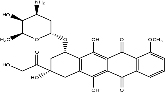 |
Figure 1 Two-dimensional (2D) structure of doxorubicin. |
Modeling of hGH Fragment 176–191 3D Structure
The hGH fragment 176–191 peptide sequence (YLRIVQCRSVEGSCGF) was submitted to the PEP-FOLD3.5 webserver. It uses the Hidden Markov Model sub-optimal conformation sampling approach for predicting small peptide 3D structures. After model generation using PEP-FOLD3.5, the 3D structure was further refined and assessed for quality using the MolProbity tool of the Swiss Model server. Finally, the suitability of the peptide structure for docking was analyzed using the Ramachandran plot function in Discovery Studio Visualizer.
Modeling of Target Protein Structures
The 3D structure files of the breast cancer biomarkers estrogen receptor (PDB:1QKU), progesterone receptor (PDB:2OVH), human epidermal growth factor receptor (PDB:3GOP), mind bomb (Mib) protein (PDB:4TSE), and Ki-67 (PDB:5J28) were then obtained from Protein Data Bank25 (Figure 2; Table 1). Published structures were subsequently edited in order to remove non-standard residues by making use of the Discovery Studio Visualizer 2021. For energy minimization, the UCSF Chimera program (www.cgk.ucsf.edu>chimera) with the following conjugate gradient minimization settings was used: removal of steric collision with steepest descent steps set to 1000, steepest descent size to 0.02 Å, conjugate gradient steps set to 1000, as well as the conjugate gradient step size to 0.02 Å. Energy minimized structures were saved as PDB files for docking simulations.26
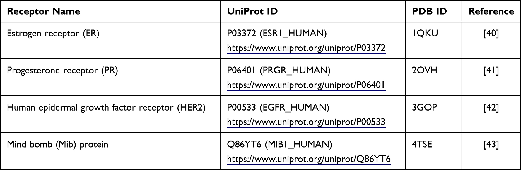 |
Table 1 Structural Information on Selected Breast Cancer Biomarkers |
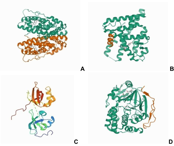 |
Figure 2 Crystal 3D structure of (A) estrogen receptor (PDB:1QKU), (B) progesterone receptor (PDB: 2OVH), (C) human epidermal growth factor receptor (PDB:3GOP), and (D) mind bomb (Mib) protein (PDB:4TSE), Ki-67 (PDB:5J28). All 3D graphics were obtained from RCSB-PDB. |
Predicting Peptide–Receptor (Breast Cancer Biomarker) Interactions
Receptor–peptide complexes were modeled by the PatchDock online docking server (https://bioinfo3d.cs.tau.ac.il/PatchDock/), which makes use of a geometry-based molecular docking algorithm as a scoring function.27 After docking analysis, complexes were additionally refined by using the FireDock online server.28
Molecular Docking Analysis
Docking of doxorubicin with tumor biomarkers (receptors) was simulated using Autodock Version 4.2.23,29 Autodock searches for the best conformation of receptor and doxorubicin were on the basis of its binding energy. Water molecules were subsequently removed from the 3D files of receptor molecules prior to docking and then later on hydrogen atoms were added to all the target proteins. Gasteiger charges were also added to the receptors. A 60×60×60 Å grid box was also set for covering the entire surface of the receptor with a default grid point spacing of 0.375 Å. The Lamarckian genetic algorithm (LGA)25 was then used for making receptor-drug flexible docking calculations. The LGA parameters were set as follows: population size (ga_pop_size) = A, energy evaluations (ga_num_generation) = B, mutation rate = C, crossover rate = D, step size = E, and number of LGA runs = 10. Molecular docking simulations of doxorubicin to peptide-bound receptors were also conducted using a 75×75×75 Å grid and default parameters.
Part - II: Nanoparticle Preparation
Doxorubicin, low molecular weight (LMW) Chitosan (95% deacetylated) and glacial acetic acid were purchased from Sigma-Aldrich (Merck group, U.S.A.) while gum Arabica (Gum Acacia) was purchased locally from Gizan, Jazan Province. Other pharmaceuticals along with analytical grade materials were used in the present study as received.
Preparation of Dual hGH Fragment 176–191 Peptide- and Doxorubicin-Loaded Chitosan Nanoparticles (Ch-hGH-DOX)
Chitosan nanoparticles were synthesized using the ionic gelation method of Avadi et al,30 with slight modification.31 Negatively charged gum Arabica was dissolved in water at normal room temperature under constant stirring, while positively charged LMW Chitosan was dissolved in 0.1% aqueous acetic acid also at normal room temperature, and under constant stirring. The pH of both solutions was subsequently adjusted to 5.5 with the addition of 0.5 M sodium hydroxide. The indicated concentrations of doxorubicin and hGH 176–191 fragment were added to the Chitosan solution. Nanoparticles were then prepared by making use of dropwise addition of gum Arabica to LMW Chitosan solution under constant magnetic stirring (200–300 rpm) at normal room temperature for three hours. Composition of the optimal dual-loaded nanoparticles is presented below in Table 2.
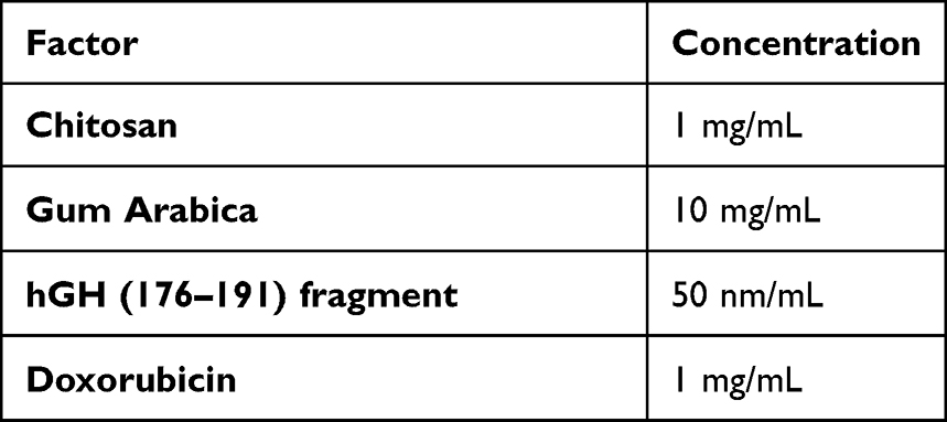 |
Table 2 Composition of Dual-Loaded Chitosan Nanoparticles |
Measurement of Particle Size, Polydispersity Index, as Well as the Zeta Potential
Photon correlation spectroscopy was used for measuring the mean particle size (PS), polydispersity index (PdI), as well as the zeta potential (ZP) (Malvern, Nano ZS90, UK). All formulations were dissolved in double-distilled water (Millipore) prior to the analysis. To avoid multi-scattering effects, the appropriate particle concentration was obtained for each dilution. All measurements were made in triplicate.
Part - III: Effects of Ch-hGH-DOX on Breast Cancer Cell Viability
Human MCF-7 breast cancer cells were procured for the present study from American Type Culture Collection (ATCC, Manassas, VI, USA) and were grown in RPMI-1640 medium (pH 7.4). It was further supplemented with 10% fetal bovine serum (FBS), 100 U/mL penicillin, 100 g/mL streptomycin. For both propagation as well as the treatment, cells were maintained in an incubator (Heraeus, GmbH, Germany) at 37°C; humidity (90%) along with CO2 (5%).
Cell Viability Assay
The cytotoxicity profiles of the indicated formulations were analyzed by making use of the 3-(4, 5-dimethylthiazol-2-yl)-2, 5-diphenyltetrazolium bromide (MTT) microculture tetrazolium viability assay.32 Cells were then seeded in multi-well plates and later incubated for 48 hours with the indicated concentration of Ch-hGH-DOX, Ch-DOX, or DOX (0–2.5 µg/mL). Each concentration was tested in triplicate, with each plate also including untreated controls along with a blank cell-free control. Subsequent to the incubation, MTT (5 mg/mL) was then added to each of the wells and the plates were further incubated for another 4 hours. The medium was then removed and DMSO (100 µL/well) was added for solubilizing the formazan crystals formed from MTT by viable cells. The optical density (OD) at 490 nm was recorded as an estimate of remaining viable cell number by making use of a microtiter plate reader (BioTek Instruments).
Growth inhibition (%) was calculated from the OD according to the following formula:

The cytotoxicity of the sample on cancer cells was expressed as the IC50 (the concentration that reduces the OD by 50% relative to untreated cells).
Statistical Analysis
Statistical Package for the Social Sciences (SPSS-10 Inc., Chicago, IL., USA) version 23 was used for data analysis. All data are presented as mean ± standard deviation (SD). One-way ANOVA was used for comparing treatment group means. p<0.05 was considered statistically significant.
Results and Discussion
In silico Analysis
The estrogen receptor (ER), progesterone receptor (PR), human epidermal growth factor receptor 2 (HER2), and Mib1/Ki-67 proliferation index are firmly established biomarkers used for the diagnosis, prognosis, and treatment guidance of primary, recurrent, and metastatic breast cancer patients. Additionally, these biomarkers are established therapeutic targets for breast cancer as well.
The 3D Structure of hGH Fragment 176–191 Peptide
In total, 63.64% of peptide residues were in the favorable zone for docking simulation according to the Ramachandran plot, while only 9.09% were in Ramachandran Outlier and 7.69% in Rotamer Outlier regions (Figure 3A).33 In addition, the MolProbity score was 2.00, and no C-Beta deviations, bad angles, or bonds were found34 (Figure 3B).35 Thus, the peptide 3D structure proved suitable for docking analysis.
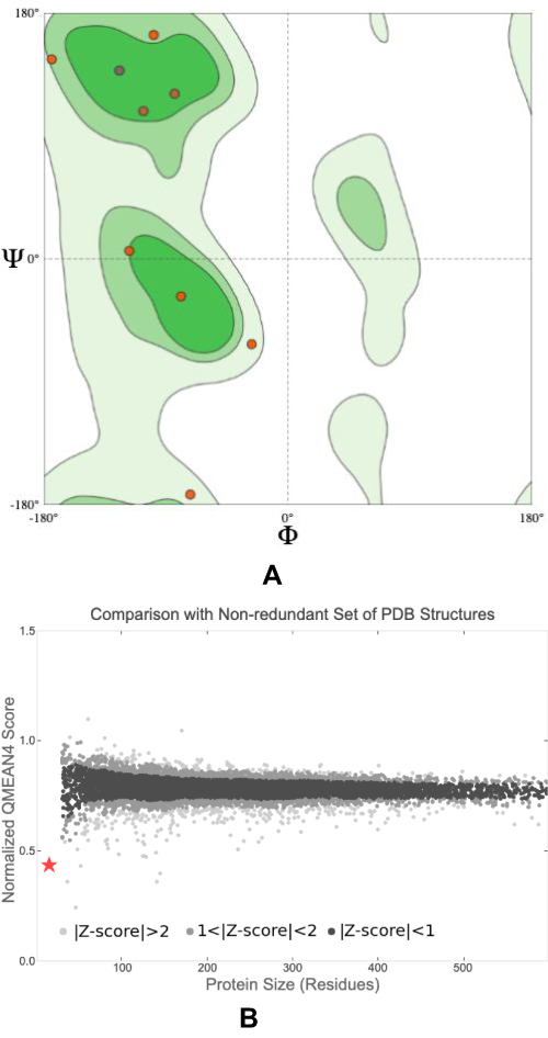 |
Figure 3 Quality of the hGH fragment 176–191 peptide 3D-model structure (A) Ramachandran plot showing distribution of peptide residues -phi (Φ) is the N(i-1), C(i), Ca(i), N(i) torsion angle and psi (Ψ) is the C(i), Ca(i), N(i), C(i+1) torsion angle. (B) The QMean score of modeled peptide after comparing with Non redundant set of PDB structures (★) is showing model on Z score plot when comparing it with structure available in PDB database. |
The docking simulations in our study depicted that the hGH fragment 176–191 peptide binds with high affinity to Ki-67, MiB protein, and the estrogen receptor, but not with the progesterone receptor or HER2 (Table 3). The peptide also influenced simulated doxorubicin binding to these breast cancer receptors (Table 4). For example, the binding energy score for doxorubicin to the progesterone receptor was −10.09 kcal/mol in the absence of hGH fragment 176–191 peptide, but was −11.31 kcal/mol after peptide binding. Furthermore, peptide binding reduced the doxorubicin inhibition constant for the progesterone receptor from 139.01 to 14.41 nM. In addition to the progesterone receptor, hGH fragment 176–191 peptide binding also augmented doxorubicin binding to HER2 and MiB protein, but slightly reduced binding to the estrogen receptor and Ki-67.
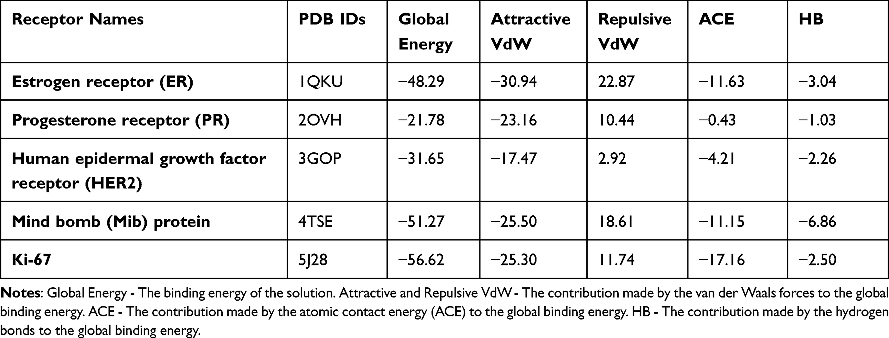 |
Table 3 Docking Results of the hGH Fragment 176–191 Peptide Model with Breast Cancer Receptor Models After Refinement Using the FireDock Server |
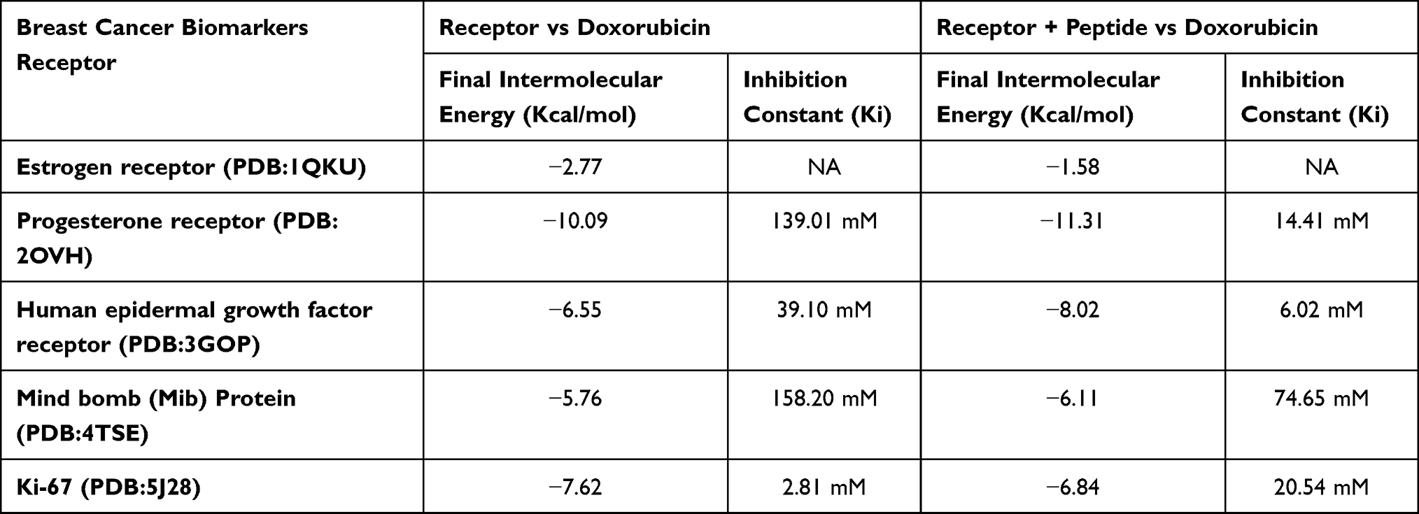 |
Table 4 Docking Results for Doxorubicin with Unbound Breast Cancer Receptors and hGH Fragment 176–191 Peptide-Bound Breast Cancer Receptors |
There is compelling evidence indicating that progesterone receptors (PRs) playing a hierarchical role in breast cancer growth and therefore they might potentially be useful in improving the success of endocrine treatments.36 Likewise, Ki-67 and MiB proteins promoting the proliferation in breast cancer can be the choice of targeting breast cancer treatment (Soliman and Yussif, 2016). Increased affinity of doxorubicin to the progesterone receptor, Ki-67 and MiB protein hence proves worthiness of the hGH fragment 176–191 peptide in targeting breast cancer cells with increased efficacy.
In silico work presents with a good predictive model of what may actually happen in the cellular environment. Therefore, the efficacy of the hGH fragment 176–191 peptide was further evaluated in in-vitro condition.
Nanoparticle Preparation
Ch-hGH-DOX was successfully prepared by the ionic gelation technique developed by Avadi et al to produce Chitosan nanoparticles from a mixture of Chitosan and gum Arabica.31 The electrostatic interactions occurring between the positively charged Chitosan as well as the negatively charged Arabica gum also facilitate the incorporation of other charged compounds such as peptides. The formula for the synthesis of Ch-hGH-DOX (Table 3) was optimized on the basis of PS, uniformity of dispersion, and ZP.
PS, Polydispersity Index, and ZP
These Ch-hGH-DOX nanoparticles were first examined for PdI value, ZP, and PS distribution using a Malvern Zetasizer. Mean PS (90.25 nm), PdI (0. 0.197), and ZP (31.7 mV) were all considered acceptable for subsequent testing in biological preparations (Table 5; Figure 4A and B).
 |
Table 5 Physiochemical Characterization of Ch-hGH-DOX by Photon Correlation Spectroscopy |
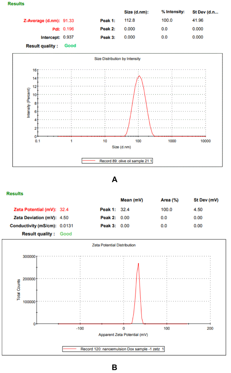 |
Figure 4 (A) Size distribution of Ch-hGH-DOX nanoparticles (size = 91.33 d.nm) (n = 1), (B) Zeta potential distribution of Ch-hGH-DOX nanoparticles (ZP = 32.4 mV) (n = 1). |
In the present study, we found the particle size to be less than 100 nm and hence we can suggest that these nanoparticles can be considered suitable for the effective delivery of the chemotherapeutic agents. According to Danaei et al 2018,37 nanocarriers having a particle size lesser than 150nm (or 200 nm) are considered to be extremely beneficial and effective for the treatment of cancers as they passively target the tumor cells by means of improved permeability as well as retention.37 The polydispersity index (PdI) of Ch-hGH-DOX nanoparticles was found to be less than 0.2 this indicates that our formulation not only has a monodispersity conduct but it also has a much lesser tendency towards aggregation.38 With regard to the Zeta potential it was observed in the present study that the Ch-hGH-DOX nanoparticles showed high positive values. With a ZP greater than 30 mV it can be said that Ch-hGH-DOX nanoparticles have a tendency of stabilizing colloidal preparations and thus preventing particle aggregation.39
With the aforementioned findings, we can conclude that the Ch-hGH-DOX nanoparticles prepared by the present method have shown to possess characteristics that are favorable for cellular uptake as well as having colloidal stability, including suitable diameter, surface charge along with a low polydispersity index that which is indicative of a reasonably homogeneous size distribution.
Anticancer Efficacy of Ch-hGH-DOX Nanoparticles
Finally, we compared the cytotoxic effects of these Ch-hGH-DOX nanoparticles to Ch-DOX nanoparticles. MCF-7 breast cancer cells when treated with varied concentrations of Ch-hGH-DOX nanoparticles for 48 hours yielded an IC50 value lower than those for doxorubicin alone and doxorubicin-loaded Chitosan nanoparticles (1.5 µg/mL vs 1.85 µg/mL as well as 1.7 µg/mL, receptively) (Figure 5). In contrast, neither unloaded Chitosan nanoparticles nor peptide-loaded Chitosan nanoparticles demonstrated substantial cytotoxicity (Figure 6). Furthermore, within the intermediate dose range (0.15 to 1.25 mg/mL), the proportion of viable MCF-7 cells remaining was significantly lower after 48 hours of Ch-hGH-DOX treatment than after Ch-DOX or DOX treatment (Figure 5). Therefore, hGH fragment 176–191 peptide appears to substantially enhance the toxicity of doxorubicin-loaded Chitosan nanoparticles, possibly by enhancing doxorubicin binding to breast cancer target proteins (Table 4).
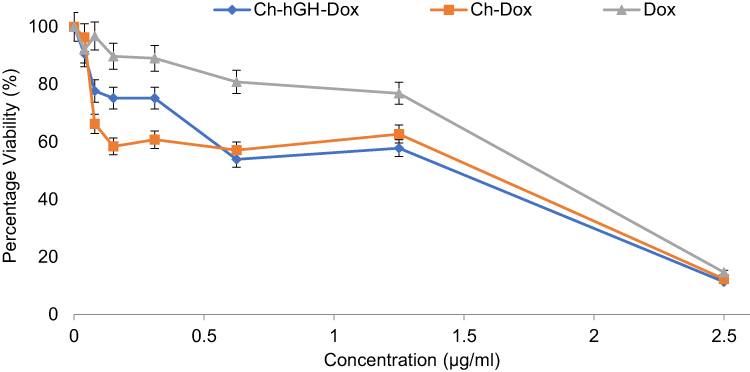 |
Figure 5 Cytotoxicity of the different Chitosan nanoparticle formulations against MCF-7 cells. Cells were treated for 48 hours and remaining viable cells estimated by MTT assay. |
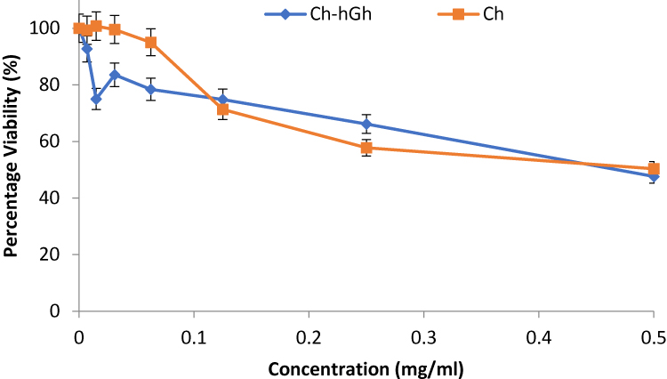 |
Figure 6 Cytotoxicity of different blank formulations against MCF-7 cells. Cells were treated and viability measured as described in Figure 4. |
Some of the potential limitations of the study are its inability to presently conduct any morphological studies, entrapment efficiency, as well as in-vitro release study.
Conclusion
Chitosan nanoparticles loaded with both doxorubicin and hGH fragment 176–192 peptide demonstrate both favorable physicochemical properties and enhanced cytotoxic efficacy against breast cancer cells. Molecular docking simulations suggest that the enhanced cytotoxicity stems from greater binding of doxorubicin to peptide-bound target proteins compared to unbound targets. Moreover, the inclusion of hGH fragment 176–192 peptide may promote accumulation of doxorubicin-loaded Chitosan nanoparticles in breast tumors while reducing non-target tissue exposure, thereby enhancing therapeutic efficacy and reducing side effects.
Acknowledgments
The author N.K.S., would like to dedicate this manuscript to his beloved and most dear father Mr. Alhaj Syed Maqbool who recently passed away. M.A.K would also like to dedicate this manuscript to his beloved father Dr Ahmed Al-kasim who passed away. The authors would like to dedicate the manuscript to Prof. Mamdouh and Mr. Mohammed Baity, our respected colleagues who passed away very recently.
Funding
This research was funded by the Deanship of Scientific Research, Jazan University, Jazan, Saudi Arabia; grant number JUP8/318.
Disclosure
The authors report no conflicts of interest in this work.
References
1. Sung H, Ferlay J, Siegel RL, et al. Global cancer statistics 2020: GLOBOCAN estimates of incidence and mortality worldwide for 36 cancers in 185 countries. CA Cancer J Clin. 2021;71(3):209–249. doi:10.3322/caac.21660
2. Thundimadathil J. Cancer treatment using peptides: current therapies and future prospects. J Amino Acids. 2012;2012:967347. doi:10.1155/2012/967347
3. Kakde DD, Kakde RB, Patil AT, Jain D, Shrivastava VK. Cancer therapeutics- opportunities, challenges and advances in drug delivery. J Appl Pharma Sci. 2011;1:1–10.
4. Enback J, Laakkonen P. Tumour-homing peptides: tools for targeting, imaging and destruction. Biochem Soc Trans. 2007;35(Pt 4):780–783. doi:10.1042/BST0350780
5. Dorsam RT, Gutkind JS. G-protein-coupled receptors and cancer. Nat Rev Cancer. 2007;7(2):79–94. doi:10.1038/nrc2069
6. Aina OH, Sroka TC, Chen ML, Lam KS. Therapeutic cancer targeting peptides. Biopolymers. 2002;66(3):184–199. doi:10.1002/bip.10257
7. Meng L, Yang L, Zhao X, et al. Targeted delivery of chemotherapy agents using a liver cancer-specific aptamer. PLoS One. 2012;7(4):e33434. doi:10.1371/journal.pone.0033434
8. Zhang XX, Eden HS, Chen X. Peptides in cancer nanomedicine: drug carriers, targeting ligands and protease substrates. J Control Release. 2012;159(1):2–13. doi:10.1016/j.jconrel.2011.10.023
9. Vlieghe P, Lisowski V, Martinez J, Khrestchatisky M. Synthetic therapeutic peptides: science and market. Drug Discov Today. 2010;15(1–2):40–56. doi:10.1016/j.drudis.2009.10.009
10. Anandhakumar S, Krishnamoorthy G, Ramkumar KM, Raichur AM. Preparation of collagen peptide functionalized chitosan nanoparticles by ionic gelation method: an effective carrier system for encapsulation and release of doxorubicin for cancer drug delivery. Mater Sci Eng C Mater Biol Appl. 2017;70(Pt 1):378–385. doi:10.1016/j.msec.2016.09.003
11. Varshosaz J, Hassanzadeh F, Aliabadi HS, Khoraskani FR, Mirian M, Behdadfar B. Targeted delivery of doxorubicin to breast cancer cells by magnetic LHRH chitosan bioconjugated nanoparticles. Int J Biol Macromol. 2016;93(Pt A):1192–1205. doi:10.1016/j.ijbiomac.2016.07.025
12. Hemati Azandaryani A, Kashanian S, Derakhshandeh K. Folate conjugated hybrid nanocarrier for targeted letrozole delivery in breast cancer treatment. Pharm Res. 2017;34(12):2798–2808. doi:10.1007/s11095-017-2260-x
13. Gomhor J, Alqaraghuli H, Kashanian S, Rafipour R, Mahdavian E, Mansouri K. Development and characterization of folic acid-functionalized apoferritin as a delivery vehicle for epirubicin against MCF-7 breast cancer cells. Artif Cells, Nanomed Biotechnol. 2018;46(sup3):S847–S854. doi:10.1080/21691401.2018.1516671
14. Strobl JS, Thomas MJ. Human growth hormone. Pharmacol Rev. 1994;46(1):1–34.
15. Møller N, Jørgensen JO. Effects of growth hormone on glucose, lipid, and protein metabolism in human subjects. Endocr Rev. 2009;30(2):152–177. doi:10.1210/er.2008-0027
16. Vijayakumar A, Novosyadlyy R, Wu Y, Yakar S, LeRoith D. Biological effects of growth hormone on carbohydrate and lipid metabolism. Growth Hormone IGF Res. 2010;20(1):1–7. doi:10.1016/j.ghir.2009.09.002
17. Zekri A, Ghaffari SH, Yousefi M, et al. Autocrine human growth hormone increases sensitivity of mammary carcinoma cell to arsenic trioxide-induced apoptosis. Mol Cell Endocrinol. 2013;377(1–2):84–92. doi:10.1016/j.mce.2013.07.002
18. de Vos AM, Ultsch M, Kossiakoff AA. Human growth hormone and extracellular domain of its receptor: crystal structure of the complex. Science. 1992;255(5042):306–312. doi:10.1126/science.1549776
19. Heffernan M, Summers RJ, Thorburn A, et al. The effects of human GH and its lipolytic fragment (AOD9604) on lipid metabolism following chronic treatment in obese mice and beta(3)-AR knock-out mice. Endocrinology. 2001;142(12):5182–5189. doi:10.1210/endo.142.12.8522
20. Heffernan MA, Jiang WJ, Thorburn AW, Ng FM. Effects of oral administration of a synthetic fragment of human growth hormone on lipid metabolism. Am J Physiol Endocrinol Metab. 2000;279(3):E501–7. doi:10.1152/ajpendo.2000.279.3.E501
21. Ng FM, Sun J, Sharma L, Libinaka R, Jiang WJ, Gianello R. Metabolic studies of a synthetic lipolytic domain (AOD9604) of human growth hormone. Horm Res. 2000;53(6):274–278. doi:10.1159/000053183
22. Wishart DS, Knox C, Guo AC, et al. DrugBank: a knowledgebase for drugs, drug actions and drug targets. Nucleic Acids Res. 2008;36(Databaseissue):D901–6. doi:10.1093/nar/gkm958
23. Morris GM, Huey R, Lindstrom W, et al. AutoDock4 and AutoDockTools4: automated docking with selective receptor flexibility. J Comput Chem. 2009;30(16):2785–2791. doi:10.1002/jcc.21256
24. Brooks BR, Brooks CL, Mackerell AD, et al. CHARMM: the biomolecular simulation program. J Comput Chem. 2009;30(10):1545–1614. doi:10.1002/jcc.21287
25. Sehnal D, Bittrich S, Deshpande M, et al. Mol* Viewer: modern web app for 3D visualization and analysis of large biomolecular structures. Nucleic Acids Res. 2021;49(W1):W431–W437. doi:10.1093/nar/gkab314
26. Pettersen EF, Goddard TD, Huang CC, et al. UCSF Chimera–a visualization system for exploratory research and analysis. J Comput Chem. 2004;25(13):1605–1612. doi:10.1002/jcc.20084
27. Mashiach E, Schneidman-Duhovny D, Andrusier N, Nussinov R, Wolfson HJ. FireDock: a web server for fast interaction refinement in molecular docking. Nucleic Acids Res. 2008;36(WebServer issue):W229–32. doi:10.1093/nar/gkn186
28. Ansari MA, Jamal QMS, Rehman S, et al. TAT-peptide conjugated repurposing drug against SARS-CoV-2 main protease (3CLpro): potential therapeutic intervention to combat COVID-19. Arab J Chem. 2020;13(11):8069–8079. doi:10.1016/j.arabjc.2020.09.037
29. Goodsell DS, Morris GM, Olson AJ. Automated docking of flexible ligands: applications of AutoDock. J Mol Recognit. 1996;9(1):1–5. doi:10.1002/(sici)1099-1352(199601)9:1<1::aid-jmr241>3.0.co;2-6
30. Avadi MR, Sadeghi AM, Mohamadpour Dounighi N, Dinarvand R, Atyabi F, Rafiee-Tehrani M. Ex vivo evaluation of insulin nanoparticles using chitosan and Arabic gum. ISRN Pharm. 2011;2011:860109. doi:10.5402/2011/860109
31. Avadi MR, Zohuriaan-Mehr MJ, Younessi P, Amini M, Tehrani MR, Shafiee A. Optimized synthesis and characterization of N-triethyl chitosan. J Bioact Compat Polym. 2003;18(6):469–479. doi:10.1177/0883911503040432
32. Syam S, Abdelwahab SI, Al-Mamary MA, Mohan S. Synthesis of chalcones with anticancer activities. Molecules. 2012;17(6):6179–6195. doi:10.3390/molecules17066179
33. Chen VB, Arendall WB, Headd JJ, et al. MolProbity: all-atom structure validation for macromolecular crystallography. Acta Crystallogr D Biol Crystallogr. 2010;66(Pt 1):12–21. doi:10.1107/S0907444909042073
34. Benkert P, Biasini M, Schwede T. Toward the estimation of the absolute quality of individual protein structure models. Bioinformatics. 2011;27(3):343–350. doi:10.1093/bioinformatics/btq662
35. Waterhouse A, Bertoni M, Bienert S, et al. Swiss-MODEL: homology modelling of protein structures and complexes. Nucleic Acids Res. 2018;46(W1):W296–W303. doi:10.1093/nar/gky427
36. Giulianelli S, Molinolo A, Lanari C. Targeting progesterone receptors in breast cancer. Vitam Horm. 2013;93:161–184. doi:10.1016/B978-0-12-416673-8.00009-5
37. Danaei M, Dehghankhold M, Ataei S, et al. Impact of particle size and polydispersity index on the clinical applications of lipidic nanocarrier systems. Pharmaceutics. 2018;10(2):57. doi:10.3390/pharmaceutics10020057
38. Jirgensons B, Straumanis ME. A Short Textbook of Colloid Chemistry. Elsevier Science; 2013.
39. Gonzalez-Mira E, Egea MA, Souto EB, Calpena AC, García ML. Optimizing flurbiprofen-loaded NLC by central composite factorial design for ocular delivery. Nanotechnology. 2011;22(4):045101. doi:10.1088/0957-4484/22/4/045101
40. Gangloff M, Ruff M, Eiler S, Duclaud S, Wurtz JM, Moras D. Crystal structure of a mutant hERα ligand-binding domain reveals key structural features for the mechanism of partial agonism. J Biol Chem. 2001;276(18):15059–15065. doi:10.1074/jbc.M009870200
41. Madauss KP, Grygielko ET, Deng SJ, et al. A structural and in vitro characterization of asoprisnil: a selective progesterone receptor modulator. Mol Endocrinol. 2007;21(5):1066–1081. doi:10.1210/me.2006-0524
42. Red Brewer M, Choi SH, Alvarado D, et al. The juxtamembrane region of the EGF receptor functions as an activation domain. Mol Cell. 2009;34(6):641–651. doi:10.1016/j.molcel.2009.04.034
43. McMillan BJ, Schnute B, Ohlenhard N, et al. A tail of two sites: a bipartite mechanism for recognition of notch ligands by mind bomb E3 ligases. Mol Cell. 2015;57(5):912–924. doi:10.1016/j.molcel.2015.01.019
 © 2022 The Author(s). This work is published and licensed by Dove Medical Press Limited. The
full terms of this license are available at https://www.dovepress.com/terms
and incorporate the Creative Commons Attribution
- Non Commercial (unported, 3.0) License.
By accessing the work you hereby accept the Terms. Non-commercial uses of the work are permitted
without any further permission from Dove Medical Press Limited, provided the work is properly
attributed. For permission for commercial use of this work, please see paragraphs 4.2 and 5 of our Terms.
© 2022 The Author(s). This work is published and licensed by Dove Medical Press Limited. The
full terms of this license are available at https://www.dovepress.com/terms
and incorporate the Creative Commons Attribution
- Non Commercial (unported, 3.0) License.
By accessing the work you hereby accept the Terms. Non-commercial uses of the work are permitted
without any further permission from Dove Medical Press Limited, provided the work is properly
attributed. For permission for commercial use of this work, please see paragraphs 4.2 and 5 of our Terms.