Back to Journals » International Journal of Nanomedicine » Volume 21
How Transformative Are ROS Responsive Hydrogel Nanoformulations to Treat Refractory Infected Wounds
Authors Liu Z, Xiang H, Xian K, Cai L, Yin D ![]() , Zhong J
, Zhong J
Received 23 October 2025
Accepted for publication 5 March 2026
Published 16 April 2026 Volume 2026:21 576258
DOI https://doi.org/10.2147/IJN.S576258
Checked for plagiarism Yes
Review by Single anonymous peer review
Peer reviewer comments 3
Editor who approved publication: Professor Farooq A. Shiekh
Zongjunlin Liu,1,* Hongqiong Xiang,1,* Ke Xian,2,* Lina Cai,1,* Dui Yin,1 Jianqiao Zhong1
1Department of Dermatology, the Affiliated Hospital of Southwest Medical University, Luzhou, 646000, People’s Republic of China; 2Clinical Medical College, Southwest Medical University, Luzhou, 646000, People’s Republic of China
*These authors contributed equally to this work
Correspondence: Jianqiao Zhong, Department of Dermatology, Affiliated Hospital of Southwest Medical University, No. 25 Tai Ping Jie, Luzhou, Sichuan, 646000, People’s Republic of China, Tel +86-15082088598, Fax +86-830-3198173, Email [email protected]
Abstract: Controlling infection in bacterial-infected wounds presents a formidable challenge, especially when compounded by additional high-risk factors that hinder the healing process during clinical interventions. Currently, the efficacy of standard therapeutic approaches often falls short, largely attributed to persistent local infections and inflammatory responses that impede healing cascades. In recent years, increasing reports on the management of hard-to-heal infected wounds have highlighted the potential of reactive oxygen species (ROS)-responsive hydrogels to facilitate wound recovery. The capacity of these hydrogels could be greatly enhanced by integrating them with traditional treatments, thereby addressing the complexities of healing in refractory infected wounds. Therefore, based on a thorough review of latest literatures, this paper comprehensively outlines the molecular mechanisms associated with hard-to-heal infected wounds, the role of ROS-responsive hydrogels in promoting healing, and a combined therapeutic strategy for wound control. It is designed to offer valuable insights to inform and advance future investigative efforts in this research field.
Keywords: hard-to-heal infected wounds, reactive oxygen species, ROS, hydrogel, nanoformulations, combination therapy strategy
Introduction
Bacterial infection, a common complication of skin wounds, ranks among the most substantial factors in hindering wound healing.1,2 Once a wound is formed, it sequentially undergoes four critical stages – hemostasis, inflammation, proliferation and remodeling – to ensure a successful healing process.3–5 If bacterial infection occurs in a wound and suffers an ineffective treatment, however, the wound would stay in the inflammatory stage for an extended time.6,7 At present, the therapy of infected wound mainly relies on the use of local or systemic antibiotics.8 Recently, some new treatment modalities have developed, including photothermal/photodynamic therapy, gas therapy and metal ion therapy, which really promote the healing of infected wounds to a certain extent;9–12 but several clinical treatment periods have witnessed that infected wounds with high-risk factors like immunocompromised conditions, multi-drug-resistant bacterial infections, large wound areas and poor local nutritional supply, are often difficult to control by using current treatment methods, thereby leading to their chronic refractoriness.13,14 Worse still, prolonged inflammation in these wounds exacerbates the excessive production of local reactive oxygen species (ROS) that surpass the antioxidant capacity of cells, in turn damaging cellular membranes, proteins and DNA, further hindering vascularization and tissue regeneration.15–18 Consequently, the wound festers and evolves into a stubborn, infected wound that resists healing. Therefore, efforts from numerous researchers are persistent to develop effective strategies for above wounds, encompassing new antibiotics, more effective antibacterial material, among which ROS-responsive hydrogel dressings are specially outstanding owing to their unique physicochemical property and ability to scavenge ROS effectively.19,20
ROS-responsive hydrogel, as an innovative macromolecular material with a three-dimensional (3D) network structure, exhibits remarkable properties containing superior water absorption, swelling capabilities, permeability, histocompatibility, biodegradability, adjustability, as well as a composition that closely mimics the extracellular matrix.21,22 Moreover, it has a lack of cytotoxicity and an excellent adhesion to wounds.21 Leveraging their special ability to interact with ROS, ROS-responsive hydrogels enable the elimination of excessive ROS from refractory wounds. More importantly, based on the 3D structure, they experience ROS-responsive “autolysis” and sequentially release encapsulated drugs, thereby integrating anti-inflammatory, antimicrobial and pro-restorative effects to greatly accelerate the complex wounds healing.23 Owing to above distinctive attributes, ROS-responsive hydrogels are emerging as promising candidates for ideal wound dressings.24 Taken together, we hence discuss the molecular mechanisms and strategies for refractory infected wounds, as well as advancements of ROS-responsive hydrogels in wounds healing.
What Do We Understand About Refractory Infected Wounds?
Difficult-to-heal infected wounds are defined by their resistance to achieving satisfactory therapeutic outcomes with current clinical approaches.25,26 These wounds have suffered a history of failed treatments, which perpetuates the presence of inflammatory cells that persistently generate excessive ROS, culminating in oxidative stress.27–29 Both radical and non-radical forms of ROS, including hydrogen peroxide and singlet oxygen, impede cell migration and proliferation, thereby prolonging the inflammatory phase of wound healing.30–32 The potential molecular mechanisms involving ROS-mediated apoptosis and necrosis that contribute to the delay in wound healing process are described as following.
ROS Directly Mediates Apoptosis
ROS has the ability to directly influence the activity of apoptotic effectors such as cysteinyl asparaginase (caspases), Bcl-2, and cytochrome c, thereby initiating apoptosis.33 The catalytic function of caspases is dependent on the reduced state of Cys in their active site. Studies have shown that low levels of H2O2 can activate caspases, leading to the cleavage of a wide range of substrates (eg, PARP), DNA damage, loss of membrane integrity, and ultimately cell death through apoptosis.34,35 Mild elevated ROS is shown to induce the expression of Bcl-2 through activation of transcription factors such as NF-κB, as an adaptive response to promote cell survival.36 In contrast, in response to excessive ROS, JNK can phosphorylate and inhibit Bcl-2 function, allowing apoptotic processes to occur. Excessive ROS can also trigger cytochrome c peroxidase activity, which leads to cardiolipin peroxidation. Oxidized cardiolipin loses its binding affinity for cytochrome c, causing the dissociation and release of cytochrome c into the cytoplasm, ultimately inducing apoptosis.37,38
ROS Indirectly Mediates Apoptosis
Excessive ROS can indirectly mediate apoptosis by affecting many signaling pathways. A moderate increase in ROS can hinder apoptosis by activating the NF-κB pathway, thereby maintaining redox balance. However, a substantial increase in ROS can lead to the inactivation of NF-κB, resulting in limited expression of anti-apoptotic proteins (eg, Bcl-xL and XIAP).39–41 Furthermore, it inhibits the expression of antioxidant genes such as MnSOD which ultimately promotes cell death execution.42,43 The activation of the SAPK pathway, induced by ROS, triggers apoptosis in cells.44 Apoptosis-regulated signaling kinase 1 (ASK1) is normally inhibited by the reduced forms of thioredoxin (Trx) or glutamine-reducing protein (Grx). However, increased levels of ROS cause oxidation of Trx and Grx, resulting in the release and subsequent activation of ASK1.45 This activation leads to the phosphorylation and activation of c-Jun n-terminal kinase (JNK) and p38-MAPK, further activating P53, pro-apoptotic proteins, ultimately inducing cell death.46,47 JNK also plays a role in inhibiting Bcl-2 function through phosphorylation and promoting the release of cytochrome c from mitochondria, collectively initiating the apoptotic process.48,49
ROS-Mediated Cell Necrosis
Normal levels of ROS can regulate the activity of various macromolecules in cells through oxidative modification, thus controlling normal cellular metabolic processes.50,51 However, chronic infected wounds often exhibit an excessive amount of ROS in local cells, which continuously attack lipids, proteins and DNA, leading to severe and irreversible oxidative damage.52,53 This directly results in the inactivation of intracellular macromolecules, cell necrosis, and significantly impedes wound healing.54
In summary, the chronic non-healing wounds are marked by persistently elevated ROS that scramble cellular metabolism and inflict widespread damage on DNA, proteins and lipids, ultimately driving cells into apoptosis or necrosis.55 This oxidative assault cripples wound neovascularization, blocks fibroblast proliferation and paralyzes immune-cell phagocytosis, allowing wound infection to gain the upper hand and healing to stall indefinitely. The mechanism by which ROS cause delayed wound healing is shown in Figure 1.
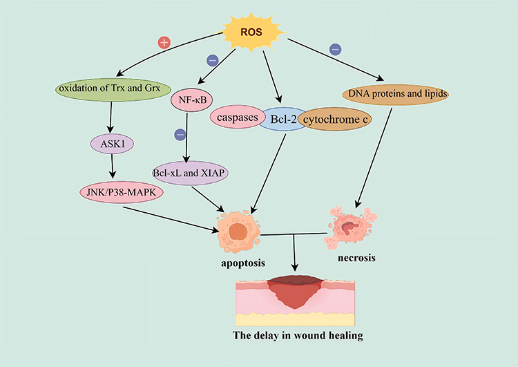 |
Figure 1 ROS-Driven Mechanisms That Delay Wound Healing. Excessive ROS in the local wound trigger apoptosis through three pathways: oxidizing thioredoxin (Trx) and glutaredoxin(Grx), silencing NF-κB signaling pathway, and directly disabling apoptotic effectors like caspases, Bcl-2, and cytochrome c.56,57 Additionally, superfluous ROS impair various intracellular macromolecules, eg. DNA, proteins and lipids, through oxidative modification, forcing cells into necrosis. The resulting patchwork of apoptosis and necrosis hinders neovascularization and matrix deposition, leaving the wound trapped in a chronic, non-healing state.58 |


Role of ROS-Responsive Hydrogels in Infected Wound Healing
Up to now, infected wounds have frequently relied on the application of topical or systemic antibiotics, supplemented with photothermal/photodynamic therapy,59 gas therapy10 and metal ion therapy.60 Although antibiotics effectively run in enhancing the healing rate of infected wounds, the prolonged use of antibiotics may elicit genetic mutations in some bacteria that would facilitate the production of drug-resistant genes.61,62 This has rendered the healing of certain infected wounds challenging, resulting in the development of chronic non-healing wounds. Even with the presence of wound dressings that possess antibacterial capabilities and ensure proper breathability, the complete healing of infected wounds that harbor drug-resistant bacteria continues to be a formidable challenge across various treatment modalities. These chronic, refractory wounds often exhibit high-level ROS locally, which substantially impede wound healing process.25,63 Considering this, researchers have put forward an innovative dressing – ROS-responsive hydrogel – that is designed to effectively control infection by scavenging excessive ROS and simultaneously releasing various active substances, thus facilitating the healing of these particularly refractory wounds.64
Characterization of ROS-Responsive Hydrogels
ROS-responsive hydrogel is a novel type of hydrogel dressing designed to eliminate excessive ROS from wounds.65 This unique hydrogel is characterized by its intricate, three-dimensional, reticulated macromolecular structure, which affords it carrying a diverse array of active ingredients.66–68 Its inherent ability to adapt its own structure and physical properties facilitates a precise, controlled release of drugs or targeted effects, remarkably enhancing the wound healing process.69,70 Boasting exceptional water absorption and swelling capabilities, along with breathability, histocompatibility, biodegradability, adjustability, extracellular matrix-like morphology, non-cytotoxicity, and strong wound adhesion, this hydrogel stands out as a cutting-edge solution in wound healing.68,71,72 The instinct of ROS-responsive hydrogels interaction with ROS enables them to eliminate excessive ROS from difficult-to-heal infected wounds;73,74 they, moreover, accurately and sustainably release the encapsulated drugs due to their dynamic structural changes, allowing for a potent combination of anti-inflammatory, antibacterial and pro-restorative effects that in turn expedite the healing process of refractory infected wounds.75 Consequently, ROS-responsive hydrogels emerge as a compelling choice for optimal wound dressings in the realm of wound care.76,77
Mechanism of ROS-Responsive Hydrogels in Scavenging ROS
The defining characteristic that differentiates ROS-responsive hydrogels from conventional hydrogels lies in their ROS responsiveness, which allows them to scavenge excessive ROS at infectious wound sites.78 The incorporation of ROS-sensitive chemical bonds as responsive moieties constitutes the core strategy for the synthesis of ROS-responsive hydrogels.79,80 These ROS-sensitive bonds can function either as crosslinkers within the hydrogel network or as integral components of the hydrogel polymer backbone/side chains.81 In the following section, we will provide a brief overview of the ROS-sensitive bonds/groups that have been reported in the current literature.
Phenylboronic Acid
In the preparation of hydrogels, phenylboronic acid is incorporated as a crosslinker to form boronic ester bonds with the hydrogel polymer backbone.82,83 When the hydrogel is exposed to a high ROS environment, ROS can coordinate with the boron atom, leading to the formation of phenol and the cleavage of boronic ester bonds.84 This cleavage disrupts the hydrogel network structure, subsequently triggering the release of other active therapeutic agents encapsulated within the hydrogel, thereby promoting wound healing. Concurrently, phenylboronic acid can also conjugate with drugs, and upon response to reactive oxygen species, the active drugs are released.85 Li et al integrated phenylboronic acid-modified hyaluronic acid (HP), metal-phenolic networks (CaTA), methacryloylated carboxymethyl chitosan (MA-CMCS), and platelet-rich plasma (PRP) into a hydrogel system. Mediated by the CaTA component linked via dynamic borate ester bonds, this system enables ROS-responsive regulation of inflammatory responses.69
Thioacetal/Thioketal
The thione bond is a ROS-responsive covalent bond that can react with several canonical reactive oxygen species (ROS), such as superoxide anion (•O2−), hydroxyl radical (•OH), and hydrogen peroxide (H2O2), even at a threshold concentration as low as 100 μM, leading to its degradation into acetone and thiol.86,87 Incorporation of thione bonds into hydrogels confers ROS responsiveness to the hydrogel system.88 Specifically, upon exposure to ROS at a certain concentration, the hydrogel undergoes cleavage or other reactions, which scavenges excess ROS and triggers the intelligent release of other encapsulated therapeutic agents from the hydrogel matrix.23,89
Metal Nanoparticles
The incorporation of specific metal ions into hydrogels enables the scavenging of excess ROS at the wound site via redox cycling between the oxidized and reduced states of the metal ions. Metal ions are frequently incorporated into ROS-responsive hydrogels in the form of nanoparticles, including cerium oxide nanoparticles, ferrocene, manganese dioxide nanoparticles, and the like. For example, Cheng et al embedded cerium oxide nanoparticles within hydrogel dressings. Leveraging the redox transition between Ce3⁺ (reduced state) and Ce4⁺ (oxidized state), the system effectively diminishes ROS levels, thereby conferring ROS responsiveness to the hydrogel.90 Tian et al engineered a smart hydrogel using carboxymethyl chitosan (CMCS) as the primary polymeric scaffold Dynamic, reversible cross-links were established via host–guest interactions between β-cyclodextrin (βCD) and ferrocene (Fc).91 Leveraging ferrocene’s redox-active nature—capable of switching between oxidized and reduced forms—the hydrogel effectively scavenges surplus ROS, thereby accelerating wound repair.91–93 Song et al successfully incorporated manganese dioxide microparticles into a multifunctional hydrogel.94 Manganese dioxide not only exerts bactericidal effects via photothermal therapy (PTT) but also catalyzes the decomposition of hydrogen peroxide and releases oxygen, thereby effectively ameliorating the inflammatory microenvironment.94 Consequently, the hydrogel significantly promotes cell proliferation, migration, angiogenesis, collagen deposition, and tissue regeneration.94
Mechanisms of ROS-Responsive Hydrogels Promoting Wound Healing
Numerous studies have demonstrated that ROS-responsive hydrogel dressings significantly enhance the healing process of various difficult-to-heal infected wounds. The ROS-responsive hydrogel encouraging wound recovery involves a variety of specific mechanisms, mainly containing excessive ROS elimination, redox balance restoration, antimicrobial activity, and the provision of a environment conducive to wound healing.25,95,96 The mechanism by which ROS-responsive hydrogels accelerate infected wound healing is shown in Figure 2.
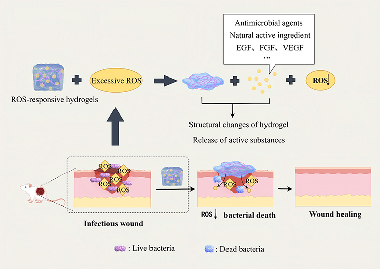 |
Figure 2 Mechanisms of ROS-Responsive Hydrogels Accelerating Infected-Wound Healing. Upon exposure to the ROS-rich milieu of infected wounds, ROS-responsive hydrogels interact with ROS, scavenge excess radicals and undergo in-situ structural rearrangement.54 This “on-demand” reaction steadily releases antibacterial agents, bioactive natural compounds and growth factors, simultaneously deactivating bacteria and quelling oxidative stress—creating a microenvironment that resists inflammation response and accelerates tissue repair.97,98 Note: ↓ indicates downregulation. |
Scavenging Excessive ROS and Regulating Redox Balance
The unique structure of ROS-responsive hydrogel specifically favors the removal of excessive ROS from the wound site, whether in vivo or vitro. By restoring ROS levels to the normal state, this intervention halts the assault on lipids and other macromolecules, thereby averting the formation of free radicals and the ensuing chain reactions.99 The mild elevation of ROS initially triggers the NF-κB pathway, which then enhances the expression of anti-apoptotic proteins like Bcl-xL and XIAP, effectively arresting the execution of cell death.100,101 Concurrently, the SAPK pathway remains inactive, while the MAPKK-JNK one does active, resulting in diminished production of P53 and pro-apoptotic proteins.102 This cascade of events leads to a reduction in local necrosis and apoptosis within the wound area, thereby facilitating wound healing.
Upregulation of M2-Phenotype Macrophages Polarization
Macrophages chiefly differentiate into pro-inflammatory (M1) and anti-inflammatory (M2) types in the process of wound healing.103 M1 macrophages are essential for clearing microorganisms and mediating inflammation at injury sites, while M2 macrophages play a crucial role in promoting anti-inflammatory effects, modulating wound adhesion, diminishing inflammation, and facilitating angiogenesis and tissue regeneration. In the late inflammatory phase of normal wounds, a gradual transition from M1 to M2 macrophages emerge along with healing progresses.104 However, some detrimental factors like bacterial infections could disrupt this polarization, resulting in persistent inflammation within the wound environment. Such interference would impair epithelial regeneration, collagen deposition and angiogenesis.104–106 The use of ROS-responsive hydrogels effectively scavenges local ROS and above harmful factors, which in turn encourages M2 macrophages polarization and amplifies anti-inflammatory responses, eventually accelerating the wound healing process.107,108
Establishment of a Microenvironment Conducive to Wound Healing
Besides eliminating ROS, ROS-responsive hydrogels also possess all the characteristics common to general hydrogels, featuring hydrophilicity and water retention, being non-cytotoxic, and adhering well to wounds and being easily detached from the wound surface.109 When applied to infectious wounds, ROS-responsive hydrogels can absorb the excessive local exudate, maintain a moist environment, and protect the wounds from further infection.20 In a word, ROS-responsive hydrogels, while protecting wounds, also offer a favorable local environment for wound healing.
Loading Active Pharmaceuticals
ROS-responsive hydrogels are provided with superior adjustability.66,70 Consequently, researchers have incorporated active substances such as antibiotics, natural antibacterial substances, nanoparticles, and growth factors into ROS-responsive hydrogels via various means, including encapsulation, during the preparation process, thereby enabling the sustained local release of various active drugs for wound healing. This targeted carrier approach focuses drugs at the infected wound site, preventing drug inactivation and minimizing the potential adverse effects typically associated with systemic medication to the greatest extent. We will further describe this combined treatment in the next section.
How Advanced are ROS Responsive Hydrogels to Treat Infected Wounds?
Although ROS-responsive hydrogels exhibit excellent biocompatibility and notable anti-inflammatory/antibacterial properties, their standalone capacity to combat bacteria and promote wound healing tends to be insufficient, thus leading to uniform therapeutic outcomes. To increase the effectiveness of these hydrogels in infected wound healing, researchers spare no effort in their integration with traditional treatment modalities. Therefore, some latest developments in combination therapies, ie, ROS-responsive hydrogels dressings combined with other vehicles such as antibiotics, photothermal/photodynamic therapy, gas therapy, metal ion therapy and pro-angiogenic therapy, are described as follows in order to guide their practical application in clinic. ROS-responsive hydrogel combination therapy is shown in Figure 3.
 |
Figure 3 ROS-Responsive Hydrogel Combination Therapy Strategies for Refractory Infected Wounds. ROS-responsive hydrogels are now integrated into five dominant combination paradigms: ROS-responsive hydrogels with antibiotics, ROS-responsive hydrogels with photothermal/ photodynamic therapy, ROS-responsive hydrogels with gas therapy, ROS-responsive hydrogels with metal ion therapy, and ROS-responsive hydrogels with angiogenesis therapy—each exploiting the ROS-triggered “on-demand” switch to synchronize antimicrobial potency with tissue repair.110,111 |
ROS-Responsive Hydrogels in Combination with Antibiotics
Antibiotics, whether systemic or topical application, are the mainstay for treating infections; however, the increase of drug-resistant bacteria has diminished the drug’s efficacy.112 Incorporating antibacterial agents into ROS-responsive hydrogels offers a targeted and controlled release strategy, effectively scavenging excess ROS and maintaining redox balance within infected regions. This innovative approach has been exemplified by the work of Qiao et al who prepared ROS-responsive HA-PBA/PVA (HPA) hydrogel through the crosslinking of hyaluronic acid grafted 3-aminophenylboronic acid (HA-PBA) and polyvinyl alcohol (PVA) via phenylboronic acid ester bonds.113 Then, they loaded hydrophilic moxifloxacin (M) and hydrophobic curcumin (Cur) into the HPA hydrogel to achieve the combined treatment of antibiotics and ROS-responsive hydrogel, obtaining HPA/M&Cur-PF.113 Through in vitro drug release experiments, they found that HPA/M&Cur-PF could rapidly release moxifloxacin and curcumin in the presence of H2O2, eliminating ROS while controlling bacterial infection. By establishing a mouse skin wound model infected with MRSA, they found that on the 7th and 14th days after administration, the wound closure rate in the HPA/M&Cur-PF group was significantly higher than that in the HPA group, suggesting that the combination of ROS-responsive hydrogel with antibiotics or other antibacterial substances has a significantly better therapeutic effect on refractory infectious wounds than using ROS-responsive hydrogel alone.113 Hu et al engineered a novel ROS-responsive nano-hydrogel by grafting phenylboronic acid onto alginate polymer chains, creating a system that provides localized antimicrobial and anti-inflammatory effects via preloaded with amikacin (AM) and naproxen (Nap).114 Their hydrogel has been demonstrated remarkable efficacy, reaching an inhibition rate of 90% against Staphylococcus aureus and 98% against Pseudomonas aeruginosa.114 Meanwhile, Zhao et al presented a hydrogel synthesized by cross-linking polyvinyl alcohol (PVA) with ROS-responsive linkers, designed to scavenge ROS.25 This hydrogel is impregnated with mupirocin and granulocyte-macrophage colony-stimulating factor (GM-CSF), and it gradually disintegrates under conditions of high reactive oxygen species (ROS), thereby releasing mupirocin and GM-CSF, that are vital contributors to infected wounds healing.25 It can effectively kill Staphylococcus aureus in the simulated microenvironment of a wound in vitro.25 The in vivo experiments revealed that after the hydrogel loaded with mupirocin was applied to the Staphylococcus aureus-infected wounds on the backs of mice for 16 hours, the colonization of Staphylococcus aureus at the wound site was significantly decreased.25 Cao et al deposited [2-(acryloyloxy)ethyl]trimethylammonium chloride (Bio-IL) and gelatin methacryloyl (GelMA) onto a doxycycline hydrochloride (DOXH)-loaded, reactive oxygen species (ROS)-degradable polyurethane (PFKU) nanofiber membrane via three-dimensional (3D) printing technology. Subsequent ultraviolet (UV) irradiation yielded conductive hydrogel strips. Notably, in a high ROS environment, the release rate of DOXH was significantly enhanced.115
ROS-Responsive Hydrogels in Combination with Photothermal/Photodynamic Therapy
Conventional photothermal therapy (PTT) and photodynamic therapy (PDT) often generate excessive ROS, which, while effective in inhibiting bacterial growth, may also trigger inflammation that further delays tissue regeneration and wound healing. However, integrating photosensitizing materials into ROS-responsive hydrogels achieves a synergistic therapeutic strategy, namely merging these hydrogels with photosensitizers employed in PTT or PDT. The incorporation of these materials confers a multitude of advantages, such as hemostasis in wounds, modulation of immune microenvironment, promotion of M2 macrophage polarization, antibacterial activity, enhancement of local angiogenesis and improvement of epithelial regeneration. Critically, this strategy favors the maintenance of redox balance, as it prevents the excessive ROS-induced damage to tissue cells, holding promise for applications in wound recovery. The typical combined approach was implemented by Wang et al, who developed an innovative, injectable bio-excited MnO2 hybrid hydrogel that responded to both redox and light stimuli (BMH).116 This cutting-edge hydrogel has demonstrated remarkable efficacy in accelerating wound healing, particularly in cases of multidrug-resistant infections, mainly through efficiently eradicating bacterial invasion and concurrently mitigating OS and inflammation within the wound’s microenvironment.116 Furthermore, reports from Wang et al showed an Ag/TiO2-doped PVA-hybridized hydrogel with potent bacteriostatic activity against Escherichia coli and Staphylococcus aureus, attributed to ROS release from its photodynamic effect under 660 nm visible light.116 In addition, Luo et al have crafted a novel near-infrared (NIR) photothermal hydrogel by incorporating α-lipoic acid-modified palladium nanoparticles into calcium ion-crosslinked sodium alginate matrix.117 This injectable hydrogel adeptly convers NIR light into localized hyperthermia, achieving the elimination of over 80% of E. coli or S. aureus and more than 60% of ROS in cells;117 it meanwhile downregulates the gene levels of TNF-α and IL-18 by 52.9% and 53.3%, respectively.117 Herein, Zhu et al designed PDA@MnO@CuO (PMC) nanoparticles using the high-efficiency photothermal agent polydopamine (PDA) as the core, along with manganese dioxide (MnO2) and copper oxide (CuO).118 The PMC nanoparticles and taurine were encapsulated in a polysaccharide-based hydrogel (FH) consisting of Falcaria vulgaris gum (FG) and hyaluronic acid (HA), thereby forming the FH-PMC-T hydrogel.118 The capacity of the FH-PMC-T hydrogel combined with mild photothermal therapy (PTT) to promote wound closure and functional skin regeneration was validated using a full-thickness rat wound model.118
ROS-Responsive Hydrogels in Combination with Gas Therapy
Although traditional gas therapy has positive effects on wound healing, it faces numerous limitations including the risk of sudden release, a short half-life, high reactivity and the possibility of producing carcinogenic by-products. However, through incorporating materials that generate therapeutic gases (eg, nitric oxide, hydrogen, oxygen, carbon monoxide and hydrogen sulfide) into ROS-responsive hydrogels, this innovative approach surpasses the conventional, intermittent ROS-triggered one owing to its prolonging the gas therapeutic effect and accelerating wound healing, thus offering a more sustained and effective prospect. Similarly, Yu et al have devised a cutting-edge sustainable wound healing system that leverages a H2O2-degradable hydrogel paired with a biosafe NO donor, l-Arginine, and employs low concentrations of H2O2 to rapidly address bacterial-infected open wounds.119 Within this system, l-Arginine is perpetually stimulated by H2O2, leading to the production of NO and in turn facilitating the chemotaxis of macrophage and fibroblast towards the wound site, as well promoting collagen synthesis.119 Consequently, this breakthrough approach rapidly enhances wound healing and skin regeneration.119 Moreover, the dynamic duo of H2O2 and NO exhibits a potent synergistic interaction, bolstering antimicrobial capabilities against the notoriously stubborn ampicillin-resistant Escherichia coli.119 In a groundbreaking study, Chen et al unveiled a hydrogen-producing hydrogel crafted from live Chlorella vulgaris and bacteria, capable of sustaining hydrogen generation for an impressive 60 hours—a feat that outshines the fleeting efficacy of traditional therapies;120 this hydrogel not only exhibits outstanding antioxidative properties but also efficiently scavenges ROS, paving the way for effective healing in chronically infected wounds.120 Coincidentally, Li pioneered a novel approach by enveloping the photosynthetic bacterium Spirulina platensis (SP) with carboxymethyl chitosan to create SP gel;121 this gel continuously produces oxygen, effectively combating acute or chronic tissue hypoxia.121 When subjected to 650 nm laser irradiation, SP releases chlorophyll, acting as a natural photosensitizer that generates ROS to photodynamically destroy bacteria in infected areas, thereby hastening wound healing.121 Ye et al developed a ROS-responsive DNA hydrogel (LGAH), into which ginseng-derived exosomes (G-Exos) and the nitric oxide (NO) donor L-arginine (L-Arg) were incorporated.110 Upon laser irradiation, L-Arg generates NO, and the synergistic effect of these components promotes the healing of infected wounds.110
ROS-Responsive Hydrogels in Combination with Metal Ion Therapy
For preferable treatment of refractory infected wounds, researchers have proposed incorporating some bacteriostatic metal ions (eg, Ag+, Zn2+, Cu2+) into ROS-responsive hydrogels, in favor of boosting antimicrobial properties and minimizing tissue irritation and toxicity. As a typical example, Hu et al have developed a novel ROS-responsive antimicrobial hydrogel, PAAg-PGFe, by integrating poly (acrylic acid) (PA), silver nanoparticles (AgNP) and polyglutamic acid (PG) enriched with Fe2+/Fe3+ ions;122 this hydrogel maintains its stability under normal conditions but swiftly disaggregates in the presence of ROS, triggering substantial oxidative stress that effectively eradicates bacteria--particularly Pseudomonas aeruginosa.122 Similarly, a versatile hydrogel (HA@Cur@Ag) has been ingeniously engineered by Shi and Chen et al who incorporated curcumin liposomes and AgNPs trading on the crosslinking between mercapto hyaluronic acid (SH-HA) and disulfide-bonded hyperbranched poly (ethylene glycol) (HB-PBHE) via a Michael addition reaction, boasting a suite of impressive properties, including ROS scavenging, bacteriostatic activity, anti-inflammatory effects, and angiogenesis promotion.123 Yet again, Bochani et al have developed a chitosan-based hydrogel, CT-TA-Fe-MnO2, that incorporates tannic acid (TA), iron (Fe) and manganese dioxide nanosheets; findings from experiments in vitro and in vivo not only confirmed its potent antimicrobial properties and wound-healing capabilities, but also revealed that CT-TA-Fe-MnO2 rarely affected vital organs, instead, reducing hemostasis time, enhancing anti-inflammatory responses and accelerating wound healing within 14 days.124 Zhang et al developed an infection-responsive antibacterial hydrogel (CBGCT), which is reinforced with copper ion-tannic acid nanosheets (Cu@TA) and guar gum. Upon exposure to acidic pH conditions or elevated reactive oxygen species (ROS) levels, the pH-responsive degradation behavior of Cu@TA nanosheets enables the sequential controlled release of copper ions and tannic acid from the CBGCT hydrogel, thereby potentiating angiogenesis and anti-inflammatory effects.125
ROS-Responsive Hydrogels in Combination with Pro-Angiogenic Therapy
In the proliferation stage, wounds are contingent upon cellular proliferation to encourage the formation of new granulation tissue. Apart from factors like infection and oxidative stress, local angiogenesis disorders are also crucial factors hindering the healing of refractory infectious wounds. The topical application of various growth factors contributes to elevation of cell proliferation, promotion of neovascularization and acceleration of wound healing. However, when applied directly, these growth factors quickly become inactive, thereby hindering their therapeutic efficacy.126 To enhance the proliferation phase of wound healing, numerous researchers spare no effort in developing a ROS-responsive hydrogel that incorporates diverse growth factors to promote angiogenesis. Encapsulating epidermal growth factors, transforming growth factors, fibroblast growth factors and others within ROS-responsive hydrogels affords them long-term maintenance of biological activity and a sustained release into wounds, hence optimizing their wound healing potential. This strategy effectively stimulates cell proliferation and angiogenesis, and enhances the nutritional supply to the wound area through upregulating angiopoietin-1 expression in wound cells and recruiting endothelial progenitor cells. The local continuous release of active growth factors is accomplished by the combination therapy of ROS-responsive hydrogels and growth factors, thereby accelerating wound healing. Zhu et al reported a ROS-responsive hydrogel loaded with fibroblast growth factor and metformin.127 In their research, it was uncovered that this hydrogel could potently recruit endothelial progenitor cells (EPCs) to promote angiogenesis and wound healing via upregulating ang1 expression.127 Likewise, Wang et al have designed an integrated hydrogel, called ITG-PEGDA@SA that is composed of polyethylene glycol/alginate, which inside serves as a ROS scavenger whereas the external sodium alginate layer degrades into a platform that delivers recombinant human epidermal growth factor (rhEGF), exhibiting a substantial promise for enhancing wound repairing and healing128 An et al synthesized a polyethylene glycol (PEG) responsive injectable hydrogel containing a thioketal bond (PEG-TK hydrogel), and combined it with epidermal growth factor (EGF) to obtain EGF@PEG-TK hydrogel.76 This hydrogel can eliminate excessive reactive oxygen species (ROS) at the local wound site and achieve sustained release of EGF.76 The in vivo experimental results indicated that the wound closure rates of PEG-TK and EGF@PEG-TK hydrogels for full-thickness skin defect wounds in Sprague-Dawley (SD) rats reached 88% and 90%, respectively, both higher than that of the control group (79%).76
The Future Prospective
Recent years have seen a surge in interest towards hydrogels, particularly ROS-responsive hydrogels, for treating infected wounds. These cutting-edge wound dressings go beyond the conventional capabilities of traditional hydrogels through effectively scavenging excess ROS, stimulating angiogenesis, exhibiting antimicrobial effects, and boosting M2 macrophage activity, all of which contribute significantly to enhanced wound recovery. Researchers are now looking into blending ROS-responsive hydrogels with traditional therapies to unlock synergistic benefits and enhance their therapeutic impact. However, the field of these hydrogels is still in its infancy. Major challenges include striking a wider balance of ROS, maintaining the hydrogel’s stability at the wound site, and refining the dressing’s precision and responsiveness. In summary, ROS-responsive hydrogel dressings still face several critical challenges and hurdles prior to clinical translation. Firstly, long-term safety assessments remain insufficient.129 ROS-responsive hydrogels typically have complex compositions, and the cleavage of ROS-responsive linkages may generate novel small-molecule byproducts—yet the potential adverse effects of these metabolites on host tissues have not been fully elucidated.130 Secondly, precise spatiotemporal regulation of drug release remains elusive. Given the substantial inter-individual and intra-tissue variability in endogenous ROS levels, active therapeutic agents encapsulated within the hydrogels are at risk of either “burst release” (premature rapid release) or “incomplete release” (insufficient drug delivery over the treatment window).131 Thirdly, clinical validation data are currently lacking. Most existing studies rely solely on animal models to evaluate safety profiles and therapeutic efficacy, with limited evidence from well-designed clinical trials to support their translational potential.132
Owing to the limitations in the authors’ expertise and professional scope, this study does not encompass the chemical synthesis methods and specific mechanisms of reactive oxygen species (ROS)-responsive hydrogels. Thorough research is crucial for developing effective ROS-responsive hydrogels suitable for clinical applications. Future research may further focus on the development of hydrogel formulations with enhanced stability, sensitivity, and safety, so as to accelerate the clinical translation of reactive oxygen species (ROS)-responsive hydrogels.133
Acknowledgments
All the figures in the text were drawn by Figdraw. The authors thank the Figdraw platform for its assistance in improving the quality of this article.
Author Contributions
Hongqiong Xiang, Zongjunlin Liu and Jianqiao Zhong were responsible for the design of conception and design. Hongqiong Xiang and Lina Cai contributed to the formation of the draft of the manuscript. Ke Xian, Jianqiao Zhong and Zongjunlin Liu are responsible for the design of figures in the manuscript. All authors made a significant contribution to the work reported, whether that is in the conception, study design, execution, acquisition of data, analysis and interpretation, or in all these areas; took part in drafting, revising or critically reviewing the article; gave final approval of the version to be published; have agreed on the journal to which the article has been submitted; and agree to be accountable for all aspects of the work.
Funding
The study was supported by the Science & Technology Bureau of Luzhou, Sichuan Province, China (Grant no: 2023JYJ039), Xuyong CountySouthwest Medical University Cooperation Project (2024XYXNYD08) and Natural Science Foundation of Sichuan Province (2026NSFSC1522).
Disclosure
The authors report no conflicts of interest in this work.
References
1. Negut I, Grumezescu V, Grumezescu AM. Treatment strategies for infected wounds. Molecules. 2018;23(9). doi:10.3390/molecules23092392
2. Resina L, Caballero P, Guggenbiller G, Weems AC, Pérez-Madrigal MM, Alemán C. Multifunctional scaffold biosensor and drug delivery system for bacterial infection prevention during skin wound healing. Macromol Biosci. 2025;25(12):e00247. doi:10.1002/mabi.202500247
3. Wan XP, Chen YY, Geng FN, Sheng YM, Wang F, Guo JL. Narrative review of the mechanism of natural products and scar formation in wound repair. Ann Translat Med. 2022;10(4):236. doi:10.21037/atm-21-7046
4. Guo S, Dipietro LA. Factors affecting wound healing. J Dent Res. 2010;89(3):219–15. doi:10.1177/0022034509359125
5. Liang X, Chen H, Zhang R, et al. Herbal micelles-loaded ROS-responsive hydrogel with immunomodulation and microenvironment reconstruction for diabetic wound healing. Biomaterials. 2025;317:123076. doi:10.1016/j.biomaterials.2024.123076
6. Sharifi S, Hajipour MJ, Gould L, Mahmoudi M. Nanomedicine in healing chronic wounds: opportunities and challenges. Mol Pharmaceut. 2021;18(2):550–575. doi:10.1021/acs.molpharmaceut.0c00346
7. Wang X, Dong J, Kang J, et al. Self-adaptive release of stem cell-derived exosomes from a multifunctional hydrogel for accelerating MRSA-infected diabetic wound repair. J Am Chem Soc. 2025;147(19):16362–16378. doi:10.1021/jacs.5c02184
8. Dissemond J, Rembe JD, Assenheimer B, et al. Systematics, diagnosis and treatment of wound infections in chronic wounds: a position paper from WundDACH. J Dtsch Dermatol Ges. 2025;23(5):565–574. doi:10.1111/ddg.15649
9. Pang Q, Jiang ZL, Wu KH, Hou RX, Zhu YB. Nanomaterials-based wound dressing for advanced management of infected wound. Antibiotics-Basel. 2023;12(2):351. doi:10.3390/antibiotics12020351
10. Wang TY, Zhu XY, Wu FG. Antibacterial gas therapy: strategies, advances, and prospects. Bioact Mater. 2023;23:129–155. doi:10.1016/j.bioactmat.2022.10.008
11. Li H, Xie H, Zhang J, et al. Hydrogel-based sequential photodynamic therapy promotes wound healing by targeting wound infection and inflammation. Nano Lett. 2025;25(32):12107–12117. doi:10.1021/acs.nanolett.5c00014
12. Hou X, Lu Y, Xu T, et al. Preparation of sodium alginate hydrogels with bioadhesive and photothermal effects to suppress bacterial infection and promote wound healing. ACS Appl Mater Interfaces. 2025;17(30):42674–42687. doi:10.1021/acsami.5c07288
13. Uberoi A, McCready-Vangi A, Grice EA. The wound microbiota: microbial mechanisms of impaired wound healing and infection. Nat Rev Microbiol. 2024;22(8):507–521. doi:10.1038/s41579-024-01035-z
14. Dinić M, Verpile R, Burgess JL, et al. Multi-drug resistant Staphylococcus epidermidis from chronic wounds impair healing in human wound model. Wound Repair Regen. 2024;32(6):799–810. doi:10.1111/wrr.13231
15. Sanchez MC, Lancel S, Boulanger E, Neviere R. Targeting oxidative stress and mitochondrial dysfunction in the treatment of impaired wound healing: a systematic review. Antioxidants. 2018;7(8):98. doi:10.3390/antiox7080098
16. Sun L, Yin H, Li YT, et al. Shengjihuayu formula ameliorates the oxidative injury in human keratinocytes via blocking JNK/c-Jun/MMPs signaling pathway. J Ethnopharmacol. 2024;326:117938. doi:10.1016/j.jep.2024.117938
17. Mallick S, Nag M, Lahiri D, et al. Engineered nanotechnology: an effective therapeutic platform for the chronic cutaneous wound. Nanomaterials. 2022;12(5). doi:10.3390/nano12050778
18. Xia W, Shan J, Lutsenko V, et al. Inactivation of antibiotic resistant bacteria by ruthenium-doped carbon dots capable of photodynamic generation of intracellular and extracellular reactive oxygen species. Biomater Adv. 2025;176:214344. doi:10.1016/j.bioadv.2025.214344
19. Yang XY, Li JY, Chen X, et al. Multifunctional hydrogels for wound healing. J Polymer Eng. 2024;44(3):173–194. doi:10.1515/polyeng-2023-0148
20. Guo C, Wu Y, Li W, Wang Y, Kong Q. Development of a microenvironment-responsive hydrogel promoting chronically infected diabetic wound healing through sequential hemostatic, antibacterial, and angiogenic activities. ACS Appl Mater Interfaces. 2022;14(27):30480–30492. doi:10.1021/acsami.2c02725
21. Ahmed EM. Hydrogel: preparation, characterization, and applications: a review. J Adv Res. 2015;6(2):105–121. doi:10.1016/j.jare.2013.07.006
22. Guan T, Li J, Chen C, Liu Y. Self-assembling peptide-based hydrogels for wound tissue repair. Adv Sci. 2022;9(10):e2104165. doi:10.1002/advs.202104165
23. Liu J, Jia B, Li Z, Li W. Reactive oxygen species-responsive polymer drug delivery systems. Front Bioeng Biotechnol. 2023;11:1115603. doi:10.3389/fbioe.2023.1115603
24. Su JJ, Li JK, Liang JH, Zhang K, Li JA. Hydrogel preparation methods and biomaterials for wound dressing. Life-Basel. 2021;11(10):1016. doi:10.3390/life11101016
25. Zhao H, Huang J, Li Y, et al. ROS-scavenging hydrogel to promote healing of bacteria infected diabetic wounds. Biomaterials. 2020;258:120286. doi:10.1016/j.biomaterials.2020.120286
26. Jo C, Choi YJ, Lee TJ. Therapeutic potential of stem cell-derived exosomes in skin wound healing. Biomimetics. 2025;10(8). doi:10.3390/biomimetics10080546
27. Zhao N, Yuan W. Injectable and self-healable hydrogel based on pullulan polysaccharide loading platelet-rich plasma and metal-phenol network nanoparticles for infectious wound healing. Int J Biol Macromol. 2024;279(Pt 3):135361. doi:10.1016/j.ijbiomac.2024.135361
28. Sarandy MM, Gonçalves RV, Valacchi G. Cutaneous redox senescence. Biomedicines. 2024;12(2). doi:10.3390/biomedicines12020348
29. Zhang Y, Zhang Y, Liang R, Zou J, Pei R, Chen X. Targeted ROS scavenging for disease therapies using nanomaterials. Adv Mater. 2025;37(50):e04435. doi:10.1002/adma.202504435
30. Zhao J, Xu T, Sun J, et al. Multifunctional nanozyme-reinforced copper-coordination polymer nanoparticles for drug‑resistance bacteria extinction and diabetic wound healing. Biomater Res. 2023;27:2152–2167. doi:10.1186/s40824-023-00429-z
31. Freinbichler W, Colivicchi MA, Stefanini C, et al. Highly reactive oxygen species: detection, formation, and possible functions. Cell Mol Life Sci. 2011;68(12):2067–2079. doi:10.1007/s00018-011-0682-x
32. Chen YS, Tian HX, Rong DC, et al. ROS homeostasis in cell fate, pathophysiology, and therapeutic interventions. Mol Biomed. 2025;6(1):89. doi:10.1186/s43556-025-00338-8
33. Hoon JJ, Cho-Young P, Eon-Gi S, et al. Lactucin induces apoptosis through reactive oxygen species‑mediated BCL‑2 and CFLARL downregulation in Caki‑1 cells. Genes Genomics. 2021;43(10):1199–1207. doi:10.1007/s13258-021-01142-8
34. Hampton MB, Orrenius S. Dual regulation of caspase activity by hydrogen peroxide: implications for apoptosis. FEBS Lett. 1997;414(3): 552–556.
35. Koo M-S, Kwon Y-G, Park J-H, Choi W-J, Billiar T, Kim Y-M. Signaling and function of caspase and c-Jun N-terminal kinase in cisplatin-induced apoptosis. Mol Cells. 2002;13(2):194–201.
36. Catz SD, Johnson JL. Transcriptional regulation of bcl-2 by nuclear factor kappa B and its significance in prostate cancer. Oncogene. 2001;20(50):7342–7351.
37. Hüttemann M, Pecina P, Rainbolt M, et al. The multiple functions of cytochrome c and their regulation in life and death decisions of the mammalian cell: from respiration to apoptosis. Mitochondrion. 2011;11(3):369–381. doi:10.1016/j.mito.2011.01.010
38. Kalpage HA, Bazylianska V, Recanati MA, et al. Tissue-specific regulation of cytochrome c by post-translational modifications: respiration, the mitochondrial membrane potential, ROS, and apoptosis. FASEB J. 2019;33(2):1540–1553. doi:10.1096/fj.201801417R
39. Morgan MJ, Liu ZG. Crosstalk of reactive oxygen species and NF-κB signaling. Cell Res. 2011;21(1):103–115. doi:10.1038/cr.2010.178
40. Wang T, Zhang X, Li JJ. The role of NF-kappaB in the regulation of cell stress responses. Int Immunopharmacol. 2002;2(11):1509–1520. doi:10.1016/s1567-5769(02)00058-9
41. Kabe Y, Ando K, Hirao S, Yoshida M, Handa H. Redox regulation of NF-kappaB activation: distinct redox regulation between the cytoplasm and the nucleus. Antioxid Redox Signal. 2005;7(3–4):395–403. doi:10.1089/ars.2005.7.395
42. Janssen YM, Van Houten B, Borm PJ, Mossman BT. Cell and tissue responses to oxidative damage. Lab Invest. 1993;69(3):261–274.
43. Calabrese V, Lodi R, Tonon C, et al. Oxidative stress, mitochondrial dysfunction and cellular stress response in Friedreich’s ataxia. J Neurol Sci. 2005;233(1–2):145–162. doi:10.1016/j.jns.2005.03.012
44. Dhupal M, Oh JM, Tripathy DR, Kim SK, Koh SB, Park KS. Immunotoxicity of titanium dioxide nanoparticles via simultaneous induction of apoptosis and multiple toll-like receptors signaling through ROS-dependent SAPK/JNK and p38 MAPK activation. Int J Nanomed. 2018;13:6735–6750. doi:10.2147/ijn.S176087
45. Song JJ, Lee YJ. Differential role of glutaredoxin and thioredoxin in metabolic oxidative stress-induced activation of apoptosis signal-regulating kinase 1. Biochem J. 2003;373(Pt 3):845–853. doi:10.1042/bj20030275
46. Beyfuss K, Hood DA. A systematic review of p53 regulation of oxidative stress in skeletal muscle. Redox Rep. 2018;23(1):100–117. doi:10.1080/13510002.2017.1416773
47. Hanson RL, Batchelor E. Coordination of MAPK and p53 dynamics in the cellular responses to DNA damage and oxidative stress. Mol Syst Biol. 2022;18(12):e11401. doi:10.15252/msb.202211401
48. Berglund CM, Radesäter AC, Persson MA, Budd Haeberlein SL. UV-induced apoptosis in SH-SY5Y cells: contribution to apoptosis by JNK signaling and cytochrome c. J Neurosci Res. 2004;78(4):580–589. doi:10.1002/jnr.20273
49. McManus MJ, Franklin JL. Dissociation of JNK activation from elevated levels of reactive oxygen species, cytochrome c release, and cell death in NGF-deprived sympathetic neurons. Mol Neurobiol. 2018;55(1):382–389. doi:10.1007/s12035-016-0332-2
50. Zarkovic N. Roles and functions of ROS and RNS in cellular physiology and pathology. Cells. 2020;9(3). doi:10.3390/cells9030767
51. Fransen M, Nordgren M, Wang B, Apanasets O. Role of peroxisomes in ROS/RNS-metabolism: implications for human disease. Biochim Biophys Acta. 2012;1822(9):1363–1373. doi:10.1016/j.bbadis.2011.12.001
52. Redza-Dutordoir M, Averill-Bates DA. Activation of apoptosis signalling pathways by reactive oxygen species. Biochim Biophys Acta. 2016;1863(12):2977–2992. doi:10.1016/j.bbamcr.2016.09.012
53. Sinha K, Das J, Pal PB, Sil PC. Oxidative stress: the mitochondria-dependent and mitochondria-independent pathways of apoptosis. Arch Toxicol. 2013;87(7):1157–1180. doi:10.1007/s00204-013-1034-4
54. Sendtner N, Seitz R, Brandl N, Müller M, Gülow K. Reactive oxygen species across death pathways: gatekeepers of apoptosis, ferroptosis, pyroptosis, paraptosis, and beyond. Int J Mol Sci. 2025;26(20). doi:10.3390/ijms262010240
55. Xia Y, Li X, Huang F, Wu Y, Liu J, Liu J. Design and advances in antioxidant hydrogels for ROS-induced oxidative disease. Acta Biomater. 2025;194:80–97. doi:10.1016/j.actbio.2025.01.057
56. Hong L, Li M, Fan Y. Oxidative stress and programmed cell death in diabetic wounds: a comprehensive review. Sci Prog. 2025;108(3):368504251370676. doi:10.1177/00368504251370676
57. Del Re DP, Amgalan D, Linkermann A, Liu Q, Kitsis RN. Fundamental mechanisms of regulated cell death and implications for heart disease. Physiol Rev. 2019;99(4):1765–1817. doi:10.1152/physrev.00022.2018
58. An X, Yu W, Liu J, Tang D, Yang L, Chen X. Oxidative cell death in cancer: mechanisms and therapeutic opportunities. Cell Death Dis. 2024;15(8):556. doi:10.1038/s41419-024-06939-5
59. Mao C, Xiang Y, Liu X, et al. Local photothermal/photodynamic synergistic therapy by disrupting bacterial membrane to accelerate reactive oxygen species permeation and protein leakage. ACS Appl Mater Interfaces. 2019;11(19):17902–17914. doi:10.1021/acsami.9b05787
60. Vosmanska V, Kolarova K, Svorcik V. Comparison of antibacterial activity of silver wound dressings. Article. Chem Listy. 2017;111(12):804–808.
61. Lorenzo B, Luca S, Antonio M, Alberto D, Cesare F, Omar C. Effects of probiotics in the management of infected chronic wounds: from cell culture to human studies. Current Clin Pharmacol. 2020;15(3):193–206. doi:10.2174/1574884714666191111130630
62. Kucisec-Tepes N. Uloga antiseptika i strategija uklanjanja biofilma kronicne rane [The role of antiseptics and strategy of biofilm removal in chronic wound]. Acta Med Croatica. 2016;70(1):33–42. Croatian.
63. Hao J, Liu C, Zhou L, et al. Enhancing diabetic wound healing with a pH/glucose dual-responsive hydrogel for ROS clearance and antibacterial activity. Int J Biol Macromol. 2024;272(Pt 2):132935. doi:10.1016/j.ijbiomac.2024.132935
64. Zou CY, Lei XX, Hu JJ, et al. Multi-crosslinking hydrogels with robust bio-adhesion and pro-coagulant activity for first-aid hemostasis and infected wound healing. Bioact Mater. 2022;16:388–402. doi:10.1016/j.bioactmat.2022.02.034
65. Ran C, Wang J, He Y, et al. Recent advances in bioinspired hydrogels with environment-responsive characteristics for biomedical applications. Macromol Biosci. 2022;22(6):e2100474. doi:10.1002/mabi.202100474
66. Ho TC, Chang CC, Chan HP, et al. Hydrogels: properties and applications in biomedicine. Molecules. 2022;27(9). doi:10.3390/molecules27092902
67. Shukla A, Syaifie PH, Rochman NT, Jaya Syaifullah S, Jauhar MM, Mardliyati E. A recent study of natural hydrogels: improving mechanical properties for biomedical applications. Biomed Mater. 2025;20(2). doi:10.1088/1748-605X/adb2cd
68. Yang Y, Xu L, Wang J, et al. Recent advances in polysaccharide-based self-healing hydrogels for biomedical applications. Carbohydr Polym. 2022;283:119161. doi:10.1016/j.carbpol.2022.119161
69. Li J, Deng Z, Liang X, et al. A smart ROS-responsive hydrogel for on-demand antibacterial and platelet-rich plasma (PRP) activation in diabetic wound healing. J Control Release. 2025;388(Pt 2):114392. doi:10.1016/j.jconrel.2025.114392
70. Hameed H, Faheem S, Paiva-Santos AC, Sarwar HS, Jamshaid M. A comprehensive review of hydrogel-based drug delivery systems: classification, properties, recent trends, and applications. AAPS Pharm Sci Tech. 2024;25(4):64. doi:10.1208/s12249-024-02786-x
71. Zhao X, Wu H, Guo B, Dong R, Qiu Y, Ma PX. Antibacterial anti-oxidant electroactive injectable hydrogel as self-healing wound dressing with hemostasis and adhesiveness for cutaneous wound healing. Biomaterials. 2017;122:34–47. doi:10.1016/j.biomaterials.2017.01.011
72. Arabpour Z, Abedi F, Salehi M, Baharnoori SM, Soleimani M, Djalilian AR. Hydrogel-Based Skin Regeneration. Int J Mol Sci. 2024;25(4). doi:10.3390/ijms25041982
73. Li M, Dong Y, Shang Y, et al. Metformin Syncs CeO(2) to recover intra- and extra-cellular ROS homeostasis in diabetic wound healing. Small. 2024;20(52):e2407802. doi:10.1002/smll.202407802
74. Hu JJ, Yu XZ, Zhang SQ, et al. Hydrogel with ROS scavenging effect encapsulates BR@Zn-BTB nanoparticles for accelerating diabetic mice wound healing via multimodal therapy. iScience. 2023;26(6):106775. doi:10.1016/j.isci.2023.106775
75. Yin B, Fan Y, Li J, et al. ROS-triggered hydrophilicity switching synergizes with pH-responsive nanocarriers for therapy of diabetic wound. Regen Biomater. 2025;12:rbaf098. doi:10.1093/rb/rbaf098
76. An Z, Zhang L, Liu Y, et al. Injectable thioketal-containing hydrogel dressing accelerates skin wound healing with the incorporation of reactive oxygen species scavenging and growth factor release. Biomater Sci. 2021;10(1):100–113. doi:10.1039/d1bm01179k
77. Jiang L, Yang X, Zhang Y, et al. Novel ROS-scavenging hydrogel with enhanced anti-inflammation and angiogenic properties for promoting diabetic wound healing. Biomater Adv. 2023;144:213226. doi:10.1016/j.bioadv.2022.213226
78. Ye H, Zhou Y, Liu X, et al. Recent advances on reactive oxygen species-responsive delivery and diagnosis system. Biomacromolecules. 2019;20(7):2441–2463. doi:10.1021/acs.biomac.9b00628
79. Lee JB, Shin YM, Kim WS, Kim SY, Sung HJ. ROS-responsive biomaterial design for medical applications. Adv Exp Med Biol. 2018;1064:237–251. doi:10.1007/978-981-13-0445-3_15
80. Wu J, Wu X, Yang F, et al. Multiply cross-linked poly(vinyl alcohol)/cellulose nanofiber composite ionic conductive hydrogels for strain sensors. Int J Biol Macromol. 2023;225:1119–1128. doi:10.1016/j.ijbiomac.2022.11.173
81. Pu M, Cao H, Zhang H, et al. ROS-responsive hydrogels: from design and additive manufacturing to biomedical applications. Mater Horiz. 2024;11(16):3721–3746. doi:10.1039/d4mh00289j
82. Li Y, Gong H, Gan T, et al. Smart hydrogel dressing enhances the healing of chronic infectious diabetic wounds through dual-barrier drug delivery action. Biomacromolecules. 2024;25(10):6814–6829. doi:10.1021/acs.biomac.4c01041
83. Nair RR, Råberg L, Mårtensson H, et al. Advances in phenylboronic acid and phenylboronic ester-based responsive systems for precision medicine. Biomater Sci. 2026;14(3):661–683. doi:10.1039/d5bm01624j
84. Ryu JH, Lee GJ, Shih YV, Kim TI, Varghese S. Phenylboronic acid-polymers for biomedical applications. Curr Med Chem. 2019;26(37):6797–6816. doi:10.2174/0929867325666181008144436
85. Ma C, Ding W, Li P, et al. ROS-responsive hydrogel with M2 macrophage nanovesicles for diabetic wound healing: targeted CO delivery to regulate mitochondrial metabolism and reprogram wound microbiota. ACS Appl Mater Interfaces. 2025;17(52):70468–70483. doi:10.1021/acsami.5c18140
86. Criado-Gonzalez M, Mecerreyes D. Thioether-based ROS responsive polymers for biomedical applications. J Mater Chem B. 2022;10(37):7206–7221. doi:10.1039/d2tb00615d
87. Regato-Herbella M, Morhenn I, Mantione D, et al. ROS-responsive 4D printable acrylic thioether-based hydrogels for smart drug release. Chem Mater. 2024;36(3):1262–1272. doi:10.1021/acs.chemmater.3c02264
88. Zhou W, Yang K, Zhao B, Zhang L, Zhang Y. [Reactive oxygen species stimuli-responsive nanocarriers]. Se Pu. 2021;39(2):118–124. Chinese. doi:10.3724/sp.J.1123.2020.11014
89. Gao F, Xiong Z. Reactive oxygen species responsive polymers for drug delivery systems. Front Chem. 2021;9:649048. doi:10.3389/fchem.2021.649048
90. Cheng H, Shi Z, Yue K, et al. Sprayable hydrogel dressing accelerates wound healing with combined reactive oxygen species-scavenging and antibacterial abilities. Acta Biomater. 2021;124:219–232. doi:10.1016/j.actbio.2021.02.002
91. Tian X, Wen Y, Zhang Z, et al. Smart multifunctional ROS-responsive supramolecular hydrogel for simultaneously regulating oxidative stress, immune dysregulation, and bacterial infection in diabetic wound healing. Biomaterials. 2026;330:124006. doi:10.1016/j.biomaterials.2026.124006
92. Saravanakumar G, Kim J, Kim WJ. Reactive-oxygen-species-responsive drug delivery systems: promises and challenges. Adv Sci. 2017;4(1):1600124. doi:10.1002/advs.201600124
93. Walawalkar MG, Pandey P, Murugavel R. The redox journey of iconic ferrocene: ferrocenium dications and ferrocenate anions. Angew Chem Int Ed Engl. 2021;60(23):12632–12635. doi:10.1002/anie.202101770
94. Song Y, Zhu J, Lv Y, et al. Temperature-triggered reversible adhesion hydrogel with responsive drug release, mild photothermal therapy, and biofilm clearance for skin infection healing. ACS Appl Mater Interfaces. 2025;17(13):19417–19435. doi:10.1021/acsami.4c22647
95. Liu W, Liu S, Sun M, et al. Glycopeptide-based multifunctional nanofibrous hydrogel that facilitates the healing of diabetic wounds infected with methicillin-resistant Staphylococcus aureus. Acta Biomater. 2024;181:161–175. doi:10.1016/j.actbio.2024.04.035
96. Li Z, Chen L, Yang S, et al. Glucose and pH dual-responsive hydrogels with antibacterial, reactive oxygen species scavenging, and angiogenesis properties for promoting the healing of infected diabetic foot ulcers. Acta Biomater. 2024;190:205–218. doi:10.1016/j.actbio.2024.10.020
97. Wang J, Lin Y, Fan H, Cui J, Wang Y, Wang Z. ROS/pH dual-responsive hydrogel dressings loaded with amphiphilic structured nano micelles for the repair of infected wounds. Int J Nanomed. 2025;20:8119–8142. doi:10.2147/ijn.S522589
98. Tehrany PM, Rahmanian P, Rezaee A, et al. Multifunctional and theranostic hydrogels for wound healing acceleration: an emphasis on diabetic-related chronic wounds. Environ Res. 2023;238(Pt 1):117087. doi:10.1016/j.envres.2023.117087
99. Qiao LP, Liang YP, Chen JY, et al. Antibacterial conductive self-healing hydrogel wound dressing with dual dynamic bonds promotes infected wound healing. Bioact Mater. 2023;30:129–141. doi:10.1016/j.bioactmat.2023.07.015
100. Wang YM, Huang XZ, Cang H, et al. The endogenous reactive oxygen species promote NF-κB activation by targeting on activation of NF-κB-inducing kinase in oral squamous carcinoma cells. Free Rad Res. 2007;41(9):963–971. doi:10.1080/10715760701445045
101. Bubici C, Papa S, Pham CG, Zazzeroni F, Franzoso G. The NF-kappaB-mediated control of ROS and JNK signaling. Histol Histopathol. 2006;21(1):69–80. doi:10.14670/hh-21.69
102. Chu Q, Yang K, Wang A. Research progress on oxidative stress and apoptosis. Wei Sheng Yan Jiu. 2003;32(3):276–279.
103. Kim SY, Nair MG. Macrophages in wound healing: activation and plasticity. Immunol Cell Biol. 2019;97(3):258–267. doi:10.1111/imcb.12236
104. Nazari M, Taremi S, Elahi R, Mostanadi P, Esmeilzadeh A. Therapeutic properties of M2 macrophages in chronic wounds: an innovative area of biomaterial-assisted M2 macrophage targeted therapy. Stem Cell Rev Rep. 2025;21(2):390–422. doi:10.1007/s12015-024-10806-3
105. Saleh B, Dhaliwal HK, Portillo-Lara R, et al. Local immunomodulation using an adhesive hydrogel loaded with miRNA-laden nanoparticles promotes wound healing. Small. 2019;15(36):1902232. doi:10.1002/smll.201902232
106. Li M, Hou Q, Zhong L, Zhao Y, Fu X. Macrophage related chronic inflammation in non-healing wounds. Front Immunol. 2021;12:681710. doi:10.3389/fimmu.2021.681710
107. Wei XL, Liu CK, Li ZQ, Gu ZX, Yang JX, Luo K. Chitosan-based hydrogel dressings for diabetic wound healing via promoting M2 macrophage-polarization. Carbohydr Polym. 2024;331121873. doi:10.1016/j.carbpol.2024.121873
108. Hu Y, Xie N, Shi C, et al. Temporal-spatial hierarchical immunomodulating dressing promotes high-quality wound healing through M2 macrophage balancing. Biomaterials. 2026;327:123775. doi:10.1016/j.biomaterials.2025.123775
109. Ma C, Qiu H, Chen Y, Mueed A, You L. 3D-bioprinting of ROS-responsive curcumin-loaded hydrogel scaffolds via low-temperature extrusion for enhancing diabetic wound healing. ACS Appl Bio Mater. 2026;9(3):1733–1749. doi:10.1021/acsabm.5c02248
110. Ye Y, Liu Y, Ma S, et al. Multifunctional DNA hydrogels with light-triggered gas-therapy and controlled G-Exos release for infected wound healing. Bioact Mater. 2025;52:422–437. doi:10.1016/j.bioactmat.2025.06.004
111. Xin J, Yang Z, Zhang S, et al. Fast fabrication of “all-in-one” injectable hydrogels as antibiotic alternatives for enhanced bacterial inhibition and accelerating wound healing. J Nanobiotechnol. 2024;22(1):439. doi:10.1186/s12951-024-02657-4
112. Zhang L, Wang K, Zhou L, et al. Self-assembled ROS-triggered Bletilla striata polysaccharide-releasing hydrogel dressing for inflammation-regulation and enhanced tissue-healing. Int J Biol Macromol. 2024;278(Pt 4):135194. doi:10.1016/j.ijbiomac.2024.135194
113. Qiao BW, Wang JX, Qiao LP, Maleki A, Liang YP, Guo BL. ROS-responsive hydrogels with spatiotemporally sequential delivery of antibacterial and anti-inflammatory drugs for the repair of MRSA-infected wounds. Regen Biomater. 2024;11:rbad110. doi:10.1093/rb/rbad110
114. Hu C, Zhang FJ, Long LY, Kong QS, Luo RF, Wang YB. Dual-responsive injectable hydrogels encapsulating drug-loaded micelles for on-demand antimicrobial activity and accelerated wound healing. J Control Release. 2020;324:204–217. doi:10.1016/j.jconrel.2020.05.010
115. Cao W, Peng S, Yao Y, et al. A nanofibrous membrane loaded with doxycycline and printed with conductive hydrogel strips promotes diabetic wound healing in vivo. Acta Biomater. 2022;152:60–73. doi:10.1016/j.actbio.2022.08.048
116. Wang SQ, Zheng H, Zhou L, et al. Injectable redox and light responsive MnO2 hybrid hydrogel for simultaneous melanoma therapy and multidrug-resistant bacteria-infected wound healing. Biomaterials. 2020:260120314. doi:10.1016/j.biomaterials.2020.120314
117. Xie CM, Luo JQ, Luo YJ, Zhou J, Guo XC, Lu X. Electroactive hydrogels with photothermal/photodynamic effects for effective wound healing assisted by polydopamine-modified graphene oxide. ACS Appl Mater Interfaces. 2023;15(36):42329–42340. doi:10.1021/acsami.3c09860
118. Zhu Y, Li J, Abbaszadeh S, et al. Combined M1 macrophage inhibition and thermotherapy for controlled fibroplasia and accelerated wound repair via an oxygenating ROS-responsive hydrogel. J Control Release. 2026;390:114554. doi:10.1016/j.jconrel.2025.114554
119. Yu J, Zhang RL, Chen BH, et al. Injectable reactive oxygen species-responsive hydrogel dressing with sustained nitric oxide release for bacterial ablation and wound healing. Adv Funct Mater. 2022;32(33):2202857. doi:10.1002/adfm.202202857
120. Chen H, Guo Y, Zhang Z, et al. Symbiotic algae-bacteria dressing for producing hydrogen to accelerate diabetic wound healing. article. Nano Lett. 2022;22(1):229–237. doi:10.1021/acs.nanolett.1c03693
121. Li WL, Wang SJ, Zhong DN, Du Z, Zhou M. A bioactive living hydrogel: photosynthetic bacteria mediated hypoxia elimination and bacteria-killing to promote infected wound healing. Adv Ther. 2021;4(1):2000107. doi:10.1002/adtp.202000107
122. Hu JX, Liu ZJ, Yu QL, Ma T. Preparation of reactive oxygen species-responsive antibacterial hydrogels for efficient anti-infection therapy. Mater Lett. 2020;263127254. doi:10.1016/j.matlet.2019.127254
123. Shi C, Zhang Y, Wu G, et al. Hyaluronic acid-based reactive oxygen species-responsive multifunctional injectable hydrogel platform accelerating diabetic wound healing. Article. Adv. Healthcare Mater. 2024;13(4). doi:10.1002/adhm.202302626
124. Bochani S, Zarepour A, Kalantari-Hesari A, et al. Injectable, antibacterial, and oxygen-releasing chitosan-based hydrogel for multimodal healing of bacteria-infected wounds. J Mat Chem B. 2023;11(33):8056–8068. doi:10.1039/d3tb01278f
125. Zhang L, Ai M, Zhuang H, et al. Angiogenesis-driven hybrid hydrogel with pH/ROS-activated anti-infection and enhanced cellular metabolism for efficient MRSA-impaired wound repair. Acta Biomater. 2026;209:241–259. doi:10.1016/j.actbio.2025.11.002
126. Zheng MH, Song WX, Huang PP, et al. Drug conjugates crosslinked bioresponsive hydrogel for combination therapy of diabetic wound. J Control Release. 2024;376:701–716. doi:10.1016/j.jconrel.2024.10.046
127. Zhu H, Xu J, Zhao M, et al. Adhesive, injectable, and ROS-responsive hybrid polyvinyl alcohol (PVA) hydrogel co-delivers metformin and fibroblast growth factor 21 (FGF21) for enhanced diabetic wound repair. Front Bioeng Biotechnol. 2022;10968078. doi:10.3389/fbioe.2022.968078
128. Wang N, Yu -K-K, Li K, Yu X-Q. A biocompatible polyethylene glycol/alginate composite hydrogel with significant reactive oxygen species consumption for promoting wound healing. Article. J Mat Chem B. 2023;11(29):6934–6942. doi:10.1039/d3tb00771e
129. Luo W, Cheng W, Ni S, Zhu X, Wu M. A review of stimuli-responsive hydrogels for RNA delivery: from material innovations to clinical barriers. Int J Biol Macromol. 2025;318(Pt 1):144862. doi:10.1016/j.ijbiomac.2025.144862
130. Delgado-Pujol EJ, Martínez G, Casado-Jurado D, et al. Hydrogels and nanogels: pioneering the future of advanced drug delivery systems. Pharmaceutics. 2025;17(2). doi:10.3390/pharmaceutics17020215
131. Xia L, Xiong X, Wang Y, Li Z. Dynamic covalent design of oxidized hyaluronic acid hydrogels: tailoring programmable biofunctions toward clinical translation in regenerative medicine. Int J Biol Macromol. 2026;336:149395. doi:10.1016/j.ijbiomac.2025.149395
132. Li B, Song S, Zhou Y, et al. Biopolymer hydrogels in biomedicine: bridging chemistry, biology, and clinical translation. Int J Biol Macromol. 2025;318(Pt 2):145048. doi:10.1016/j.ijbiomac.2025.145048
133. Calderon Moreno JM, Chelu M, Popa M. Biocompatible stimuli-sensitive natural hydrogels: recent advances in biomedical applications. Gels. 2025;11(12). doi:10.3390/gels11120993
 © 2026 The Author(s). This work is published and licensed by Dove Medical Press Limited. The
full terms of this license are available at https://www.dovepress.com/terms
and incorporate the Creative Commons Attribution
- Non Commercial (unported, 4.0) License.
By accessing the work you hereby accept the Terms. Non-commercial uses of the work are permitted
without any further permission from Dove Medical Press Limited, provided the work is properly
attributed. For permission for commercial use of this work, please see paragraphs 4.2 and 5 of our Terms.
© 2026 The Author(s). This work is published and licensed by Dove Medical Press Limited. The
full terms of this license are available at https://www.dovepress.com/terms
and incorporate the Creative Commons Attribution
- Non Commercial (unported, 4.0) License.
By accessing the work you hereby accept the Terms. Non-commercial uses of the work are permitted
without any further permission from Dove Medical Press Limited, provided the work is properly
attributed. For permission for commercial use of this work, please see paragraphs 4.2 and 5 of our Terms.