Back to Journals » Clinical Ophthalmology » Volume 16
How to Manage the Cortex After CTR Insertion
Authors Matsuura K , Miyoshi T, Yoshida H, Shimowake T
Received 27 January 2022
Accepted for publication 22 March 2022
Published 8 April 2022 Volume 2022:16 Pages 1085—1089
DOI https://doi.org/10.2147/OPTH.S359319
Checked for plagiarism Yes
Review by Single anonymous peer review
Peer reviewer comments 2
Editor who approved publication: Dr Scott Fraser
Supplementary video 1 - Flushing in Ordinary Cataract Surgery.
Views: 190
Kazuki Matsuura,1 Teruyuki Miyoshi,2 Hironori Yoshida,2 Takahiro Shimowake3
1Nojima Hospital, Kurayoshi-city, Tottori Pref, 6820863, Japan; 2Miyoshi Eye Center, Fukuyama-city, Hiroshima Pref, 7200053, Japan; 3Shimowake Eye Clinic, Matsuyama-city, Ehime Pref, 7900043, Japan
Correspondence: Teruyuki Miyoshi, Miyoshi Eye Center, 2-39 Daikoku-cho, Fukuyama-city, Hiroshima Pref, 7200053, Japan, Tel +81-84-927-2222, Fax +81-84-927-2223, Email [email protected]
Abstract: Capsular tension ring (CTR) realizes safe cataract surgery. However, residual cortex removal becomes difficult with CTR. Originally, the flushing technique was developed for intracameral antibiotic administration. Using this technique with larger amounts of solution enables surgeons to 1) deliver antibiotics to the anterior chamber and area behind the intraocular lens, resulting in stable, scheduled antibiotic concentration and 2) entirely irrigate and displace the area, leading to the effective cleansing of residual substances and bacterial pollution. When performing the flushing technique, the residual cortex and debris that were not eliminated by ordinary irrigation and aspiration can be pushed out to the anterior chamber. Applying flushing technique to CTR cases, the residual cortex and debris trapped between the CTR loop and capsular equator is lifted into the anterior chamber and easily removed. If the capsular bag is polluted by bacteria, it may also be lifted to the anterior chamber.
Keywords: capsular tension ring, cortex removal, intracameral moxifloxacin, flushing technique
Introduction
Cataract surgery in patients with zonular weakness can be challenging. There is an increased risk of complications, such as posterior capsular rupture and zonular dialysis. A capsular tension ring (CTR) stabilizes the capsular bag intraoperatively, facilitating safe cataract surgery in patients with zonular weakness. Optimal positioning and Centering of the intraocular lens (IOL) are important, especially in cases with premium IOLs, such as toric and multifocal IOLs. Coimplantation of a CTR increases the rotational stability of a toric IOL, improving corneal astigmatism and visual acuity.1,2 Coimplantation of a CTR with a multifocal IOL significantly improves refractive predictability and intermediate visual outcomes.3,4 Miyoshi et al reported that co-implanted CTRs reduce decentration and axis misalignment of toric IOLs and the tilt of multifocal IOLs, improving postoperative visual function in Eyes with suspected zonular weakness.5
After capsulorhexis, a CTR can be implanted at any stage of phacoemulsification. Early CTR implantation following capsulorhexis and hydrodissection is popular because it supports the area of zonular weakness during cataract extraction.6 However, it is difficult to remove the residual cortex trapped in the space between the CTR loop and the capsular equator. To combat this difficulty, modified CTR, which creates the space between CTR and the capsular bag equator, was invented.7 However, this method has not gained popularity.
As an intracameral antibiotics procedure, we have developed a method of flushing the anterior chamber, area behind the IOL, and equator capsular using diluted moxifloxacin (MFLX) at the conclusion of surgery.8 Using this technique with larger amounts of solution will enable surgeons to 1) Deliver antibiotics to the anterior chamber and area behind the IOL resulting in stable, scheduled antibiotic concentrations; 2) Entirely irrigate and displace the area, leading to effective cleansing of residual substances and bacterial pollution. We applied the irrigating and cleansing benefits of the flushing technique to the elimination of residual cortex in cases with CTRs.
Not only the cortex but also the polluted ophthalmic viscosurgical device (OVD) may be trapped between the CTR loop and the capsular equator. Thus, we have also referred to the second benefit of applying the flushing technique to CTR cases: endophthalmitis prophylaxis. This study was approved by the Miyoshi Eye Center Review Board. A written informed consent for the use of intracameral MFLX with flushing technique was obtained from the patients.
Flushing in Ordinary Cataract Surgery
Preparation
A hydrodissection cannula or disposable dull needle for OVD injection is attached to a disposable 5 mL syringe filled with commercially available MFLX (Vigamox, Alcon Inc., Hünenberg, Switzerland) 10x diluted with an intraocular irrigating solution (Opeguard MA, Senju Pharmaceutical Co. Ltd., Osaka) or (BSS ® Sterile Irrigating Solution, Alcon laboratories, Inc. Texas). The hydrodissection cannula (M-1524 23G, Inami, Tokyo) is curved, with a rounded tip to prevent posterior capsular rupture. When a disposable needle is used, the tip must be bent 2 mm for safety and efficient irrigation. (Supplementary video 1, Figure 1A–E).8
Surgical Procedure
Following IOL implantation and removal of the OVD, 2–4 mL of diluted moxifloxacin is injected into the anterior chamber for approximately 10–15 s using a 5 mL hydration syringe. A cannula is inserted via a side port, and the anterior chamber is flushed for several seconds (Figures 1A and B). The opposite edge or side edge of the IOL is lifted using the tip of the needle and the stream of solution is directed behind the IOL (Figure 1C). Once the anterior chamber has been flushed for several seconds, the needle is removed while maintaining the stream of solution (Figures 1D and E).
Flushing for Cortex Removal in CTR Patients
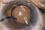
Since CTR is often used in cases with weak zonules, the anterior chamber and posterior capsular can become unstable during irrigation and aspiration (I/A). If this begins to occur, the surgeon should stop I/A and implant the IOL first. Then, after flushing several times, the residual cortex will be raised to the anterior chamber and can be easily removed with I/A. This procedure prevents unexpected posterior capsular rupture and zonular dialysis will be avoided (Supplementary video 2, Figure 2).
 |
Figure 2 Applying flush to cortex removal for the cases with CTR. When the residual cortex is hard to be lifted up. You should put top of the syringe carefully, under the IOL with continuous flow. |
Over the last 5 years, intracameral MFLX with flushing technique was performed in more than 15,000 eyes including at least 1000 eyes with CTR. There were no apparent intraoperative complications related to this technique.
Discussion
Intracameral antibiotics are generally administered by injecting small doses (0.05–0.2 mL) of either undiluted or diluted solutions. However, there is a possibility that pharmaceuticals will not reach the areas that are most susceptible to endophthalmitis: the back surface of the IOL and equator of the bag capsular. Thus, we invented this flushing technique to eliminate postoperative endophthalmitis. We always use a 10x dilution of MFLX and have had no cases of endophthalmitis among over 25,542 patients.9
When performing our flushing technique, residual cortex and debris that was not eliminated by ordinary I/A are pushed out to the anterior chamber (Supplementary video 2, Case 3). The flushing current immediately spreads into the capsular bag. The residual substances (particles) in the capsular bag under the iris are pushed into the accessible area. They can then be easily removed with I/A (Supplementary video 3, Figures 3A–D).
In general, objects can be moved by both aspiration and flow using either water or air. We conducted an experiment to compare the force exerted by aspiration (suction) with that exerted by blowing (pressure) on an object. Vacuum cleaner and air spray gun were prepared as suction and pressure forces, respectively. Our experiment indicated that the pressure from blowing is far more powerful than suction from aspiration and exerts a greater force on an object. With pressure, an object could be moved from a distance. With aspiration, the same object did not move until the vacuum gun was brought close enough to touch it (Supplementary video 4).
When ordinary I/A is performed during cataract surgery, the tip of the I/A handpiece must be placed very close to, or touching the cortex attached to the capsular bag. Further away, the aspiration force does not affect the cortex. In contrast, the force induced by the pressure from a flush stream can affect remote objects.
During cataract surgery with CTRs, residual cortex often becomes trapped between the CTR loop and the equator of the capsular bag. A flush stream creates pressure in the anterior chamber and inside the capsular bag. This causes the bag to expand, including the equator area. The trapped cortex is freed, and the cortex can be peeled from the capsule and lifted into the anterior chamber from a distance. If the capsular bag is polluted by bacteria, it may also be lifted to the anterior chamber.
Following irrigation by ordinary I/A, there is always a possibility that polluted OVDs can remain in the equator of the capsular bag. This seems to occur more frequently in cases with CTRs. Since the number of cases with CTRs is increasing, this flushing technique is an efficient and practical method of the thorough removal of debris and residual cortex and may reduce or even eliminate the risk of postoperative endophthalmitis.
Abbreviations
CTR, Capsular tension ring; IOL, intraocular lens; MFLX, moxifloxacin; OVD, ophthalmic viscosurgical device; I/A, irrigation and aspiration.
Disclosure
The authors report no conflicts of interest in this work.
References
1. Rastogi A, Khanam S, Goel Y, Kamlesh, Thacker P, Kumar P. Comparative evaluation of rotational stability and visual outcome of toric intraocular lenses with and without a capsular tension ring. Indian J Ophthalmol. 2018;66:411–415.
2. Zhao Y, Li J, Yang K, Li X, Zhu S. Combined Special Capsular Tension Ring and Toric IOL Implantation for Management of Astigmatism and High Axial Myopia with Cataracts. Semin Ophthalmol. 2018;33:389–394.
3. Alió JL, Elkady B, Ortiz D, Bernabeu G. Microincision multifocal intraocular lens with and without a capsular tension ring: optical quality and clinical outcomes. J Cataract Refract Surg. 2008;34:1468–1475.
4. Alió JL, Plaza-Puche AB, Piñero DP. Rotationally asymmetric multifocal IOL implantation with and without capsular tension ring: refractive and visual outcomes and intraocular optical performance. J Refract Surg. 2012;28:253–258.
5. Miyoshi T, Fujie S, Yoshida H, Iwamoto H, Tsukamoto H, Oshika T. Effects of capsular tension ring on surgical outcomes of premium intraocular lens in patients with suspected zonular weakness. PLoS One. 2020;15:e0228999. doi:10.1371/journal.pone.0228999
6. Ozturk E, Gunduz A. Optimal timing of capsular tension ring implantation in pseudoexfoliation syndrome. Arq Bras Oftalmol. 2021;84:158–162.
7. Hendrson BA, Kim JY. Modified capsular tension ring for cortical removal after implantation. J Cataract Refract Surg. 2007;34:1688–1690.
8. Matsuura K, Suto C, Akura J, Inoue Y. Bag and chamber flushing: a new method of using intracameral moxifloxacin to irrigate the anterior chamber and the area behind the intraocular lens. Graefes Arch Clin Exp Ophthalmol. 2013;251:81–87.
9. Matsuura K, Uotani R, Sasaki S. Irrigation, incision hydration, and eye pressurization with antibiotic-containing solution. Clin Ophthalmol. 2015;9:1767–1769.
 © 2022 The Author(s). This work is published and licensed by Dove Medical Press Limited. The
full terms of this license are available at https://www.dovepress.com/terms.php
and incorporate the Creative Commons Attribution
- Non Commercial (unported, v3.0) License.
By accessing the work you hereby accept the Terms. Non-commercial uses of the work are permitted
without any further permission from Dove Medical Press Limited, provided the work is properly
attributed. For permission for commercial use of this work, please see paragraphs 4.2 and 5 of our Terms.


