
Back to Journals » Cancer Management and Research » Volume 17

Ginseng-Derived Exosomes Attenuate Immune Evasion in NSCLC via PD-L1 Modulation
Authors Zhu LJ  , Chen XQ, Lin QY, Feng JN, Yuan SF
Received 14 May 2025
Accepted for publication 20 June 2025
Published 24 July 2025 Volume 2025:17 Pages 1503—1512
DOI https://doi.org/10.2147/CMAR.S540462
Checked for plagiarism Yes
Review by Single anonymous peer review
Peer reviewer comments 5
Editor who approved publication: Professor Yong Teng
Lin-Jia Zhu,1 Xiao-Qiang Chen,2 Qiu-Yan Lin,2 Jie-Ni Feng,2 Shao-Fei Yuan2
1Department of Respiratory Medicine, The Third Affiliated Hospital of Wenzhou Medical University, Ruian, Zhejiang, 325200, People’s Republic of China; 2Department of Medical Oncology, The Third Affiliated Hospital of Wenzhou Medical University; Liji Medical Research Academy; Life and Health Research Academy of Wenzhou Medical University, Ruian, Zhejiang, 325200, People’s Republic of China
Correspondence: Lin-Jia Zhu, Email [email protected]
Background: Non-small cell lung cancer (NSCLC) is a major cause of cancer-related death worldwide. While PD-1/PD-L1 immune checkpoint blockade has shown promise, its efficacy is often limited by tumor-induced immune evasion. Ginseng-derived exosomes (G-Exos), as natural plant-based nanocarriers, may offer a novel strategy for immunomodulation. This study investigated the potential of G-Exos to regulate PD-L1 expression and enhance anti-tumor immunity in NSCLC.
Methods: Exosomes were isolated from ginseng cell cultures and characterized via transmission electron microscopy and nanoparticle tracking analysis. Uptake by NSCLC cells was confirmed using PKH26 labeling. In vitro, NSCLC cells were co-cultured with activated T cells to evaluate cytotoxicity (colony formation), cytokine secretion [enzyme-linked immunosorbent assay (ELISA)], and T-cell activation (flow cytometry). PD-L1 expression was assessed by quantitative polymerase chain reaction (qPCR) and Western blot. In vivo, C57BL/6 mice (n = 20) bearing Lewis lung carcinoma (LLC) tumors were randomized into four groups (n = 5/group): PBS, G-Exos (10 μg), anti-PD-L1 (8 μg), or combination therapy. Treatments were administered intravenously every other day for 20 days. Tumor growth was measured, and tissues were analyzed by immunohistochemistry and flow cytometry.
Results: G-Exos were efficiently internalized by NSCLC cells and demonstrated immunostimulatory properties in vitro. They enhanced T-cell-mediated cytotoxicity, as reflected by reduced tumor colony formation, and promoted immune activation, evidenced by increased IL-2 and IFN-γ secretion and a higher proportion of CD8⁺ T cells expressing TNF-α and perforin. Mechanistically, G-Exos downregulated PD-L1 expression at both transcriptional and translational levels in NSCLC cells. In vivo, G-Exos treatment significantly inhibited tumor growth and, when combined with anti-PD-L1 monoclonal antibody, exhibited a synergistic effect characterized by greater tumor suppression and increased infiltration of cytotoxic CD8⁺ T cells in the tumor microenvironment.
Conclusion: Ginseng-derived exosomes downregulate PD-L1 and enhance T-cell function, counteracting immune evasion in NSCLC. Their synergy with anti-PD-L1 therapy supports their potential as adjuvant nanotherapeutics in cancer immunotherapy.
Keywords: non-small cell lung cancer (NSCLC), PD-1/PD-L1 axis, ginseng-derived exosomes, immune checkpoints
Corrigendum for this paper has been published.
Introduction
Lung cancer remains the leading cause of cancer-related mortality worldwide, with approximately 2.2 million new cases diagnosed annually according to GLOBOCAN 2020 data.1 It accounts for nearly 11.4% of global cancer incidence and 18% of cancer deaths, posing a significant public health burden. Non-small cell lung cancer (NSCLC) represents 85–90% of all lung malignancies. Due to frequent late-stage diagnosis and limited therapeutic options, the 5-year survival rate for advanced NSCLC remains below 20%.2,3 While early-stage NSCLC may be managed surgically, systemic therapies including chemotherapy, targeted agents, and immune checkpoint inhibitors (ICIs) are mainstays for advanced disease.4–6 Despite advances, primary and acquired resistance to ICIs necessitate novel strategies to overcome immune evasion.
The PD-1/PD-L1 axis is a pivotal immune checkpoint pathway in NSCLC. Tumor cell surface PD-L1 binds PD-1 on T cells, suppressing cytotoxic activity and enabling immune escape.7 Inflammatory cytokines (eg, IFN-γ) in the tumor microenvironment further upregulate PD-L1, establishing an immunosuppressive niche.8 Clinically, monoclonal antibodies targeting PD-1 (nivolumab, pembrolizumab) or PD-L1 (atezolizumab, durvalumab) improve survival in subsets of NSCLC patients.9,10 However, response rates remain suboptimal, prompting research into upstream regulators of PD-L1 (eg, PTEN/AKT, NF-κB) to enhance ICI efficacy.11,12
Exosomes—nanoscale extracellular vesicles (30–150 nm)—have emerged as promising therapeutic vehicles due to their biocompatibility, low immunogenicity, and capacity to deliver bioactive cargo (proteins, nucleic acids) to specific cell types.13,14 In oncology, exosomes can modulate tumor immunity by shuttling immunoregulatory molecules; for instance, plant-derived exosomes may deliver phytochemicals with antitumor properties while evading rapid clearance.15–17 Notably, recent studies highlight exosomes as natural nanocarriers capable of reprogramming the tumor immune microenvironment, including immune checkpoint regulation.18
Ginseng (Panax ginseng) exhibits documented antitumor and immunomodulatory effects, primarily attributed to ginsenosides and polysaccharides.19 Critically, ginseng-derived exosomes (G-Exos) offer unique advantages: (1) high stability in circulation, (2) intrinsic bioactivity from ginseng phytocompounds, and (3) potential for targeted delivery to tumor sites. While animal exosomes have been extensively studied in cancer immunotherapy, plant exosomes—particularly from ginseng—remain underexplored in NSCLC. Given the established role of PD-L1 in NSCLC immune evasion and the therapeutic potential of exosomal modulation, we hypothesized that G-Exos could deliver bioactive ginseng compounds (eg, ginsenosides) or regulatory RNAs into tumor cells to suppress PD-L1 expression, thereby attenuating PD-L1-mediated immunosuppression. Herein, we investigate the ability of G-Exos to inhibit immune evasion in NSCLC via targeted PD-L1 pathway modulation, a previously unreported mechanism that may synergize with existing ICIs.
Materials and Methods
Cell Lines and Treatment
The human NSCLC cell lines A549 and H1299 and mouse lung cancer cell line LLC were purchased from the Shanghai Cell Bank of the Chinese Science Academy. A549 and H1299 cells were maintained in culture medium consisting of 90% Roswell Park Memorial Institute-1640 (RPMI-1640) medium (Hyclone, Logan, UT, USA) supplemented with 10% fetal bovine serum (FBS; Gibco, Carlsbad, CA, USA) and 1% penicillin/streptomycin solution. LLC cells were cultured in (Dulbecco’s modified Eagle’s medium (DMEM); Hyclone, Logan, UT, USA) containing 10% FBS and 1% penicillin/streptomycin. All cells were incubated at 37°C in a humidified 5% CO2 atmosphere. All cell lines were obtained from a certified cell bank and were confirmed to be mycoplasma-free. The use of human and murine cell lines complied with institutional biosafety and research guidelines.
Exosome Isolation and Characterization
Suspension cultures of Panax ginseng root cells were established from sterilized root explants of 4-year-old plants (collected from Jilin Province, China). The explants were cultured in Murashige and Skoog (MS) basal medium supplemented with 1.0 mg/L 2,4-dichlorophenoxyacetic acid (2,4-D) and 0.1 mg/L kinetin, maintained at 25°C under a 16-h light/8-h dark photoperiod with orbital shaking at 110 rpm for 14 days. Exosomes were subsequently isolated from the cell culture supernatants using ExoQuick-TC™ Precipitation Solution (System Biosciences, #EXOTC10A-1) according to the manufacturer’s instructions. For transmission electron microscopy (TEM), exosomes were fixed with 2% glutaraldehyde, adsorbed onto carbon-coated grids, negatively stained with 1% uranyl acetate, and visualized using a Hitachi HT7800 microscope. Nanoparticle size distribution and concentration were determined by nanoparticle tracking analysis (NTA) using a NanoSight NS300 (Malvern). Expression of exosomal surface markers CD63 and CD81 was confirmed by Western blot using anti-CD63 (Abcam, #ab134045) and anti-CD81 (Abcam, #ab109201) antibodies.
Exosome Labeling and Internalization Assay
Isolated exosomes were labeled with 4 μM PKH26 (Sigma #PKH26GL) in Diluent C for 5 min at RT. Labeling was quenched with 1% BSA, followed by ultracentrifugation (100,000 ×g, 2 h) to remove free dye. PKH26-labeled exosomes (10 μg/mL) were incubated with NSCLC cells for 4 h. Nuclei were counterstained with DAPI (Sigma), and uptake was visualized by confocal microscopy (Leica TCS SP8).
T Cell-Mediated Cytotoxicity Assay
Peripheral blood mononuclear cells (PBMCs) were isolated from healthy donors (Ethics approval: WMU-2023-011) using Ficoll density gradient centrifugation. Monocytes were differentiated into dendritic cells (DCs) by culturing with granulocyte-macrophage colony-stimulating factor (GM-CSF, 100 ng/mL) and interleukin-4 (IL-4, 50 ng/mL) for 7 days. Tumor lysates were prepared by subjecting NSCLC cells (A549 and H1299) to five freeze-thaw cycles, followed by centrifugation at 12,000 ×g for 10 minutes; the supernatant was collected as lysate. DCs were pulsed with tumor lysate at a 1:5 ratio (based on cell count equivalents of DCs to tumor cells) for 24 hours and then co-cultured with autologous PBMCs to prime T cells. For cytotoxicity assessment, activated T cells (effectors) were co-cultured with tumor cells (targets) at an effector-to-target (E:T) ratio of 10:1 for 24 hours. Tumor cell viability was evaluated using colony formation assay (10-day culture with 0.5% crystal violet staining), Annexin V-FITC/PI apoptosis assay (BD Biosciences), and MTT assay (Sigma-Aldrich).
For T-cell activation analysis, co-cultured T cells were stimulated with Cell Activation Cocktail containing Brefeldin A (BioLegend) for 5 hours and stained for surface CD8 (BioLegend #301014), as well as intracellular TNF-α (BioLegend #502909) and perforin (BioLegend #308104), using the same intracellular staining protocol applied to tumor-infiltrating lymphocyte (TIL) analysis. T cell activation experiments were independently repeated three times.
Enzyme-Linked Immunosorbent Assay (ELISA)
The cell culture supernatants were collected and centrifuged to remove cell debris. The levels of cytokines, including human IFN-γ (Thermo Fisher, #88-7316-88), TNF-α (Thermo Fisher, #88-7346-88), and IL-2 (Thermo Fisher, #88-7025-88), were measured using commercial ELISA kits according to the manufacturer’s instructions. Positive controls using recombinant cytokines were included in each assay to ensure accuracy and comparability of the results. All ELISA assays were performed in three independent biological replicates.
Quantitative Real-Time PCR (qPCR) Assay
Total RNA was extracted with TRIzol (Beyotime). cDNA synthesized using TransScript® First-Strand cDNA Synthesis Kit (#AT301-02). qPCR utilized SYBR Green (Takara #RR420A) with primers: PD-L1: F 5′-GCTGCACTAACTGTGGTAGTG-3′, R 5′-AGGACCCAGTTTGGATTGGT-3′; GAPDH: F 5′-GGAGCGAGATCCCTCCAAAAT-3′, R 5′-GGCTGTTGTCATACTTCTCATGG-3′; Relative mRNA expression was calculated by 2−ΔΔCt method (Livak & Schmittgen, 2001).
Western Blot
Cells were lysed with radioimmunoprecipitation assay (RIPA) buffer (Beyotime) to extract total protein, and protein concentrations were determined using the bicinchoninic acid (BCA) assay (Thermo Fisher Scientific). Equal amounts of protein (35 μg per sample) were separated by 10% SDS-polyacrylamide gel electrophoresis (SDS-PAGE) and transferred onto polyvinylidene fluoride (PVDF) membranes (Millipore, Billerica, MA, USA). Membranes were blocked with 5% skimmed milk in TBST (Tris-buffered saline with 0.1% Tween-20) for 1 hour at room temperature, followed by overnight incubation at 4°C with primary antibodies: anti-PD-L1 (1:1000; Abcam, #ab213480) and anti-β-actin (1:5000; Abcam, #ab8226), the latter serving as a loading control. After three washes with TBST, membranes were incubated with HRP-conjugated secondary antibodies (1:5000; Abcam) for 1 hour at room temperature. Protein bands were visualized using enhanced chemiluminescence (ECL) reagent (Pierce, Thermo Fisher Scientific) and imaged with a ChemiDoc MP system (Bio-Rad). Band intensity was quantified using ImageJ software (NIH), and PD-L1 expression was normalized to β-actin. Each experiment was independently repeated at least three times using biologically independent samples to ensure reproducibility.
Syngeneic Tumor Model and Treatment
Female C57BL/6 mice (6 weeks old, n = 20) were purchased from the Vital River Laboratory Animal Technology (Beijing, China). After a 7-day acclimation period, the mice were subcutaneously injected with mouse Lewis lung carcinoma (LLC) cells (5 × 10⁶ cells in 50 µL PBS). Tumor volume was calculated using the formula: volume = (length × width2)/2. When tumors reached approximately 100 mm3, the mice were randomized into four groups (n = 5 per group): Control: PBS (50 µL, intravenous injection, every 2 days); G-Exos: 10 µg exosomes (intravenous injection, every 2 days); Anti-PD-L1: 8 µg PD-L1 antibody (intravenous injection, every 2 days); Exos+Anti-PD-L1: 10 µg exosomes + 8 µg PD-L1 antibody (intravenous injection, every 2 days). Treatments were administered via tail vein injection every two days for a total of 20 days (10 total injections). Tumor width and length were measured every three days. At the end of treatment, mice were sacrificed by cervical dislocation, and tumors were excised and weighed. Tumor tissues were collected for subsequent experiments, including immunohistochemistry (IHC) and flow cytometry to assess PD-L1 expression and immune cell infiltration. All animal procedures were approved by the Animal Ethical Committee of the Third Affiliated Hospital of Wenzhou Medical University and conducted in accordance with the Guide for the Care and Use of Laboratory Animals (8th edition, National Research Council, USA).
Analysis of Tumor-Infiltrating Lymphocytes
Tumor tissues collected from the syngeneic tumor model were cut into small pieces and digested with PBS containing 0.2% collagenase IV and 0.002% deoxyribonuclease I (Invitrogen) at 37°C for 1 hour. The homogenate was centrifuged, and tumor-infiltrating lymphocytes (TILs) were isolated using a Percoll gradient (Solarbio, China) according to the manufacturer’s instructions. Isolated cells were stained with antibodies against surface markers: CD45-APC (103111, BioLegend), CD3-BV421™ (100227, BioLegend), CD4-PE (100407, BioLegend), and CD8b.2-PE/Cyanine7 (140415, BioLegend). For intracellular cytokine detection, cells were fixed and permeabilized using intracellular staining buffer (421002, BioLegend), followed by staining with IFN-γ-FITC (clone XMG1.2, 163511, BioLegend) and TNF-α-PE (clone MP6-XT22, 11–7321-82, Invitrogen). A Zombie R718™ Fixable Viability Kit (423115, BioLegend) was used to exclude dead cells. Data were acquired using a BD FACSymphony™ flow cytometer and analyzed with FlowJo software (v10.8.1).
Statistical Analysis
All data are presented as mean ± standard deviation (SD). Statistical significance between two groups was determined using the unpaired Student’s t-test. For comparisons among three or more groups, one-way ANOVA followed by Tukey’s post hoc test was performed. All analyses were conducted using GraphPad Prism software (version 9.0). A P value < 0.05 was considered statistically significant.
Results
Isolation and Identification of Exosomes
Transmission electron microscopy (TEM) revealed spherical vesicles with double-layer membranes (Figure 1A). Nanoparticle tracking analysis (NTA) showed a size distribution peak at approximately 150 nm (Figure 1B). Confocal microscopy confirmed internalization of PKH26-labeled ginseng exosomes into A549 and H1299 cells (Figure 1C). Western blot further validated exosomal identity through positive detection of CD63 and CD81 markers (Figure 1D).
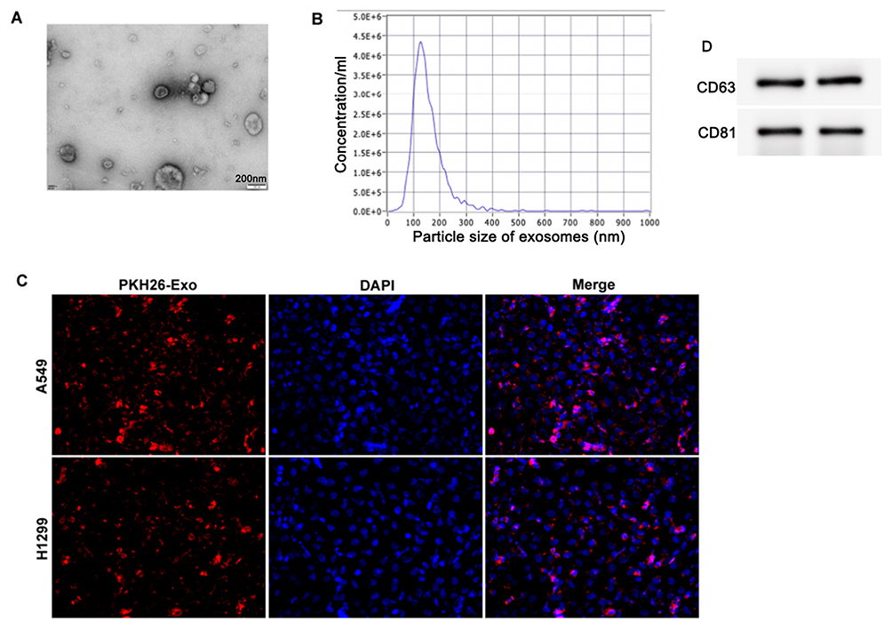 |
Figure 1 Isolation and identification of exosomes. (A) TEM morphology of exosomes. (B) NTA size distribution (x-axis: Diameter nm; y-axis: Particle Concentration). (C) PKH26-labeled exosomes (red) internalized by NSCLC cells (blue: DAPI-stained nuclei). (D) Western blot confirmed expression of CD63 and CD81. Data are expressed as mean ± SD of three independent experiments. No statistical differences were observed between groups since this figure mainly demonstrates qualitative characterization of exosomes. |
Ginseng-Derived Exosomes Attenuate Immune Evasion In vitro
To investigate the in vitro effects of ginseng-derived exosomes on tumor immune evasion, non-small cell lung cancer (NSCLC) cells were treated with exosomes and co-cultured with human PBMCs. The proliferation of cancer cells was assessed by a colony formation assay. As shown in Figure 2A, co-culture with exosomes markedly reduced the number of tumor colonies, indicating enhanced T cell–mediated anti-proliferative cytotoxicity.
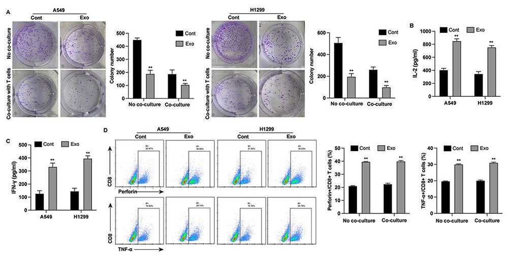 |
Figure 2 Ginseng-derived exosomes attenuate in vitro immune evasion in NSCLC. NSCLC cells were co-cultured with PBMCs under the following conditions: (A) Colony formation assay: NSCLC cells + PBMCs treated with PBS (Control) vs G-Exos (10 μg/mL). (B and C) Cytokine secretion: IL-2 (B) and IFN-γ (C) levels measured by ELISA in co-culture supernatants (Control vs G-Exos-treated). (D) T-cell activation: Flow cytometry analysis of CD8+TNF-α+ and CD8+perforin+ T cells (Control vs G-Exos-treated). Data represent mean ± SD of three independent experiments. Statistical significance was determined by two-tailed Student’s t-test (**p<0.01 vs Control). |
Cytokine levels in the co-culture supernatant were quantified by ELISA. Treatment with ginseng exosomes led to significantly increased production of immune-activating cytokines IL-2 and IFN-γ (Figure 2B and C), with recombinant cytokines included as positive controls. T-cell activation was further evaluated by flow cytometry, which revealed elevated proportions of cytotoxic CD8⁺TNF-α+ and CD8+ perforin+ T cells in the exosome-treated group (Figure 2D).
Ginseng Exosomes Downregulate PD-L1 Expression
To further elucidate the molecular mechanisms underlying the immune activation effects of ginseng-derived exosomes, we examined PD-L1 expression in non-small cell lung cancer (NSCLC) cells. Quantitative PCR and Western blot analysis revealed a significant reduction in PD-L1 mRNA and protein levels following exosome treatment (Figure 3A). Consistently, flow cytometry analysis demonstrated decreased proportions of PD-L1⁺ cells in both A549 and H1299 cell lines (Figure 3B).
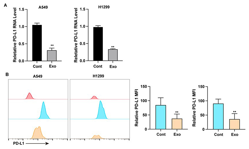 |
Figure 3 Ginseng-derived exosomes regulate lung cancer cell PD-L1 expression. (A) PD-L1 mRNA (qPCR) and protein (Western blot): NSCLC cells treated with PBS (Control) vs G-Exos (10 μg/mL). (B) Flow cytometry of PD-L1+ cells: NSCLC cells treated with PBS (Control) vs G-Exos (10 μg/mL). Data represent mean ± SD of three independent experiments. Statistical significance was determined by two-tailed Student’s t-test (**p<0.01 vs Control). |
Synergistic Antitumor Effects In vivo
To investigate the in vivo efficacy of ginseng-derived exosomes, we established a syngeneic tumor model using the murine lung cancer cell line LLC. Mice were treated with ginseng exosomes, anti-PD-L1 antibody, or a combination of both. Exosome treatment alone resulted in reduced PD-L1 expression and moderate tumor growth inhibition, while the combination therapy synergistically enhanced antitumor effects compared to monotherapy (Figure 4A–C). Immunohistochemical staining revealed a marked increase in tumor-infiltrating CD8⁺ T cells in the combination group (Figure 4D). Furthermore, flow cytometry analysis of tumor tissues demonstrated elevated production of cytotoxic molecules, including perforin and TNF-α, following combined treatment (Figure 4E).
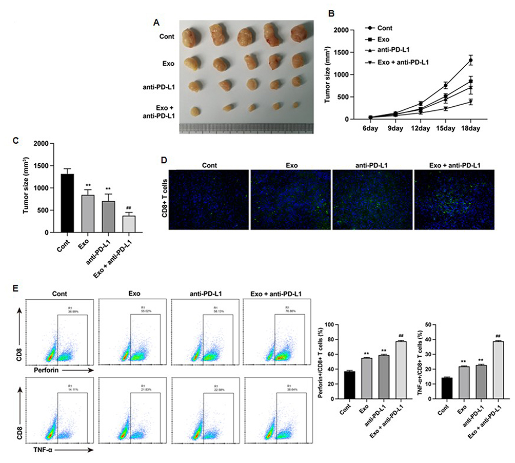 |
Figure 4 Ginseng-derived exosomes inhibits in vivo tumor growth via regulating immune response. (A–C) Tumor growth and weight: PBS (Control), G-Exos (10 μg), Anti-PD-L1 mAb (8 μg), G-Exos + Anti-PD-L1 (Combination). (D) IHC of CD8+ T cells (brown) in tumor sections. (E) Flow cytometry of perforin and TNF-α in tumor tissues. Data represent mean ± SD of three independent experiments. Statistical significance was determined by one-way ANOVA followed by Tukey’s post hoc test. **p<0.01 vs control; ##p<0.01 vs Exo group. |
Discussion
Cancer cells possess an intrinsic ability to evade immune surveillance through a variety of mechanisms, making immune evasion a hallmark of malignancy.20 Immune checkpoint inhibitors (ICIs), particularly those targeting the PD-1/PD-L1 axis, have revolutionized cancer therapy by restoring T-cell activity against tumor cells.21,22 Despite their remarkable clinical success, a significant proportion of patients do not respond to ICIs, suggesting that PD-L1 expression alone is insufficient as a predictive biomarker.23–25 Moreover, the heterogeneity of PD-L1 regulation among tumors contributes to inconsistent treatment outcomes, underscoring the urgent need for novel approaches that both improve immune activation and enhance ICI efficacy.
In the current study, we investigated the role of ginseng-derived exosomes (G-Exos) in regulating immune evasion and modulating PD-L1 expression in non-small cell lung cancer (NSCLC). Our findings demonstrated that G-Exos significantly suppressed the proliferation of NSCLC cells in vitro, as evidenced by reduced colony formation when co-cultured with PBMCs (Figure 2A). This antiproliferative effect was associated with enhanced T-cell activation, indicated by increased production of immune-stimulatory cytokines IL-2 and IFN-γ (Figure 2B and C), and elevated proportions of cytotoxic CD8+TNF-α+ and CD8+perforin+ T cells (Figure 2D).
Importantly, we observed that G-Exos downregulated PD-L1 expression in NSCLC cells at both the mRNA and protein levels (Figure 3A), and flow cytometry analysis confirmed a decreased proportion of PD-L1+ A549 and H1299 cells (Figure 3B). These findings suggest that G-Exos not only enhance T-cell cytotoxicity but also diminish tumor-mediated immunosuppression by targeting PD-L1. The mechanisms by which G-Exos regulate PD-L1 expression are not yet fully elucidated, but potential involvement of bioactive exosomal components such as miRNAs or phytochemicals merits further investigation.
Our in vivo studies further confirmed the immunomodulatory and anti-tumor effects of G-Exos. In a lung cancer syngeneic tumor model, G-Exos significantly inhibited tumor growth, and this effect was amplified when combined with anti–PD-L1 antibody therapy (Figure 4A–C). Co-treatment led to increased infiltration of CD8+ T cells in tumor tissues (Figure 4D) and upregulation of key cytotoxic factors, including TNF-α and perforin (Figure 4E), indicating a synergistic enhancement of antitumor immunity.
These results are consistent with and expand upon prior research on plant-derived exosome-like nanoparticles (PELNs), which have emerged as promising agents in cancer immunotherapy.17 PELNs isolated from edible plants are capable of modulating inflammatory responses and reprogramming the tumor microenvironment with minimal immunogenicity and cost-effective production.17,26 Recent studies, such as Olson, have highlighted the potential of edible plant exosomes in delivering bioactive molecules that suppress tumor progression and modulate immune responses, supporting our observations of enhanced T-cell activation and PD-L1 downregulation in NSCLC models treated with G-Exos.27
Despite these promising findings, several limitations should be acknowledged. First, the specific cargo within G-Exos responsible for PD-L1 suppression remains unidentified. Second, only one in vivo model (LLC syngeneic tumor) was used, without long-term survival data. Third, optimal exosome dosing, biodistribution, and safety profiles were not fully assessed. These limitations warrant further research to validate our findings across different tumor types and experimental systems. Future studies should employ multi-omics approaches to characterize the functional components of G-Exos and elucidate their regulatory networks, particularly in relation to PD-L1 modulation.
Furthermore, the absence of robust predictive biomarkers continues to hinder the effective application of ICIs. While PD-L1 expression is currently the most utilized biomarker, it often fails to correlate with therapeutic response.28 Ginseng-derived exosomes may offer an adjunct or alternative strategy to enhance immune responsiveness, potentially improving patient outcomes regardless of baseline PD-L1 status. However, additional clinical studies are necessary to explore the translational potential of G-Exos and assess their performance across patient populations.
In conclusion, our study provides novel evidence that ginseng-derived exosomes enhance anti-tumor immune responses by simultaneously promoting T-cell activation and downregulating PD-L1 expression in NSCLC. These dual effects not only attenuate immune evasion but also sensitize tumors to immune checkpoint blockade therapy. G-Exos thus represent a promising and biocompatible adjunct to current immunotherapeutic strategies.
Conclusion
In conclusion, our study demonstrates that exosomes derived from ginseng (G-Exos) effectively attenuate immune evasion in non-small cell lung cancer (NSCLC) by simultaneously modulating PD-L1 expression and enhancing T cell-mediated cytotoxicity. Specifically, G-Exos significantly downregulated PD-L1 at both the transcriptional and translational levels, thereby disrupting the tumor’s ability to suppress immune surveillance. Additionally, G-Exos promoted T-cell activation, as evidenced by elevated production of immune-stimulatory cytokines such as IL-2 and IFN-γ, as well as increased expression of cytotoxic markers including TNF-α and perforin in CD8⁺ T cells, which contributed to reduced tumor cell proliferation in vitro.
Importantly, combination therapy using G-Exos and an anti-PD-L1 monoclonal antibody exhibited a synergistic antitumor effect in vivo, resulting in enhanced tumor suppression and increased infiltration of CD8⁺ T cells in the tumor microenvironment. These results align closely with our initial hypothesis that plant-derived exosomes can serve as functional immunomodulators and support their potential role as adjuvants to current immune checkpoint blockade therapies.
While these findings suggest a promising therapeutic strategy, this study also highlights areas that warrant further investigation. The precise bioactive molecules within G-Exos responsible for PD-L1 suppression remain unidentified, and the regulatory pathways through which they exert their effects are yet to be elucidated. Moreover, comprehensive assessments of G-Exos in patient-derived syngeneic tumor (PDX) models, as well as evaluations of long-term safety, pharmacokinetics, and biodistribution, are essential for translational development.
Overall, this study provides important mechanistic and preclinical insights into the immunotherapeutic potential of ginseng-derived exosomes and lays the groundwork for future clinical exploration of plant-derived nanocarriers in cancer immunotherapy.
Disclosure
The authors report no conflicts of interest in this work.
References
1. Duma N, Santana-Davila R, Molina JR. Non-small cell lung cancer: epidemiology, screening, diagnosis, and treatment. Mayo Clin Proc. 2019;94(8):1623–1640. doi:10.1016/j.mayocp.2019.01.013
2. Chen M, Lu H, Copley SJ, et al. A novel radiogenomics biomarker for predicting treatment response and pneumotoxicity from programmed cell death protein or ligand-1 inhibition immunotherapy in NSCLC. J Thorac Oncol. 2023;18(6):718–730. doi:10.1016/j.jtho.2023.01.089
3. Riely GJ, Wood DE, Ettinger DS, et al. Non-small cell lung cancer, version 4.2024, NCCN clinical practice guidelines in oncology. J Natl Compr Canc Netw. 2024;22(4):249–274. doi:10.6004/jnccn.2204.0023
4. Herbst RS, Morgensztern D, Boshoff C. The biology and management of non-small cell lung cancer. Nature. 2018;553(7689):446–454. doi:10.1038/nature25183
5. Salgia R, Pharaon R, Mambetsariev I, et al. The improbable targeted therapy: KRAS as an emerging target in non-small cell lung cancer (NSCLC). Cell Rep Med. 2021;2(1):100186. doi:10.1016/j.xcrm.2020.100186
6. Saw SPL, Ong BH, Chua KLM, et al. Revisiting neoadjuvant therapy in non-small-cell lung cancer. Lancet Oncol. 2021;22(11):e501–e516. doi:10.1016/S1470-2045(21)00383-1
7. Dermani FK, Samadi P, Rahmani G, et al. PD-1/PD-L1 immune checkpoint: potential target for cancer therapy. J Cell Physiol. 2019;234(2):1313–1325. doi:10.1002/jcp.27172
8. Yamaguchi H, Hsu JM, Yang WH, et al. Mechanisms regulating PD-L1 expression in cancers and associated opportunities for novel small-molecule therapeutics. Nat Rev Clin Oncol. 2022;19(5):287–305. doi:10.1038/s41571-022-00601-9
9. Yi M, Zheng X, Niu M, et al. Combination strategies with PD-1/PD-L1 blockade: current advances and future directions. Mol Cancer. 2022;21(1):28. doi:10.1186/s12943-021-01489-2
10. Tang Q, Chen Y, Li X, et al. The role of PD-1/PD-L1 and application of immune-checkpoint inhibitors in human cancers. Front Immunol. 2022;13:964442. doi:10.3389/fimmu.2022.964442
11. Cha JH, Chan LC, Li CW, et al. Mechanisms controlling PD-L1 expression in cancer. Mol Cell. 2019;76(3):359–370. doi:10.1016/j.molcel.2019.09.030
12. Gou Q, Dong C, Xu H, et al. PD-L1 degradation pathway and immunotherapy for cancer. Cell Death Dis. 2020;11(11):955. doi:10.1038/s41419-020-03140-2
13. Reck M, Remon J, Hellmann MD. First-line immunotherapy for non-small-cell lung cancer. J Clin Oncol. 2022;40(6):586–597. doi:10.1200/JCO.21.01497
14. Tu E, McGlinchey K, Wang J, et al. Anti-PD-L1 and anti-CD73 combination therapy promotes T cell response to EGFR-mutated NSCLC. JCI Insight. 2022;7(3). doi:10.1172/jci.insight.142843
15. Khan NA, Asim M, Biswas KH, et al. Exosome nanovesicles as potential biomarkers and immune checkpoint signaling modulators in lung cancer microenvironment: recent advances and emerging concepts. J Exp Clin Cancer Res. 2023;42(1):221. doi:10.1186/s13046-023-02753-7
16. Li Q, Song Q, Zhao Z, et al. Genetically engineered artificial exosome-constructed hydrogel for ovarian cancer therapy. ACS Nano. 2023;17(11):10376–10392. doi:10.1021/acsnano.3c00804
17. Yi Q, Xu Z, Thakur A, et al. Current understanding of plant-derived exosome-like nanoparticles in regulating the inflammatory response and immune system microenvironment. Pharmacol Res. 2023;190:106733. doi:10.1016/j.phrs.2023.106733
18. Patel B, Gaikwad S, Prasad S. Exploring the significance of extracellular vesicles: key players in advancing cancer and possible theranostic tools. Cancer Pathogenesis Ther. 2025;3(2):109–119. doi:10.1016/j.cpt.2024.04.005
19. Liu H, Lu X, Hu Y, et al. Chemical constituents of panax ginseng and panax notoginseng explain why they differ in therapeutic efficacy. Pharmacol Res. 2020;161:105263. doi:10.1016/j.phrs.2020.105263
20. Hegde PS, Chen DS. Top 10 challenges in cancer immunotherapy. Immunity. 2020;52(1):17–35. doi:10.1016/j.immuni.2019.12.011
21. Pardoll DM. The blockade of immune checkpoints in cancer immunotherapy. Nat Rev Cancer. 2012;12(4):252–264. doi:10.1038/nrc3239
22. Lee HH, Wang YN, Xia W, et al. Removal of N-linked glycosylation enhances PD-L1 detection and predicts Anti-PD-1/PD-L1 therapeutic efficacy. Cancer Cell. 2019;36(2):168–178.e164. doi:10.1016/j.ccell.2019.06.008
23. Gubens MA, Davies M. NCCN guidelines updates: new immunotherapy strategies for improving outcomes in non-small cell lung cancer. J Natl Compr Canc Netw. 2019;17(5.5):574–578. doi:10.6004/jnccn.2019.5005
24. Gao X, Xu N, Li Z, et al. Safety and antitumour activity of cadonilimab, an anti-PD-1/CTLA-4 bispecific antibody, for patients with advanced solid tumours (COMPASSION-03): a multicentre, open-label, phase 1b/2 trial. Lancet Oncol. 2023;24(10):1134–1146. doi:10.1016/S1470-2045(23)00411-4
25. Clingan P, Ladwa R, Brungs D, et al. Efficacy and safety of cosibelimab, an anti-PD-L1 antibody, in metastatic cutaneous squamous cell carcinoma. J Immunother Cancer. 2023;11(10):e007637. doi:10.1136/jitc-2023-007637
26. Kim J, Zhu Y, Chen S, et al. Anti-glioma effect of ginseng-derived exosomes-like nanoparticles by active blood-brain-barrier penetration and tumor microenvironment modulation. J Nanobiotechnology. 2023;21(1):253. doi:10.1186/s12951-023-02006-x
27. Yu Y, Duan X, Wang X, et al. Enhanced therapeutic effects of ginseng-derived exosome-like nanoparticles loaded hyaluronic acid injectable hydrogels for breast tumor treatment. Int J Biol Macromol. 2025;310(Pt 3):142914. doi:10.1016/j.ijbiomac.2025.142914
28. Kim ST, Cristescu R, Bass AJ, et al. Comprehensive molecular characterization of clinical responses to PD-1 inhibition in metastatic gastric cancer. Nat Med. 2018;24(9):1449–1458. doi:10.1038/s41591-018-0101-z
 © 2025 The Author(s). This work is published and licensed by Dove Medical Press Limited. The
full terms of this license are available at https://www.dovepress.com/terms
and incorporate the Creative Commons Attribution
- Non Commercial (unported, 4.0) License.
By accessing the work you hereby accept the Terms. Non-commercial uses of the work are permitted
without any further permission from Dove Medical Press Limited, provided the work is properly
attributed. For permission for commercial use of this work, please see paragraphs 4.2 and 5 of our Terms.
© 2025 The Author(s). This work is published and licensed by Dove Medical Press Limited. The
full terms of this license are available at https://www.dovepress.com/terms
and incorporate the Creative Commons Attribution
- Non Commercial (unported, 4.0) License.
By accessing the work you hereby accept the Terms. Non-commercial uses of the work are permitted
without any further permission from Dove Medical Press Limited, provided the work is properly
attributed. For permission for commercial use of this work, please see paragraphs 4.2 and 5 of our Terms.