Back to Journals » Diabetes, Metabolic Syndrome and Obesity » Volume 13
Ginger Extract Increases GLUT-4 Expression Preferentially Through AMPK Than PI3K Signalling Pathways in C2C12 Muscle Cells
Authors Tajik Kord M, Pourrajab F, Hekmatimoghaddam S ![]()
Received 29 April 2020
Accepted for publication 11 August 2020
Published 15 September 2020 Volume 2020:13 Pages 3231—3238
DOI https://doi.org/10.2147/DMSO.S260224
Checked for plagiarism Yes
Review by Single anonymous peer review
Peer reviewer comments 2
Editor who approved publication: Dr Konstantinos Tziomalos
Marjan Tajik Kord,1 Fatemeh Pourrajab,1,2 Seyedhossein Hekmatimoghaddam3,4
1Department of Biochemistry and Molecular Biology, School of Medicine, Shahid Sadoughi University of Medical Sciences, Yazd, Iran; 2Nutrition and Food Security Research Center, Shahid Sadoughi University of Medical Sciences, Yazd, Iran; 3Yazd Cardiovascular Research Center, Afshar Hospital, Shahid Sadoughi University of Medical Sciences, Yazd, Iran; 4Department of Advanced Medical Sciences and Technologies, School of Paramedicine, Shahid Sadoughi University of Medical Sciences, Yazd, Iran
Correspondence: Seyedhossein Hekmatimoghaddam Tel + 0098-9133518314
Fax + 0098-3517256458
Email [email protected]
Purpose: There are two signal transduction pathways related to glucose metabolism in C2C12 mouse myoblast cells; one through AMP-activated protein kinase (AMPK), and the other through phosphoinositide 3-kinase (PI3K). Ginger is reported to have hypoglycemic effects. The aim of this study was to determine the exact mechanism of action of ginger in those pathways.
Methods: C2C12 cells were seeded to four separate experimental groups; Control: treated with 50 μg/mL DMSO in the absence of any inhibitor; Treatment 1: treated with 50 μg/mL ethyl acetate ginger extract without any inhibitor; Treatment 2: treated with 50 μg/mL extract in the presence of 20 μM AMPK inhibitor; Treatment 3: treated with 50 μg/mL extract in the presence of 25 μM PI3K inhibitor. The amount of GLUT-4 protein (an important glucose transporter) was determined in cytosolic and membrane fractions using sodium dodecyl sulfate polyacrylamide gel electrophoresis and Western blotting.
Results: GLUT-4 concentration was significantly higher in the membrane fraction of cells treated with ethyl acetate ginger extract in the absence of any inhibitor in comparison with cells treated with this extract in the presence of each of the inhibitors (P-value < 0.05). GLUT-4 quantity in the membrane fractions in all groups was more than cytosolic fractions. The amount of GLUT-4 in membrane fraction of treated cells in the presence of PI3K inhibitor was higher than in the cells treated with this extract in the presence of AMPK inhibitor (P-value < 0.05).
Conclusion: Ethyl acetate ginger extract affects the amount of GLUT-4 protein in membrane and cytosolic fractions of C2C12 myoblast cells mostly through AMPK pathway but less via PI3K.
Keywords: glucose, metabolism, myoblasts, signal transduction
Introduction
Diabetes is a metabolic disorder affecting millions of people1 and has turned into a global problem nowadays.2,3 Control and management of this disease is a challenge.4 Several factors are involved in its pathogenesis, and some of its more prevalent symptoms are increased serum glucose, frequent urination and dehydration.5 Chronic hyperglycemia in diabetes causes damage to and dysfunction of other vital organs such as liver, heart and kidneys, in addition to retinopathy, neuropathy and atherosclerosis, among the many.6 The ability of the body in utilizing the energy of food is impaired in diabetes. Production of insulin or its function is impaired in most cases, so cells cannot efficiently respond to insulin by uptake of glucose, leading to its accumulation in blood.1 Glucose uptake is the rate-limiting step in glucose metabolism. Two pathways play roles in glucose metabolism in skeletal muscles; one is through AMP-activated protein kinase (AMPK), and the other is stimulated by phosphoinositide 3 kinase (PI3K). Researchers suggest that these two pathways have some roles in translocation of GLUT-4,7,8 which is the main insulin-responsive glucose transporter.7 Binding of insulin to its membrane receptor results in insulin receptor substrate 1 (IRS-1) phosphorylation and IRS-1 associated PI3K activation.
PI3K has both regulatory (p38) and catalytic subunits. Interaction of IRS with p38 leads to activation of p38 which has downstream effects such as Akt through which GLUT-4 is finally activated and transmitted into the membrane.9–11 AMPK is a serin-protease enzyme and a key sensor in the cell, with important implications in the regulation of metabolism.12,13 AMPK is activated in response to cellular stress, decreased energy, hypoxia, increase in cellular AMP/ATP ratio and also some upstream kinases such as LKB1 (a tumour suppressor) and CaMKt (Ca-calmodulin dependent kinases).12,14,15 Activated AMPK stimulates catabolic pathways for producing ATP and inhibits anabolic pathways.8,11,15 Activated AMPK increases GLUT-4 expression and translocation as well.8,14,16
There are some researches on herbal medicine for the treatment of some common diseases including diabetes mellitus. For example, green tea decreases the risk of coronary artery disease and is introduced as a useful antioxidant that may prevent some cancers. Both leaf and beans of fenugreek are useful for the treatment of diabetes. Cinnamon decreases the severity of diabetes and coronary artery diseases.4 Before the introduction of therapeutic insulin, diabetes was treated with traditional plant remedies.17 Some patients cannot tolerate the side effects of some diabetes drugs, and some of them are expensive so that patients have turned to traditional herbal plants.4
Ginger is the rhizome of Zingiber officinale Roscoe, a perennial herbaceous plant6,18 with narrow bright green, grass-like leaves and yellowish-green flowers.5 Ginger is used as a cooking spice and also has therapeutic effects.6,18 It contains several substances including phenolic compounds. The main substance responsible for the spicy taste of ginger is (6)-gingerol, which turns to shogaols upon dehydration.5,18,19 Ginger is used for the treatment of catarrh, rheumatism and asthma.19 It has been shown that ginger has hypoglycemic effects and improves diabetes.5,6,8,17,18 Due to the role of GLUT-4 and muscle tissue in glucose uptake, we assessed the effect of ethyl acetate extract of ginger on amount of GLUT-4 protein in C2C12 muscle cells (embryonic precursor cells of the myogenic lineage that develop from the mesoderm), and applied AMPK inhibitor (compound C) and PI3K inhibitor (LY) to determine the involved signalling pathway.
Materials and Methods
The study was approved by Shahid Sadoughi University of medical sciences, Yazd, Iran, and done in laboratories of the school of medicine. Dulbecco’s modified Eagle’s medium (DMEM), fetal bovine serum (FBS), horse serum and trypsin were purchased from Gibco-BRL (Grand Island, NY, USA). Dimethyl sulfoxide (DMSO), AMPK inhibitor 6-(4-[2-piperidin-1-ylethoxy]phenyl)-3-pyridin-4-ylpyrazolo(1,5-a)pyrimidine (also known as dorsomorphin) and PI3K inhibitor (2-[4-morpholinyl]8 phenyl-1[4H] benzopyran.4-one hydrochloride) were obtained from Sigma-Aldrich (St Louis, Mo, USA). First antibody against GLUT-4 (mouse monoclonal IgG) and second antibody (goat anti-mouse IgG-HRP) were purchased from Santa Cruz Biotechnology, USA. Penicillin and streptomycin were obtained from Biochrome (Germany). The nitrocellulose membrane was purchased from Merck Millipore, USA. Enhanced chemiluminescence (ECL) Western blotting apparatus was from Amersham (GE Healthcare, UK).
Fresh ginger rhizomes, Zingiber officinale, were purchased from a market in Yazd, Iran. The rhizome of ginger is the native plant of India and is imported from this country to Iran, The herbarium number of Zingiber officinale in India is No.014/2009. It was clipped and dried at 50 ºC with a drier. Dried ginger powder (with 10% moisture) was extracted at 27 ºC with ethyl acetate. The solvent was evaporated to yield an ethyl acetate extract EAG (yield, 6.59 g, 1.93%). Then, the stock solution (5 mg/mL) was prepared with DMSO as solvent.6,18
The C2C12 cell line (ATCC: CRL-1772) was purchased from the cell bank of Institute Pasture, Iran, then counted, and 8–10×104 cells were chosen for culture in 50 mL special flasks which were maintained in glucose DMEM supplemented with 10% fetal bovine serum and 1% penicillin-streptomycin antibiotics at 37 ºC with 5% CO2. The C2C12 myoblasts were allowed to proliferate until 80% confluence, followed by passage in 4 flasks and differentiated into multinucleated myotubes in DMEM containing 2% horse serum and antibiotics. After 6–8 days, differentiated cells were treated with 50 µg/mL ginger ethyl acetate extract for 3 hours, after which AMPK inhibitor (20 µM) and PI3K inhibitor (25 μM) were separately added 30 minutes before extraction.8
The nontoxic concentration of ginger ethyl acetate extract was determined with the MTT assay. Cell viability was assessed by cell count using trypan blue. Different concentrations of extract (10, 50, 100 and 200 µg/mL) and DMSO were used as control. One hundred µL of MTT solution (5 mg/mL MTT in phosphate-buffered saline, PBS) was added to each well and incubated for 3h at 37 ºC. After removing the supernatant, the formazan crystal generated was dissolved in DMSO, and absorbance was determined at 540 nm using a spectrophotometric reader. To determine cell viability, the control groups were compared with treated groups. Finally, 50 µg dose of extract was selected.6
After cytotoxicity assay and assessment of other related researches, 50 µg/mL of ginger ethyl acetate extract was chosen for cell treatment for 3 h while AMPK and PI3K inhibitors were added 30 minutes before extract in those samples designed to be affected with inhibitors.
C2C12 cells were seeded to four separate experimental groups: Control: treated cells with 50 μg/mL DMSO in the absence of any inhibitor; Treatment 1: treated cells with 50 μg/mL ethyl acetate ginger extract without any inhibitor; Treatment 2: treated cells with 50 μg/mL extract in the presence of 20 μM AMPK inhibitor; Treatment 3: treated cells with 50 μg/mL extract in the presence of 25 μM PI3K inhibitor (Table 1).
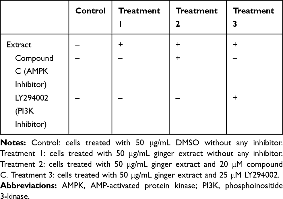 |
Table 1 Different Groups of Cell Treatment |
Myotubes were treated with ginger ethyl acetate extract for 3 h and then washed 3 times with PBS, pH 7.4. Tissue homogenizer (HES buffer, pH 7.4; 225 mM sucrose; 4 mM Na2-EDTA, 20 mM HEPES; 1 mM phenylmethyl sulfonyl fluoride, and 1 tablet/dL anti-protease cocktail) was added to cells. Some similar studies had used Tortorella method to prepare fractions;20 in this study, this method was modified: centrifugation was performed at 15,000 rpm for 45 minutes at 4ºC, the supernatant of this stage was taken as a cytosolic fraction, and the resulted pellet was used as membrane fraction and homogenized in HES buffer.
Protein concentration in the membrane and cytoplasmic fractions was determined according to the Bradford method.
Proteins in the cell lysates (70 µg) were separated on 12% sodium dodecyl sulfate-polyacrylamide gel electrophoresis (SDS PAGE) and transferred to nitrocellulose membrane (24 h). The blotted membranes were blocked using 2% blocking agent in PBS and washed with 1% Tween 20 in PBS. The blotted paper was immersed in 2% blocking agent containing mouse anti-GLUT-4 antibody IF8 (dilution 1:400) for 24 h, followed by a wash stage, and finally incubated with goat anti-mouse IgG-HRP secondary antibody with a dilution of 1:1500 for 3 h.
The bands were clarified using the ECL kit. GLUT-4 bands were visualized by gel documentation device. The amounts of GLUT-4 protein were determined by Gene Tools (Syngene, USA) image analysis software.
Statistical Analysis
Data were analyzed by the Shapiro–Wilk test and the Kolmogorov–Smirnov test for normality and equality of continuous distributions. By p = 0.001 it was shown that they do not have a normal distribution. So, the Mann–Whitney U-test was used for comparison of the membrane vs cytosolic fractions regarding the amount of GLUT-4, which yielded p = 0.001, ie, a statistically significant difference. To compare the 8 groups regarding the median amount of GLUT-4 we applied Kruskal–Wallis test which showed p = 0.002 and prompted us to do post hoc test Bonferroni. The results are expressed as mean ± standard deviation (SD). Any P ˂ 0.05 was considered to be statistically significant.
Results
Cytotoxicity Assay
With the MTT assay, we determined the nontoxic concentration of ginger ethyl acetate extract. Different concentrations (10, 50, 100 and 200 µg/mL) and DMSO were used. By comparing the results of MTT assay and other similar investigations, no toxicity was observed at these doses. Finally, the dose 50 µg was selected.6
Evaluation of Cell Growth
An inverted microscope was used (Model TS100, Nikon Co., Japan) to assess the growth of C2C12 cells. Figure 1 shows the cells.
Determination of GLUT-4 Protein Content in Different Groups
After preparation of the cytosolic and plasma membrane fractions, the Bradford method was performed (to load equal amount of protein in Western blotting), SDS PAGE electrophoresis was done, and GLUT-4 bands in cytosolic fractions and membrane fractions were detected using Western blotting and gel documentation software analysis (Figure 2).
The amount of GLUT-4 protein was analyzed. In each experiment, the maximum amount of GLUT-4 belonged to the membrane fraction of the second sample. The maximum amount of GLUT-4 obtained through analysis was considered 100, and the quantities of other fractions were determined relative to the membrane fraction of sample 2 (Figure 3). Bonferroni test showed that the difference in GLUT-4 quantity between ginger extract membrane fraction and ginger extract cytosol fraction is significant (p = 0.012).
GLUT-4 Protein in Membrane Fractions
The amounts of GLUT-4 protein in membrane fractions were compared (Figure 4). It was 20.17 ± 2.02, 100 ± 0, 33.25 ± 2.82, and 42.15 ± 1.01 in the control, treatment 1, 2 and 3 groups, respectively. Bonferroni test revealed that the amount of GLUT-4 protein in the membrane fraction of cells in the treatment 1 (ginger extract) group was significantly higher compared to the DMSO group (P = 0.013).
GLUT-4 Protein in Cytosolic Fractions
The amounts of GLUT-4 protein in cytosolic fractions were compared (Figure 5). It was found to be 13.45 ± 0.6, 12.68 ± 0.9, 17.86 ± 1.04 and 16.39 ± 0.8 in the control, treatment 1, 2 and 3 groups, respectively. As is seen, its amount in the treatment 1 and control groups was significantly increased compared to the cells in other two groups (P = 0.039 by Bonferroni test when comparing ginger extract with ginger extract plus AMPK inhibitor).
Changes in Protein Levels in the Presence of Inhibitors
The presence of each inhibitor leads to a significant decrease in the amount of GLUT-4 protein in the membrane fractions compared to its absence (P < 0.05). Conversely, the presence of each inhibitor significantly increased the amount of GLUT-4 protein in the cytosolic fractions in comparison with its absence (P < 0.05).
The amount of GLUT-4 protein in the membrane fraction of cells in the treatment 3 group (GLUT-4 quantity = 42.15 ± 1.01) was significantly more compared to that in the treatment 2 group (GLUT-4 quantity = 33.25 ± 2.82), P < 0.05, while there was no significant difference between the amount of GLUT-4 protein in the cytosolic fraction of cells in the treatment 2 group (GLUT-4 quantity = 17.86 ± 1.04) and the treatment 3 group (GLUT-4 quantity = 16.39 ± 0.8), P > 0.05.
Discussion
Diabetes is a metabolic disorder, control of which has proved to be challenging.1 New drug candidates for its treatment should be fully evaluated in terms of mechanism of action.21 GLUT-4 is an important transporter of glucose, especially in muscle cells.9,10 There are two key pathways in glucose metabolism in skeletal muscles, namely AMPK and PI3K pathways.7,8
Many herbal plants have health benefits, most of them being phenolic compounds. During the last decade, many researchers have investigated the biochemical actions and health benefits of them, such as anti-obesity, anti-cancer and anti-diabetes effects.8 Adherence to the prescribed drugs is very important22 and prescription of herbal drugs may positively influence patients’ compliance. Plants are well known for their complex actions due to various constituents.23 In many researches, it has been shown that ginger has hypoglycemic effects.6,17,18
Ginger can increase glucose uptake in L6 cells and can be consumed as oral medicine. Ginger has been used in traditional medicine for some diseases such as arthritis, rheumatism and muscular aches.6 Methanol and ethyl acetate ginger extracts decrease obesity in mice, decrease serum glucose levels, and improve insulin resistance.19 Ginger extract has potential to inhibit key enzymes relevant to type 2 diabetes and inflammation, such as α-glucosidase and α-amylase.18 Ginger decreases serum concentration of glucose, cholesterol and triglycerides in rats with streptozotocin-induced diabetes.5 (6)-gingerol increases glucose uptake in skeletal muscle by increasing GLUT4 membrane expression.24
It probably acts through two pathways: AMPK pathway and PI3K pathway. When PI3K was activated it activates Akt which leads to translocation of GLUT-4 into the membrane; LY292004 (PI3K inhibitor) inhibits this pathway. Also, when AMPK is activated it mediates translocation of GLUT-4 into the membrane; compound C (AMPK inhibitor) does not allow activation of this pathway.25 A comparable study concerning the metabolic effects of ginger was done by Toda et al.26 They used extract of black ginger (Kaempferia parviflora, Thai ginseng, not grown in Iran), while we used Zingiber officinale Roscoe (a different genus from the family Zingiberaceae). Moreover, in our study, the specific inhibitors used against each of the 2 metabolic pathways showed the exact way ginger acts.
The results of this study showed that ethyl acetate ginger extract significantly increases GLUT-4 protein amount in the membrane fraction of differentiated C2C12 cells through two pathways, AMPK and PI3K pathways, but with greater involvement of AMPK pathway because when there is no inhibitor, ethyl acetate ginger extract can increase GLUT-4 protein in the membrane fraction, and this effect becomes less in the presence of each inhibitor. On the other hand, in the presence of AMPK inhibitor ethyl acetate ginger extract has less effect on increasing GLUT-4 protein amount in membrane fraction compared to the presence of PI3K inhibitor. This fact shows that AMPK pathway is the more effective signalling pathway in C2C12 cells treated with ginger extract.
There was a higher amount of GLUT-4 protein in cytosolic fractions of cells treated with ethyl acetate ginger extract in the presence of each of AMPK inhibitor and PI3K inhibitor in comparison with the cells treated with ethyl acetate ginger extract without any inhibitor. This significant difference confirms that GLUT-4 protein cannot completely translocate from the cytosol into the membrane in the presence of those two inhibitors.
In clinical trials, doses of 170 mg to 1 g of crude ginger 3 to 4 times daily have been used for its diverse medicinal effects.27 A limitation on our study was the lack of supportive data from gene expressions. In vivo studies would also be complementary. The study could be completed by the application of HPLC-grade gingerol (the presumed active ingredient of ginger extract) on C2C12 cells and other kinds of cells important in glucose metabolism.
Conclusion
Ginger extract can affect glucose metabolism in muscle cells in vitro. The anti-diabetic effects of ginger maybe by increasing GLUT-4 in the membrane fraction of differentiated C2C12 cells through AMPK and PI3K pathways, but the role of AMPK pathway is more important.
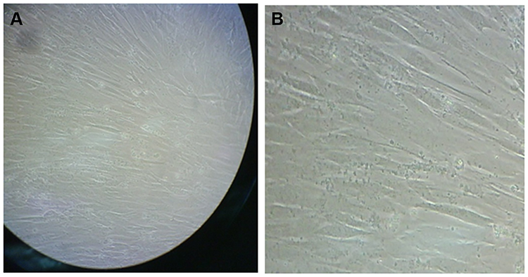 |
Figure 1 C2C12 cells on the third day of differentiation, inverted microscopy (A) ×100; (B) ×200. |
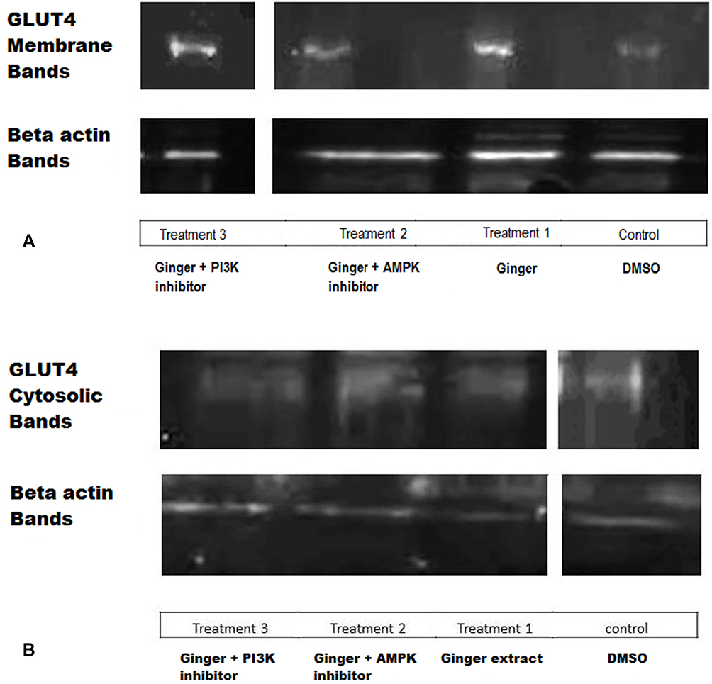 |
Figure 2 (A) GLUT-4 bands of membrane fractions, (B) GLUT-4 bands of cytosolic fractions. Control: cells treated with 50 μg/mL DMSO, but without any inhibitor. Treatment 1: cells treated with 50 μg/mL ginger extract without any inhibitor. Treatment 2: cells treated with 50 μg/mL ginger extract and 20 μM AMPK inhibitor. Treatment 3: cells treated with 50 μg/mL ginger extract and 25 μM PI3K inhibitor. |
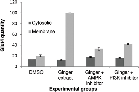 |
Figure 3 Comparison of GLUT-4 quantity between cytosolic and membrane fractions. In each treatment group there was significantly more amount of GLUT-4 protein in membrane fractions in comparison to cytosolic fractions, P value = 0.012 by Bonferroni post hoc test. |
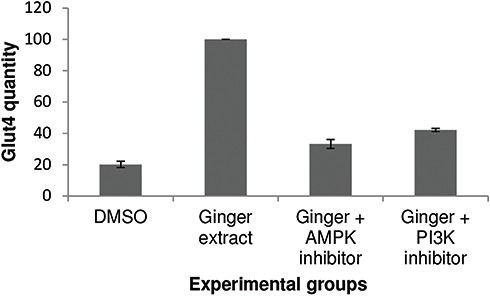 |
Figure 4 Comparison of GLUT-4 quantity between membrane fractions of different treatment groups. The amount of GLUT-4 protein was significantly more in membrane fraction of treatment 1 (cells treated with 50 μg/mL ginger extract without any inhibitors) compared to the control (cells treated with 50 μg/mL DMSO without any inhibitors) (p = 0.013), and also higher in comparison with the treatment 2 (cells treated with 50 μg/mL ginger extract and 20 μM compound C) and treatment 3 (cells treated with 50 μg/mL ginger extract and 25 μM LY294002) groups. |
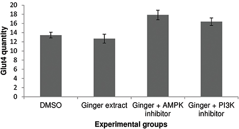 |
Figure 5 Comparison of GLUT-4 quantity between cytosolic fractions of different treatment groups. There was significantly more amount of GLUT-4 protein in cytosolic fraction of treatment 2 (cells treated with 50 μg/mL ginger extract and 20 μM compound C) in comparison with the treatment 1, P value = 0.039. A similar situation was seen in cytosolic fraction of treatment 3 (cells treated with 50 μg/mL ginger extract and 25 μM LY294002) compared to the treatment 1, but there was no significant different between treatment 2 and treatment 3, and also between control and treatment 1 (P value > 0.05). |
Disclosure
The authors report no conflicts of interest in this work.
References
1. Deepthi B, Sowjanya K, Lidiya B, Bhargavi RS, Babu PS. A modern review of diabetes mellitus: an annihilatory metabolic disorder. J In Silico In Vitro Pharmacol. 2017;3(1):14.
2. Harbilas DHD, Martineau LCMLC, Harris CSHCS, et al. Evaluation of the antidiabetic potential of selected medicinal plant extracts from the Canadian boreal forest used to treat symptoms of diabetes: part II. Can J Physiol Pharma. 2009;87(6):479–492. doi:10.1139/Y09-029
3. Zhang M, Chen L. Berberine in type 2 diabetes therapy: a new perspective for an old antidiarrheal drug? Acta Pharmaceutica Sin B. 2012;4(2):379–386. doi:10.1016/j.apsb.2012.06.004
4. Xie W, Zhao Y, Zhang Y. Traditional Chinese medicines in treatment of patients with type 2 diabetes mellitus. Evid-Based Compl Alt. 2011;2011(1–13):388–393.
5. Al-Amin ZM, Thomson M, Al-Qattan KK, Peltonen-Shalaby R, Ali M. Anti-diabetic and hypolipidaemic properties of ginger [Zingiber officinale] in streptozotocin-induced diabetic rats. Brit J Nutr. 2006;96(4):660–666. doi:10.1079/BJN20061849
6. Rani MP, Krishna MS, Padmakumari KP, Raghu KG, Sundaresan A. Zingiber officinale extract exhibits antidiabetic potential via modulating glucose uptake, protein glycation and inhibiting adipocyte differentiation: an in vitro study. J Sci Food Agr. 2012;92(9):1948–1955. doi:10.1002/jsfa.5567
7. Hao C, Hao J, Wang W, et al. Insulin sensitizing effects of oligomannuronate-chromium [III] complexes in C2C12 skeletal muscle cells. PLoS One. 2011;6(9):e24598. doi:10.1371/journal.pone.0024598
8. Kang C, Kim E. Synergistic effect of curcumin and insulin on muscle cell glucose metabolism. Food Chem Toxicol. 2010;48(8):2366–2373. doi:10.1016/j.fct.2010.05.073
9. Thong FSL, Dugani CB, Klip A. Turning signals on and off: GLUT4 traffic in the insulin-signaling highway. Physiology. 2005;20(4):271–284. doi:10.1152/physiol.00017.2005
10. Bhattacharya S, Dey D, Roy SS. Molecular mechanism of insulin resistance. J Biosciences. 2007;32(2):405–413. doi:10.1007/s12038-007-0038-8
11. Cheng Z, Pang T, Gu M, et al. Berberine-stimulated glucose uptake in L6 myotubes involves both AMPK and p38 MAPK. Bba-Gen Subjects. 2006;1760(11):1682–1689. doi:10.1016/j.bbagen.2006.09.007
12. Viollet B, Lantier L, Devin-Leclerc J, et al. Targeting the AMPK pathway for the treatment of Type 2 diabetes. Front Biosci-Landmark. 2009;14:33–80.
13. Tara M, Rogers NH, Stancheva ZS, Greenberg AS. Estradiol and the estradiol metabolite, 2-hydroxyestradiol, activate AMP-activated protein kinase in C2C12 myotubes. Obesity. 2008;16(6):1284–1288. doi:10.1038/oby.2008.50
14. Lim CT, Kola B, Korbonits M. AMPK as a mediator of hormonal signaling. J Mol Endocrinol. 2010;44(2):87–97. doi:10.1677/JME-09-0063
15. Kim JH, Park JM, Kim EK, et al. Curcumin stimulates glucose uptake through AMPK‐p38 MAPK pathways in L6 myotube cells. J Cell Physiol. 2010;223(3):771–778. doi:10.1002/jcp.22093
16. Musi N, Hayashi T, Fujii N, Hirshman MF, Witters LA, Goodyear LJ. AMP-activated protein kinase activity and glucose uptake in rat skeletal muscle. Am J Physiol Endocrinol Metab. 2001;280(5):E677–E684.
17. Noipha K, Ratanachaiyavong S, Ninla-Aesong P. Enhancement of glucose transport by selected plant foods in muscle cell line L6. Diabetes Res Clin Pr. 2010;89(2):e22–e26. doi:10.1016/j.diabres.2010.04.021
18. Priya Rani M, Padmakumari K, Sankarikutty B, Lijo Cherian O, Nisha V, Raghu K. Inhibitory potential of ginger extracts against enzymes linked to type 2 diabetes, inflammation and induced oxidative stress. Int J Food Sci Nutr. 2011;62(2):106–110. doi:10.3109/09637486.2010.515565
19. Ali BH, Blunden G, Tanira MO, Nemmar A. Some phytochemical, pharmacological and toxicological properties of ginger [Zingiber officinale Roscoe]: a review of recent research. Food Chem Toxicol. 2008;46(2):409–420. doi:10.1016/j.fct.2007.09.085
20. Tortorella LL, Pilch PF. C2C12 myocytes lack an insulin-responsive vesicular compartment despite dexamethasone-induced GLUT4 expression. Am J Physiol Endocrinol Metab. 2002;283(3):E514–524. doi:10.1152/ajpendo.00092.2002
21. Gill P, Gill V, Singh N. In silico quantitative structure pharmacokinetic relationship modeling on antidiabetic drugs: time to reach peak plasma concentration. Am J Adv Drug Deliv. 2013;3(1):277–284.
22. Sharifi Rad J, Hoseini Alfatemi M, Sharifi Rad M, Jyoti Sen D. Phytochemical and antimicrobial evaluation of the essential oils and antioxidant activity of aqueous extracts from flower and stem of sinapis arvensis L. Am J Adv Drug Deliv. 2013;1(1):1–10.
23. Inamdar SZ, Kulkarni RV, Karajgi SR, Manvi FV, Ganachari MS, Mahendra Kumar BJ. [2013] Medication adherence in diabetes mellitus: an overview on pharmacist role. Am J Adv Drug Deliv. 2013;3(1):238–250.
24. Samad MB, Mohsin MNAB, Razu BA, et al. [6]-Gingerol, from Zingiber officinale, potentiates GLP-1 mediated glucose-stimulated insulin secretion pathway in pancreatic beta-cells and increases RAB8/RAB10-regulated membrane presentation of GLUT4 transporters in skeletal muscle to improve hyperglycemia in Lepr[db/db] type 2 diabetic mice. BMC Complement Altern Med. 2017;17([1]):395.
25. Jin Son M, Miura Y, Yagasaki K. Mechanisms for antidiabetic effect of gingerol in cultured cells and obese diabetic model mice. Cytotechnology. 2015;67(4):641–652. doi:10.1007/s10616-014-9730-3
26. Toda K, Takeda S, Hitoe S, Nakamura S, Matsuda H, Shimoda H. Enhancement of energy production by black ginger extract containing polymethoxy flavonoids in myocytes through improving glucose, lactic acid and lipid metabolism. J Nat Med. 2016;70(2):163–172. doi:10.1007/s11418-015-0948-y
27. http://www.drugs.com/npp/ginger.html.
 © 2020 The Author(s). This work is published and licensed by Dove Medical Press Limited. The
full terms of this license are available at https://www.dovepress.com/terms
and incorporate the Creative Commons Attribution
- Non Commercial (unported, 3.0) License.
By accessing the work you hereby accept the Terms. Non-commercial uses of the work are permitted
without any further permission from Dove Medical Press Limited, provided the work is properly
attributed. For permission for commercial use of this work, please see paragraphs 4.2 and 5 of our Terms.
© 2020 The Author(s). This work is published and licensed by Dove Medical Press Limited. The
full terms of this license are available at https://www.dovepress.com/terms
and incorporate the Creative Commons Attribution
- Non Commercial (unported, 3.0) License.
By accessing the work you hereby accept the Terms. Non-commercial uses of the work are permitted
without any further permission from Dove Medical Press Limited, provided the work is properly
attributed. For permission for commercial use of this work, please see paragraphs 4.2 and 5 of our Terms.