Back to Journals » International Journal of Nanomedicine » Volume 21
Gestational Intranasal Exposure to Silicon Dioxide Nanoparticles in Rats
Authors Jeong JS, Kim D, Kim W, Kim SY, Lee SY, Park JD, Wi IS, Park C, Kim JH, Jeong W, Suh HN, Kang MS, Kim IH, Kim SH, Lee J ![]()
Received 5 March 2026
Accepted for publication 22 May 2026
Published 11 June 2026 Volume 2026:21 604622
DOI https://doi.org/10.2147/IJN.S604622
Checked for plagiarism Yes
Review by Single anonymous peer review
Peer reviewer comments 3
Editor who approved publication: Dr Sachin Mali
Ji-Seong Jeong,1,* Donghee Kim,2,* Woojin Kim,3 Sang Yun Kim,1 Sun-Young Lee,1 Jeong-Dong Park,1 In-Su Wi,1 Cheoljin Park,2 Jeong Hwan Kim,2 Wonjun Jeong,2 Han Na Suh,4 Min-Sung Kang,4 In-Hyeon Kim,4,5 Sung-Hwan Kim,4 Jinsoo Lee2
1Laboratory of Developmental and Reproductive Toxicology Research, Korea Institute of Toxicology, Daejeon, Republic of Korea; 2Laboratory Animal Medicine, College of Veterinary Medicine, Chungnam National University, Daejeon, Republic of Korea; 3Toxicologic Pathology Research Group, Korea Institute of Toxicology, Daejeon, Republic of Korea; 4Jeonbuk Branch Institute, Korea Institute of Toxicology, Jeongeup, Republic of Korea; 5College of Veterinary Medicine, Chonnam National University, Gwangju, Republic of Korea
*These authors contributed equally to this work
Correspondence: Sung-Hwan Kim, Jeonbuk Branch Institute, Korea Institute of Toxicology, Jeongeup, Republic of Korea, Tel +82-63-570-8757, Email [email protected] Jinsoo Lee, Laboratory Animal Medicine, College of Veterinary Medicine, Chungnam National University, Daejeon, Republic of Korea, Tel +82-42-821-6778, Email [email protected]
Background: Silicon dioxide nanoparticles (SiO2NPs) are among the most widely manufactured nanomaterials, used in diverse industrial, pharmaceutical, and consumer products. Although the respiratory tract represents one of the major routes of human exposure, the potential reproductive and developmental toxicity of SiO2NPs via respiratory exposure remains poorly characterized.
Methods: This study investigated the maternal and developmental toxicity of SiO2NPs in pregnant Sprague-Dawley rats. Pregnant animals were intranasally administered SiO2NPs once daily during gestation day (GD) 6 to 20 at doses of 0, 2, 6, and 20 mg/animal, followed by caesarean section on GD 21.
Results: Treatment-related changes were observed in the respiratory tract, appearing as mild inflammatory and regenerative alterations. No mortality or systemic maternal toxicity was observed, and evaluation of caesarean section parameters and fetal morphology demonstrated no evidence of adverse developmental effects.
Conclusion: Repeated intranasal exposure to SiO2NPs during pregnancy induced localized histopathological changes in the respiratory tract, with no evidence of systemic maternal or embryo-fetal toxicity at the assessed endpoints under the present experimental conditions. These findings provide experimental data relevant to the assessment of respiratory exposure to SiO2NPs during pregnancy; however, further studies are warranted considering the diverse properties of nanoparticles and the potential for varied exposure scenarios.
Keywords: silicon dioxide nanoparticles, developmental toxicity, maternal toxicity, nanotoxicology
Introduction
Silicon dioxide nanoparticles (SiO2NPs) are among the most widely manufactured nanomaterials, with uses extending across industrial and biomedical fields. The chemical stability, biocompatibility, and intrinsic surface chemistry of SiO2NPs have enabled incorporation as functional additives in food and pharmaceutical formulations, as well as in catalytic and environmental systems.1–3 In parallel with these expanding applications, global production and commercial use of SiO2NPs have increased substantially, with recent market analyses estimating a global value of approximately USD 1.01 billion in 2024 and projecting growth to USD 3.65 billion by 2033.4 The rapid growth in production and utilization highlights the growing need to assess potential impacts on human health.
Toxicological concerns have emerged, leading to extensive research investigating the biological hazards of SiO2NPs. Numerous in vitro studies have demonstrated that SiO2NPs induce cytotoxicity through the excessive generation of reactive oxygen species (ROS), resulting in oxidative stress, mitochondrial dysfunction, inflammation, and apoptotic cell death.5,6 These toxic responses have been consistently observed across multiple cell types, including lung epithelial cells, macrophages, and other mammalian cell lines. Additionally, in vivo investigations have confirmed that SiO2NPs exposure elicits oxidative injury, inflammatory cell infiltration, and tissue abnormalities in multiple organs such as the brain,7 cardiovascular system,8 lung,9 liver,10 and kidney.11 Hong et al12 reported that single intratracheal exposure to SiO2NPs (surface area 640 m2/g, 6.25, 12.5, and 25 mg/animal) or microscale SiO2 particles (1–5 µm, 25 mg/animal) induced lung inflammation and fibrosis in rats. Boudard et al13 reported that long-term oral exposure to SiO2NPs (20 nm, 30 mg/L in drinking water for 18 months) led to silica accumulation and histopathological lesions in the kidneys and liver of mice. It is worth noting, however, that not all studies report toxicity. Some investigations have found mild to no adverse effects under certain exposure conditions. Ninety-day dermal exposure to 20 nm SiO2NPs (500, 1000, and 2000 mg/kg) demonstrated no systemic toxicity in rats.14 Single intravenous administration of 150 nm SiO2NPs (1, 2.5, 5, 10, 100, 200, and 300 mg/kg) caused no mortality, clinical abnormalities, or histopathological changes in mice.15 These divergent findings emphasize the importance of conducting in vivo studies under controlled and well-characterized experimental conditions.
Variability in reported toxic effects of SiO2NPs is often attributable to differences in particle properties and exposure scenarios. Factors such as particle size, agglomeration state, surface chemistry, dose, and route of exposure can critically influence the biological response.16–18 These factors likely contribute to the inconsistent toxicity findings reported across previous studies. Among these, the route of exposure deserves particular attention, as human contact with nanoparticles can occur through multiple pathways, including inhalation, ingestion, dermal contact, or injection.19,20 Notably, respiratory exposure is considered a major route of concern, especially under environmental and workplace settings.21 Song et al22 reported the presence of 20 nm SiO2NPs in the lungs of printing factory workers, detected in macrophages and within the pulmonary vascular system. SiO2NPs primarily accumulate in the respiratory tract, and smaller particles may translocate across the alveolar–capillary barrier, leading to systemic distribution.5,23 In this context, intranasal administration is considered a relevant experimental approach to mimic respiratory exposure and to evaluate both local and systemic effects of SiO2NPs under controlled conditions.
Previous studies have primarily focused on systemic effects of SiO2NPs, leaving reproductive and developmental aspects underexplored. This gap is particularly important because pregnancy is a unique physiological state that can alter susceptibility to nanoparticle exposure. The ability of nanoparticles to cross the placental barrier and reach the fetus raises the possibility of direct or indirect effects on fetal development.24–26 Supporting this concern, experimental studies have reported developmental abnormalities following SiO2NPs exposure. For example, in the frog embryo developmental toxicity assay, SiO2NPs exhibited teratogenic effects, inducing malformations and elevated ROS production.27 In mice, prenatal dietary exposure to food-grade SiO2NPs (S200, approximately 10–30 nm, 225 and 5000 mg/kg) resulted in fetal loss and inflammation in both maternal and fetal livers, accompanied by broad DNA methylation changes.28 In addition, prenatal intravenous exposure to SiO2NPs caused female offspring-specific subfertility through disrupted meiotic recombination and oocyte apoptosis in mice.29 Conversely, several studies consistently reported an absence of reproductive or developmental toxicity after SiO2NPs exposure.30,31 Together, these conflicting results underscore the need for further investigations to clarify the reproductive and developmental risks of SiO2NPs under realistic exposure conditions.
Therefore, this study was designed to comprehensively evaluate the maternal and developmental toxicity of SiO2NPs following repeated intranasal exposure during pregnancy in rats. Pregnant Sprague-Dawley rats were administered SiO2NPs intranasally once daily from gestation day (GD) 6 to 20 at doses of 0, 2, 6, and 20 mg/animal. Detailed assessments of maternal clinical observations, histopathological examinations including the respiratory system, and embryo-fetal developmental endpoints were conducted. To ensure relevance for human health risk assessment, the study design was informed by the international regulatory authorities test guidelines on the toxicity32 from a regulatory toxicology standpoint. The results of this study deepen the current understanding of SiO2NP-induced reproductive vulnerability, providing evidence to support route-aware safety evaluations.
Materials and Methods
SiO2NPs and Physicochemical Characterization
SiO2NPs (nanopowder, 99.5% purity) were sourced from Sigma-Aldrich (USA). According to the supplier’s specifications, their nominal primary particle size was 15 nm. Additional physicochemical characterization of the SiO2NPs was conducted in our previous study, and the same batch was used in the present work to ensure consistency of the results.33 Briefly, the primary particle size and morphology were confirmed by transmission electron microscopy (TEM, JEM-2100F, JEOL, Japan), and elemental composition and purity were verified by energy-dispersive X-ray spectroscopy (EDS; Oxford Instruments, UK). The hydrodynamic diameter in the vehicle was measured using dynamic light scattering (DLS; ELS-8000, Otsuka Electronics, Japan), confirming that the particles were present as a suspension in saline.
In our previous study, the SiO2NPs exhibited an amorphous morphology with a mean primary size of 13.6 ± 5.6 nm and a hydrodynamic diameter of approximately 421.3 nm when suspended in saline (6 mg/mL), indicating the formation of agglomerates in the vehicle.
Animals and Housing
Sprague-Dawley rats (Crl:CD(SD), specific-pathogen-free; Orient Bio, Republic of Korea) were obtained at approximately 9 weeks of age. Animals were acclimated for 5 days, and animals without clinical abnormalities were subsequently used for mating. Females were mated 1:1 with males overnight. Sperm positive vaginal smears marked gestation day (GD) 0. Each confirmed pregnant female was housed individually in a polysulfone cage (W×L×H; 260×420×180 mm3) containing sterilized aspen bedding and enrichment items. Environmental parameters were controlled at approximately 20–26°C, with 30–70% humidity, 150–300 Lux, 10–20 times/h ventilation, and a 12-h light/dark cycle. Irradiated standard rodent diet (PMI Nutrition International, USA) and UV-sterilized tap water were available ad libitum. Group allocation occurred on GD 0 using body-weight-based randomization to balance means across groups.
All experimental procedures were conducted at the Korea Institute of Toxicology (KIT), an AAALAC International-accredited facility operating under Good Laboratory Practice (GLP) standards. The study received prior approval from the Institutional Animal Care and Use Committees (IACUC) of KIT (Approval No. 2112–0060) based on the Animal Protection Act of Korea and the Guide for the Care and Use of Laboratory Animals.34 This study was performed in a GLP-compliant facility and under standard operating procedures, although this investigation was designated as non-GLP for exploratory research.
Experimental Design and Dose Preparation
Forty-eight female SD rats were assigned to four groups, consisting of a vehicle control and three SiO2NPs exposure groups (12 mating-proven females per group). A brief overview of the experimental design and dosing schedule is illustrated in Figure 1. Doses of 2, 6, and 20 mg/animal were administered using formulations prepared at concentrations of 20, 60, and 200 mg/mL, respectively. The dose levels were selected based on the maximum feasible dose for intranasal administration in rats. This was determined considering both the maximum administrable volume (approximately 100 µL per animal), which is commonly used in rodent intranasal studies, and the highest concentration at which SiO2NPs could be stably dispersed in the vehicle. In a preliminary dose range-finding study, no treatment-related effects on clinical signs, body weight, food consumption, or organ weights were observed up to 20 mg/animal. The lower dose levels were set using an approximately three-fold geometric spacing. Animals in the vehicle control group received sterile saline alone using the same intranasal dosing procedure and volume. From GD 6 to 20, each female received once-daily intranasal administration under light isoflurane anesthesia. A total volume of 100 µL was given per animal, equally divided into 50 µL per nostril. Test formulations were freshly prepared each day by dispersing SiO2NPs in sterile saline. The high dose concentration (200 mg/mL) was prepared first and sonicated for 8 minutes at 25% amplitude using an ultrasonic processor (VC 505, Sonics & Materials Inc., USA) and subsequently diluted with saline to obtain the middle- and low-dose formulations. All suspensions were maintained under gentle magnetic stirring until administration to ensure homogeneity.
 |
Figure 1 Schematic representation of the study design. |
Maternal Clinical Observations and Measurements
Mortality and morbidity were monitored twice daily, at the beginning and end of routine animal room procedures. Clinical observation, including changes in appearance and behavior, was conducted once daily during the pre-dosing period and twice daily (before and after dosing) during the dosing period. Particular attention was paid to detecting signs of premature parturition or abortion throughout gestation. Maternal body weight and food consumption were recorded on GD 0, 6, 9, 12, 15, 18, and 21.
At termination on GD 21, all surviving females were euthanized by CO2 inhalation and subjected to a complete gross necropsy. Careful examination was directed toward the thoracic and abdominal cavities, with special inspection of reproductive organs and any macroscopic abnormalities. Gravid uterine weight was recorded to calculate the corrected terminal weight (body weight on GD 21 – gravid uterine weight) and the net body weight change (corrected terminal body weight – body weight on GD 6). Absolute and relative organ weights were determined for adrenal glands, brain, heart, kidneys, liver, lungs, spleen, and thymus.
Histopathological Assessment
Maternal tissues, including the brain, liver, kidneys, adrenal glands, heart, lungs, nasal cavity, trachea, larynx, spleen, thymus, lymph node (the hilar region of the lung) and placenta, were preserved in 10% neutral buffered formalin for histopathological evaluation. To ensure a detailed assessment of respiratory toxicity, airway tissues were trimmed according to multilevel criteria: the larynx at three levels including the base of the epiglottis; the lungs at a single plane encompassing all lobes and the main bronchi; the nasal cavity at a minimum of four levels, one of which included the nasopharyngeal duct, teeth, and nasal associated lymphoid tissue; and the trachea at two levels comprising one longitudinal and one transverse section. The fixed tissues were processed routinely, embedded in paraffin, sectioned, and stained with hematoxylin and eosin (H&E), and examined under a light microscope. Histopathological assessment was conducted by an experienced toxicologic pathologist. Although blinding was not implemented, the assessments were performed in an objective and consistent manner. Histopathological evaluations were initially performed in vehicle control and high-dose groups. When treatment-related lesions were observed in the high-dose group, the same organs from the low- and mid-dose groups were subsequently examined to determine whether a dose–response relationship was present.
Caesarean Section and Developmental Parameters
On GD 21, all females underwent caesarean section for developmental parameters assessment. The ovaries and uterine horns were examined to determine the number of corpora lutea, implantation sites, and the status of each implantation (live fetus, dead fetus, and early or late resorption). Pre-implantation loss, post-implantation loss, and fetal death rates were calculated. Each live fetus was weighed and sexed, and each placenta was weighed and examined macroscopically.
Fetal Birth Defect Examinations
All live fetuses obtained at caesarean section were subjected to a comprehensive morphological evaluation for birth defect detection. Immediately after removal from the uterus, each fetus was inspected externally for abnormalities. Within each litter, fetuses were sequentially numbered along the uterine horns, with odd-numbered fetuses proceeding for skeletal evaluation and even-numbered fetuses for visceral assessment. For visceral examination, fetuses were fixed in Bouin’s solution and sectioned using established methods: the head was examined using the modified Wilson’s method, thoracic organs by Nishimura’s method, and abdominal viscera by Staples’s method.35–37 For skeletal examination, the remaining fetuses were fixed with 70% ethanol, cleared with potassium hydroxide, and stained with alizarin red using modified Dawson’s method.38 Fetal morphological findings were categorized by severity as either variations or malformations and reported using the descriptive terminology for abnormalities in common laboratory animal models.39
Statistical Analysis
Statistical analyses were performed using Statistical Analysis software (SAS Institute Inc., Cary, NC, USA) and the Pristima System (Xybion Medical System, USA), as described in our previous studies.40,41 All data are presented as mean ± standard deviation (SD). The litter was used as the experimental unit for all statistical analyses. Homogeneity of variance was assessed using Bartlett’s test. For data with homogeneous variances, one-way analysis of variance (ANOVA) was performed, followed by Dunnett’s test for comparisons between the control and treated groups. For data with heterogeneous variances, the Kruskal–Wallis test was used, followed by Dunn’s rank sum test for pairwise comparisons. One-way analysis of covariance (ANCOVA) was applied to fetal and placental weight data, with litter size included as a covariate. A p-value < 0.05 was considered statistically significant.
Results
Maternal Health Examinations
General Clinical Sign Observations
All female rats survived throughout the study without any unscheduled deaths or moribund conditions. During clinical monitoring conducted throughout the dosing and observation periods, no treatment-related abnormalities were observed in any group. Although sporadic minor fur loss on the forelimbs occurred in a few females from the control and low-dose groups, these changes lacked dose dependency and were regarded as spontaneous findings.
Body Weight and Food Consumption
Maternal body weight and body weight gain were unaffected by treatment, with no significant differences across groups throughout gestation (Figure 2). Food consumption was comparable between the treated and control group animals (Table 1). Although minor intergroup fluctuations were occasionally observed, they showed no dose-related trends and were considered incidental and within the range of normal biological variation.
 |
Table 1 Food Consumption of Intranasally SiO2NPs-Exposed Pregnant Females During Pregnancy |
 |
Figure 2 Maternal body weight (A) and body weight gain (B) during gestation in rats intranasally exposed to SiO2NPs. Pregnant rats were administered 0, 2, 6, and 20 mg/animal/day via intranasal administration from gestation day (GD) 6 to 20. Maternal body weight was measured on GD 0, 6, 9, 12, 15, 18, and 21, and body weight gain (mean ± SD, n = 12 per group) was calculated for each interval. VC, vehicle control. |
Organ Weight and Macroscopic Findings
No treatment-related macroscopic abnormalities were observed at scheduled necropsy. A single minimal focal liver scar was noted in one control female and considered incidental, consistent with spontaneous background lesions. Absolute organ weights for all major organs did not differ among groups, with sporadic variations without dose dependency (Table 2). Relative organ weights, expressed as a percentage of terminal body weight, were likewise unaffected.
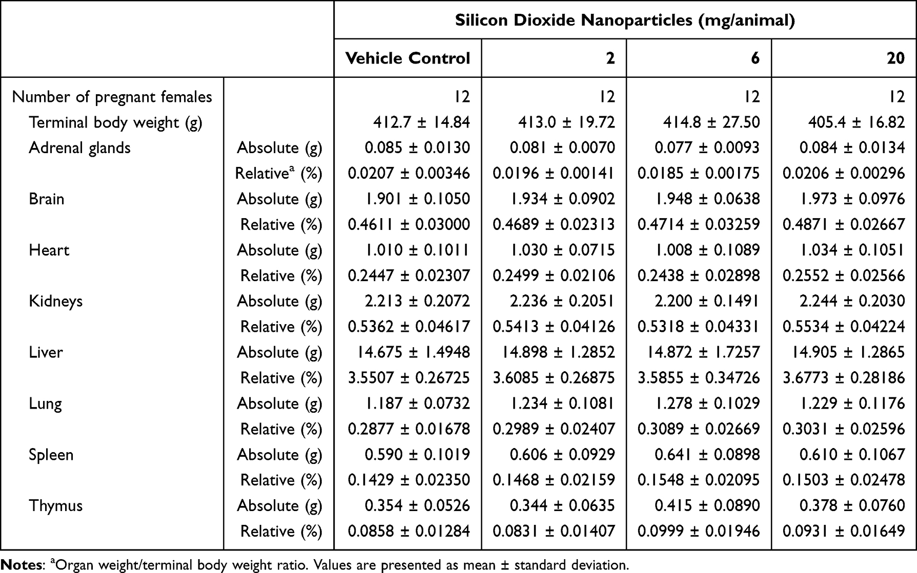 |
Table 2 Absolute and Relative Organ Weights of Intranasally SiO2NPs-Exposed Pregnant Females During Pregnancy |
Maternal Histopathology
Microscopic examination revealed that the primary SiO2NPs-related effects were localized to the respiratory tract. Histopathological abnormalities in the nasal cavity, lungs, and trachea are presented in Table 3. In the nasal cavity, erosion, transitional epithelium hyperplasia, mucous cell hyperplasia/metaplasia, neutrophilic infiltration, and regeneration were observed. Erosion was observed in all treated groups. Transitional epithelium hyperplasia was confined to level 1 (the posterior part of the incisors) and was observed only in the mid- and high-dose groups, with mucosal thickening at the lateral meatus. Mucous cell hyperplasia/metaplasia was evident in all treated groups, predominantly at level 1 in the low- and mid-dose groups. In contrast, in the high-dose group, it was mainly observed at levels 3 (second palatine crest) and 4 (first molar teeth), accompanied by an increased number of mucous cells compared with controls. Neutrophilic infiltration was noted in all treated groups, with the highest incidence in the high-dose group. Regeneration was primarily observed at level 2 (incisive papilla) across all dose groups and was characterized by basophilic cytoplasm, epithelial thickening, and an irregular luminal contour.
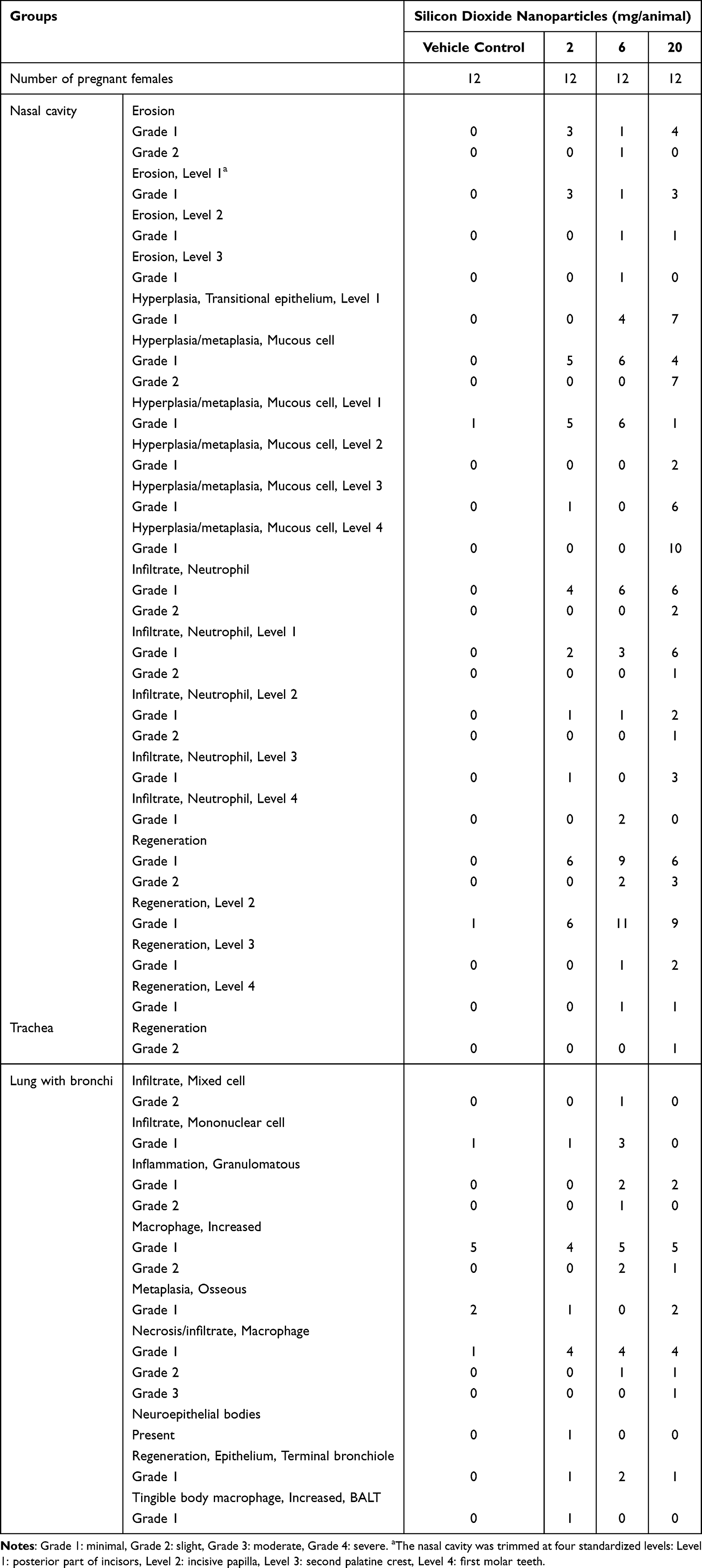 |
Table 3 Histopathological Respiratory Abnormalities of Intranasally SiO2NPs-Exposed Pregnant Females During Pregnancy |
In the lungs, SiO2NPs-related lesions consisted mainly of granulomatous inflammation and necrosis/infiltrate of macrophages. Granulomatous inflammation occurred predominantly in the mid- and high-dose groups, with macrophages and mononuclear cells distributed in periductal and perivascular patterns. Necrosis with macrophage infiltration was observed across treated groups in a dose-dependent manner, involving focal to multifocal alveolar lesions with eosinophilic pneumocyte cytoplasm and loss of cellular detail. Epithelial regeneration in the terminal bronchioles was occasionally observed; however, the change showed no apparent relationship to SiO2NPs exposure. In the trachea, slight mucosal regeneration was observed only in a single high-dose animal, characterized by increased mucosal thickness with basophilic cytoplasm and infiltration of neutrophils. Representative histopathological abnormalities are shown in Figure 3.
 |
Figure 3 Representative histopathological findings in the respiratory tract of pregnant rats intranasally exposed to SiO2NPs. (A) Nasal cavity, Level 4 (trimmed at the first molar teeth): mucous cell hyperplasia and metaplasia (arrow) at the nasopharyngeal meatus, characterized by increased mucosal thickness. (B) Nasal cavity, Level 2 (trimmed at the incisive papilla): epithelial regeneration (dotted line) at the dorsal nasal meatus, characterized by thickened basophilic epithelial cells and irregular mucosal surface. (C) Lung: focal necrosis with macrophage infiltration (asterisks) at the alveolar wall. H&E staining; scale bars = 50 µm. |
No treatment-related histopathological changes were identified in other organs. Minimal findings in visceral tissues were sporadic and showed no consistent dose-related patterns. In the kidney, occasional minimal tubular and interstitial changes were seen only in control and high-dose group animals and were regarded as incidental. Similarly, in the liver, minimal fibrosis and mononuclear cell infiltration were confined to the control and high-dose groups, without dose dependency, and were interpreted as background lesions. The placenta showed an isolated minimal mineralization in a single high-dose group female and was considered spontaneous.
Caesarean Section and Fetal Birth Defect Examinations
Caesarean Section and Gravid Uterine Weight
No treatment-related changes were observed in gravid uterine weight, corrected maternal body weight, or net body weight changes (Table 4). Caesarean section outcomes, including corpora lutea, implantation, early and late resorption, fetal deaths, and live fetuses, sex ratio, implantation loss, fetal weight, and placental weight, showed no treatment-related differences among groups (Table 5).
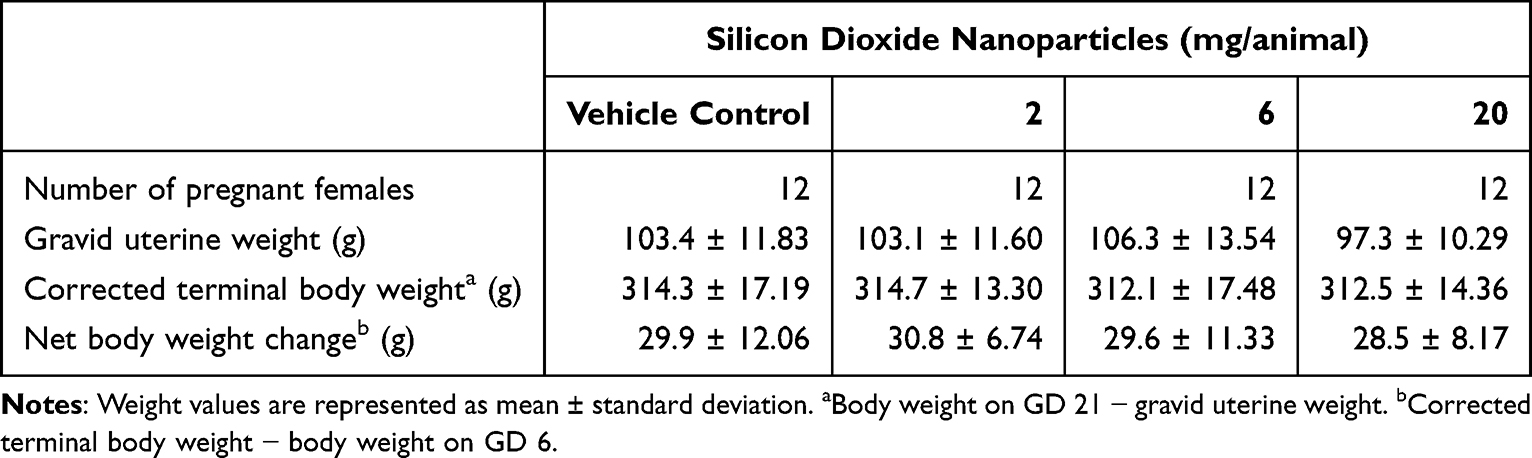 |
Table 4 Gravid Uterine Weight, Corrected Terminal Body Weight, and Net Body Weight Change of Intranasally SiO2NPs-Exposed Pregnant Females During Pregnancy |
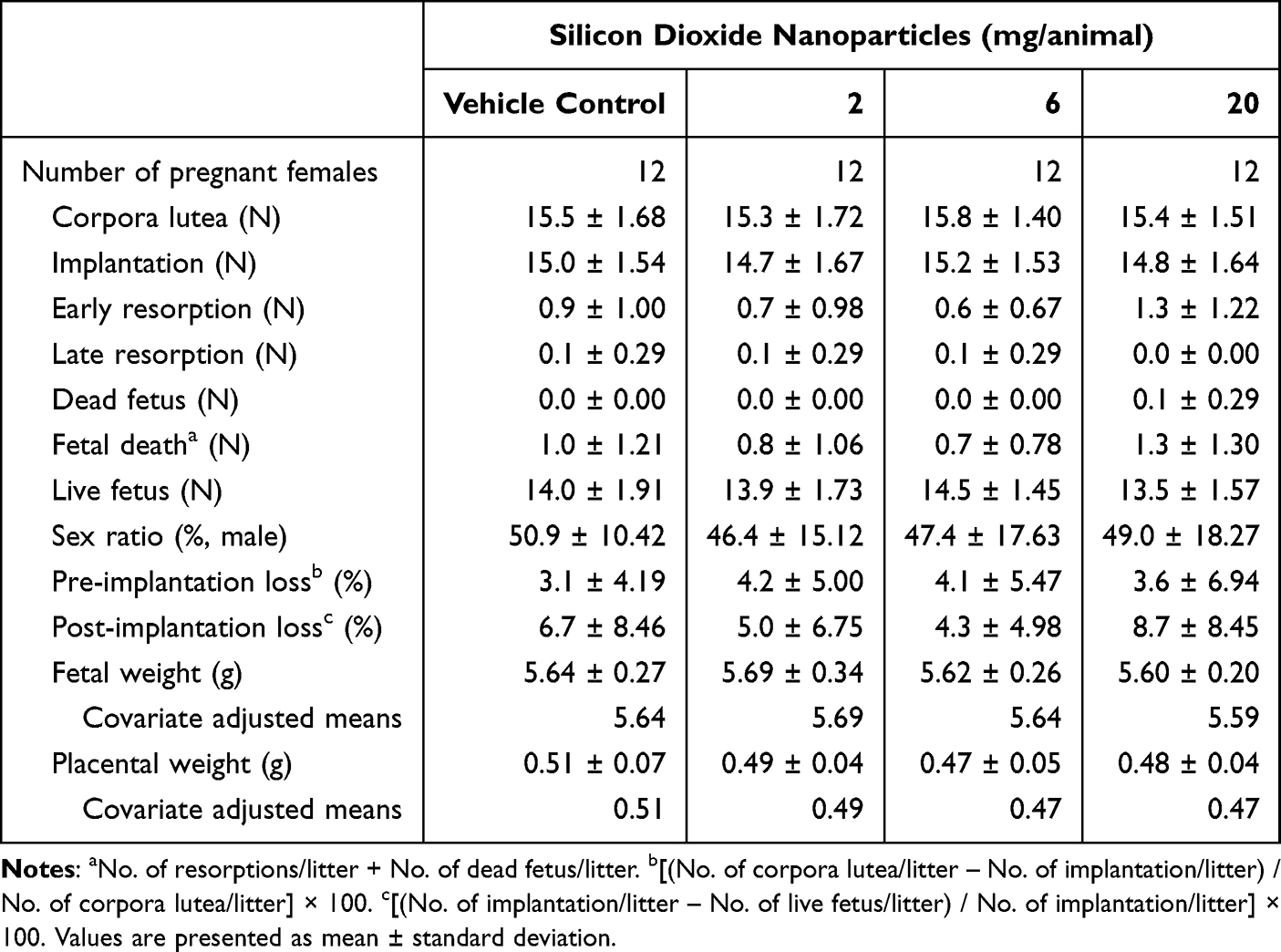 |
Table 5 Caesarean Section Results of Intranasally SiO2NPs-Exposed Pregnant Females During Pregnancy |
Fetal Birth Defect Examinations
No treatment-related external or visceral malformations were observed (Table 6). External variations were limited to a single case of thread-like tail in one fetus from the mid-dose group, which was considered incidental. Visceral variations, such as thymic cords and convoluted or dilated ureters, occurred sporadically across all groups, including controls, without dose dependency and were within the expected background range. Skeletal variations, such as supernumerary ribs and incomplete ossification of the skull or vertebrae, were observed at similar incidences in control and treated groups, with no dose-related trends. A single case of short rib was noted in one high-dose fetus but was regarded as incidental (Table 7).
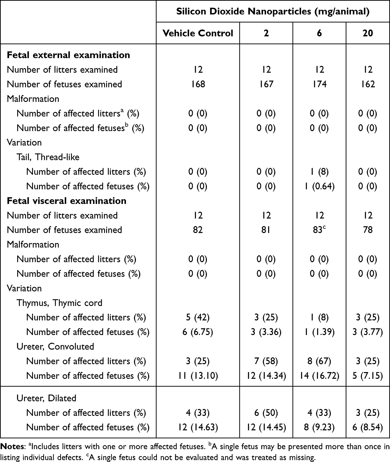 |
Table 6 Fetal External and Visceral Examinations of Intranasally SiO2NPs-Exposed Females During Pregnancy |
 |
Table 7 Fetal Skeletal Examination of Intranasally SiO2NPs-Exposed Females During Pregnancy |
Discussion
The expanding use of SiO2NPs in industrial, biomedical, and food applications has increased the potential for human exposure.2,5,42 This concern becomes particularly relevant during pregnancy, as inhaled nanoparticles may affect maternal health and potentially reach the developing fetus. Although placental transfer of nanoparticles has been demonstrated, evidence regarding the reproductive and developmental toxicity of SiO2NPs via respiratory exposure remains limited.43,44 Most previous studies have focused on oral or intravenous administration, with fewer examining inhalation-related outcomes. To fill this gap, we evaluated the effects of repeated intranasal SiO2NPs administration from GD 6 to 20 in pregnant rats, employing a regulatory perspective with comprehensive maternal and embryo-fetal endpoints. The primary finding of this study is that SiO2NPs induced localized pathological changes in the respiratory tract, with no evidence of systemic maternal or embryo-fetal toxicity at the measured endpoints under the present experimental conditions.
Histopathological examination revealed that the most distinct changes were localized to the respiratory tract. In the nasal cavities, lesions included epithelial damage with inflammatory cell infiltration and subsequent hyperplastic and regenerative changes in a dose-dependent manner. In the lungs, macrophage infiltration, necrosis, and granulomatous inflammation were evident. The findings align with previous reports of respiratory tract alterations following intranasal or inhalation exposure to silica nanoparticles. Yoshida et al45 showed that intranasally administered silica nanoparticles localized to the nasal mucosa with associated inflammatory cell aggregation, and Park et al46 reported acute airway inflammation characterized by epithelial thickening and inflammatory cell infiltration after 3-day intranasal exposure. Consistently, sub-chronic inhalation studies in rats reported inflammation with granuloma formation,47 whereas an intratracheal instillation model showed progression to fibrotic nodules after 56 days.48 Collectively, these findings indicate that local deposition of SiO2NPs induces epithelial injury and inflammatory responses within the respiratory tract, ultimately leading to tissue remodeling processes.
The mechanisms underlying SiO2NPs-induced toxicity are thought to be closely associated with oxidative stress and inflammatory signaling. Once deposited onto airway surfaces, SiO2NPs interact with epithelial and immune cells, disturbing intracellular redox homeostasis and promoting excessive production of reactive oxygen species (ROS).49 The elevated ROS promptly activate redox-sensitive pathways such as NF-κB and MAPK, inducing cytokine and chemokine release, amplifying local inflammatory responses.50,51 Concurrently, the imbalance between ROS generation and antioxidant defenses compromises mitochondrial stability and structural integrity, triggering apoptotic or necrotic cell death.52,53 In addition, ROS-mediated disruption of epithelial junctional complexes (such as zonula occludens and occludin) weakens barrier integrity, facilitating deeper particle-cell interactions.54 Through these oxidative and inflammatory cascades, SiO2NPs induce localized epithelial injury, promote inflammatory cell recruitment, and initiate early remodeling processes in the respiratory tract.
While localized respiratory alterations were evident, no evidence of systemic maternal toxicity was observed at the assessed endpoints. Throughout gestation, SiO2NPs exposure did not affect maternal clinical status, body weight gain, or organ weights, nor were any treatment-related histopathological changes observed outside the respiratory tract. These negative findings are consistent with previous in vivo studies reporting minimal or no systemic toxicity of SiO2NPs following various exposure routes. Single intratracheal instillation of SiO2NPs (20 and 50 nm, 200 and 400 µg/mL) produced no treatment-related toxicity in male rats.17 Shin et al55 reported only minimal changes, such as temporary body weight decrease and modest RBC increases, without toxicologically relevant pulmonary or systemic effects after a subacute 4-week inhalation (60–70 nm, 0.407, 1.439, and 5.386 mg/m3, 6 h/day, 5 days/week). In addition, by oral gavage in rats, Kim et al56 reported no systemic toxicity following 90-day administration (20 and 100 nm, 500, 1000, and 2000 mg/kg/day), and Liang et al57 similarly observed no adverse effects after 90-day exposure to 25.9 nm SiO2NPs (166.7, 500, and 1500 mg/kg/day). In contrast, several studies have reported systemic adverse effects of SiO2NPs across different particle sizes and exposure routes. Eight weekly intratracheal instillation of SiO2NPs (1.5, 3, and 6 mg/kg, once a week) in male rats elicited systemic particle distribution with dose-dependent lesions in the lung, heart, spleen, and kidney.58 Similarly, twelve weekly intratracheal instillation of SiO2NPs (1.5, 3, 6 mg/kg, once per week) in male ApoE−/− mice triggered multi-organ lesions (lung, liver, heart, and kidney).59 Additionally, repeated daily intraperitoneal injections of 10 nm SiO2NPs (2 mg/kg, 5 days per week) in rats decreased weight gain, altered serum chemistry, induced hepatic histopathology, and markedly downregulated hepatic drug-metabolizing enzyme genes.60
Although the respiratory tract is a major portal of nanoparticle exposure, reproductive and developmental toxicity studies involving respiratory administration of SiO2NPs remain scarce. Several studies have reported toxicity in reproductive organs following respiratory exposure. Tian et al61 showed that single intratracheal instillation of SiO2NPs (250 mg per mouse; 50 µL at 5 g/mL) in mice induced trophoblast apoptosis and uterine inflammation, indicating acute female reproductive toxicity. In pregnant mice, intratracheal instillation (43 nm, 7, 21, and 35 mg/kg, 5 times in 15 days) caused ovarian pathology with endocrine imbalance and elevated follicular atresia.62 Similarly, Zhang et al63 reported that repeated intratracheal instillation of 58 nm SiO2NPs (2 mg/kg, 15 times in 45 days) in male mice damaged the seminiferous epithelium and reduced sperm quality with increased abnormalities. However, in our present study, exposure to SiO2NPs did not induce any detectable reproductive or developmental toxicity. Indicators related to maternal condition, cesarean section findings, and embryo-fetal development showed no treatment-related changes. This observation is consistent with previous studies based on oral exposure routes. For example, oral gavage administration of SiO2NPs in rats, whether from GD 6 to 19 at doses of (10–25 nm, 100, 300, and 1000 mg/kg)31 or across two generations at similar dose levels (409–1664 nm, 100, 300, and 1000 mg/kg), did not produce toxicological effects on maternal health, reproductive performance, or offspring development.30 In line with this, our previous oral study in mice using the same SiO2NPs demonstrated that daily administration of males for approximately 6 weeks (starting 2 weeks before mating) and to females began 2 weeks before mating and continued until implantation (GD 6) (15 nm, 60, 250, and 1000 mg/kg) did not compromise fertility and early embryonic development.33 These contrasting findings suggest that the SiO2NP toxicity cannot be confined to a single factor.
Building upon these observations, the variability in reported outcomes for SiO2NP likely arises from the combined influence of multiple interacting factors. Particle-related properties, particularly size, surface charge, and pore architecture, are well recognized as critical determinants of cellular uptake and biodistribution in vivo.64,65 Relevantly, a systematic review of 76 in vitro studies concluded that smaller particles exhibited more cytotoxicity.66 Exposure-related aspects, such as administration route, applied dose, host species, and developmental stage at exposure, also play critical roles. For instance, developmental effects have been documented more frequently after intravenous administration. Intravenous 70 nm SiO2NPs (70, 300, and 1000 nm, 0.8 mg/mouse, for two days on GD 16 to 17) crossed the placenta causing embryo resorption and fetal body weight decrease,26 and early gestation intravenous exposure (approximately 22 nm, 5, 7.5, and 10 mg/kg/day for 7 days) impaired decidualization leading to resorption.67 Also, host susceptibility, shaped by interspecies differences and maternal physiological status, further contributes to the divergent outcomes with nanoparticle exposure.68 Pregnancy itself introduces unique physiological states that modulate nanoparticle interactions.69,70 Moreover, not only direct particle transfer, but also maternal-placental dysfunction itself, such as trophoblast apoptosis or impaired signaling, has been implicated as a potential mediator of nanoparticle-induced toxicity.71 In this context, although our intranasal exposure model did not show systemic or placental alterations, further investigation is warranted to determine whether SiO2NPs-induced developmental effects are mediated through placental dysfunction or placental signaling pathways.
In this study, we evaluated the reproductive and developmental toxicity of SiO2NPs in rats following intranasal exposure. As the toxicological effects of SiO2NPs depend on particle size, host species, and exposure conditions, the present findings should be interpreted within the context of these study-specific conditions. The evaluation was confined to the organogenesis period and did not include postnatal examinations. Given that critical developmental processes, including postnatal organ maturation and functional differentiation, continue after birth, additional studies are warranted to evaluate potential delayed or subtle effects that may not be captured by the endpoints assessed in the present study. Furthermore, the current study did not include mechanistic biomarkers or biodistribution analyses, and therefore does not address whether the observed respiratory lesions were associated with inflammatory responses, functional impairment, or particle deposition in target tissues. In addition, the study design does not allow determination of whether the observed lesions are cumulative, reversible, or progressive over time. Future studies incorporating postnatal assessments, as well as mechanistic and functional evaluations, would provide a more comprehensive understanding of the toxicological potential of SiO2NPs exposure.
Conclusion
Repeated intranasal exposure to SiO2NPs during pregnancy induced localized histopathological alterations in the maternal respiratory tract. No evidence of systemic maternal or embryo-fetal toxicity was observed at the measured endpoints under the present experimental conditions. These findings provide experimental data relevant to the evaluation of respiratory exposure to SiO2NPs during pregnancy. Further studies evaluating postnatal endpoints, particle deposition analysis, and mechanistic outcomes would contribute to a more comprehensive understanding of the toxicological effects of SiO2NPs during pregnancy.
Ethics Approval
The study received prior approval from the Institutional Animal Care and Use Committees (IACUC) of KIT (Approval No. 2112-0060) based on the Animal Protection Act of Korea and the Guide for the Care and Use of Laboratory Animals.
Acknowledgments
The authors would like to especially thank the technical staff of the Korea Institute of Toxicology for their technical support. The authors used ChatGPT (OpenAI) to improve the clarity and fluency of the English writing in this manuscript.
Author Contributions
All authors made a significant contribution to the work reported, whether that is in the conception, study design, execution, acquisition of data, analysis and interpretation, or in all these areas; took part in drafting, revising or critically reviewing the article; gave final approval of the version to be published; have agreed on the journal to which the article has been submitted; and agree to be accountable for all aspects of the work.
Funding
This research was supported by the National Research Foundation of Korea (NRF) grant funded by the Korea government (RS-2024-00348477), the research fund of Chungnam National University, and the Korea Institute of Toxicology, Republic of Korea (KK-2602).
Disclosure
The authors report no potential conflicts of interest in this article.
References
1. Dekkers S, Krystek P, Peters RJ, et al. Presence and risks of nanosilica in food products. Nanotoxicology. 2011;5(3):393–17. doi:10.3109/17435390.2010.519836
2. Nayl A, Abd-Elhamid A, Aly AA, Bräse S. Recent progress in the applications of silica-based nanoparticles. RSC Adv. 2022;12(22):13706–13726. doi:10.1039/D2RA01587K
3. Ruijter N, Zanoni I, Persson D, et al. Hazard screening of colloidal silica nanomaterials with varying degrees of silane surface functionalization: a safe-by-design case study. Part Fibre Toxicol. 2025;22(1):15. doi:10.1186/s12989-025-00629-6
4. Grand View Research. NanoSilica market size, share & trends analysis by type (P Type, S Type, Type III), by end use (rubber, food & healthcare, coatings, plastics, abrasives & refractories), by region, and segment forecasts, 2025 – 2033; 2025. Available from: https://www.grandviewresearch.com/industry-analysis/nanosilica-market.
5. Liu JY, Sayes CM. A toxicological profile of silica nanoparticles. Toxicol Res. 2022;11(4):565–582. doi:10.1093/toxres/tfac038
6. Napierska D, Thomassen LC, Lison D, Martens JA, Hoet PH. The nanosilica hazard: another variable entity. Part Fibre Toxicol. 2010;7(1):39. doi:10.1186/1743-8977-7-39
7. You R, Ho Y-S, Hung CH-L, et al. Silica nanoparticles induce neurodegeneration-like changes in behavior, neuropathology, and affect synapse through MAPK activation. Part Fibre Toxicol. 2018;15(1):28. doi:10.1186/s12989-018-0263-3
8. Li Y, Ma R, Liu X, et al. Endoplasmic reticulum stress-dependent oxidative stress mediated vascular injury induced by silica nanoparticles in vivo and in vitro. NanoImpact. 2019;14:100169. doi:10.1016/j.impact.2019.100169
9. Wang M, Li J, Dong S, et al. Silica nanoparticles induce lung inflammation in mice via ROS/PARP/TRPM2 signaling-mediated lysosome impairment and autophagy dysfunction. Part Fibre Toxicol. 2020;17(1):23. doi:10.1186/s12989-020-00353-3
10. Nishimori H, Kondoh M, Isoda K, Tsunoda S-I, Tsutsumi Y, Yagi K. Silica nanoparticles as hepatotoxicants. Eur J Pharm Biopharm. 2009;72(3):496–501. doi:10.1016/j.ejpb.2009.02.005
11. Aouey B, Boukholda K, Ciobica A, et al. Renal fibrosis and oxidative stress induced by silica nanoparticles in male rats and its molecular mechanisms. Iran J Pharm Res World. 2024;23(1):e143703. doi:10.5812/ijpr-143703
12. Hong Y, Wu QY, Li MY, Lao CS, Zhang YJ. Pulmonary toxicity in rats caused by exposure to intratracheal instillation of SiO2 nanoparticles. Biomed Environ Sci. 2017;30(4):264–279. doi:10.3967/bes2017.036
13. Boudard D, Aureli F, Laurent B, et al. Chronic oral exposure to synthetic amorphous silica (NM-200) results in renal and liver lesions in mice. Kidney Int Rep. 2019;4(10):1463–1471. doi:10.1016/j.ekir.2019.06.007
14. Ryu HJ, Seong N-W, So BJ, et al. Evaluation of silica nanoparticle toxicity after topical exposure for 90 days. Int J Nanomed. 2014;9(sup2):127–136. doi:10.2147/IJN.S57929
15. Chan W-T, Liu C-C, Chiang Chiau J-S, et al. In vivo toxicologic study of larger silica nanoparticles in mice. Int J Nanomed. 2017;12:3421–3432. doi:10.2147/IJN.S126823
16. Rajpal VR, Nongthongbam B, Bhatia M, et al. The nano-paradox: addressing nanotoxicity for sustainable agriculture, circular economy and SDGs. J Nanobiotechnol. 2025;23(1):314. doi:10.1186/s12951-025-03371-5
17. Han H-Y. Sublethal pulmonary toxicity screening of silica nanoparticles in rats after direct intratracheal instillation. Toxicol Res. 2022;38(4):523–530. doi:10.1007/s43188-022-00135-3
18. Murugadoss S, Lison D, Godderis L, et al. Toxicology of silica nanoparticles: an update. Arch Toxicol. 2017;91(9):2967–3010. doi:10.1007/s00204-017-1993-y
19. Guo C, Liu Y, Li Y. Adverse effects of amorphous silica nanoparticles: focus on human cardiovascular health. J Hazard Mater. 2021;406:124626. doi:10.1016/j.jhazmat.2020.124626
20. Sun L, Sogo Y, Wang X, Ito A. Biosafety of mesoporous silica nanoparticles: a combined experimental and literature study. J Mater Sci. 2021;32(9):102. doi:10.1007/s10856-021-06582-y
21. Xu H, Li Y, Zhao X, Guo C, Li Y. Respiratory toxicity of amorphous silica nanoparticles: a review. Environ Chem Lett. 2025;23(1):271–319. doi:10.1007/s10311-024-01787-3
22. Song Y, Li X, Wang L, et al. Nanomaterials in humans: identification, characteristics, and potential damage. Toxicol Pathol. 2011;39(5):841–849. doi:10.1177/0192623311413787
23. Mostovenko E, Canal CG, Cho M, et al. Indirect mediators of systemic health outcomes following nanoparticle inhalation exposure. Pharmacol Ther. 2022;235:108120.
24. Lee J, Jeong J-S, Kim SY, et al. Titanium dioxide nanoparticles oral exposure to pregnant rats and its distribution. Part Fibre Toxicol. 2019;16(1):31. doi:10.1186/s12989-019-0313-5
25. Wick P, Malek A, Manser P, et al. Barrier capacity of human placenta for nanosized materials. Environ Health Perspect. 2010;118(3):432–436. doi:10.1289/ehp.0901200
26. Yamashita K, Yoshioka Y, Higashisaka K, et al. Silica and titanium dioxide nanoparticles cause pregnancy complications in mice. Nature Nanotechnol. 2011;6(5):321–328. doi:10.1038/nnano.2011.41
27. Carotenuto R, Tussellino M, Ronca R, et al. Toxic effects of SiO2NPs in early embryogenesis of Xenopus laevis. Chemosphere. 2022;289:133233. doi:10.1016/j.chemosphere.2021.133233
28. Zhan Y, Lou H, Shou R, et al. Maternal exposure to E 551 during pregnancy leads to genome-wide DNA methylation changes and metabolic disorders in the livers of pregnant mice and their fetuses. J Hazard Mater. 2024;465:133233. doi:10.1016/j.jhazmat.2023.133233
29. Lei M, Zhu Z, Wei C, et al. Prenatal silicon dioxide nanoparticles exposure reduces female offspring fertility without affecting males. Adv Sci. 2025;12(3):2410353. doi:10.1002/advs.202410353
30. Wolterbeek A, Oosterwijk T, Schneider S, et al. Oral two-generation reproduction toxicity study with NM-200 synthetic amorphous silica in Wistar rats. Reprod Toxicol. 2015;56:147–154. doi:10.1016/j.reprotox.2015.03.006
31. Hofmann T, Schneider S, Wolterbeek A, van de Sandt H, Landsiedel R, van Ravenzwaay B. Prenatal toxicity of synthetic amorphous silica nanomaterial in rats. Reprod Toxicol. 2015;56:141–146. doi:10.1016/j.reprotox.2015.04.006
32. OECD. Test No. 414: Prenatal Developmental Toxicity Study. OECD; 2018:9264070826.
33. Jeong J-S, Kim SY, Lee S-Y, et al. Oral toxicity study of silicon dioxide nanoparticles on fertility and early embryonic development in mice. Int J Nanomed. 2025;20:12511–12528. doi:10.2147/IJN.S543070
34. Council NR. Guide for the Care and Use of Laboratory Animals. National Academies Press; 2011.
35. Nishimura K. A microdissection method for detecting thoracic visceral malformations in mouse and rat fetuses. Cong Anom. 1974;14:23–40.
36. Staples R. Detection of visceral alterations in mammalian fetuses. Teratology. 1974;9:A37–A38.
37. Wilson J. Methods for administering agents and detecting malformations. In: Experimental Animals. University of Chicago Press; 1965:262–277.
38. Dawson AB. A note on the staining of the skeleton of cleared specimens with alizarin red S. Stain Technol. 1926;1(4):123–124. doi:10.3109/10520292609115636
39. Makris SL, Solomon HM, Clark R, et al. Terminology of developmental abnormalities in common laboratory mammals (version 2). Cong Anomal. 2009;49(3):123–246. doi:10.1111/j.1741-4520.2009.00239.x
40. Jeong J-S, Rastogi A, Kim T-W, et al. A combined fertility and developmental toxicity study with an antisense oligonucleotide targeting murine apolipoprotein C-III mRNA in mice. Nucleic Acid Therap. 2024;34(6):285–294. doi:10.1089/nat.2024.0057
41. Lee J, Choi S-J, Jeong J-S, et al. Adverse postnatal developmental effects in offspring from humidifier disinfectant biocide inhaled pregnant rats. Chemosphere. 2022;286:131636. doi:10.1016/j.chemosphere.2021.131636
42. McCormick S, Niang M, Dahm MM. Occupational exposures to engineered nanomaterials: a review of workplace exposure assessment methods. Curr Environ Health Rep. 2021;8(3):223–234. doi:10.1007/s40572-021-00316-6
43. Muoth C, Aengenheister L, Kucki M, Wick P, Buerki-Thurnherr T. Nanoparticle transport across the placental barrier: pushing the field forward! Nanomedicine. 2016;11(8):941–957. doi:10.2217/nnm-2015-0012
44. Poulsen MS, Mose T, Maroun LL, Mathiesen L, Knudsen LE, Rytting E. Kinetics of silica nanoparticles in the human placenta. Nanotoxicology. 2015;9(sup1):79–86. doi:10.3109/17435390.2013.812259
45. Yoshida T, Yoshioka Y, Tochigi S, et al. Intranasal exposure to amorphous nanosilica particles could activate intrinsic coagulation cascade and platelets in mice. Part Fibre Toxicol. 2013;10(1):41. doi:10.1186/1743-8977-10-41
46. Park HJ, Sohn J-H, Kim Y-J, et al. Acute exposure to silica nanoparticles aggravate airway inflammation: different effects according to surface characteristics. Exp Mol Med. 2015;47(7):e173–e173. doi:10.1038/emm.2015.50
47. Toshalieva S, Hussein UA-R, Nabee A, et al. Pulmonary effects of silica nanoparticles in rats following subchronic inhalation exposure. J Nanostruct. 2024;14(1):65–72.
48. Guan Y, Liu N, Yu Y, et al. Pathological comparison of rat pulmonary models induced by silica nanoparticles and indium-tin oxide nanoparticles. Int J Nanomed. 2022;17:4277. doi:10.2147/IJN.S380259
49. Ao L-H, Wei Y-G, Tian H-R, Zhao H, Li J, Ban J-Q. Advances in the study of silica nanoparticles in lung diseases. Sci Total Environ. 2024;912:169352. doi:10.1016/j.scitotenv.2023.169352
50. Ma Y, Liang Q, Wang F, et al. Silica nanoparticles induce pulmonary autophagy dysfunction and epithelial-to-mesenchymal transition via p62/NF-κB signaling pathway. Ecotoxicol Environ Saf. 2022;232:113303. doi:10.1016/j.ecoenv.2022.113303
51. Liu X, Sun J. Endothelial cells dysfunction induced by silica nanoparticles through oxidative stress via JNK/P53 and NF-κB pathways. Biomaterials. 2010;31(32):8198–8209. doi:10.1016/j.biomaterials.2010.07.069
52. Lee K-I, Su C-C, Fang K-M, Wu C-C, Wu C-T, Chen Y-W. Ultrafine silicon dioxide nanoparticles cause lung epithelial cells apoptosis via oxidative stress-activated PI3K/Akt-mediated mitochondria-and endoplasmic reticulum stress-dependent signaling pathways. Sci Rep. 2020;10(1):9928. doi:10.1038/s41598-020-66644-z
53. Yang X, Yang P, Zhang J, et al. Silica nanoparticle exposure inhibits surfactant protein A and B in A549 cells through ROS-mediated JNK/c-Jun signaling pathway. Environ Toxicol. 2022;37(9):2291–2301. doi:10.1002/tox.23596
54. Liu Y, Wei H, Tang J, et al. Dysfunction of pulmonary epithelial tight junction induced by silicon dioxide nanoparticles via the ROS/ERK pathway and protein degradation. Chemosphere. 2020;255:126954. doi:10.1016/j.chemosphere.2020.126954
55. Shin JH, Jeon K, Kim JK, et al. Subacute inhalation toxicity study of synthetic amorphous silica nanoparticles in Sprague-Dawley rats. Inhal toxicol. 2017;29(12–14):567–576. doi:10.1080/08958378.2018.1426661
56. Kim Y-R, Lee S-Y, Lee EJ, et al. Toxicity of colloidal silica nanoparticles administered orally for 90 days in rats. Int J Nanomed. 2014;9(sup2):67–78. doi:10.2147/IJN.S57925
57. Liang CL, Xiang Q, Cui WM, et al. Subchronic oral toxicity of silica nanoparticles and silica microparticles in rats. Biomed Environ Sci. 2018;31(3):197–207. doi:10.3967/bes2018.025
58. Xu H, Zhu Y, Zhu L, et al. Warning on the inhalation of silica nanoparticles: experimental evidence for its easy passage through the air-blood barrier, resulting in systemic distribution and pathological injuries. Chem Biol Interact. 2025;409:111423. doi:10.1016/j.cbi.2025.111423
59. Li X, Li Y, Lv S, et al. Long-term respiratory exposure to amorphous silica nanoparticles promoted systemic inflammation and progression of fibrosis in a susceptible mouse model. Chemosphere. 2022;300:134633. doi:10.1016/j.chemosphere.2022.134633
60. Almansour M, Alarifi S, Jarrar B. In vivo investigation on the chronic hepatotoxicity induced by intraperitoneal administration of 10-nm silicon dioxide nanoparticles. Int J Nanomed. 2018;Volume 13:2685–2696. doi:10.2147/IJN.S162847
61. Tian J, Li J, Yin H, et al. In vitro and in vivo uterine metabolic disorders induced by silica nanoparticle through the AMPK signaling pathway. Sci Total Environ. 2021;762:143152. doi:10.1016/j.scitotenv.2020.143152
62. Liu J, Yang M, Jing L, et al. Silica nanoparticle exposure inducing granulosa cell apoptosis and follicular atresia in female Balb/c mice. Environ Sci Pollut Res. 2018;25(4):3423–3434. doi:10.1007/s11356-017-0724-5
63. Zhang J, Ren L, Zou Y, et al. Silica nanoparticles induce start inhibition of meiosis and cell cycle arrest via down-regulating meiotic relevant factors. Toxicol Res. 2016;5(5):1453–1464. doi:10.1039/C6TX00236F
64. Yu T, Hubbard D, Ray A, Ghandehari H. In vivo biodistribution and pharmacokinetics of silica nanoparticles as a function of geometry, porosity and surface characteristics. J Control Release. 2012;163(1):46–54. doi:10.1016/j.jconrel.2012.05.046
65. Albanese A, Tang PS, Chan WC. The effect of nanoparticle size, shape, and surface chemistry on biological systems. Ann Rev Biomed Eng. 2012;14(1):1–16. doi:10.1146/annurev-bioeng-071811-150124
66. Dong X, Wu Z, Li X, et al. The size-dependent cytotoxicity of amorphous silica nanoparticles: a systematic review of in vitro studies. Int J Nanomed. 2020;15:9089–9113. doi:10.2147/IJN.S276105
67. Bai Y, Li F-F, Zhang Y, Ding Y-B. Silicon dioxide nanoparticles compromise decidualization via autophagy impairment to possibly cause embryo resorption. Toxicol Lett. 2023;381:72–82. doi:10.1016/j.toxlet.2023.05.003
68. Hougaard KS, Campagnolo L, Chavatte-Palmer P, et al. A perspective on the developmental toxicity of inhaled nanoparticles. Reprod Toxicol. 2015;56:118–140. doi:10.1016/j.reprotox.2015.05.015
69. Pritchard N, Kaitu’u-Lino TU, Harris L, Tong S, Hannan N. Nanoparticles in pregnancy: the next frontier in reproductive therapeutics. Hum Reprod Update. 2021;27(2):280–304. doi:10.1093/humupd/dmaa049
70. Costantine MM. Physiologic and pharmacokinetic changes in pregnancy. Front Pharmacol. 2014;5:65. doi:10.3389/fphar.2014.00065
71. Dugershaw BB, Aengenheister L, Hansen SSK, Hougaard KS, Buerki-Thurnherr T. Recent insights on indirect mechanisms in developmental toxicity of nanomaterials. Part Fibre Toxicol. 2020;17(1):31. doi:10.1186/s12989-020-00359-x
 © 2026 The Author(s). This work is published and licensed by Dove Medical Press Limited. The
full terms of this license are available at https://www.dovepress.com/terms
and incorporate the Creative Commons Attribution
- Non Commercial (unported, 4.0) License.
By accessing the work you hereby accept the Terms. Non-commercial uses of the work are permitted
without any further permission from Dove Medical Press Limited, provided the work is properly
attributed. For permission for commercial use of this work, please see paragraphs 4.2 and 5 of our Terms.
© 2026 The Author(s). This work is published and licensed by Dove Medical Press Limited. The
full terms of this license are available at https://www.dovepress.com/terms
and incorporate the Creative Commons Attribution
- Non Commercial (unported, 4.0) License.
By accessing the work you hereby accept the Terms. Non-commercial uses of the work are permitted
without any further permission from Dove Medical Press Limited, provided the work is properly
attributed. For permission for commercial use of this work, please see paragraphs 4.2 and 5 of our Terms.