
Back to Journals » Infection and Drug Resistance » Volume 19
Fusarium solani Endophthalmitis in the Absence of Identifiable Risk Factors: A Case Report and Literature Review
Authors Wang H, Yu J  , Zhang M, Zhao Y
Received 6 December 2025
Accepted for publication 17 March 2026
Published 1 April 2026 Volume 2026:19 583259
DOI https://doi.org/10.2147/IDR.S583259
Checked for plagiarism Yes
Review by Single anonymous peer review
Peer reviewer comments 3
Editor who approved publication: Prof. Dr. Héctor Mora-Montes
Han Wang,1,2 Jinhan Yu,1 Meifen Zhang,3 Ying Zhao1
1Department of Laboratory Medicine, State Key Laboratory of Complex Severe and Rare Diseases, Peking Union Medical College Hospital, Chinese Academy of Medical Sciences and Peking Union Medical College, Beijing, People’s Republic of China; 2Graduate School, Peking Union Medical College, Chinese Academy of Medical Sciences, Beijing, People’s Republic of China; 3Department of Ophthalmology, Peking Union Medical College Hospital,Chinese Academy of Medical Sciences and Peking Union Medical College, Beijing, People’s Republic of China
Correspondence: Ying Zhao, Department of Laboratory Medicine, State Key Laboratory of Complex Severe and Rare Diseases, Peking Union Medical College Hospital, Chinese Academy of Medical Sciences and Peking Union Medical College, Beijing, People’s Republic of China, Email [email protected]
Background: Fusarium solani (F. solani) is a rare cause of endophthalmitis, most commonly occurring after ocular trauma, surgery, or in immunocompromised individuals. Fungal endophthalmitis carries a distinctly poor prognosis. Among published cases of Fusarium endophthalmitis, enucleation rates exceed 20%. Mortality in disseminated infections among immunocompromised hosts approaches 50– 80%, reaching nearly 100% in persistently neutropenic patients. Even among patients who retain the eye, significant visual impairment is nearly universal. To our knowledge, cases arising spontaneously in immunocompetent individuals without any identifiable risk factors have not been previously documented.
Case: We report a case of endophthalmitis in an otherwise healthy woman with no apparent risk factors. Initial conventional cultures of ocular samples yielded negative results. However, F. solani was ultimately identified through three consecutive next-generation sequencing (NGS) analyses and by inoculating aqueous humor into blood culture bottles. The patient was treated with vitrectomy, intravitreal injection of amphotericin B and oral voriconazole. Despite aggressive antifungal therapy, the infection progressed rapidly, ultimately necessitating evisceration of the affected eye. We report a case of endophthalmitis in a 67-year-old immunocompetent woman with no history of ocular breach or systemic immunosuppression. Initial conventional cultures of aqueous humor were negative. The pathogen was ultimately identified through repeated NGS and by inoculating aqueous humor into blood culture bottles. The isolate was confirmed as Fusarium solani by ITS and TEF-1α sequencing. Antifungal susceptibility testing showed MICs of 1 μg/mL for both amphotericin B and voriconazole, values well within the wild-type range. Despite guideline-adherent combination therapy (amphotericin B and voriconazole) and early vitrectomy, the infection progressed relentlessly and ultimately necessitating evisceration.
Conclusion: To our knowledge, this represents the first documented case of F. solani endophthalmitis occurring in the absence of identifiable risk factors in an immunocompetent host. This case underscores the diagnostic value of NGS in detecting rare ocular pathogens, and the potential utility of blood culture bottle inoculation for pathogen recovery. Besides, this case report also reminds us that in vitro antifungal susceptibility does not guarantee in vivo efficacy in Fusarium endophthalmitis. It also highlights the importance of considering filamentous fungi in the differential diagnosis of unexplained intraocular infections, even in patients without classical risk factors.
Keywords: Fusarium solani, endophthalmitis, voriconazole, amphotericin B, next-generation sequencing (NGS)
Introduction
Within the spectrum of pathogens responsible for fungal endophthalmitis, Fusarium species represent a clinically significant group. These filamentous fungi are ubiquitous in soil, plants, and aquatic environments. And they are capable of causing opportunistic infections in both immunocompetent and immunocompromised hosts.1–5 Their intrinsic resistance to multiple antifungal agents further underscores their clinical importance. In immunocompromised individuals, such as those with hematologic malignancies, neutropenia, or prolonged exposure to broad-spectrum antibiotics, Fusarium can cause invasive disease ranging from endophthalmitis and endocarditis to pneumonia and even disseminated infection in severe cases.6–10 Although most reported cases of Fusarium endophthalmitis are associated with ocular trauma, surgery or systemic immunosuppression, we describe here a unique case of F. solani endophthalmitis occurring in the absence of any identifiable risk factor.
Endophthalmitis is a vision-threatening intraocular infection most commonly caused by bacteria or fungi.11,12 Fungal cases present particular clinical challenges due to insidious onset, diagnostic difficulty, limited therapeutic options and poor prognosis. In this patient, vitrectomy of the left eye was performed, and systemic amphotericin B was combined with intravitreal voriconazole guided by antifungal susceptibility testing. Despite these measures, the infection is still difficult to cure and ultimately requires evisceration of the eyeball.
This case offers several distinct contributions to the existing literature. First of all, it documents spontaneous Fusarium endophthalmitis in a truly immunocompetent host without ocular breach or systemic immunosuppression. This presentation is so rare and we a systematic review of the past two decades identified no directly comparable case. Second, it demonstrates a striking discordance between in vitro susceptibility (MIC = 1 μg/mL for both amphotericin B and voriconazole) and complete clinical failure, challenging the assumption that “sensitive” antifungal susceptibility predicts treatment response in intraocular fusariosis. Third, it illustrates the diagnostic synergy of NGS and blood culture bottle enrichment when conventional cultures remain negative—a practical lesson for clinical microbiology laboratories.
Case Presentation
A 67-year-old woman with a 20-year history of bilateral angle-closure glaucoma presented with a one-month history of redness and pain in her left eye, accompanied by progressive visual decline over the preceding two weeks. Initially misdiagnosed as iritis at another hospital and received two subconjunctival dexamethasone injections, which led to exacerbation of the condition. She was subsequently referred to the Department of Ophthalmology at Peking Union Medical College Hospital for further evaluation and management. The clinical timeline, including diagnostic procedures and therapeutic interventions during hospitalization, is illustrated in Figure 1. The diagnosis of exogenous endophthalmitis was established based on definitive signs of intraocular inflammation, characterized by a marked anterior chamber reaction with hypopyon, intense flare (Tyn+), and cellular activity (Cell 4+), which indicated extension of the infectious process beyond the bleb. This was accompanied by evidence of vitreous involvement, as B-scan ultrasonography confirmed the presence of inflammatory vitreous opacities. This diagnosis is further supported by the patient’s severe loss of vision to light perception.
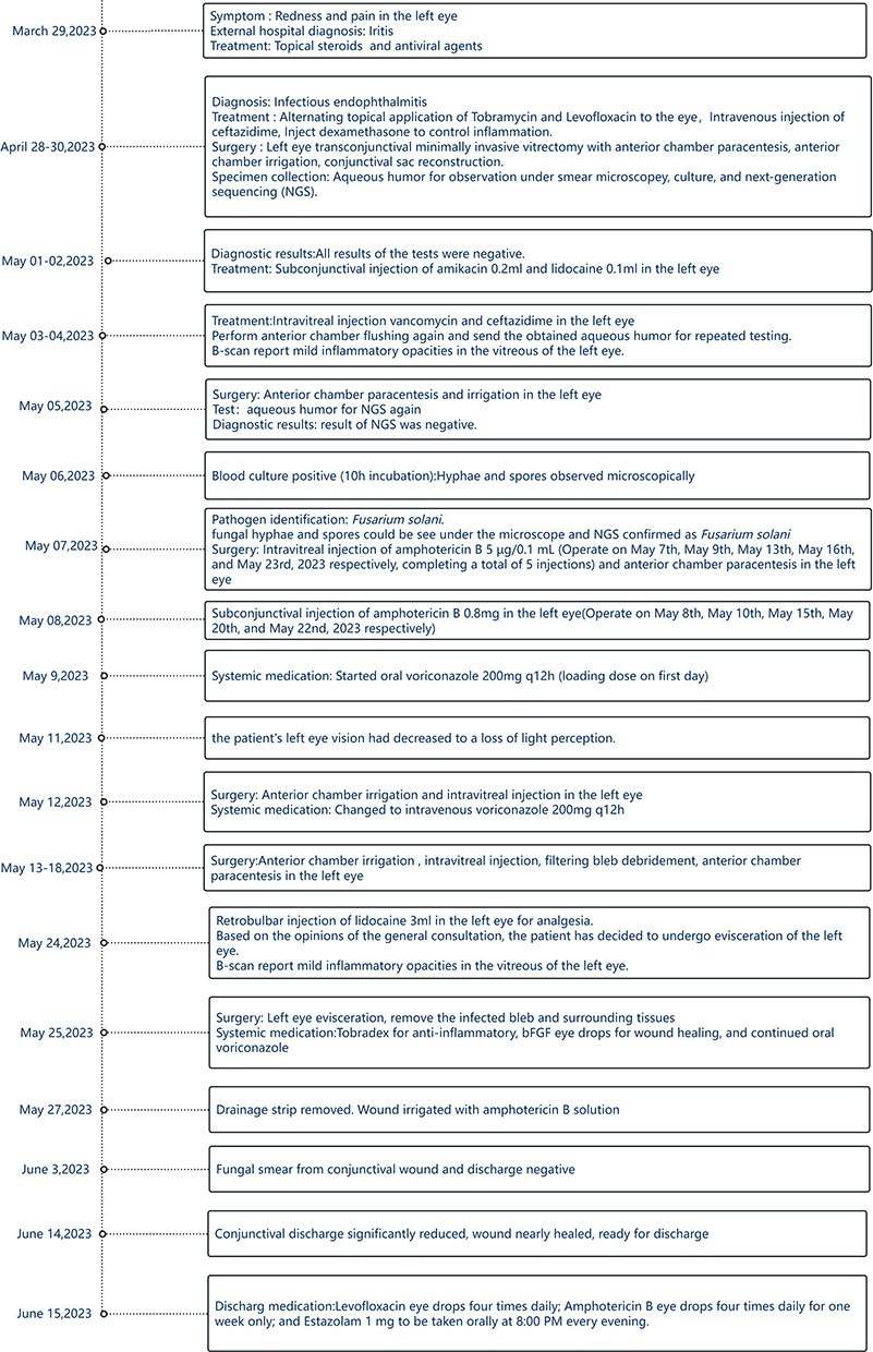 |
Figure 1 Figure depicting the patient’s clinical timeline. |
Diagnostic vitrectomy was performed upon admission, resulting in temporary improvement of visual acuity.Vitreous samples were collected during vitrectomy for microbiological examination (Gram stain, potassium hydroxide preparation, and culture on solid media). However, no organisms were detected. Intraoperative aqueous humor samples were submitted twice for fungal culture and next-generation sequencing (NGS), both yielding negative results. The patient showed no improvement after one week of topical and intravitreal antibiotics, another aqueous humor sample was submitted for NGS and inoculated into blood culture bottles for enrichment. NGS subsequently detected Fusarium solani species complex, and the blood culture turned positive within 10 hours. After five days of incubation, filamentous fungi were isolated and identified as F. solani by matrix-assisted laser desorption/ionization time-of-flight mass spectrometry (MALDI-TOF MS).
Intravitreal amphotericin B was administered every 72 hours. After three days of oral voriconazole, antifungal therapy was escalated to intravenous infusion. Despite two weeks of combined amphotericin B and voriconazole therapy, intraocular inflammation and corneal lesion continued to progress. After thorough discussion and informed consent, the patient underwent evisceration of the left eye. Postoperatively, oral voriconazole and topical antibiotics were continued prophylactically.The patient recovered uneventfully and was discharged in stable condition. Figure 2A and B depict the ocular infection at different stages of disease progression.
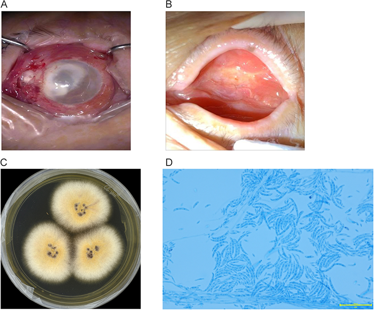 |
Figure 2 (A) Initial presentation upon admission. After 14 days of progressive infection, showing marked purulent deposits beneath the conjunctiva, partial corneal epithelial defects, corneal endothelial contamination, extensive purulent coating on the inner ocular surface, and anterior chamber exudate; (B) This figure shows the recovery of the patient’s left eye after removing the contents and resolution of the primary lesion. (C) Colony morphology after 5 days of culture on Sabouraud dextrose agar (SDA) at 35°C, showing yellow colonies with white, fluffy aerial mycelium. (D) direct microscopic examination: Lactophenol cotton blue staining demonstrated characteristic sporulation similar to Fusarium spp. (× 400). |
Traditional diagnostic methods, including direct smear microscopy, culture on Sabouraud dextrose agar (SDA), and two initial rounds of next-generation sequencing (NGS) of aqueous humor, failed to detect the pathogen. One week later, aqueous humor was recollected for a third NGS analysis and simultaneously inoculated into blood culture bottles for enrichment. NGS identified sequences specific to F. solani. The blood cultures became positive within 10 hours, revealing hyphae and spores on microscopic examination. The sample was subsequently subcultured on Columbia blood agar and SDA at 35°C in a CO2 incubator for five days. Identification by MALDI-TOF MS yielded a score of 1.78 (Genus-level confidence) for F. solani. The microscopic morphology of the colonies, visualized with lactophenol cotton blue staining, is presented in Figure 2C and D.
Laboratory Examination
Gene sequencing of the internal transcribed spacer (ITS) and translation elongation factor 1-α (TEF1-α) further confirmed the identification of F. solani. ITS sequencing demonstrated 100% identity (E value = 0.0; GenBank accession no. PV163846.1), and TEF-1α sequencing likewise showed 100% identity (E value = 0.0; GenBank accession no. PV764940.1). Reference sequences of Fusarium species were retrieved from GenBank, and those used in this study are listed in Table 1.
 |
Table 1 Reference Strains of Fusarium Species and Their GenBank Accession Numbers Used for Phylogenetic Analysis |
Maximum-likelihood (ML) phylogenetic analysis was performed in MEGA11 with 1000 bootstrap replicates (Figures S1 and S2). Phylogenetic reconstruction based on the TEF-1α region provided clearer taxonomic resolution, confirming that the isolate clustered with F. solani, consistent with the NGS results.
According to the joint guidelines of European Society of Clinical Microbiology and Infectious Diseases (ESCMID) and the European Confederation of Medical Mycology (ECMM), the preferred antifungal agents for Fusarium infections are voriconazole and liposomal amphotericin B.13–15 Antifungal susceptibility testing was performed using the broth dilution method (Thermo Fisher Scientific, UK) and broth microdilution testing (CLSI M38-A2) yielded MICs of 1.0 µg/mL for both amphotericin B and voriconazole.16 The breakpoint of the epidemiological critical value (ECV) for reference F. is shown in Table 2.
 |
Table 2 Antifungal Susceptibilities of F.solani by the CLSI Broth Dilution Method |
Discussion
Fusarium is a genus of filamentous fungi widely distributed in the environment and known for causing opportunistic infections in human.17–19 Clinically significant fusariosis primarily involves species within the Fusarium solani species complexes (FSSC), Fusarium fujikuroi species complexes (FFSC), Fusarium oxysporum species complexes (FOSC), and Fusarium dimerum species complexes (FDSC). FSSC, FFSC, and FOSC accounting for over 90% of disseminated infections in immunocompromised hosts and ocular infections in immunocompetent individuals.20–22 Endophthalmitis caused by Fusarium is a severe, vision-threatening condition that occurs predominantly in immunocompromised patients, such as those with hematologic malignancies or transplant recipients.8,9,23,24 Compared to invasive Aspergillus infections, invasive Fusarium infections more frequently yield positive blood cultures. Fusarium is recognized as a significant pathogen in patients with hematological malignancies, contributing to substantial morbidity and mortality.25,26 It carries high mortality rates ranging from 50% to 80%,12 approaching nearly 100% in persistently neutropenic patients.7
For the context of post-trabeculectomy patients, it is essential to distinguish between blebitis which is confined to the filtering bleb.Endophthalmitis involves intraocular structures. In this case, several clinical features confirmed the diagnosis of endophthalmitis rather than isolated blebitis. First, the presence of a marked anterior chamber reaction, including a hypopyon occupying one-quarter of the anterior chamber, along with intense flare (Tyn+) and cellular activity (Cell 4+), indicated that the inflammation had extended beyond the bleb into the anterior segment. Second, vitreous involvement was evident: the vitreous cavity could not be clearly visualized on admission, and subsequent B-scan ultrasonography confirmed inflammatory opacities, directly supporting posterior segment spread. Third, the patient presented with severe and progressive visual decline, from finger counting to light perception, which is atypical for blebitis and strongly suggestive of sight-threatening endophthalmitis. These findings collectively underscore the importance of differentiating between these two entities, as delayed recognition of endophthalmitis can lead to irreversible visual loss and poor outcomes.
This case describes the progression to refractory endophthalmitis in an immunocompetent patient without conventional risk factors, challenging the current epidemiological understanding regarding fungal ocular pathogenesis.
There are some explanations regarding the use of dexamethasone hormone in the early stages of the patient’s illness.Upon the patient’s admission, the primary clinical consideration was filtering bleb-associated endophthalmitis. This judgment was based on the patient’s history of trabeculectomy in the left eye 20 years prior. In ophthalmic clinical practice, filtering bleb-associated endophthalmitis is most frequently caused by bacterial infections.14
After vitrectomy, the patient exhibited marked intraocular inflammatory signs, which raised a high clinical suspicion of acute infective endophthalmitis. At this acute stage, microbiological testing for pathogen identification was still pending. The intervention was limited to a single subconjunctival injection of 5 mg dexamethasone to minimize the risk of adverse effects, with no additional systemic, topical, or repeated corticosteroid administration throughout the entire clinical course.Literature supports the use of corticosteroids as adjuvant therapy for endophthalmitis to reduce tissue damage caused by upregulation of the immune system.27
Molecular analysis remains the gold standard for species identification. Among available markers, the ITS region serves as the most universally effective barcode for fungi, providing broad applicability and distinction between interspecific and intraspecific variation.28 However, ITS does not always provide reliable species-level resolution in certain genera, such as Fusarium. Through comprehensive evaluation, a novel high-fidelity primer pair targeting the TEF-1α gene—a well-established phylogenetic marker in mycology—has demonstrated potential as an auxiliary DNA barcode, outperforming the ITS region in discriminatory power. In this study, primer pairs ITS1/ITS4, Bt-2a/Bt-2b, and EF-1H/EF-2T were used to amplify the ITS rDNA, β-tubulin (TUB2), and TEF-1α gene regions, respectively.29–31 Phylogenetic analysis revealed 100% sequence identity for both ITS and TEF-1α, while TUB2 amplification yielded a maximum identity of 99.75%. Accordingly, based on these results, the ITS (Figure S1) and TEF-1α (Figure S2) sequences were selected for phylogenetic reconstruction to confirm the species-level identification of the fungal isolate.
For confirmed Fusarium endophthalmitis, the European Society of Clinical Microbiology and Infectious Diseases (ESCMID) Fungal Infection Study Group (EFISG) and the European Confederation of Medical Mycology (ECMM) recommend liposomal amphotericin B and voriconazole as preferred therapeutic agents.13 In clinical practice, intravitreal voriconazole and topical amphotericin B are commonly used. Nevertheless, their efficacy in invasive fusariosis remains controversial, with clinical trials reporting low cure rates and treatment failure observed in the present case.32 Given the rapid progression of filamentous fungal endophthalmitis and the limited success of pharmacological therapy, vitrectomy is often required as an adjunct intervention.16,33
Antifungal resistance in Fusarium species remains incompletely characterized, with no established clinical breakpoints. Only the epidemiological cut-off value (ECV) for F.solani has been set at 8 μg/mL for amphotericin B.13 Literature review shows that amphotericin B exhibits the greatest in vitro activity (MIC range 0.5–2 μg/mL), followed by voriconazole (1–8 μg/mL). Susceptibility to posaconazole is highly variable (0.5 to >16 μg/mL), while isavuconazole demonstrates limited efficacy (MIC50 >16 μg/mL). Fluconazole and echinocandins are largely inactive. Notably, Fusarium solani and Fusarium verticillioides tend to display higher azole MICs than other species.5 Overall, published data consistently indicate amphotericin B and voriconazole as the most active agents against Fusarium spp.29,34–37
In this case, broth microdilution testing (CLSI M38-A2) yielded MICs of 1.0 µg/mL for both amphotericin B and voriconazole.16 Nonetheless, voriconazole may show limited in vivo efficacy, and therapeutic outcomes in Fusarium endophthalmitis remain suboptimal. The frequent discordance between in vitro susceptibility and clinical response highlights the urgent need for novel antifungal strategies, while prevention, timely diagnosis, and early intervention remain critical.
Spontaneous Fusarium endophthalmitis in an immunocompetent host is exceptionally rare and under-recognized. Our literature review confirms that nearly all previously reported cases occurred in the setting of ocular trauma, intraocular surgery, hematologic malignancy, or other forms of immunosuppression. The present case demonstrates that filamentous fungal endophthalmitis can arise in a healthy individual without any identifiable portal of entry.
In vitro susceptibility does not equate to clinical efficacy in Fusarium endophthalmitis. The isolate in this case exhibited MIC values of 1 ug/mL for both amphotericin B and voriconazole, which is within the range considered as wild-type by epidemiological cutoff values. Yet the infection progressed relentlessly despite guideline-adherent combination therapy and intraocular irrigation. This case provides the direct evidence of the dissociation between laboratory susceptibility and therapeutic outcome. It also underscores the urgent need for revised treatment paradigms and more potent antifungal agents for intraocular fusariosis.
Research has found that in the detection of fungal endophthalmitis pathogens, the positive rates of vitreous and aqueous humor cultures are both very low.15 One important limitation of this case is the uncertainty regarding whether the patient received any antifungal therapy prior to referral. At the time of admission to our hospital, the patient was unable to recall the exact medications administered at the outside facility, and no complete medical records could be obtained. Therefore, we cannot definitively exclude the possibility that prior exposure to antifungal agents contributed to the initial negative results on conventional fungal culture and the first two rounds of next-generation sequencing (NGS). If antifungal therapy had indeed been initiated prior to referral, it may have temporarily suppressed fungal burden or viability, thereby reducing the sensitivity of subsequent diagnostic tests. This potential confounder underscores a critical clinical principle: whenever possible, microbiological sampling should be performed before the initiation of empirical antifungal therapy. It also highlights, conversely, the robustness of our diagnostic approach—despite the possibility of prior antifungal exposure, repeated NGS and blood culture bottle enrichment ultimately succeeded in identifying the pathogen. Clinicians should be aware that a negative initial workup does not rule out fungal endophthalmitis, particularly in the setting of possible prior treatment.
The diagnostic yield of NGS and blood culture bottle inoculation can be a effective way when conventional methods fail. Two initial NGS runs and multiple fungal cultures were negative; it was only through a third NGS analysis and simultaneous inoculation of aqueous humor into blood culture bottles that the pathogen was identified. This case offers a replicable diagnostic algorithm for similar cryptic intraocular infections.This case has indeed been instructive for our team, which highlight a potential gap in our initial workflow for specific high-suspicion scenarios. As a direct result of this experience, we are actively reviewing our internal protocol and considering the implementation of parallel enrichment culture in addition to direct plating for all initial aqueous humor samples in cases with high pretest probability of infection or when fungal or atypical pathogens are suspected.
The outcome of evisceration is a tragic but important clinical benchmark. This case does not shy away from documenting treatment failure. In doing so, it provides a realistic portrayal of the aggressive nature of F. solani endophthalmitis.It reinforces that through the therapeutic window is narrow, early aggressive intervention (eg. diagnostic vitrectomy) should be strongly considered even when classical risk factors are absent.
This case also illuminates a broader and unresolved clinical question: when should empirical antifungal therapy be initiated in a patient with suspected fungal endophthalmitis, particularly when initial diagnostic tests are negative but clinical suspicion remains high?Unlike bacterial endophthalmitis, there are no equivalent consensus exists for empirical antifungal therapy in the absence of trauma, surgery, or known immunosuppression. Several factors contribute to this uncertainty: (1) antifungal agents are not routinely included in empirical regimens for acute endophthalmitis; (2) intravitreal antifungal injections carry logistical and safety considerations; (3) there is concern that empirical therapy may further reduce the diagnostic yield of subsequent cultures or molecular testing; and (4) data on the efficacy of pre-emptive antifungal therapy in this specific context are lacking. In this case, the interval between initial presentation and targeted antifungal therapy may have contributed to disease progression and eventual loss of the eye.
We do not advocate for indiscriminate empirical antifungal use. Over-treatment carries its own harms, including toxicity, cost, and potential delay in pursuing non-infectious diagnoses. However, we suggest that in selected patients with rapidly progressive endophthalmitis, negative workup, and epidemiological clues suggestive of fungal etiology. Early empirical antifungal therapy may be justified, particularly in combination with early diagnostic vitrectomy. The decision must be made jointly by ophthalmology, infectious disease, and microbiology teams, with close monitoring and an ongoing effort to secure a microbiological diagnosis.
PubMed review identified 14 reported cases of Fusarium endophthalmitis over the past two decades, with the majority presenting as keratitis in the early stages. Among these, eight cases occurred in patients with neutropenia and immunocompromised status following chemotherapy for leukemia (8/14), and one case developed after liver transplantation in a patient with cirrhosis (1/14). One case was associated with cataract surgery (1/14), and two cases resulted from corneal infection due to Fusarium graminearum that progressed to endophthalmitis (2/14). Another case involved an immunocompetent individual who developed Fusarium graminearum endophthalmitis following a sawdust-related hand injury (1/14). Additionally, one case was reported in which Fusarium infection complicating rheumatic keratitis progressed acutely to endophthalmitis during intravenous tocilizumab therapy (1/14). Outcomes were generally poor : only 8 of 14 patients recovered. 3 patients required enucleation due to uncontrolled infection, 1 experienced recurrence six months after discharge leading to secondary ocular infection, and 2 died from multisystem organ failure following treatment discontinuation. More details are summarized in Table 3.8,9,23,24,33,38–46
 |
Table 3 Summary of Endophthalmitis by Fusarium Species |
Literature indicates that Fusarium endophthalmitis occurs mainly in immunocompromised patients, though cases in immunocompetent hosts also progress aggressively and are difficult to control. Importantly, among the 14 cases identified in our review, none presented with spontaneous endophthalmitis in a completely immunocompetent individual without any identifiable ocular breach, hematologic disease, or iatrogenic immunosuppression. The sole case in an immunocompetent adult followed a thorn prick to the hand, which is a clear traumatic portal of entry. The present case is therefore distinct in its complete absence of risk factors, to our knowledge, the first documented instance of truly spontaneous F. solani endophthalmitis. Most require vitrectomy, with some ultimately undergoing enucleation. Antifungal therapy alone rarely achieves cure, and visual outcomes remain generally poor, with residual impairment common even in eyes preserved.47
Patients with prior cataract surgery appear at increased risk for Fusarium endophthalmitis. In the present case, the contribution of remote laser peripheral iridotomy remains uncertain. Post-cataract fungal endophthalmitis is typically diagnosed within weeks after surgery (reported averages: 7–31 days),20,22,48 and case reviews implicate contaminated materials that often linked to specific manufacturers, such as viscoelastic devices or intraocular dyes.20,47,49–51
This case of Fusarium endophthalmitis in an immunocompetent patient underscores the pathogen’s aggressive potential even without typical risk factors, challenging conventional epidemiology. It highlights the value of early molecular diagnosis, combined surgical and antifungal therapy, while exposing the limitations of current agents despite favorable in vitro susceptibility. Along with published reports, it reinforces the need for heightened clinical vigilance, more effective therapies, and clearly defined breakpoints for Fusarium spp.
Conclusion
In summary, this case provides three central lessons for clinical practice. First, Fusarium endophthalmitis can occur spontaneously in immunocompetent individuals and must remain in the differential diagnosis even when no classical risk factors are present. Second, a susceptible result on antifungal susceptibility testing does not guarantee clinical response, which reminds us that treatment decisions should not rely on MIC values alone. Third, early deployment of NGS and alternative culture methods (eg., blood culture bottle enrichment) can be decisive in establishing a microbiological diagnosis when conventional approaches fail.
Statement Regarding the Use of DeepSeek AI
DeepSeek was used strictly as an auxiliary tool in my writing process, primarily for English grammar checking and text polishing. I would input my own paragraphs or drafts into DeepSeek with clear instructions, such as “Please check for grammatical errors in the following passage” or “Please polish the following text to improve its fluency and professionalism.” All ideas, case information organization, arguments, conclusions, and analysis in the article were independently conceived and developed by me.
Data Sharing Statement
ITS, TEF-1αsequences are available under the following accession numbers: PV163846.1, PV764940.1 in https://www.ncbi.nlm.nih.gov/.
Ethics Approval and Informed Consent
This study was approved by the Human Research Ethics Committee of Peking Union Medical College Hospital, Approval No. 1-25PJ2140. In accordance with the institutional policies of Peking Union Medical College Hospital, the ethical approval for this study also encompasses the publication of anonymized case details.the patient provided written informed consent for the use of the anonymized data for research purposes. All data collected were stripped of personal identifiers to maintain privacy.
Informed Consent Statement
Written informed consent was obtained from the patient for publication of this case.
Consent for Publication
All authors grant permission to publish the provided materials (images, videos, audio recordings). All authors have read and agreed to the published version of the manuscript.
Acknowledgments
We would like to thank the patient reported in this article and her attending physician Linyang Gan. We express our gratitude to the Center for Bioinformatics, and the Clinical Biobank, National Infrastructures for Translational Medicine, Institute of Clinical Medicine, Peking Union Medical College Hospital, Chinese Academy of Medical Sciences for providing support in computational analysis and clinical sample collection.
Author Contributions
All authors made a significantcontribution to the work reported, whether that is in the conception, study design, execution, acquisition of data, analysis and interpretation, or in all these areas; took part in drafting, revising or critically reviewing the article; gave final approval of the version to be published; have agreed on the journal to which the article has been submitted; and agree to be accountable for all aspects of the work.
Funding
This study was funded by National High Level Hospital Clinical Research Funding (2022-PUMCH-C-052).
Disclosure
The authors declare no conflicts of interest in this work.
References
1. Balmas V, Fancellu F, Sanna S, Scherm B, Migheli Q, Malbrán I. Water distribution systems in Sardinian hospitals host invasive clonal lineages of the Fusarium oxysporum and Fusarium solani species complexes. Mycologia. 2021;113(4):725–12. doi:10.1080/00275514.2021.1905497. Epub 2021 May 14. PMID: 33989126.
2. Sun S, Hoy MJ, Heitman J. Fungal pathogens. Curr Biol. 2020;30(19):R1163–R1169. doi:10.1016/j.cub.2020.07.032. PMID: 33022261.
3. Chiewchanvit S, Chongkae S, Mahanupab P, et al. Melanization of Fusarium keratoplasticum (F. solani Species Complex) During Disseminated Fusariosis in a Patient with Acute Leukemia. Mycopathologia. 2017;182(9–10):879–885. doi:10.1007/s11046-017-0156-2. Epub 2017 Jun 14. PMID: 28616680.
4. Uemura EVG, Barbosa MDS, Simionatto S, Al-Harrasi A, Al-Hatmi AMS, Rossato L. Rossato, L. Onychomycosis Caused by Fusarium Species. Journal of Fungi. 2022;8(4):360. doi:10.3390/jof8040360
5. Nucci M, Anaissie E, Forrest GN. Invasive fusariosis. Clin Microbiol Rev. 2023;36(4):e0015922. doi:10.1128/cmr.00159-22. Epub 2023 Nov 8. PMID: 37937988; PMCID: PMC10732078.
6. Demonchy J, Biard L, Clere-Jehl R, et al. Multicenter Retrospective Study of Invasive Fusariosis in Intensive Care Units, France. Emerg Infect Dis. 2024;30(2):215–224. doi:10.3201/eid3002.231221. PMID: 38270146; PMCID: PMC10826781.
7. Nucci M, Barreiros G, Akiti T, Anaissie E, Nouér SA. Invasive Fusariosis in Patients with Hematologic Diseases. J Fungi (Basel). 2021;7(10):815. doi:10.3390/jof7100815. PMID: 34682236; PMCID: PMC8537065.
8. Ocampo-Garza J, Herz-Ruelas ME, Chavez-Alvarez S, et al. Disseminated fusariosis with endophthalmitis after skin trauma in acute lymphoblastic leukaemia. J Eur Acad Dermatol Venereol. 2016;30(11):e121–e123. doi:10.1111/jdv.13392. Epub 2015 Sep 22. PMID: 26395131.
9. Kishida Y, Sakaguchi M, Kunishige T, et al. Disseminated Fusariosis Successfully Treated with Empirical Liposomal Amphotericin B and Voriconazole Combination Followed by Ocular Therapy in an Allogenic Hematopoietic Stem Cell Transplant Recipient. Intern Med. 2025;64(19):2907–2912. doi:10.2169/internalmedicine.4657-24. Epub 2025 Apr 12. PMID: 40222926; PMCID: PMC12549048.
10. Lamoth F, Kontoyiannis DP. Therapeutic Challenges of Non-Aspergillus Invasive Mold Infections in Immunosuppressed Patients. Antimicrob Agents Chemother. 2019;63(11):e01244–19. doi:10.1128/AAC.01244-19. PMID: 31481441; PMCID: PMC6811444.
11. Lalitha P, Prajna NV, Manoharan G, et al. Trends in bacterial and fungal keratitis in South India, 2002–2012. Br J Ophthalmol. 2015;99(2):192–194. doi:10.1136/bjophthalmol-2014-305000. Epub 2014 Aug 20. PMID: 25143391; PMCID: PMC7325420.
12. Durand ML. Bacterial and Fungal Endophthalmitis. Clin Microbiol Rev. 2017;30(3):597–613. doi:10.1128/CMR.00113-16. PMID: 28356323; PMCID: PMC5475221.
13. Cornely OA, Cuenca-Estrella M, Meis JF, Ullmann AJ. European Society of Clinical Microbiology and Infectious Diseases (ESCMID) Fungal Infection Study Group (EFISG) and European Confederation of Medical Mycology (ECMM) 2013 joint guidelines on diagnosis and management of rare and emerging fungal diseases. Clin Microbiol Infect. 2014;20(3):1–4. doi:10.1111/1469-0691.12569. PMID: 24606200.
14. Wang X, Zhang P, Suo J, Li Q, Zhang Y. The diagnosis and treatment progress of infectious endophthalmitis. Eye (Lond). 2025;39(3):492–504. doi:10.1038/s41433-024-03474-7. Epub 2024 Nov 30. PMID: 39616279; PMCID: PMC11794455.
15. Ly V, Sallam A. Fungal Endophthalmitis. In: StatPearls [Internet]. Treasure Island (FL): StatPearls Publishing; 2025. PMID: 32644683.
16. John HR, Mahmoud AG, Barbara DA, et al. Reference Method for Broth Dilution Antifungal Suscepti-Bility Testing of Filamentous Fungi: Approved Standard M38-A2. Wayne, PA, USA: CLSI; 2008:1–35.
17. Munkvold GP, Proctor RH, Moretti A. Mycotoxin Production in Fusarium According to Contemporary Species Concepts. Annu Rev Phytopathol. 2021;59:373–402. doi:10.1146/annurev-phyto-020620-102825. Epub 2021 Jun 2. PMID: 34077240.
18. Husaini AM, Sakina A, Cambay SR. Host-Pathogen Interaction in Fusarium oxysporum Infections: where Do We Stand? Mol Plant Microbe Interact. 2018;31(9):889–898. doi:10.1094/MPMI-12-17-0302-CR. Epub 2018 Jul 27. PMID: 29547356.
19. Choi JH, Lee S, Nah JY, et al. Species composition of and fumonisin production by the Fusarium fujikuroi species complex isolated from Korean cereals. Int J Food Microbiol. 2018;267:62–69. doi:10.1016/j.ijfoodmicro.2017.12.006. Epub 2017 Dec 13. PMID: 29291460
20. Armer VJ, Kroll E, Darino M, Smith DP, Urban M, Hammond-Kosack KE. Navigating the Fusarium species complex: host-range plasticity and genome variations. Fungal Biol. 2024;128(8 Pt B):2439–2459. doi:10.1016/j.funbio.2024.07.004. Epub 2024 Jul 14. PMID: 39653491.
21. Montoya AM, Rodríguez-Grimaldo JE, López-Jácome LE, Bonifaz A, Enríquez-Domínguez E, Castañón-Olivares LR. Species distribution and antifungal susceptibility profiles of clinical and environmental Fusarium isolates from Mexico: a multicenter study. Mycologia. 2024;116(2):258–266. doi:10.1080/00275514.2023.2293296. Epub 2024 Jan 17. PMID: 38232343.
22. Al-Hatmi AM, Meis JF, de Hoog GS. Fusarium: molecular Diversity and Intrinsic Drug Resistance. PLoS Pathog. 2016;12(4):e1005464. doi:10.1371/journal.ppat.1005464. PMID: 27054821; PMCID: PMC4824402.
23. Kapp M, Schargus M, Deuchert T, et al. ndophthalmitis as primary clinical manifestation of fatal fusariosis in an allogeneic stem cell recipient. Transpl Infect Dis. 2011;13(4):374–379. doi:10.1111/j.1399-3062.2011.00608.x. Epub 2011 Feb 16. PMID: 21324055.
24. Zhao CS, Wai K, Koo EB, et al. Endogenous Fusarium Endophthalmitis after Bone Marrow Transplant: a Case Report and Literature Review. Vision (Basel). 2024;8(3):44. doi:10.3390/vision8030044. PMID: 39051230; PMCID: PMC11270371.
25. Tala-Ighil T, Garcia-Hermoso D, Dalle F, et al. French Mycoses Study Group. Epidemiology and Prognostic Factors Associated With Mold-Positive Blood Cultures: 10-Year Data From a French Prospective Surveillance Program (2012–2022). Clin Infect Dis. 2025;80(3):529–539. doi:10.1093/cid/ciae594. PMID: 39792000
26. Hardak E, Fuchs E, Geffen Y, Zuckerman T, Oren I. Clinical Spectrum, Diagnosis and Outcome of Rare Fungal Infections in Patients with Hematological Malignancies: experience of 15-Year Period from a Single Tertiary Medical Center. Mycopathologia. 2020;185(2):347–355. doi:10.1007/s11046-020-00436-x. Epub 2020 Feb 25. PMID: 32100219.
27. Azhari J, Tetelbom PS, Sallam AB. The Role of Adjuvant Systemic and Intravitreal Corticosteroids in Fungal Endophthalmitis Treatment. J Fungi (Basel). 2023;9(12):1147. doi:10.3390/jof9121147. PMID: 38132748; PMCID: PMC10744273.
28. Badotti F, de Oliveira FS, Garcia CF, et al. Effectiveness of ITS and sub-regions as DNA barcode markers for the identification of Basidiomycota (Fungi). BMC Microbiol. 2017;17(1):42. doi:10.1186/s12866-017-0958-x. PMID: 28228107; PMCID: PMC5322588.
29. Guo P, Chen J, Tan Y, et al. Comparison of molecular and MALDI-TOF MS identification and antifungal susceptibility of clinical Fusarium isolates in Southern China. Front Microbiol. 2022;13:992582. doi:10.3389/fmicb.2022.992582. PMID: 36386677; PMCID: PMC9664484
30. Wang MM, Crous PW, Sandoval-Denis M, et al. Fusarium and allied genera from China: species diversity and distribution. Persoonia. 2022;48:1–53. doi:10.3767/persoonia.2023.48.01. Epub 2022 Feb 21. PMID: 38234691; PMCID: PMC10792286.
31. O’Donnell K, Whitaker BK, Laraba I, et al. DNA Sequence-Based Identification of Fusarium: a Work in Progress. Plant Dis. 2022;106(6):1597–1609. doi:10.1094/PDIS-09-21-2035-SR. Epub 2022 May 10. PMID: 34907805.
32. Al-Hatmi AMS, Bonifaz A, Ranque S, Sybren de Hoog G, Verweij PE, Meis JF. Current antifungal treatment of fusariosis. Int J Antimicrob Agents. 2018;51(3):326–332. doi:10.1016/j.ijantimicag.2017.06.017. Epub 2017 Jul 10. PMID: 28705676.
33. Rizzello I, Castagnetti F, Toschi PG, et al. Successful treatment of bilateral endogenous F. solani endophthalmitis in a patient with acute lymphocytic leukaemia. Mycoses. 2018;61(1):53–60. doi:10.1111/myc.12697. Epub 2017 Sep 22. PMID: 28872724.
34. Al-Hatmi AMS, Curfs-Breuker I, de Hoog GS, Meis JF, Verweij PE. Antifungal Susceptibility Testing of Fusarium: a Practical Approach. J Fungi (Basel). 2017;3(2):19. doi:10.3390/jof3020019. PMID: 29371537; PMCID: PMC5715922.
35. Broutin A, Bigot J, Senghor Y, Moreno-Sabater A, Guitard J, Hennequin C. In Vitro Susceptibility of Fusarium to Isavuconazole. Antimicrob Agents Chemother. 2020;64(2):e01621–19. doi:10.1128/AAC.01621-19. PMID: 31767726; PMCID: PMC6985727.
36. Osratabadi M, Akhtari J, Faeli L, et al. In Vitro Antifungal Susceptibility Profile of Miltefosine against a Collection of Azole and Echinocandins Resistant Fusarium Strains. J Fungi. 2022;8(7):709. doi:10.3390/jof8070709. PMID: 35887464; PMCID: PMC9315751.
37. Gupta C, Jongman M, Das S, et al. Genotyping and In Vitro Antifungal Susceptibility Testing of Fusarium Isolates from Onychomycosis in India. Mycopathologia. 2016;181(7–8):497–504. doi:10.1007/s11046-016-0014-7. Epub 2016 May 2. PMID: 27138574.
38. Jørgensen JS, Prause JU, Kiilgaard JF. Bilateral endogenous F. solani endophthalmitis in a liver-transplanted patient: a case report. J Med Case Rep. 2014;8:101. doi:10.1186/1752-1947-8-101. PMID: 24661421; PMCID: PMC3978080.
39. Agarwal M, Shah S, Garg A, Gandhi A, Kumar B. Fusarium Causing Recalcitrant Post-operative endophthalmitis-Report of a Case with Review of Literature. Ocul Immunol Inflamm. 2022;30(4):989–991. doi:10.1080/09273948.2020.1830124. Epub 2021 Oct 12. PMID: 34637660.
40. Edelstein SL, Akduman L, Durham BH, Fothergill AW, Hsu HY. Resistant Fusarium keratitis progressing to endophthalmitis. Eye Contact Lens. 2012;38(5):331–335. doi:10.1097/ICL.0b013e318235c5af. PMID: 21993589.
41. Milligan AL, Gruener AM, Milligan ID, O’Hara GA, Stanford MR. Isolated endogenous Fusarium endophthalmitis in an immunocompetent adult after a thorn prick to the hand. Am J Ophthalmol Case Rep. 2016;6:45–47. doi:10.1016/j.ajoc.2016.12.017. PMID: 29260055; PMCID: PMC5722186.
42. Yoshida M, Kiyota N, Maruyama K, et al. Endogenous Fusarium Endophthalmitis During Treatment for Acute Myeloid Leukemia, Successfully Treated with 25-Gauge Vitrectomy and Antifungal Medications. Mycopathologia. 2018;183(2):451–457. doi:10.1007/s11046-017-0221-x. Epub 2017 Dec 16. PMID: 29249044.
43. Avisar I, Weinberger D, Kremer I. Fusarium keratitis and endophthalmitis treated by intravenous ambisome. Harefuah. 2009;148(1):28–9,88. PMID: 19320385.
44. Ichikawa S, Fukuhara N, Watanabe S, et al. Long-term survival after cord blood transplantation for acute myeloid leukemia complicated with disseminated fusariosis. J Infect Chemother. 2020;26(2):292–295. doi:10.1016/j.jiac.2019.08.022. Epub 2019 Sep 28. PMID: 31570321.
45. Kah TA, Yong KC, Rahman RA. Disseminated fusariosis and endogenous fungal endophthalmitis in acute lymphoblastic leukemia following platelet transfusion possibly due to transfusion-related immunomodulation. BMC Ophthalmol. 2011;11:30. doi:10.1186/1471-2415-11-30. PMID: 22044440; PMCID: PMC3220630.
46. Mitsuoka Y, Soma T, Maruyama K, Nishida K. Fusarium infection complicating rheumatic keratitis that acutely progressed to endophthalmitis during regular infusion of tocilizumab: a case report. BMC Ophthalmol. 2021;21(1):224. doi:10.1186/s12886-021-01981-9. PMID: 34011297; PMCID: PMC8135165.
47. Zhang M, Xu GZ, Jiang R, et al. Pediatric Infectious Endophthalmitis: a 271-case Retrospective Study at a Single Center in China. Chin Med J (Engl). 2016;129(24):2936–2943. doi:10.4103/0366-6999.195473. PMID: 27958225; PMCID: PMC5198528.
48. Singh H, Kaur H, Hunjan MS, Sharma S. Unveiling toxigenic Fusarium species causing maize ear rot: insights into fumonisin production potential. Front Plant Sci. 2025;16:1516644. doi:10.3389/fpls.2025.1516644. PMID: 40190660; PMCID: PMC11968736.
49. Yoon SJ, Kim SH, Bahk HJ, et al. Fungal Endophthalmitis Outbreak after Cataract Surgery, South Korea, 2020. Emerg Infect Dis. 2022;28(11):2226–2233. doi:10.3201/eid2811.220361. PMID: 36285883; PMCID: PMC9622257.
50. Kim SW, Kim JH, Choi M, et al. An Outbreak of Fungal Endophthalmitis After Cataract Surgery in South Korea. JAMA Ophthalmol. 2023;141(3):226–233. doi:10.1001/jamaophthalmol.2022.5927. PMID: 36656597; PMCID: PMC9857837.
51. Ahearn DG, Stulting RD. Moulds associated with contaminated ocular and injectable drugs: FDA recalls, epidemiology considerations, drug shortages, and aseptic processing. Med Mycol. 2018;56(4):389–394. doi:10.1093/mmy/myx070. PMID: 29420758.
 © 2026 The Author(s). This work is published and licensed by Dove Medical Press Limited. The
full terms of this license are available at https://www.dovepress.com/terms
and incorporate the Creative Commons Attribution
- Non Commercial (unported, 4.0) License.
By accessing the work you hereby accept the Terms. Non-commercial uses of the work are permitted
without any further permission from Dove Medical Press Limited, provided the work is properly
attributed. For permission for commercial use of this work, please see paragraphs 4.2 and 5 of our Terms.
© 2026 The Author(s). This work is published and licensed by Dove Medical Press Limited. The
full terms of this license are available at https://www.dovepress.com/terms
and incorporate the Creative Commons Attribution
- Non Commercial (unported, 4.0) License.
By accessing the work you hereby accept the Terms. Non-commercial uses of the work are permitted
without any further permission from Dove Medical Press Limited, provided the work is properly
attributed. For permission for commercial use of this work, please see paragraphs 4.2 and 5 of our Terms.