
Back to Journals » Journal of Inflammation Research » Volume 18
Functions of Macrophages, T Cells, and Neutrophils in the Synovial Microenvironment of Osteoarthritis
Received 31 August 2025
Accepted for publication 3 November 2025
Published 11 November 2025 Volume 2025:18 Pages 15671—15686
DOI https://doi.org/10.2147/JIR.S563253
Checked for plagiarism Yes
Review by Single anonymous peer review
Peer reviewer comments 3
Editor who approved publication: Dr Ujjwol Risal
Pengfei Wen, Lin Liu
Department of Joint Surgery, HongHui Hospital, Xi’an Jiaotong University, Xi’an, Shaanxi, 710054, People’s Republic of China
Correspondence: Pengfei Wen, Department of Joint Surgery, HongHui Hospital, Xi’an Jiaotong University, Xi’an, Shaanxi, 710054, People’s Republic of China, Email [email protected]
Abstract: Previous reviews on osteoarthritis have primarily focused on inflammation caused by cartilage destruction, often overlooking the role of synovitis in the progression of osteoarthritis (OA). Macrophages, T cells, and neutrophils play critical roles in the advancement of synovitis. Therefore, this article emphasizes the functions of macrophages, T cells, and neutrophils in OA-related synovitis, aiming to provide a deeper understanding of the interactions among damage-associated molecular patterns, immune cell activation, and signaling pathways (such as NF-κB and mTOR) in OA, thereby offering new therapeutic targets. In OA, the accumulation of macrophages within inflamed synovial fluid, along with elevated levels of RANKL, tumor necrosis factor-alpha, and interleukin-1, enhances the capacity of synovial macrophages to differentiate into osteoclasts. Furthermore, synovial macrophages can polarize into either pro-inflammatory M1 or anti-inflammatory M2 phenotypes, with a predominance of the M1 phenotype promoting cartilage destruction. T cells—particularly Th17 and regulatory T cells—disrupt immune homeostasis and exacerbate inflammation. Neutrophils contribute to tissue damage by releasing elastase and reactive oxygen species, which directly injure cartilage and bone. Current OA management primarily focuses on symptomatic relief through agents such as non-steroidal anti-inflammatory drugs and corticosteroids. In contrast, emerging therapeutic strategies aim to modulate immune pathways. Promising approaches include platelet-rich plasma, mesenchymal stem cell therapy, and photobiomodulation, which have shown potential in regulating inflammation and promoting cartilage repair. A deeper understanding of the interactions among damage-associated molecular patterns, immune cell activation, and signaling pathways—such as NF-κB and mTOR—offers new therapeutic targets. Ultimately, OA management may evolve toward a precision medicine approach that integrates immune modulation with regenerative strategies to restore joint homeostasis.
Keywords: immune modulation, homeostasis, pain, osteoarthritis, synovial membrane
Introduction
Osteoarthritis (OA), a chronic, nonspecific, degenerative illness that affects 595 million individuals globally, primarily causes significant difficulties in daily living.1 Some populations, including women, Asians, and older people, have relatively high incidence rates of OA.2 OA is influenced by several risk factors, including joint-level variables, such as injury, dislocation, and excessive joint loading, as well as personal-level factors, such as age, sex, heredity, obesity, and food.3,4
The distinct pathological features of OA include articular cartilage degradation, reactive hyperplasia of the joint borders and subchondral bone, and subsequent synovial inflammation. In addition, meniscus lesions can lead to structural lesions associated with OA, such as bone marrow edema, cartilage loss, and reduced subchondral bone density. However, many meniscus lesions can cause knee joint strain, which can lead to the progression of OA.5
Severe joint pain can seriously affect patients’ daily lives.6,7 OA has been considered a degenerative condition that causes cartilage loss for years.8 However, our knowledge of inflammation in OA is rapidly changing owing to ongoing advancements in molecular biology. Thus, OA may be primarily caused by inflammation.
Knee OA, is a disease of the whole joint, characterized by degeneration and pathological alterations across all articular tissues.9 These include not only the articular cartilage, synovium, and the subchondral bone, but also menisci, ligaments, and the infrapatellar fat pad (IFP).10 Each of these tissues undergoes distinct but interconnected degenerative changes that collectively drive disease progression and contribute to clinical symptoms. In addition, studies have shown that IFP becomes inflamed and fibrotic in OA and it is also involved in OA pain.11 The IFP serves as both an active site and origin of joint inflammation, contributing significantly to the pro-inflammatory synovial environment in OA. The IFP represents a valuable reservoir of mesenchymal stem cells (MSCs), which possess immunomodulatory properties and may contribute to the regeneration of joint tissues.11 Furthermore, all joint tissues including IFP undergo biomechanical changes.12
Inflammatory synovitis can result from the impact of OA on the synovial tissue of joints.13 Although synovitis is not always present in OA, it can lead to increased discomfort, joint dysfunction, and accelerated cartilage degradation.14 As OA worsens, the severity of synovitis increases. In individuals with OA, synovial inflammation is present in >90% of synovial tissues.15 The thin layer of cells that makes up the synovial lining has phenotypic traits of fibroblasts and macrophages. The complex structure formed by these cells and the vascularized connective tissue matrix underneath them are significant sources of synovial fluid components that are essential for healthy cartilage and joint function.16 The inflow of T cells and macrophages, growth of new blood vessels, and consequent release of inflammatory cytokines are typical features of synovial inflammation.16 By promoting the synthesis of matrix metalloproteinases (MMPs), inflammatory cytokines contribute significantly to the development of OA by promoting the synthesis of MMPs.17 Therapeutic targets for controlling the course of OA may involve understanding cellular functions in the immune response and inflammatory process, as well as how cells produce and regulate proinflammatory cytokines. Furthermore, the OA phenotype may influence the type and source of inflammatory mediators.18 This review focuses on the biological relevance of common immune cells, such as neutrophils, T cells, and macrophages, in the OA synovium, which also suggests potential preventative and therapeutic measures.
Clinical Features and Risk Factors for OA
Clinical Features of OA
Pain
In the early stages of OA, patients experience intense and predictable pain commonly caused by mechanical injury. (Table 1) Occasionally, high-intensity activities may be restricted; however, at this point, they do not affect function. As the disease progresses, pain in the middle stage becomes more common, and stiffness becomes unpredictable Daily life is affected by pain. In advanced stages, function is severely hampered by constant throbbing pain punctuated by brief, erratic bouts of intense agony. Specific mechanical reasons for pain include meniscal injury, ligament damage, subchondral microfractures, periosteal straining, osteophyte growth, increased intraosseous pressure, synovitis, and shrinking of the joint space.19,20 Although the biological mechanisms underlying pain remain unclear, local inflammation may play an important role.21 For example, increased IFP signal intensity on MRI, which indicates inflammation, was positively correlated with pain in osteoarthritic knees.22,23
 |
Table 1 Clinical Features and Risk Factors for OA |
Joint Stiffness
A common symptom of OA is joint stiffness, which can manifest as a lack of flexibility in the joints and cause discomfort or difficulty in movement. (Table 1) One of the main causes of joint stiffness is a lack of synovial surface-active phospholipids.24 Although joint stiffness is most noticeable in the morning, it can also occur later in the day, particularly after being inactive for a while.25 Studies have found that knee joint exercise affects IFP stiffness, and an increase in stiffness has been observed in patients with knee OA.26,27 In severe knee OA, the knee fat pad is stiffer, and greater stiffness is associated with more severe symptoms.27
Bone Hyperplasia and Swelling
Both small (such as the interphalangeal joints) and large (such as the knee joints) joints may experience bone enlargement and swelling owing to OA. It is caused by various pathologies, such as blocked blood, soft tissue edema, damaged chondrocytes, circulation, increased bone density, and cystic degeneration.28
Risk Factors for OA
Personal-level variables, such as heredity, age, obesity, sex, and diet, and joint-level variables, such as injury, joint dislocation, and aberrant joint loading are among the many postulated risk factors for OA.29 (Table 1) Although the probable mechanism underlying joint destruction remains unknown, age may be a primary risk factor for OA at the individual level. This could be attributable to several factors such as oxidative damage, cartilage thinning, muscular weakening, and impaired proprioception.30 OA of the hands, knees, and hips is more common in women than in men, and its frequency increases with menopause.31 Hormones are likely involved in the development of OA, which has led researchers to speculate on this phenomenon.32 Other factors may explain why men and women differ in terms of reduced cartilage volume, bone loss, and muscle strength deficiency.32 Genetic factors account for 60% of hand and hip OA cases and 40% of knee OA cases. Several genes may be involved in OA onset and development.33
Knee OA is more prevalent when the fracture is accompanied by cartilage injury, subchondral bone injury, lateral ligament injury, or meniscal injury, in 21%–40% of cases.34 Although cartilage loss and damage are inevitable consequences of aging, not all individuals develop OA. Obesity is another major risk factor for OA. The complex interplay between metabolic, biomechanical, and inflammatory factors caused by obesity affects OA.35,36 Studies have revealed a strong link between the pathophysiology of OA and adipose tissue growth.37,38 Fat factor receptors are present in almost all cells within the joint; thus, they may directly contribute to OA through synovitis, cartilage damage, and bone remodeling.39 In addition, IFP produces many factors that may affect disease progression through paracrine mechanisms, including cytokines, lipokines, growth factors, and fatty acid and lipid derivatives.40 In contrast to the elevated secretion of arachidonic acid (which promotes PGE2 production and exacerbates cartilage inflammation), the IFP of OA patients also released higher levels of docosahexaenoic acid (DHA), a polyunsaturated fatty acid that suppresses key inflammatory mediators including proinflammatory cytokines, cartilage-degrading enzymes, and COX-2.38 The role of specific vitamins and diet in OA is an active area of research. Among these, research on vitamin D is the most extensive, although the results are inconsistent.41,42 In addition to the studies on specific vitamins, other studies have explored the relationship between various diets and OA. For example, a higher intake of dietary fiber and soy milk and adherence to the Mediterranean diet have positive effects on various outcomes of OA. However, further research is needed to confirm these findings.43
The Immune Mechanism of OA
Inflammation is considered a major pathophysiological mechanism of OA, which is a chronic condition.44 Numerous cell and tissue types, both inside and outside the joints, are involved in the complicated pathophysiological phenomena of OA-related inflammation.45 Genetic predispositions and changes in gene expression caused by variations in mechanical stress encountered by chondrocytes in articular cartilage are two of the many interrelated variables contributing to the complicated pathophysiology of OA.46 A spatially and temporally confined inflammatory response is not only beneficial but essential for tissue healing and the restoration of homeostasis.47 For instance, at the initial stage of fracture healing, known as the inflammatory phase, resident and recruited macrophages along with other immune cells at the fracture site release various cytokines in response to injury. This early inflammatory response initiates the recruitment, proliferation, and differentiation of MSCs, promotes the synthesis of extracellular matrix proteins, stimulates angiogenesis, and ultimately leads to tissue remodeling.48 However, when inflammation becomes dysregulated and fails to resolve, it can progress into a chronic state.49 A growing body of evidence implicates such persistent, low-grade inflammation—often linked to metabolic syndrome and innate immunity—as a key driver in the pathophysiology of OA.7 One of the main elements in the pathophysiology of OA is the role of the immune system in its onset and progression.50
Synovium and Synovitis
Synovial inflammation, often known as synovitis, is a common symptom of OA. However, during joint illnesses, additional tissues, such as the articular cartilage, meniscus, and subchondral bone, also participate in inflammatory interactions.51 In 1982, Goldenberg et al conducted a histopathological assessment of synovial tissues of patients with OA. They reported that nearly 90% of synovial tissues exhibited synovitis.52 Tissue analysis and sensitivity imaging have demonstrated an extremely high incidence of synovial inflammation across all OA phases. Among patients with KL grades 2–3, the prevalence of synovitis was high. Among patients in the most severe stage (KL grade 4), up to 83% have synovitis.53 Joint swelling, discomfort, and effusion are only a few clinical symptoms and indicators of OA directly caused by synovitis, which also represent the structural development of the disease.54,55
Synovial membranes are soft tissues present in the bursae, tendon sheaths, and joints. The intima is the inner layer of the continuous cellular layer, whereas the sub-intima is the outer layer. Macrophages and fibroblasts are present in the intimal layer, whereas blood, lymphatics, and fibroblasts are found in the subintimal layer.56 As the primary barrier to molecular exchange between the articular cartilage and plasma, the synovium is essential for preserving the health of both the articular cartilage and joint. The synovial interstitium, which inhibits the diffusion of small molecules and the microvascular endothelium, which limits the transport of proteins, are dual-barrier structures that form the blood-synovial barrier.57 In addition, Eymard et al found that IFP has a potential role in the induction of synovial inflammation in patients with severe knee OA. Furthermore, secretion of PGE2 by the IFP may be involved in the OA inflammatory process.58
In the case of synovial inflammation, the influx of macrophages and T cells, formation of new blood vessels, and secretion of proinflammatory cytokines are typical characteristics.13 Comparing the main immunohistological features of inflammation in early and late OA, Benito et al have reported that synovitis severely influences disease progression.54 Synovial tissue samples with evident hyperplasia of the synovium were immunohistochemically stained with obvious positive markers indicative of inflammatory cell infiltration (CD4+ and CD68+) and angiogenesis and were highly expressed in both early and late OA. Vascular endothelial growth factor (VEGF), intercellular adhesion molecule 1 (ICAM-1), proinflammatory cytokines, tumor necrosis factor α (TNF-α), and interleukin (IL)-1β are increased, and their expression is increased in early OA.54 Sohn et al have reported that the synovial fluid of patients with OA exhibits higher proinflammatory cytokine levels (TNF-α, IL-6, and VEGF) than those of healthy individuals.59
Cellular and Molecular Mechanisms of OA Synovial Inflammation
Damage-associated molecular patterns (DAMPs), which are produced by the extracellular matrix (ECM) during cartilage degradation in the joint cavity, are key causes of both acute and chronic inflammation through the activation of the immune system in the synovium.60–62 DAMPs are endogenous stimulants produced by dead cells or the ECM.63 The biological activities of DAMPs are mediated through pattern recognition receptors (PRRs).64 The presence of these receptors in immune cells, osteoblasts, chondrocytes, and synovial cells has been previously confirmed. When DAMPs bind to these receptors, they initiate signalling pathways that trigger the activation of various transcription factors. This activation leads to the release of multiple components, such as the complement cascade, cytokines [including IL-1β, IL-6, and TNF-α], chemokines [such as CC motif chemokine ligands-2, −5, −7, and −8], catabolic factors [such as MMP-1, −3, −9, and −13], and cathepsins (B, K, and L)65 (Figure 1).
 |
Figure 1 Cellular and molecular mechanisms in osteoarthritis synovial inflammation.Immune system activation is initiated and driven by chondrocyte death, triggering damage-associated molecular patterns (DAMPs). The biological activity of DAMPs is mediated by pattern recognition receptors, which are present on the surfaces of immune cells, chondrocytes, osteoblasts, and synovial cells. The binding of DAMPs to these receptors initiates downstream signaling cascades, leading to the activation of multiple transcription factors. Figure created in BioRender. Luo, P. (2025) https://BioRender.com/n10z021. |
In summary, stimulants introduced into the synovial cavity trigger the synovial cells to generate and release inflammatory mediators into the synovial fluid. These mediators subsequently stimulate chondrocytes to produce MMPs, creating a detrimental feedback loop between the synovium and the cartilage.66 Inflammatory mediators, including cytokines, transcription factors, proteases, and ECM proteins, are linked to the definite roles of epigenetic changes and synovial inflammation in OA.67,68 Moreover, excessive production of inflammatory mediators can result in the progressive degradation of tissues, and the origins and categories of these mediators may differ based on the OA phenotype. Given the intricate cellular and molecular mechanisms described above, we identified DAMPs as crucial in the development of OA synovitis. Targeting the activity of DAMPs and their receptors in immunotherapeutic approaches represents a promising avenue for OA treatment. Elucidating DAMPs, their receptors, and associated pathological pathways is essential to address degenerative joint conditions, including OA.
The infiltration of inflammatory cells (eg, mast cells, T cells, macrophages) into the infrapatellar fat pad (IPFP) and synovium is a hallmark of progressive osteoarthritis (KOA), leading to the secretion of various inflammatory mediators.40 Among these, prostaglandin E2 (PGE2) plays a critical role in the crosstalk between the IPFP and synovium by stimulating the inflammatory response in fibroblast-like synovial cells.58 In addition, leptin released by IPF can induce IL-8 expression via leptin receptor, IRS-1, PI3K, Akt cascade and promotion of NF-κB/p300 binding in human synovial fibroblasts.69
Immune Cells in OA Synovial Inflammation
Numerous immune cells from the innate and adaptive immune systems have been identified in the synovial tissues of patients with OA. According to Lindblad et al, inflammation in the synovium close to the cartilage may cause a more intense inflammatory response, which is characterized by T cells surrounded by B and plasma cells.70 Compared with the synovium of healthy individuals, the synovium of patients with OA has fewer anti-inflammatory regulatory T cells and more proinflammatory immune cells, such as M1 macrophages, Th1 cells, and Th17 cells.71–73 When synovial samples from patients with knee OA were examined, cells lining the joint were highly immunoreactive and phagocytic. Synovial fluid from end-stage osteoarthritic knees during total arthroplasty showed significant enrichment of macrophages, T cells, and neutrophils.74
To initiate tissue healing, different inflammatory cells are drawn to the site of damage and release proinflammatory and catabolic mediators.75 To facilitate tissue healing, macrophages first move to the inflammatory site and then undergo phenotypic changes under the pathological conditions of OA. Furthermore, pathogens or DAMPs may be released by injured tissues and cells, which can trigger macrophage inflammatory signalling pathways, produce various cytokines, and attract additional inflammatory cells, such as T cells, neutrophils, and lymphocytes.76 Researchers discovered that T-cells and macrophages account for 22% and 65% of the synovial tissue in patients with OA, respectively.77 The neutrophil population was the smallest. The functions of neutrophils, T cells, and macrophages in OA are discussed in detail below.
Role of Macrophages in the Synovium in OA
As a basic defense system acquired by humans from invertebrates, the innate immune system recognizes infections using germline-encoded proteins.78 Innate immune cells either directly destroy pathogens or trigger a cascade of events that activate the adaptive immune system. Natural killer (NK) cells, dendritic cells, and macrophages are cells of the innate immune system.79 Innate immune cells called macrophages exist in nearly every tissue and affect homeostasis, wound healing, and immune response.80 “Synovial macrophages” usually refer to tissue-resident macrophages located in the synovial membrane and subintima membrane.81 The synovial membrane consists of two different cell types: synovial macrophages (type A synovial cells) and fibroblast-like synovial cells (type B synovial cells).78 Their propensity for phagocytic behavior is their most distinctive trait. However, they also perform several other tasks, including producing and secreting many secretory chemicals and starting and controlling hormones and cellular immunity.82 Synovial macrophages differentiate into osteoclasts with the ability to resorb lacunar material, although this process requires the involvement of M-CSF or TNF-α/IL-1 in addition to RANKL.83 Macrophages also play a significant role in the production of MMPs, which are enzymes that break down all ECM proteins. In OA and other joint disorders, macrophage infiltration in the IFP has been observed to increase.84 Studies have identified the presence of both M1-like (CD11c+) and M2-like (CD206+) macrophage markers in this tissue.85,86 According to Wei et al, macrophages isolated from the IPFP of affected joints suppress the chondrogenic differentiation of MSCs,86 indicating a potentially adverse effect of these macrophages on cartilage repair. Nonetheless, the exact roles and origins of macrophages within the IPFP require further investigation.
The inflammatory response necessary for musculoskeletal tissue healing is primarily mediated by macrophages.87 Macrophages are the most prevalent invading cells and important mediators of inflammation, making them important players in OA. The microenvironment determines whether macrophages are typically activated (M1-type macrophages) or alternatively activated (M2-type macrophages).88 Induced by proinflammatory mediators such as interferon-γ and lipopolysaccharide, M1-type macrophages secrete cytokines and contribute significantly to the host’s defence against infection. In response to IL-4 and IL-13, M2-type macrophages secrete anti-inflammatory IL-10, the IL-1 decoy receptor, and arginase, further inhibiting the action of IL-1β and inducible Nitric Oxide Synthase (iNOS), and are associated with tissue remodeling. Along with fibroblasts, macrophages often remain dormant in healthy synovial joints. However, they can polarize into proinflammatory M1-type macrophages when OA stimulates inflammation.89 In OA synovial fluid, M1-polarized macrophages release growth factors, cytokines, MMPs, TIMs, and other substances that cause inflammation and cartilage deterioration90 (Figure 2). Furthermore, the ability of M1 and M2 macrophages to dynamically adjust their polarization in response to changes in the microenvironment suggests that macrophage polarization is plastic.91 Thus, targeting macrophage polarization may prove to be a successful treatment strategy during the onset of OA, as it may decrease the production of detrimental inflammatory cues to chondrocytes by converting proinflammatory M1 macrophages into anti-inflammatory M2 macrophages. The direct in vivo involvement of macrophages in human OA was validated in another study.92 Thus, inflammation and macrophages are intimately linked to joint symptoms and the radiological severity of OA in the knee. The presence of active macrophages in the periarticular tissues of the knee joint and their correlation with OA symptoms suggest that inflammation caused by macrophages is frequent and might be the root cause or exacerbating factor of OA symptoms in other joints throughout the body.93
 |
Figure 2 Mechanism of macrophages in osteoarthritis progression.Pathogen-associated molecular patterns, damage-associated molecular patterns (DAMPs), and inflammasomes are microenvironmental stimuli that promote the activation and polarization of synovial macrophages. This process is also regulated by signaling pathways, such as mammalian target of rapamycin, nuclear factor-κB (NF-κB), c-Jun N-terminal kinase (JNK), and phosphatidylinositol 3-kinase/protein kinase B (PI3K/Akt). The increased number of M1-polarized macrophages in the synovium of osteoarthritis secretes various factors, including cytokines, growth factors, matrix metalloproteinases, and tissue inhibitors of metalloproteinases, thereby triggering inflammation and subsequent cartilage degeneration and osteophyte formation. Besides autocrine interactions, polarized macrophages alter intracellular signaling pathways in chondrocytes, including transforming growth factor-β, JNK, Akt, NF-κB, and β-catenin signaling pathways, promoting the degradation of extracellular matrix components. As a DAMP, the ECM further stimulates the activation and polarization of macrophages, forming a vicious cycle of inflammation and cartilage degeneration. Polarized synovial macrophages and macrophage reprogramming may be suitable targets for the prevention and early treatment of osteoarthritis. Copyright 2020. Reproduced from Zhang H, Cai D, Bai X. Macrophages regulate the progression of Osteoarthritis. Osteoarthritis and cartilage. May 2020;28(5):555–561. doi:10.1016/j.joca.2020.01.007.90 |
Role of T Cells in the Synovium in OA
T cells, including memory T cells, cytotoxic T cells, and helper T cells, play a key role in OA pathophysiology. T lymphocytes are the primary cause of synovial infiltration in patients with OA.94 T cell infiltration in synovial tissue is dominated by CD4+ T cells, and patients with OA have more CD4+ T cells in the lower layer of the synovium than healthy individuals.94 The CD4 antibody is a cytokine present in the surface of CD4+ T cells, which are crucial immunological cells in the human immune system. According to the enzyme-linked immunosorbent assay (ELISA) data, patients with OA had higher blood levels of soluble CD4 antibodies, according to ELISA controls. These findings suggest a role for peripheral T helper (Th) cells in the pathophysiology of OA.77 Furthermore, T lymphocytes expressing HLA antigen D-related (DR) were found in the synovium of patients with OA, according to immunohistochemical investigations.95 These results corroborated the observation that activated T cells are present in the synovium of patients with OA; nearly all T cells in the synovium of patients with OA expressed the activation markers HLA-DR and CD69.96
The interleukin-2 receptor (IL-2R) alpha chain CD25 is expressed mostly on the surface of immune-related cells such as B cells, NK cells, and activated T cells.35 After interacting with its ligand IL-2, CD25 forms a high-affinity heterotrimeric receptor with β and γ chains and participates in a range of immunological responses.97,98 According to the findings of an MR investigation by Luo et al, hip OA is causally associated with seven CD25-related characteristics.99 The development of inflammation in hip OA can be reduced by either increasing the number of T lymphocytes expressing CD25 or increasing the expression level of CD25. A greater abundance of CD25, which competitively intercepts IL-2, a crucial component necessary for the survival of nearby activated T cells, is responsible for this effect.99 Overall, substantial alterations in T cell profiles have been observed in the synovial fluid, peripheral blood, and synovium of patients with OA.95 Thus, T cells may be associated with OA etiology.
Numerous elements are important determinants of T cell destiny, including intracellular signal transduction [such as mechanistic Target Of Rapamycin (mTOR) complex 1 (mTORC1)], external stimuli (such as antigens), and cellular metabolism (such as amino acid metabolism).100 For example, mTORC1 controls Th17 differentiation and IL-17 production via a number of pathways, including Signal Transducer and Activator of Transcription 3 (STAT3), Hypoxia-Inducible Factor-1-αlpha subunit (HIF-1α), Ribosomal protein S6 kinase 1 (S6K1), and Ribosomal protein S6 kinase 2 (S6K2), which suggests that mTORC1 signalling may be used to modify the pathophysiology of OA.101 Furthermore, the activation and differentiation of T cells, particularly Th1 and Th17 cells, are significantly affected by amino acid metabolism and transport, suggesting that amino acid metabolism influences OA pathophysiology. Additionally, the balance between Th17 and Treg cells is disrupted throughout the degenerative phase of OA, resulting in inflammatory reactions and articular cartilage degeneration.102 Patients can experience pain relief and improved joint function by regulating the Th17/Treg balance to reduce the inflammatory response. Therefore, controlling the Th17/Treg cell imbalance is a key target for the treatment of OA (Figure 3). Many levels of intervention, such as the modulation of different transcriptional regulators, cytokines, and epigenetic changes, can influence the activation and function of Th17/Treg cells. These interventions can reduce OA symptoms by altering the symptoms of OA. These targets should be considered promising novel therapeutic approaches for treating OA.
 |
Figure 3 Imbalance of Th17/Treg cells is an important mechanism for OA occurrence.Th17 cells promote inflammatory responses. Meanwhile, Tregs inhibit inflammatory responses and antagonize the effects of Th17 cells. An imbalance in the Th17/Treg cell ratio is involved in the pathophysiology of OA. Copyright 2024, Reproduced from Wen Z, Qiu L, Ye Z et al. The role of Th/Treg immune cells in osteoarthritis. Frontiers in immunology. 2024;15:1393418. doi:10.3389/fimmu.2024.1393418.102 |
Role of Neutrophils in the Synovium of Patients with OA
A subset of polymorphonuclear leukocytes and neutrophils may be the first white blood cells drawn to the site of inflammation, serving as the first line of defense against invasive microbes.103 Neutrophils participate in cytokine and chemokine cascades linked to inflammation and utilize intercellular interactions to regulate immune responses.104 Neutrophils have the greatest potential for cytotoxicity among cells associated with OA because they produce reactive oxygen species and degrade enzymes.105 Neutrophils comprise 8% of cells in synovial fluid.106 In OA, neutrophils are among the first immune cells to enter the synovium. They also contribute to many chemokines and cytokines released by the immune cells into the synovial fluid107 (Figure 4). The synovium and synovial fluid of patients with OA contain chemokines that recruit immune cells and activate signaling cascades that accelerate the progression of OA. In synovial fluid, neutrophils form complexes with neutrophil gelatinase-associated lipocalin and MMP-9, which accelerates cartilage degradation.74 VEGF is present in neutrophils, and a significant amount of VEGF is present in the supernatant following vesicle release upon stimulation.108 VEGF exacerbates OA pain, increases vascular density, and promotes endothelial cell proliferation within the OA synovium.109
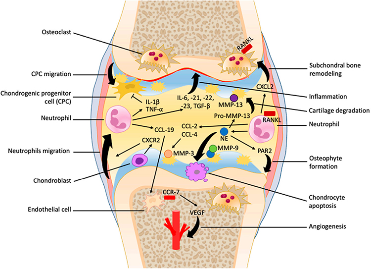 |
Figure 4 Role of neutrophils in the osteoarthritis progression. Neutrophils are recruited to the synovial bursa and promote the secretion of numerous cytokines and chemokines in the synovial fluid, which facilitate inflammatory responses and vascular infiltration, and inhibit the migration of chondroprogenitor cells. Neutrophil elastase formation exacerbates cartilage degradation, chondrocyte apoptosis, imbalance of subchondral bone remodeling, and osteophyte formation. Copyright 2022. Reproduced from Chaney S, Vergara R, Qiryaqoz Z, Suggs K, Akkouch A. The Involvement of Neutrophils in the Pathophysiology and Treatment of Osteoarthritis. Biomedicines. Jul 6 2022;10(7)doi:10.3390/biomedicines10071604.107 |
On the one hand, degradative elastase is strongly related to the severity of OA, with neutrophils being the primary source.74 Chondrocyte enlargement and compensatory synthesis throughout the ECM occur naturally during endochondral ossification, but are inappropriately stimulated at the onset of OA. Moreover, in advanced OA, chondrocyte differentiation, proliferation, and hypertrophy are unrestrained, leading to subchondral bone calcification and hardening, accompanied by articular surface fibrosis.110,111 Hypertrophic chondrocytes die and stop growing as OA progresses. Apoptosis-induced cavity emptying causes articular cartilage degradation, eventually leading to osteophytes.74 Neutrophil elastase (NE) can drive neutrophils and other immune cell types to move through the ECM, thereby increasing inflammation. In vitro studies have demonstrated that NE, even at low doses, can rapidly destroy cartilage collagen.112 Overactivation of MMP-13 may be the medium that cause permanent cartilage degradation in patients with OA.113 Neutrophils release NE, which activates latent pro-MMP-13. Pro-MMP-13 is a critical regulatory node between maintaining cartilage homeostasis and triggering pathological destruction. In the study of OA pathogenesis, detecting the level of pro-MMP-13 is often used as a biomarker for the activity of tissue remodeling, particularly cartilage degradation.114 In vitro, NE directly and robustly activated pro-MMP-13, and N-terminal sequencing identified cleavage close to the cysteine switch at70 MKKPR, ultimately resulting in the fully active form with the neo-N terminus of83 YNVFP, which is closely related to cartilage collagen destruction in OA patients with synovitis.112
Treatment for Synovitis in OA
Current Treatment Methods
Currently, the cause of OA remains unresolved. Controlling symptoms, slowing disease progression, and improving the quality of life are the goals of all OA treatments. Currently, OA is treated with medication, surgery, physical therapy, exercise therapy, and weight loss (if applicable).
Most patients with OA take various medications, including glucocorticoids, opioids, nonsteroidal anti-inflammatory drugs, and cytokine inhibitors, which can reduce inflammation to some degree.115,116 Intra-articular injection of corticosteroids effectively relieves OA pain, although its effect is short-lived.117 Intra-articular injection of the corticosteroid FX006 has short-term efficacy. Researchers have examined the long-term effects of a triamcinolone acetonide (TA) extended-release formulation prepared using microsphere technology in patients with knee OA. According to several randomized, double-blind, controlled dose optimization studies, TA-ER can considerably improve the Western Ontario and McMaster Universities Osteoarthritis Index (WOMAC) ratings for physical function, pain, and stiffness. Compared to the placebo, it also improved knee injury and OA outcome scores, particularly the Knee Osteoarthritis Outcome Scale - Quality Of Life (KOOS-QOL) score. Thus, it has been authorized by the Food and Drug Administration to treat OA-related knee discomfort. An additional benefit of the TA-ER mode of action is that systemic exposure is lower than that of rapid-release triamcinolone acetonide because of its slower intra-articular release rate.118 Currently, intramuscular and intra-articular steroid injections offer temporary relief from OA symptoms. In the United States, sustained-release formulations for intra-articular steroid injections have recently been approved.
In cases where all therapeutic and pharmacological intrusions have proven ineffective, clinical procedures, including arthroscopic lavage, debridement, bone microfracture, and abrasion arthroplasty of the worn joint, may be necessary.119 The most common physical therapies for patients with OA are electrotherapy, acupuncture, ultrasound, and cold and heat therapy.120 Exercise therapy can improve changes in pain scores in patients with OA, increase muscle strength, and increase the stability of the affected joint; however, no difference in inflammatory activity has been observed.121
Potential Treatment Methods
Drugs have long been injected intra-articularly to treat patients with OA, and this method works well because it produces short-term analgesic and anti-inflammatory benefits.122 Extended and frequent injections can cause cartilage deterioration or damage to the other joint components. Consequently, investigating the biological regulatory factors that can stimulate cartilage regeneration and maintain joint homeostasis is crucial and beneficial for advancing novel intra-articular therapeutic approaches. Further investigation of the molecular mechanisms that trigger and maintain synovial inflammation is required. A deeper understanding of these pathways might reveal novel therapeutic targets. Macrophage polarization offers valuable insights for the development of novel interventions against OA. Clinical studies have demonstrated that the intra-articular administration of cells and their derivatives is promising for promoting the polarization of synovial macrophages within osteoarthritic joints. The use of platelet-rich plasma (PRP) is a strategy that is becoming increasingly popular in therapeutic settings. (Table 2) PRP, which is made from a patient’s blood, stimulates tissue-building processes, reduces inflammation, and relieves pain to maintain joint health.123 Preclinical studies have shown that PRP increases M2 macrophage polarization and suppresses M1 macrophage polarization in OA. PRP has anti-inflammatory properties by altering the canonical NF-κB signalling pathway in various cell types, including chondrocytes, synovial cells, and macrophages.124 Adipose tissue-derived mesenchymal stem cells (ADMSCs) are a promising cell source.
 |
Table 2 Summary of Novel Therapeutic Strategies for Osteoarthritis (OA) |
These cells display substantial immunomodulatory and anti-inflammatory properties via the prostaglandin E2 (PGE2)/cyclooxygenase 2 (COX) 2 pathway.125 (Table 2) ADMSCs reduce NF-κB1 and NF-κBIA (NF-κB signal inhibitors) and therefore may primarily activate the NF-κB pathway to suppress macrophages.126 Furthermore, Ma et al engineered groundbreaking artificial M2 macrophages, which have remarkable therapeutic potential as M2 macrophages in OA treatment.127 Macrophage membranes make up the “shell”, whereas the “yolk” consists of nanogels that are responsive to inflammation. This construct showed targeted delivery and prolonged retention in inflammatory areas while suppressing macrophage immune activity and has potential as a future intra-articular treatment for OA. Photobiomodulation therapy (PBMT), also known as low-level laser therapy, is an emerging therapeutic strategy. PBMT has been shown in vivo to reduce pain, decrease the levels of inflammatory mediators (eg, TNF-α, IL-1β, and IL-6) and decrease joint swelling in various animal models of OA.128 Zhang et al showed for the first time that PBMT can alter synovial macrophage polarization in collagenase-induced mouse knee OA.129 For example, we exposed the right knees of OA mice to a 630-nm LED device at different power densities for 4 weeks, and histological and immunostaining analyses revealed significant improvements in synovitis, cartilage degradation, and osteophyte formation. The transition of synovial macrophage polarization from M1 to M2 phenotype may partially contribute to therapeutic outcomes.129
Two important proinflammatory cytokines that can initiate the synthesis of inflammatory mediators in the joint microenvironment are TNF and IL-1.131,133 Broadly expressed MMPs and other matrix-degrading enzymes are released directly leading to cartilage degradation.132 A family of zinc-dependent proteolytic enzymes known as MMPs is essential for the breakdown of ECM components. Among them, MMP-13 has emerged as a promising therapeutic target. Wang et al examined the effect of an MMP-13 inhibitor, CL82198, on MMP-13 activity in a preclinical OA model.130 Beginning on the first postoperative day, the surgically generated OA model mice received either CL82198 or saline control via daily intraperitoneal injection.130 After CL82198 treatment, OA progression was significantly alleviated. However, no clinical studies have investigated this compound.130
Outlook
The novelty of our review lies in its systematic construction of the “immune-inflammation-destruction” vicious cycle network model in OA, which advances beyond the traditional mechanical wear paradigm. It highlights the paradigm shift towards immune-targeted therapies over symptomatic treatment and prospectively outlines an evolving path toward precision medicine, thereby providing a new theoretical framework and therapeutic strategy for OA research. The future paradigm of OA treatment is expected to shift toward precision medicine based on immune subtyping, achieving joint homeostasis restoration through chronological interventions (eg, early-stage immunomodulation combined with late-stage regenerative strategies).
Currently, the broad consensus is that OA is a complex condition influenced by multiple factors and that synovitis and immune cells are key contributors to its progression. Investigating the role of immune cells in OA pathogenesis can help preserve joint homeostasis and mitigate pathological damage. Consequently, novel targeted therapies are gaining increasing attention. Additionally, many emerging treatment strategies focus on slowing the degeneration of articular cartilage; however, the capacity to restore joint structure is deemed essential for individuals with advanced OA.134 Regenerative medicine, a field that leverages cell and gene therapy, along with tissue engineering, has emerged as one of the most promising areas in biotechnology. It offers significant potential for researchers, healthcare professionals, and patients.135 Moreover, regenerative medicine holds the potential for personalized therapy and for addressing complex, multifactorial conditions, including OA.
Data Sharing Statement
Data availability is not applicable as no data was generated for this paper.
Acknowledgments
Thanks to Professor Luo Pan from Beijing Chaoyang Hospital Affiliated to Capital Medical University for his help with the pictures in the review.
Author Contributions
PW - Conceptualization, Formal Analysis, Writing - Original draft; LL - Methodology, Investigation, Formal Analysis, Writing – Original draft. All authors took part in drafting, revising or critically reviewing the article; gave final approval of the version to be published; have agreed on the journal to which the article has been submitted; and agree to be accountable for all aspects of the work.
Funding
This work was supported by the Youth Cultivation Project of Xi’an Health Commission (Program No. 2023qn17) and the Key Research and Development Program of Shaanxi Province (Program No. 2023-YBSF-099).
Disclosure
The authors have no conflicts of interest to disclose.
References
1. CG SJD, Haile LM, Rafferty Q, et al. Global, regional, and national burden of osteoarthritis, 1990-2020 and projections to 2050: a systematic analysis for the global burden of disease study 2021. Lancet Rheumatol. 2023;5(9):e508–e522. doi:10.1016/s2665-9913(23)00163-7
2. Courties A, Kouki I, Soliman N, Mathieu S, Sellam J. Osteoarthritis year in review 2024: epidemiology and therapy. Osteoarthritis Cartilage. 2024;32(11):1397–1404. doi:10.1016/j.joca.2024.07.014
3. Olivotto E, Trisolino G, Belluzzi E, et al. Macroscopic synovial inflammation correlates with symptoms and cartilage lesions in patients undergoing arthroscopic partial meniscectomy: a clinical study. J Clin Med. 2022;11(15). doi:10.3390/jcm11154330
4. Palazzo C, Nguyen C, Lefevre-Colau MM, Rannou F, Poiraudeau S. Risk factors and burden of osteoarthritis. Ann Phys Rehabil Med. 2016;59(3):134–138. doi:10.1016/j.rehab.2016.01.006
5. Atik I, Gul E, Atik S. Evaluation of the relationship between knee osteoarthritis and meniscus pathologies. Malawi Med. J. 2024;36(1):48–52. doi:10.4314/mmj.v36i1.8
6. Nguyen LT, Sharma AR, Chakraborty C, Saibaba B, Ahn ME, Lee SS. Review of prospects of biological fluid biomarkers in osteoarthritis. Int J Mol Sci. 2017;18(3). doi:10.3390/ijms18030601
7. Berenbaum F. Osteoarthritis as an inflammatory disease (osteoarthritis is not osteoarthrosis!). Osteoarthritis Cartilage. 2013;21(1):16–21. doi:10.1016/j.joca.2012.11.012
8. Muthu S, Korpershoek JV, Novais EJ, Tawy GF, Hollander AP, Martin I. Failure of cartilage regeneration: emerging hypotheses and related therapeutic strategies. Nat Rev Rheumatol. 2023;19(7):403–416. doi:10.1038/s41584-023-00979-5
9. Mahmoudian A, Lohmander LS, Mobasheri A, Englund M, Luyten FP. Early-stage symptomatic osteoarthritis of the knee - time for action. Nat Rev Rheumatol. 2021;17(10):621–632. doi:10.1038/s41584-021-00673-4
10. Peters H, Potla P, Rockel JS, et al. Cell and transcriptomic diversity of infrapatellar fat pad during knee osteoarthritis. Ann Rheumatic Dis. 2025;84(2):351–367. doi:10.1136/ard-2024-225928
11. Wang MG, Seale P, Furman D. The infrapatellar fat pad in inflammaging, knee joint health, and osteoarthritis. Npj Aging. 2024;10(1):34. doi:10.1038/s41514-024-00159-z
12. Pettenuzzo S, Berardo A, Belluzzi E, et al. Mechanical insights into fat pads: a comparative study of infrapatellar and suprapatellar fat pads in osteoarthritis. Connective Tissue Res. 2025;66(4):272–283. doi:10.1080/03008207.2025.2502591
13. Sellam J, Berenbaum F. The role of synovitis in pathophysiology and clinical symptoms of osteoarthritis. Nat Rev Rheumatol. 2010;6(11):625–635. doi:10.1038/nrrheum.2010.159
14. Mathiessen A, Conaghan PG. Synovitis in osteoarthritis: current understanding with therapeutic implications. Arthritis Res Ther. 2017;19(1):18. doi:10.1186/s13075-017-1229-9
15. Scanzello CR, Goldring SR. The role of synovitis in osteoarthritis pathogenesis. Bone. 2012;51(2):249–257. doi:10.1016/j.bone.2012.02.012
16. Zhao K, Ruan J, Nie L, Ye X, Li J. Effects of synovial macrophages in osteoarthritis. Front Immunol. 2023;14:1164137. doi:10.3389/fimmu.2023.1164137
17. Mukherjee A, Das B. The role of inflammatory mediators and matrix metalloproteinases (MMPs) in the progression of osteoarthritis. Biomater. Biosyst. 2024;13:100090. doi:10.1016/j.bbiosy.2024.100090
18. Bijlsma JW, Berenbaum F, Lafeber FP. Osteoarthritis: an update with relevance for clinical practice. Lancet. 2011;377(9783):2115–2126. doi:10.1016/s0140-6736(11)60243-2
19. Yu H, Huang T, Lu WW, Tong L, Chen D. Osteoarthritis Pain. Int J Mol Sci. 23(9). doi:10.3390/ijms23094642
20. Neogi T. The epidemiology and impact of pain in osteoarthritis. Osteoarthritis Cartilage. 2013;21(9):1145–1153. doi:10.1016/j.joca.2013.03.018
21. Geraghty T, Winter DR, Miller RJ, Miller RE, Malfait AM. Neuroimmune interactions and osteoarthritis pain: focus on macrophages. Pain Reports. 2021;6(1):e892. doi:10.1097/pr9.0000000000000892
22. Ballegaard C, Riis RG, Bliddal H, et al. Knee pain and inflammation in the infrapatellar fat pad estimated by conventional and dynamic contrast-enhanced magnetic resonance imaging in obese patients with osteoarthritis: a cross-sectional study. Osteoarthritis Cartilage. 2014;22(7):933–940. doi:10.1016/j.joca.2014.04.018
23. Han W, Aitken D, Zhu Z, et al. Hypointense signals in the infrapatellar fat pad assessed by magnetic resonance imaging are associated with knee symptoms and structure in older adults: a cohort study. Arthritis Res Ther. 2016;18(1):234. doi:10.1186/s13075-016-1130-y
24. Salaffi F, Ciapetti A, Carotti M. The sources of pain in osteoarthritis: a pathophysiological review. Reumatismo. 2014;66(1):57–71. doi:10.4081/reumatismo.2014.766
25. Abhishek A, Doherty M. Diagnosis and clinical presentation of osteoarthritis. Rheum Dis Clin North Am. 2013;39(1):45–66. doi:10.1016/j.rdc.2012.10.007
26. Okada S, Taniguchi M, Yagi M, et al. Infrapatellar fat pad stiffness is associated with knee symptoms in patients with knee osteoarthritis. Clin Rheumatol. 2025;44(6):2455–2462. doi:10.1007/s10067-025-07442-8
27. Ogaya S, Horiguchi S, Kaizu R, et al. Relationship between knee joint angles and infrapatellar fat pad stiffness in patients with knee osteoarthritis. J. Med. Ultrasound. 2025. doi:10.1007/s10396-025-01584-7
28. Li M, Yin H, Yan Z, et al. The immune microenvironment in cartilage injury and repair. Acta Biomater. 2022;140:23–42. doi:10.1016/j.actbio.2021.12.006
29. Duong V, Abdel Shaheed C, Ferreira ML, et al. Risk factors for the development of knee osteoarthritis across the lifespan: a systematic review and meta-analysis. Osteoarthritis Cartilage. 2025;33(10):1162–1179. doi:10.1016/j.joca.2025.03.003
30. Litwic A, Edwards MH, Dennison EM, Cooper C. Epidemiology and burden of osteoarthritis. Br Med Bul. 2013;105(1):185–199. doi:10.1093/bmb/lds038
31. Srikanth VK, Fryer JL, Zhai G, Winzenberg TM, Hosmer D, Jones G. A meta-analysis of sex differences prevalence, incidence and severity of osteoarthritis. Osteoarthritis Cartilage. 2005;13(9):769–781. doi:10.1016/j.joca.2005.04.014
32. Gulati M, Dursun E, Vincent K, watt FE. The influence of sex hormones on musculoskeletal pain and osteoarthritis. Lancet Rheumatol. 2023;5(4):e225–e238. doi:10.1016/s2665-9913(23)00060-7
33. Boer CG, Hatzikotoulas K, Southam L, et al. Deciphering osteoarthritis genetics across 826,690 individuals from 9 populations. Cell. 2021;184(18):4784–4818.e17. doi:10.1016/j.cell.2021.07.038
34. van Meer BL, Meuffels DE, van Eijsden WA, et al. Which determinants predict tibiofemoral and patellofemoral osteoarthritis after anterior cruciate ligament injury? A systematic review. Br. J. Sports Med. 2015;49(15):975–983. doi:10.1136/bjsports-2013-093258
35. Zhu J, Ruan G, Cen H, et al. Association of serum levels of inflammatory markers and adipokines with joint symptoms and structures in participants with knee osteoarthritis. Rheumatology. 2022;61(3):1044–1052. doi:10.1093/rheumatology/keab479
36. Collins KH, Lenz KL, Pollitt EN, et al. Adipose tissue is a critical regulator of osteoarthritis.
37. Belluzzi E, Macchi V, Fontanella CG, et al. Infrapatellar fat pad gene expression and protein production in patients with and without osteoarthritis. Int J Mol Sci. 2020;21(17). doi:10.3390/ijms21176016
38. Mustonen AM, Käkelä R, Lehenkari P, et al. Distinct fatty acid signatures in infrapatellar fat pad and synovial fluid of patients with osteoarthritis versus rheumatoid arthritis. Arthritis Res Ther. 2019;21(1):124. doi:10.1186/s13075-019-1914-y
39. Xie C, Chen Q. Adipokines: new therapeutic target for osteoarthritis?. Curr Rheumatol Rep. 2019;21(12):71. doi:10.1007/s11926-019-0868-z
40. Klein-Wieringa IR, de Lange-Brokaar BJ, Yusuf E, et al. Inflammatory cells in patients with endstage knee osteoarthritis: a comparison between the synovium and the infrapatellar fat pad. J Rheumatol. 2016;43(4):771–778. doi:10.3899/jrheum.151068
41. Perry TA, Parkes MJ, Hodgson R, Felson DT, TW O, Arden NK. Effect of Vitamin D supplementation on synovial tissue volume and subchondral bone marrow lesion volume in symptomatic knee osteoarthritis. BMC Musculoskelet. Disord. 2019;20(1):76. doi:10.1186/s12891-019-2424-4
42. Jin X, Jones G, Cicuttini F, et al. Effect of vitamin d supplementation on tibial cartilage volume and knee pain among patients with symptomatic knee osteoarthritis: a randomized clinical trial. JAMA. 2016;315(10):1005–1013. doi:10.1001/jama.2016.1961
43. Allen KD, Golightly YM. State of the evidence. Curr Opin Rheumatol. 2015;27(3):276–283. doi:10.1097/bor.0000000000000161
44. Knights AJ, Redding SJ, Maerz T. Inflammation in osteoarthritis: the latest progress and ongoing challenges. Curr Opin Rheumatol. 2023;35(2):128–134. doi:10.1097/bor.0000000000000923
45. Motta F, Barone E, Sica A, Selmi C. Inflammaging and Osteoarthritis. Clin Rev Allergy Immunol. 2023;64(2):222–238. doi:10.1007/s12016-022-08941-1
46. Hunter DJ, Bierma-Zeinstra SO. Lancet. Lancet (London, England). 2019;393(10182):1745–1759. doi:10.1016/s0140-6736(19)30417-9
47. Duda GN, Geissler S, Checa S, Tsitsilonis S, Petersen A, Schmidt-Bleek K. The decisive early phase of bone regeneration. Nat Rev Rheumatol. 2023;19(2):78–95. doi:10.1038/s41584-022-00887-0
48. Bahney CS, Zondervan RL, Allison P, et al. Cellular biology of fracture healing. J Orthopaedic Res. 2019;37(1):35–50. doi:10.1002/jor.24170
49. Medzhitov R. Origin and physiological roles of inflammation. Nature. 2008;454(7203):428–435. doi:10.1038/nature07201
50. Goldring MB, Otero M. Inflammation in osteoarthritis. Curr Opin Rheumatol. 2011;23(5):471–478. doi:10.1097/BOR.0b013e328349c2b1
51. Panichi V, Costantini S, Grasso M, Arciola CR, Dolzani P. Innate immunity and synovitis: key players in osteoarthritis progression. Int J Mol Sci. 2024;25(22). doi:10.3390/ijms252212082
52. Goldenberg DL, Egan MS, Cohen AS. Inflammatory synovitis in degenerative joint disease. J Rheumatol. 1982;9(2):204–209.
53. Raghu H, Lepus CM, Wang Q, et al. CCL2/CCR2, but not CCL5/CCR5, mediates monocyte recruitment, inflammation and cartilage destruction in osteoarthritis. Ann Rheumatic Dis. 2017;76(5):914–922. doi:10.1136/annrheumdis-2016-210426
54. Benito MJ, Veale DJ, FitzGerald O, van den Berg WB, Bresnihan B, van den Berg WB. Synovial tissue inflammation in early and late osteoarthritis. Ann Rheumatic Dis. 2005;64(9):1263–1267. doi:10.1136/ard.2004.025270
55. Sakurai Y, Fujita M, Kawasaki S, et al. Contribution of synovial macrophages to rat advanced osteoarthritis pain resistant to cyclooxygenase inhibitors. Pain. 2019;160(4):895–907. doi:10.1097/j.pain.0000000000001466
56. Smith MD. The normal synovium. Open Rheumatol J. 2011;5:100–106. doi:10.2174/1874312901105010100
57. Prieto-Potin I, Largo R, Roman-Blas JA, Herrero-Beaumont G, Walsh DA. Characterization of multinucleated giant cells in synovium and subchondral bone in knee osteoarthritis and rheumatoid arthritis. BMC Musculoskelet. Disord. 2015;16:226. doi:10.1186/s12891-015-0664-5
58. Eymard F, Pigenet A, Citadelle D, et al. Induction of an inflammatory and prodegradative phenotype in autologous fibroblast-like synoviocytes by the infrapatellar fat pad from patients with knee osteoarthritis. Arthritis & Rheumatol. 2014;66(8):2165–2174. doi:10.1002/art.38657
59. Sohn DH, Sokolove J, Sharpe O, et al. Plasma proteins present in osteoarthritic synovial fluid can stimulate cytokine production via Toll-like receptor 4. Arthritis Res Ther. 2012;14(1):R7. doi:10.1186/ar3555
60. Lopes EBP, Filiberti A, Husain SA, Humphrey MB. Immune contributions to osteoarthritis. Curr Osteoporosis Rep. 2017;15(6):593–600. doi:10.1007/s11914-017-0411-y
61. de Lange-Brokaar BJ, Ioan-Facsinay A, van Osch GJ, et al. Synovial inflammation, immune cells and their cytokines in osteoarthritis: a review. Osteoarthritis Cartilage. 2012;20(12):1484–1499. doi:10.1016/j.joca.2012.08.027
62. Wang H, Wang Q, Yang M, et al. Histomorphology and innate immunity during the progression of osteoarthritis: does synovitis affect cartilage degradation? J Cell Physiol. 2018;233(2):1342–1358. doi:10.1002/jcp.26011
63. Frevert CW, Felgenhauer J, Wygrecka M, Nastase MV, Schaefer L. Danger-associated molecular patterns derived from the extracellular matrix provide temporal control of innate immunity. J Histochem Cytochem. 2018;66(4):213–227. doi:10.1369/0022155417740880
64. Chung HY, Kim DH, Lee EK, et al. Redefining chronic inflammation in aging and age-related diseases: proposal of the senoinflammation concept. Aging and Disease. 2019;10(2):367–382. doi:10.14336/ad.2018.0324
65. Rahmati M, Mobasheri A, Mozafari M. Inflammatory mediators in osteoarthritis: a critical review of the state-of-the-art, current prospects, and future challenges. Bone. 2016;85:81–90. doi:10.1016/j.bone.2016.01.019
66. Nefla M, Holzinger D, Berenbaum F, Jacques C. The danger from within: alarmins in arthritis. Nat Rev Rheumatol. 2016;12(11):669–683. doi:10.1038/nrrheum.2016.162
67. Goldring MB, Otero M, Plumb DA, et al. Roles of inflammatory and anabolic cytokines in cartilage metabolism: signals and multiple effectors converge upon MMP-13 regulation in osteoarthritis. Eur Cells Mater. 2011;21:202–220. doi:10.22203/ecm.v021a16
68. Barter MJ, Bui C, Young DA. Epigenetic mechanisms in cartilage and osteoarthritis: DNA methylation, histone modifications and microRNAs. Osteoarthritis Cartilage. 2012;20(5):339–349. doi:10.1016/j.joca.2011.12.012
69. Yue S, Zhai G, Zhao S, et al. The biphasic role of the infrapatellar fat pad in osteoarthritis. Biomed Pharmacothe. 2024;179:117364. doi:10.1016/j.biopha.2024.117364
70. Lindblad S, Klareskog L, Hedfors E, Forsum U, Sundström C. Phenotypic characterization of synovial tissue cells in situ in different types of synovitis. Arthritis Rheum. 1983;26(11):1321–1332. doi:10.1002/art.1780261104
71. Wood MJ, Leckenby A, Reynolds G, et al. Macrophage proliferation distinguishes 2 subgroups of knee osteoarthritis patients. JCI Insight. 2019;4(2). doi:10.1172/jci.insight.125325
72. Moradi B, Schnatzer P, Hagmann S, et al. CD4⁺CD25⁺/highCD127low/− regulatory T cells are enriched in rheumatoid arthritis and osteoarthritis joints--analysis of frequency and phenotype in synovial membrane, synovial fluid and peripheral blood. Arthritis Res Ther. 2014;16(2):R97. doi:10.1186/ar4545
73. Li YS, Luo W, Zhu SA, Lei GH. T cells in osteoarthritis: alterations and beyond. Front Immunol. 2017;8:356. doi:10.3389/fimmu.2017.00356
74. Hsueh MF, Zhang X, Wellman SS, Bolognesi MP, Kraus VB. Synergistic roles of macrophages and neutrophils in osteoarthritis progression. Arthritis Rheumatol. 2021;73(1):89–99. doi:10.1002/art.41486
75. Korkmaz B, Horwitz MS, Jenne DE, Gauthier F. Neutrophil elastase, proteinase 3, and cathepsin G as therapeutic targets in human diseases. Pharmacol Rev. 2010;62(4):726–759. doi:10.1124/pr.110.002733
76. Robinson WH, Lepus CM, Wang Q, et al. Low-grade inflammation as a key mediator of the pathogenesis of osteoarthritis. Nat Rev Rheumatol. 2016;12(10):580–592. doi:10.1038/nrrheum.2016.136
77. Pessler F, Chen LX, Dai L, et al. A histomorphometric analysis of synovial biopsies from individuals with Gulf War Veterans’ Illness and joint pain compared to normal and osteoarthritis synovium. Clin Rheumatol. 2008;27(9):1127–1134. doi:10.1007/s10067-008-0878-0
78. Giorgetti OB, CP O, Schorpp M, Boehm T. Origin and evolutionary malleability of T cell receptor α diversity. Nature. 2023;619(7968):193–200. doi:10.1038/s41586-023-06218-x
79. Lambert C, Zappia J, Sanchez C, Florin A, Dubuc JE, Henrotin Y. The damage-associated molecular patterns (Damps) as potential targets to treat osteoarthritis: perspectives from a review of the literature. Front Med. 2020;7:607186. doi:10.3389/fmed.2020.607186
80. Gentek R, Molawi K, Sieweke MH. Tissue macrophage identity and self-renewal. Immunol Rev. 2014;262(1):56–73. doi:10.1111/imr.12224
81. Bai LK, Su YZ, Wang, et al. Synovial macrophages: past life, current situation, and application in inflammatory arthritis. Front Immunol. 2022;13:905356. doi:10.3389/fimmu.2022.905356
82. Athanasou NA. Synovial macrophages. Ann Rheumatic Dis. 1995;54(5):392–394. doi:10.1136/ard.54.5.392
83. Adamopoulos IE, Sabokbar A, Wordsworth BP, Carr A, Ferguson DJ, Athanasou NA. Synovial fluid macrophages are capable of osteoclast formation and resorption. J Pathol. 2006;208(1):35–43. doi:10.1002/path.1891
84. Schmidli MR, Fuhrer B, Kurt N, et al. Inflammatory pattern of the infrapatellar fat pad in dogs with canine cruciate ligament disease. BMC Vet Res. 2018;14(1):161. doi:10.1186/s12917-018-1488-y
85. Barboza E, Hudson J, Chang WP, et al. Profibrotic infrapatellar fat pad remodeling without m1 macrophage polarization precedes knee osteoarthritis in mice with diet-induced obesity. Arthritis Rheumatol. 2017;69(6):1221–1232. doi:10.1002/art.40056
86. Wei W, Rudjito E, Fahy N, et al. The infrapatellar fat pad from diseased joints inhibits chondrogenesis of mesenchymal stem cells. Eur Cells Mater. 2015;30:303–314. doi:10.22203/ecm.v030a21
87. Goodman SB, Qin L. Inflammation and the musculoskeletal system. Journal of Orthopaedic Translation. 2017;
88. McLaughlin T, Ackerman SE, Shen L, Engleman E. Role of innate and adaptive immunity in obesity-associated metabolic disease. J Clin Invest. 2017;127(1):5–13. doi:10.1172/jci88876
89. Sun AR, Panchal SK, Friis T, et al. Obesity-associated metabolic syndrome spontaneously induces infiltration of pro-inflammatory macrophage in synovium and promotes osteoarthritis. PLoS One. 2017;12(8):e0183693. doi:10.1371/journal.pone.0183693
90. Zhang H, Cai D, Bai X. Macrophages regulate the progression of osteoarthritis. Osteoarthritis Cartilage. 2020;28(5):555–561. doi:10.1016/j.joca.2020.01.007
91. Chang JW, Tang CH. The role of macrophage polarization in rheumatoid arthritis and osteoarthritis: pathogenesis and therapeutic strategies. Int Immunopharmacol. 2024;142(Pt A):113056. doi:10.1016/j.intimp.2024.113056
92. Liu Y, Hao R, Lv J, et al. Targeted knockdown of PGAM5 in synovial macrophages efficiently alleviates osteoarthritis. Bone Res. 2024;12(1):15. doi:10.1038/s41413-024-00318-8
93. Kraus VB, McDaniel G, Huebner JL, et al. Direct in vivo evidence of activated macrophages in human osteoarthritis. Osteoarthritis Cartilage. 2016;24(9):1613–1621. doi:10.1016/j.joca.2016.04.010
94. Haynes MK, Hume EL, Smith JB. Phenotypic characterization of inflammatory cells from osteoarthritic synovium and synovial fluids. Clin Immunol. 2002;105(3):315–325. doi:10.1006/clim.2002.5283
95. Rosshirt N, Trauth R, Platzer H, et al. Proinflammatory T cell polarization is already present in patients with early knee osteoarthritis. Arthritis Res Ther. 2021;23(1):37. doi:10.1186/s13075-020-02410-w
96. Marco-Salvador M, Binvignat M, Stys P, Sellam J, Mariotti-Ferrandiz E. T-cell receptor repertoire and HLA in osteoarthritis: a systematic literature review. Osteoarthritis Cartilage. 2025. doi:10.1016/j.joca.2025.08.018
97. Li Y, Li X, Geng X, Zhao H. The IL-2A receptor pathway and its role in lymphocyte differentiation and function. Cytokine Growth Factor Rev. 2022;67:66–79. doi:10.1016/j.cytogfr.2022.06.004
98. Hernandez R, LaPorte KM, Hsiung S, Santos Savio A, Malek TR. High-dose IL-2/CD25 fusion protein amplifies vaccine-induced CD4 + and CD8 neoantigen-specific T cells to promote antitumor immunity. J. Immunother. Cancer. 2021;9(9):e002865. doi:10.1136/jitc-2021-002865
99. Luo H, Zhu Y, Guo B, et al. Causal relationships between CD25 on immune cells and Hip osteoarthritis. Front Immunol. 2023;14:1247710. doi:10.3389/fimmu.2023.1247710
100. Wu M, Xiao H, Liu G, et al. Glutamine promotes intestinal SIgA secretion through intestinal microbiota and IL-13. Mol Nutr Food Res. 2016;60(7):1637–1648. doi:10.1002/mnfr.201600026
101. Perl A. Activation of mTOR (mechanistic target of rapamycin) in rheumatic diseases. Nat Rev Rheumatol. 2016;12(3):169–182. doi:10.1038/nrrheum.2015.172
102. Wen Z, Qiu L, Ye Z, et al. The role of Th/Treg immune cells in osteoarthritis. Front Immunol. 2024;15:1393418. doi:10.3389/fimmu.2024.1393418
103. Mihlan M, Wissmann S, Gavrilov A, et al. Neutrophil trapping and nexocytosis, mast cell-mediated processes for inflammatory signal relay. Cell. 2024;187(19):5316–5335.e28. doi:10.1016/j.cell.2024.07.014
104. Wright HL, Moots RJ, Edwards SW. The multifactorial role of neutrophils in rheumatoid arthritis. Nat Rev Rheumatol. 2014;10(10):593–601. doi:10.1038/nrrheum.2014.80
105. Wang EC, Newton Z, Hayward OA, et al. Regulation of early cartilage destruction in inflammatory arthritis by death receptor 3. Arthritis Rheumatol. 2014;66(10):2762–2772. doi:10.1002/art.38770
106. Kriegova E, Manukyan G, Mikulkova Z, et al. Gender-related differences observed among immune cells in synovial fluid in knee osteoarthritis. Osteoarthritis Cartilage. 2018;26(9):1247–1256. doi:10.1016/j.joca.2018.04.016
107. Chaney S, Vergara R, Qiryaqoz Z, Suggs K, Akkouch A. The involvement of neutrophils in the pathophysiology and treatment of osteoarthritis. Biomedicines. 2022;10(7). doi:10.3390/biomedicines10071604
108. Gong Y, Koh DR. Neutrophils promote inflammatory angiogenesis via release of preformed VEGF in an in vivo corneal model. Cell Tissue Res. 2010;339(2):437–448. doi:10.1007/s00441-009-0908-5
109. Hamilton JL, Nagao M, Levine BR, Chen D, Olsen BR, Im HJ. Targeting VEGF and its receptors for the treatment of osteoarthritis and associated pain. J Bone Miner Res. 2016;31(5):911–924. doi:10.1002/jbmr.2828
110. Rim YA, Nam Y, Ju JH. The role of chondrocyte hypertrophy and senescence in osteoarthritis initiation and progression. Int J Mol Sci. 2020;21(7). doi:10.3390/ijms21072358
111. Man GS, Mologhianu G. Osteoarthritis pathogenesis - a complex process that involves the entire joint. J Med Life. 2014;7(1):37–41.
112. Wilkinson DJ, Falconer AMD, Wright HL, et al. Matrix metalloproteinase-13 is fully activated by neutrophil elastase and inactivates its serpin inhibitor, alpha-1 antitrypsin: implications for osteoarthritis. FEBS J. 2022;289(1):121–139. doi:10.1111/febs.16127
113. Hu Q, Ecker M. Overview of MMP-13 as a Promising Target for the Treatment of Osteoarthritis. Int J Mol Sci. 22(4). doi:10.3390/ijms22041742
114. Mehana EE, Khafaga AF, El-Blehi SS. The role of matrix metalloproteinases in osteoarthritis pathogenesis: an updated review. Life Sci. 2019;234:116786. doi:10.1016/j.lfs.2019.116786
115. Ayhan E, Kesmezacar H, Akgun I. Intraarticular injections (corticosteroid, hyaluronic acid, platelet rich plasma) for the knee osteoarthritis. World J. Orthop. 2014;5(3):351–361. doi:10.5312/wjo.v5.i3.351
116. Georgiev T. Multimodal approach to intraarticular drug delivery in knee osteoarthritis. Rheumatol Int. 2020;40(11):1763–1769. doi:10.1007/s00296-020-04681-7
117. da Costa BR, Hari R, Jüni P. Intra-articular corticosteroids for osteoarthritis of the knee. JAMA. 2016;316(24):2671–2672. doi:10.1001/jama.2016.17565
118. Kraus VB, Conaghan PG, Aazami HA, et al. Synovial and systemic pharmacokinetics (PK) of triamcinolone acetonide (TA) following intra-articular (IA) injection of an extended-release microsphere-based formulation (FX006) or standard crystalline suspension in patients with knee osteoarthritis (OA). Osteoarthritis Cartilage. 2018;26(1):34–42. doi:10.1016/j.joca.2017.10.003
119. Skou ST, Roos EM. Physical therapy for patients with knee and Hip osteoarthritis: supervised, active treatment is current best practice. Clin Exp Rheumatol. 2019;37(5):112–117.
120. Dantas LO, Salvini TF, McAlindon TE. Knee osteoarthritis: key treatments and implications for physical therapy. Braz. J. Phys. Ther. 2021;25(2):135–146. doi:10.1016/j.bjpt.2020.08.004
121. Bandak E, Boesen M, Bliddal H, et al. The effect of exercise therapy on inflammatory activity assessed by MRI in knee osteoarthritis: secondary outcomes from a randomized controlled trial. The Knee. 2021;28:256–265. doi:10.1016/j.knee.2020.12.022
122. Li K, Yan G, Huang H, et al. Anti-inflammatory and immunomodulatory effects of the extracellular vesicles derived from human umbilical cord mesenchymal stem cells on osteoarthritis via M2 macrophages. J Nanobiotechnol. 2022;20(1):38. doi:10.1186/s12951-021-01236-1
123. Uchiyama R, Toyoda E, Maehara M, et al. Effect of platelet-rich plasma on M1/M2 macrophage polarization. Int J Mol Sci. 22(5). doi:10.3390/ijms22052336
124. Andia I, Maffulli N. Platelet-rich plasma for managing pain and inflammation in osteoarthritis. Nat Rev Rheumatol. 2013;9(12):721–730. doi:10.1038/nrrheum.2013.141
125. Manferdini C, Maumus M, Gabusi E, et al. Adipose-derived mesenchymal stem cells exert antiinflammatory effects on chondrocytes and synoviocytes from osteoarthritis patients through prostaglandin E2. Arthritis Rheum. 2013;65(5):1271–1281. doi:10.1002/art.37908
126. Paolella F, Manferdini C, Gabusi E, et al. Effect of microfragmented adipose tissue on osteoarthritic synovial macrophage factors. J Cell Physiol. 2019;234(4):5044–5055. doi:10.1002/jcp.27307
127. Ma Y, Yang H, Zong X, et al. Artificial M2 macrophages for disease-modifying osteoarthritis therapeutics. Biomaterials. 2021;274:120865. doi:10.1016/j.biomaterials.2021.120865
128. Alves AC, Vieira R, Leal-Junior E, et al. Effect of low-level laser therapy on the expression of inflammatory mediators and on neutrophils and macrophages in acute joint inflammation. Arthritis Res Ther. 2013;15(5):R116. doi:10.1186/ar4296
129. Zhang Y, Ji Q, Chen D, Chang B, Xu Y, HJCJLMS Q. Efficacy of 630 nm LED-based non-damage phototherapy on early stage knee osteoarthritis in mice. J. Laser Med. Surg. 2022;31:61–66.
130. Wang M, Sampson ER, Jin H, et al. MMP13 is a critical target gene during the progression of osteoarthritis. Arthritis Res Ther. 2013;15(1):R5. doi:10.1186/ar4133
131. El Mansouri FE, Chabane N, Zayed N, et al. Contribution of H3K4 methylation by SET-1A to interleukin-1-induced cyclooxygenase 2 and inducible nitric oxide synthase expression in human osteoarthritis chondrocytes. Arthritis Rheum. 2011;63(1):168–179. doi:10.1002/art.27762
132. Chow YY, Chin KY. The role of inflammation in the pathogenesis of osteoarthritis. Mediators Inflammation. 2020;2020:8293921. doi:10.1155/2020/8293921
133. Jones SW, Brockbank SM, Clements KM, et al. Mitogen-activated protein kinase-activated protein kinase 2 (MK2) modulates key biological pathways associated with OA disease pathology. Osteoarthritis Cartilage. 2009;17(1):124–131. doi:10.1016/j.joca.2008.05.001
134. Dzobo K, Thomford NE, Senthebane DA, et al. Advances in regenerative medicine and tissue engineering: innovation and transformation of medicine. Stem Cells International. 2018;2018:2495848. doi:10.1155/2018/2495848
135. Roseti L, Desando G, Cavallo C, Petretta M, Grigolo B. Articular cartilage regeneration in osteoarthritis. Cells. 2019;8(11). doi:10.3390/cells8111305
 © 2025 The Author(s). This work is published and licensed by Dove Medical Press Limited. The
full terms of this license are available at https://www.dovepress.com/terms
and incorporate the Creative Commons Attribution
- Non Commercial (unported, 4.0) License.
By accessing the work you hereby accept the Terms. Non-commercial uses of the work are permitted
without any further permission from Dove Medical Press Limited, provided the work is properly
attributed. For permission for commercial use of this work, please see paragraphs 4.2 and 5 of our Terms.
© 2025 The Author(s). This work is published and licensed by Dove Medical Press Limited. The
full terms of this license are available at https://www.dovepress.com/terms
and incorporate the Creative Commons Attribution
- Non Commercial (unported, 4.0) License.
By accessing the work you hereby accept the Terms. Non-commercial uses of the work are permitted
without any further permission from Dove Medical Press Limited, provided the work is properly
attributed. For permission for commercial use of this work, please see paragraphs 4.2 and 5 of our Terms.