Back to Journals » Therapeutics and Clinical Risk Management » Volume 14
Factors associated with union time of acute middle-third scaphoid fractures: an observational study
Authors Zhao H, Tian S, Kong L, Bai J, Lu J, Zhang B, Tian D
Received 26 March 2018
Accepted for publication 8 May 2018
Published 19 June 2018 Volume 2018:14 Pages 1127—1131
DOI https://doi.org/10.2147/TCRM.S169310
Checked for plagiarism Yes
Review by Single anonymous peer review
Peer reviewer comments 2
Editor who approved publication: Professor De Yun Wang
Hongfang Zhao,* Siyu Tian,* Lingde Kong,* Jiangbo Bai,* Jian Lu, Bing Zhang, Dehu Tian
Department of Orthopedics, The Third Hospital of Hebei Medical University, Shijiazhuang, Hebei 050051, People’s Republic of China
*These authors contributed equally to this work
Background: The aim of this study was to investigate the union time of acute middle-third scaphoid fractures following treatments and to analyze the effect of different factors on late union.
Patients and methods: We retrospectively reviewed patients with acute middle-third scaphoid fracture at our institution between January 2013 and December 2017. Patient demographics, fracture characteristics, and treatment strategy, such as age, gender, body mass index, habit of smoking, sides of injury, dominant hand, ulnar variance, multiple fractures, and treatment methods, were investigated. Univariate and multivariate analyses were used to identify possible predictive factors.
Results: A total of 132 patients with scaphoid fracture were included in our study. Operation was performed in 67 patients (50.8%), and conservative treatment was performed in the other 65 patients (49.2%). The union time was 7.2±0.5 weeks. In the multivariate logistic regression analysis, late diagnosis (odds ratio, 1.247; 95% CI, 1.022–1.521) and conservative treatment method (odds ratio, 1.615; 95% CI, 1.031–2.528) were identified as 2 independent predictors of late union in scaphoid fractures patients. Other parameters were not demonstrated to be predictive factors.
Conclusion: Late diagnosis and conservative treatment were two factors associated with late union. Long time of follow-up is necessary for patient with these factors.
Keywords: predictive factors, union time, nonunion, scaphoid fractures, multivariable analysis
Background
As the keystone for wrist stability by connecting the proximal row of carpal bones to the distal row, the scaphoid is the most commonly fractured carpal bone in active adolescents and young adults. It is estimated to account for up to 90% of carpal fractures and 2%–7% of all fractures,1,2 and the annual incidence is about 3.8–4.3 fractures per 10,000 people.3,4 Fractures are generally described as proximal-third, middle-third, or distal-third according to the site of fracture line. Among them, the middle-third is the most common site of fracture, accounting for ~75% of scaphoid fracture.5 Classification of scaphoid fractures is essential for surgeons to guide treatment and to evaluate prognosis. According to the Herbert classification, acute middle-third fractures of the scaphoid can be classified into B1 (distal oblique fractures) and B2 (complete waist fractures) types.6
Traditionally, nondisplaced and minimally displaced scaphoid fractures were considered to be stable, and cast immobilization was regarded as the first choice of treatment. Several studies have demonstrated satisfactory healing rates in association with conservative treatment for these fractures, ranging from 90% to 100%.2,7,8 In recent years, as the patients’ expectations increase significantly, surgeons have been prone to choose early surgical treatment for these kinds of fractures. Due to the delicate vascular supply, injury to the scaphoid or its attachments may lead to severe long-term complications, such as bone nonunion and delay in union.9,10 Though risk factors for the development of nonunion have been well studied in previous literatures,11,12 little information is available regarding factors influencing time to healing.
We hypothesized that multiple factors might be associated with time to union of acute middle-third scaphoid fractures. The purpose of this study was to detect the union time of acute middle-third scaphoid fractures following conservative and surgical treatment and to analyze the effect of patient demographics, fracture characteristics, and treatment strategy on the union time.
Patients and methods
Patient population
All patients with an acute middle-third scaphoid fracture (within 4 weeks) who were referred to our hospital from January 2013 to December 2017 were considered for participation in this retrospective study. The inclusion criteria were adolescent (≥16 years old) and adult patients with type B1 or B2 scaphoid fractures according to the Herbert classification (Figure 1).6 Besides, only those with nondisplaced or minimally displaced fractures (<1 mm) as seen on plain radiographs were included. Patients with a history of previous scaphoid fractures and those with fractures that had occurred more than 4 weeks before treatment were excluded. The ethics committee of the Third Hospital of Hebei Medical University approved this research and waived the informed consent because this was a retrospective observational study, and all data were collected and analyzed anonymously.
 | Figure 1 The Herbert classification of middle-third fractures: B1 distal oblique fracture and B2 complete waist fracture. |
Treatment strategy
Initially, patients underwent wrist radiographs to determine fracture site, presence of displacement, ulnar variance, and multiple fractures. Computed tomographic (CT) scans were performed to assess details of fractures if plain radiographs cannot show it. Magnetic resonance imaging (MRI) scans were also used to determine the presence of insidious fractures and occult injuries when other imaging modalities cannot determine. Patients received either nonoperative treatment with a cast or operative treatment with a Herbert screw.6
The nonoperative treatment is mainly cast immobilization in a below-the-elbow cast with the thumb held in palmar abduction, the interphalangeal joint free, and the wrist in neutral or slight extension position. Hand therapy training was initiated when indicated. The cast was removed when fractures were considered completely united, based on the clinical findings, standard radiographs, and CT scans. After that, mobilization was encouraged.
The operative treatment for acute scaphoid fractures is performed by the volar approach. In brief, patients are placed in a supine position, and the wrist placed in extension and maximal ulnar deviation during surgical procedure. The prominence of the scaphoid tubercle is marked first, and then, a Kirschner wire is drilled about 45° ulnarly and 45° dorsally in relation to the neutral plain, entering the distal scaphoid, passing through the fracture plane, and entering the proximal scaphoid under a C-arm fluoroscope. The intraosseous position was checked in all planes before a self-drilling Herbert Cannulated Bone Screw (Zimmer, Warsaw, IN, USA) was introduced into the scaphoid. At last, the Kirschner wire was removed. Postoperative cast immobilization was not necessary for these patients.
Data collection and follow-up
The following patients’ demographics were collected, including age, gender, body mass index (BMI), habit of smoking, sides of injury, and dominant hand. Besides, the exact time between injury and making a definite diagnosis was also recorded. We defined it as late diagnosis if the time from injury to making a definite diagnosis was >5 days.
From wrist radiographs, the vertical distance was measured between the line drawn parallel to the proximal surface of lunate facet of radius and another line drawn parallel to articular surface of ulnar head. A distance >2 mm was considered as the presence of ulnar variance. Multiple fractures were defined as the presence of other fractures except for scaphoid fracture. Type B1 or B2 fractures were classified according to Herbert classification (Table 1).6
 | Table 1 Classification of type B scaphoid fractures according to the Herbert classification |
Patients were followed up routinely every 2 weeks after treatment. Fracture union was defined based on radiographic findings showing more than 50% bridging trabeculae on radiographs in more than 1 view of plain radiographs. The time from treatment to fracture union was recorded. A union time more than 8 weeks was considered as late union, otherwise, it was considered as early union. Final confirmatory CT scans were obtained to confirm complete healing, and follow-up was stopped.
Statistical analysis
Descriptive analysis of the variables was performed by use of mean and SD for continuous variables and frequencies and percentages for categorical variables. The difference between groups was determined by univariate analysis, which included Fisher’s exact tests and independent-samples t-tests. After univariate analysis, variables that might be potentially associated with union time of acute scaphoid fractures (P<0.20) were entered into the multiple logistic regression analysis and P-value <0.05 was considered as statistical significance. SPSS version 18.0 for Windows (SPSS Inc., Chicago, IL, USA) was used for data analyses.
Results
A total of 132 patients with scaphoid fracture were included in our study. There were 103 males (78.0%) and 29 females (22.0%). The age of these patients was 25.0±5.1 years. Advanced imaging before treatment was performed for 61 fractures (46.2%), with 55 fractures (90.2%) having at least 1 CT scan and 6 (9.8%) having at least 1 MRI scan. Operation was performed in 67 patients (50.8%), and conservative treatment was performed in the other 65 patients (49.2%).
There were 3 complications in the conservative group. Two of them were persistent wrist pain syndrome and resolved gradually after physical therapy. The other one was diagnosed as nonunion after 4 months of follow-up, and so the patient underwent internal fixation and bone grafting surgery. There were 4 complications in the operative group, which were a partial injury of the flexor carpi radialis tendon or trapezium bone during surgery. There were no infections or nonunion in operative group.
Patients were followed up for 11.3±2.1 weeks. The union time was 7.2±0.5 weeks. According to the criteria of fracture union, there were 89 patients in early union group and 43 patients in late union group. In the univariate analysis, there were 9 patients (10.1%) with late diagnosis in early union group and 11 patients (25.6%) in late union group. There was a statistically significant difference (P=0.036) between groups. Similarly, operation was performed in 52 patients (58.4%) in early union group and in 15 (34.9%) in late union group, and the difference is highly statistically significant (P=0.015). Furthermore, the difference in Herbert classification between groups is significant (P<0.20). However, there were no significant differences in age, gender, BMI, smoking, sides of injury, dominant hand, ulnar variance, and multiple fractures between early union and late union patients. The details of the result are listed in Table 2. In the multivariate logistic regression analysis, only late diagnosis (odds ratio [OR], 1.247; 95% CI, 1.022–1.521) and conservative treatment method (OR, 1.615; 95% CI, 1.031–2.528) were identified as independent predictors of late union in scaphoid fractures patients (Table 3). Other parameters were not demonstrated to be independent predictive factors.
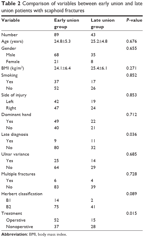 | Table 2 Comparison of variables between early union and late union patients with scaphoid fractures |
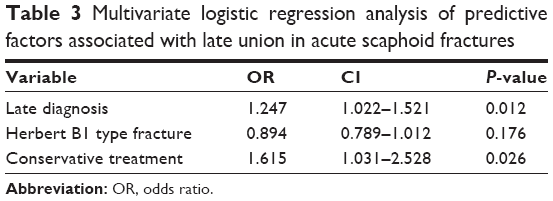 | Table 3 Multivariate logistic regression analysis of predictive factors associated with late union in acute scaphoid fractures |
Discussion
In this study, we reviewed 132 patients with acute middle-third scaphoid fractures and found that both treatments can lead to satisfactory union rate. After the multivariate analysis, our main finding confirmed that late diagnosis and conservative treatment were 2 risk factors independently associated with late union. Therefore, long-term follow-up is necessary for patient with these factors.
Formerly, the first choice for nondisplaced and minimally displaced fractures was conservative treatment with long periods of cast immobilization. Despite the well-recognized problems of scaphoid nonunion after conservative treatment, it was demonstrated by previous studies that the majority of these fractures healed completely without complications.2,7,8 In this study, we only included middle-third scaphoid fracture, which had better vascular supply than proximal-third fracture, and the results showed high rate of fracture union. However, conservative treatment with long-term immobilization is often associated with severe joint stiffness and decreased grip strength, as well as resulting in delays of patients’ return to work and resumption of normal daily activities.
An alternative surgical treatment was introduced by Filan and Herbert6 with use of a new headless fixation screw. The double-threaded Herbert screw had differential pitch at the loading and trailing ends to provide rigid fixation, and thus, cast immobilization was rarely required. In their study, all acute fractures in their series had united by 12 months. Nevertheless, the true long-term benefits of internal fixation have not been adequately determined in recent randomized controlled trials or meta-analysis.13–15 In our current study, though we cannot determine a significant difference in the union rate, we did find relatively shorter union time in the operative group in comparison with the conservative group. This result was consistent with previous studies. For example, Bond et al16 and McQueen et al17 compared operative treatment with casting in mostly nondisplaced scaphoid fractures, and no significant difference in the union rate was found in either study, but both studies showed shorter union time and faster recovery in the operative group.
Meanwhile, our study found that late diagnosis might also lead to longer union time. Previous studies have demonstrated that delay in diagnosis is an important risk factor for the development of nonunion.18,19 There might be numerous reasons for the delayed diagnosis. Some patients might have felt that their symptoms did not warrant medical attention, but most patients encountered a missed diagnosis, because many nondisplaced scaphoid fractures presenting in the acute phase have normal initial radiographs. A previous study has revealed that 16% of scaphoid fractures may be radiographically occult.20 Stevenson et al21 recommended early CT test to manage suspected occult scaphoid fractures for its accurate diagnosis, while Fallahi et al22 considered that MRI scan should play a major role in the management of clinically suspected scaphoid fracture. For this reason, one should have a high level of suspicion when assessing young patients with radial wrist pain, and CT or MRI should be obtained to further assess for a nondisplaced scaphoid fracture.
B1 and B2 type fractures have a similar fracture line on the volar aspect of the scaphoid waist, but they show differences on the dorsal aspect of the scaphoid waist. In B1 fractures, the fracture line extends proximal to the scaphoid apex, but in B2 fractures, the fracture line extends distal to the scaphoid apex.23 In comparison with B2 fractures, B1 type fractures are considered more stable, and interfragmentary motion is considerably lesser in B1 fractures.24 In this series, B1 and B2 type fractures did not show significant difference in union time. However, we cannot make a definite conclusion yet because of the relatively small sample size, and further studies on basis of large population are still required to confirm this result.
The strength of this study is the absence of significant heterogenicity among patients. However, this study has several limitations. First of all, the retrospective design is the typical restriction of our study. Prospective studies are still required to confirm our results in future. Second, the results were concluded from patients with only acute middle-third scaphoid fractures, so the findings of our study need verification with different patient samples. Finally, we only analyzed a limited number of factors, and the inclusion of other factors may provide more valuable information to us.
Conclusion
In summary, though conservative treatment can lead to satisfactory union rate in patients with acute middle-third scaphoid fractures, it is associated with long union time. Besides, late diagnosis was also associated with late union. Long-term follow-up is necessary for patients with these factors.
Acknowledgment
We are grateful to Dr Bo Peng for his assistance in the statistical analysis.
Disclosure
The authors report no conflicts of interest in this work.
References
Pillai A, Jain M. Management of clinical fractures of the scaphoid: results of an audit and literature review. Eur J Emerg Med. 2005;12(2):47–51. | ||
Kozin SH. Incidence, mechanism, and natural history of scaphoid fractures. Hand Clin. 2001;17(4):515–524. | ||
Hove LM. Epidemiology of scaphoid fractures in Bergen, Norway. Scand J Plast Reconstr Surg Hand Surg. 1999;33(4):423–426. | ||
Brondum V, Larsen CF, Skov O. Fracture of the carpal scaphoid: frequency and distribution in a well-defined population. Eur J Radiol. 1992;15(2):118–122. | ||
Haisman JM, Rohde RS, Weiland AJ; American Academy of Orthopaedic Surgeons. Acute fractures of the scaphoid. J Bone Joint Surg Am. 2006;88(12):2750–2758. | ||
Filan SL, Herbert TJ. Herbert screw fixation of scaphoid fractures. J Bone Joint Surg Br. 1996;78(4):519–529. | ||
Bhat M, McCarthy M, Davis TR, Oni JA, Dawson S. MRI and plain radiography in the assessment of displaced fractures of the waist of the carpal scaphoid. J Bone Joint Surg Br. 2004;86(5):705–713. | ||
Gellman H, Caputo RJ, Carter V, Aboulafia A, Mckay M. Comparison of short and long thumb-spica casts for non-displaced fractures of the carpal scaphoid. J Bone Joint Surg Am. 1989;71(3):354–357. | ||
Janowski J, Coady C, Catalano LW 3rd. Scaphoid fractures: nonunion and malunion. J Hand Surg Am. 2016;41(11):1087–1092. | ||
Cooney WP, Linscheid RL, Dobyns JH. Scaphoid fractures. Problems associated with nonunion and avascular necrosis. Orthop Clin North Am. 1984;15(2):381–391. | ||
Wong K, von Schroeder HP. Delays and poor management of scaphoid fractures: factors contributing to nonunion. J Hand Surg Am. 2011;36(9):1471–1474. | ||
Mandaleson A, Tham SK, Lewis C, Ackland DC, Ek ET. Scaphoid fracture fixation in a nonunion model: a biomechanical study comparing 3 types of fixation. J Hand Surg Am. 2018;43(3):221–228. | ||
Shen L, Tang J, Luo C, Xie X, An Z, Zhang C. Comparison of operative and non-operative treatment of acute undisplaced or minimally-displaced scaphoid fractures: a meta-analysis of randomized controlled trials. PLoS One. 2015;10(5):e125247. | ||
Al-Ajmi TA, Al-Faryan KH, Al-Kanaan NF, et al. A systematic review and meta-analysis of randomized controlled trials comparing surgical versus conservative treatments for acute undisplaced or minimally-displaced scaphoid fractures. Clin Orthop Surg. 2018;10(1):64–73. | ||
Vinnars B, Pietreanu M, Bodestedt A, Ekenstam F, Gerdin B. Nonoperative compared with operative treatment of acute scaphoid fractures. A randomized clinical trial. J Bone Joint Surg Am. 2008;90(6):1176–1185. | ||
Bond CD, Shin AY, McBride MT, Dao KD. Percutaneous screw fixation or cast immobilization for nondisplaced scaphoid fractures. J Bone Joint Surg Am. 2001;83-A(4):483–488. | ||
McQueen MM, Gelbke MK, Wakefield A, Will EM, Gaebler C. Percutaneous screw fixation versus conservative treatment for fractures of the waist of the scaphoid: a prospective randomised study. J Bone Joint Surg Br. 2008;90(1):66–71. | ||
Langhoff O, Andersen JL. Consequences of late immobilization of scaphoid fractures. J Hand Surg Br. 1988;13(1):77–79. | ||
Kawamura K, Chung KC. Treatment of scaphoid fractures and nonunions. J Hand Surg Am. 2008;33(6):988–997. | ||
Hunter JC, Escobedo EM, Wilson AJ, Hanel DP, Zink-Brody GC, Mann FA. MR imaging of clinically suspected scaphoid fractures. AJR Am J Roentgenol. 1997;168(5):1287–1293. | ||
Stevenson JD, Morley D, Srivastava S, Willard C, Bhoora IG. Early CT for suspected occult scaphoid fractures. J Hand Surg Eur Vol. 2012;37(5):447–451. | ||
Fallahi F, Oliver R, Mandalia SS, Jonker L. Early MRI diagnostics for suspected scaphoid fractures subsequent to initial plain radiography. Eur J Orthop Surg Traumatol. 2014;24(7):1161–1166. | ||
Moritomo H. Radiographic clues for determining carpal instability and treatment protocol for scaphoid fractures. J Orthop Sci. 2014;19(3):379–383. | ||
Leventhal EL, Wolfe SW, Moore DC, Akelman E, Weiss AP, Crisco JJ. Interfragmentary motion in patients with scaphoid nonunion. J Hand Surg Am. 2008;33(7):1108–1115. |
 © 2018 The Author(s). This work is published and licensed by Dove Medical Press Limited. The
full terms of this license are available at https://www.dovepress.com/terms
and incorporate the Creative Commons Attribution
- Non Commercial (unported, 3.0) License.
By accessing the work you hereby accept the Terms. Non-commercial uses of the work are permitted
without any further permission from Dove Medical Press Limited, provided the work is properly
attributed. For permission for commercial use of this work, please see paragraphs 4.2 and 5 of our Terms.
© 2018 The Author(s). This work is published and licensed by Dove Medical Press Limited. The
full terms of this license are available at https://www.dovepress.com/terms
and incorporate the Creative Commons Attribution
- Non Commercial (unported, 3.0) License.
By accessing the work you hereby accept the Terms. Non-commercial uses of the work are permitted
without any further permission from Dove Medical Press Limited, provided the work is properly
attributed. For permission for commercial use of this work, please see paragraphs 4.2 and 5 of our Terms.