Back to Journals » International Journal of Nanomedicine » Volume 20
Extracellular Vesicle-Based Therapeutic Cargo Delivery for Cancer Therapy
Authors Huang Z, Cheng J, Deng Z, Liu C, Huang T, Lin W
Received 25 June 2025
Accepted for publication 15 October 2025
Published 27 October 2025 Volume 2025:20 Pages 13007—13037
DOI https://doi.org/10.2147/IJN.S548006
Checked for plagiarism Yes
Review by Single anonymous peer review
Peer reviewer comments 5
Editor who approved publication: Dr Yan Shen
Zhiben Huang,1,* Jiaqing Cheng,2,* Zhimin Deng,1 Chunjiang Liu,1 Tianying Huang,2 Wansong Lin3,4
1College of Chemistry, Fuzhou University, Fuzhou, Fujian, 350108, People’s Republic of China; 2The School of Basic Medical Sciences, Fujian Medical University, Fuzhou, Fujian, 350122, People’s Republic of China; 3Laboratory of Immuno-Oncology, Clinical Oncology School of Fujian Medical University, Fujian Cancer Hospital, Fuzhou, Fujian, 350014, People’s Republic of China; 4Fujian Key Laboratory of Translational Cancer Medicine, Fuzhou, Fujian, 350014, People’s Republic of China
*These authors contributed equally to this work
Correspondence: Wansong Lin, Laboratory of Immuno-Oncology, Clinical Oncology School of Fujian Medical University, Fujian Cancer Hospital, Fuzhou, Fujian, 350014, People’s Republic of China, Email [email protected]
Abstract: Extracellular vesicles (EVs) have become attractive nanoscale delivery systems in oncology because of their inherent biological advantages and distinct physicochemical properties. These cell-derived nanoparticles exhibit low immunogenicity, inherent targeting capacities, and the natural ability to facilitate precise intercellular transfer of bioactive molecules, including proteins and nucleic acids. Recently, remarkable progress has been made in EV engineering. These advancements have improved the accuracy and efficiency of delivering therapeutic agents to tumor sites. In this review, we outline the biogenesis, composition, and unique advantages of EVs compared with conventional nanocarriers, with an emphasis on their therapeutic potential in cancer treatment. Furthermore, we discuss EV cargo–loading methods and advanced engineering approaches designed to enhance tumor-specific targeting. In addition, we systematically summarize diverse therapeutic applications, including chemotherapeutic drug delivery, nucleic acid–based therapies, cancer vaccines, and multifunctional theranostic platforms. The review also discusses current clinical trials and highlights the critical challenges in clinical translation, such as limited clinical-scale manufacturing and unclear regulatory pathways. Future efforts to overcome these challenges could transform EVs into precise and personalized tools for cancer treatment. This concise overview provides valuable insights into the current developments and future perspectives of EV-mediated therapeutic delivery systems in oncology.
Keywords: nanocarriers, drug loading, targeted delivery, immunotherapy, clinical translation
Introduction
Despite significant advancements in treatment, cancer remains a major health issue worldwide.1 Although current cancer treatments (eg, chemotherapy, radiotherapy, targeted therapy, and immunotherapy) have substantially improved patient survival, they face several challenges, including severe systemic toxicity, limited tumor specificity, and therapeutic resistance, underscoring the need for safer, more precise, and effective strategies for the targeted delivery of therapeutic cargo in oncology.2
To address these limitations, nanoparticle-based drug delivery systems (eg, liposomes, polymeric nanoparticles, and other synthetic nanocarriers) have been extensively explored.3,4 These systems can encapsulate drugs and preferentially accumulate in tumors via enhanced permeability or targeting ligands, reducing off-target effects.5,6 Emerging porous and hollow nanomaterials, such as mesoporous silica and metal–organic frameworks, have expanded this toolkit: their high surface areas and tunable pore sizes enable exceptional drug loading and controlled release.7 For instance, hollow polydopamine nanobowls encapsulate tirapazamine and tether MoS2 nanozymes, enabling high-capacity delivery that suppresses tumor growth and limits metastasis via coordinated multimodal mechanisms.8 Similarly, advanced polymeric nanoparticles have progressed considerably. Biodegradable polymers (eg, PLGA) enable precise control over particle stability, degradation rates, and surface functionalization for active targeting.9 Despite these advances, the use of synthetic nanoparticles faces challenges, including rapid clearance by the mononuclear phagocyte system, limited barrier penetration, potential immunogenicity, and inefficient cellular uptake.10–13
In this context, extracellular vesicles (EVs) have attracted considerable attention as promising bio-derived nanocarriers.14 EVs are naturally occurring lipid bilayer-bound vesicles that are secreted by virtually all cell types, and their properties differ by cellular source (Table 1 summarizes EVs from common cellular origins and their key features). EVs can be classified into the following subtypes: exosomes (30–150 nm), microvesicles (100–1000 nm), and apoptotic bodies (up to several microns).15–17 Among them, exosomes and microvesicles have attracted considerable interest because of their nanoscale dimensions, excellent biocompatibility, low immunogenicity, and intrinsic ability to mediate targeted intercellular communication.18 These native features position EVs as a promising alternative to conventional nanocarriers for therapeutic delivery in oncology, with the potential to overcome key limitations associated with current cancer therapies.
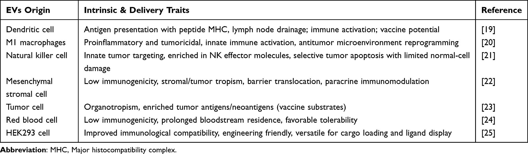 |
Table 1 Key Features of EVs from Different Cellular Origins |
In this review, we outline the biogenesis and molecular composition of EVs and discuss cargo-loading strategies and surface engineering approaches to improve tumor-specific targeting. Furthermore, we evaluate the diverse therapeutic applications of engineered EVs, from the targeted delivery of chemotherapeutics and nucleic acids to cancer vaccines and multimodal theranostics. Finally, we summarize the current efforts toward clinical translation, highlight ongoing challenges, and provide insights into future opportunities for advancing EV-based precision cancer therapy.
EVs: A Concise Overview
Biogenesis of EVs
The biogenesis of EVs involves sequential membrane remodeling events within the endosomal network (Figure 1). This process begins with the endocytosis of plasma membrane components, which generates vesicular structures that fuse to form early endosomes. As these endosomes mature into late-stage compartments, inward membrane invagination leads to the formation of intraluminal vesicles (ILVs) within multivesicular bodies (MVBs). As exosome precursors, ILVs are either degraded lysosomally or released extracellularly upon the fusion of MVBs with the plasma membrane.26,27 The formation of ILVs is predominantly controlled by the endosomal sorting complex required for transport (ESCRT) machinery. ESCRT-0, ESCRT-I, and ESCRT-II recognize and sequester ubiquitinated cargo, whereas ESCRT-III facilitates membrane remodeling and ILV scission, in coordination with accessory proteins (eg, VPS4 and ALIX).28,29 Aside from the ESCRT-dependent pathway, there are alternative mechanisms, which involve ceramides, tetraspanins, and Rab GTPases that contribute to ILV formation and cargo sorting.26 The interplay between these pathways determines the composition and functional properties of EVs, which vary depending on the cell type and physiological context.
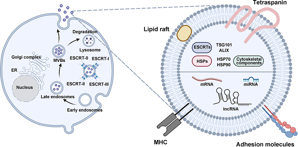 |
Figure 1 EVs are formed through endosomal pathways, originating from early endosomes that mature into multivesicular bodies (MVBs) before release. These vesicles contain diverse array of membrane-associated proteins, lipids, and nucleic acids. |
Composition of EVs
EVs have a unique molecular composition that reflects their cellular origin and biogenesis pathways. This composition enables EVs to effectively facilitate intercellular communication and serve as promising therapeutic delivery vectors.30 EVs are structurally encapsulated by a lipid bilayer, which primarily comprises cholesterol, sphingomyelin, and phosphatidylcholine that account for approximately 80% of the total lipids and confer membrane rigidity and stability. This bilayer preserves vesicular integrity, safeguards internal cargo, and facilitates interactions with target cells.31 Membrane-associated proteins are key determinants of EV functionality. EVs are enriched in tetraspanins (eg, CD9, CD63, and CD81), which contribute to vesicle formation, membrane organization, and cellular internalization.32,33 Moreover, they contain adhesion molecules, particularly integrins that facilitate target cell recognition and tissue-specific distribution. Among them, α6β4 and α6β1 promote lung tropism, whereas αvβ5 mediates liver-specific targeting.34,35
In addition, the intravesicular cargo contains various biomolecules, including heat shock proteins (eg, HSP70 and HSP90), ESCRT-associated proteins (TSG101 and ALIX), and cytoskeletal components. Collectively, these molecules maintain vesicular stability and regulate cargo trafficking.36 EVs also contain nucleic acids, including microRNAs (miRNAs), long noncoding RNAs, and messenger RNAs (mRNAs), which modulate gene expression in recipient cells.37 Given their molecular heterogeneity and natural ability to protect and transport bioactive molecules, EVs have a significant potential as therapeutic delivery vehicles.38
EV Isolation and Characterization
EVs can be isolated through various strategies that leverage differences in size, density, or surface markers. Common isolation methods include differential ultracentrifugation, density gradient centrifugation, polymer-based precipitation, membrane ultrafiltration, size-exclusion chromatography, immunoaffinity capture, and emerging microfluidic-based techniques, each with distinct yield–purity tradeoffs.39 The key attributes of these methods are summarized in Table 2. After isolation, EV preparations are characterized to confirm vesicle size, morphology, and marker profiles. Generally, nanoparticle tracking analysis is used to quantify particle concentration and size distribution.40 Transmission electron microscopy is used to characterize EV morphology at the nanometer scale.40 Western blotting is used to verify EV identity by detecting enriched markers (eg, CD63, CD81, and TSG101) and to assess purity by confirming the absence of cellular contaminants (eg, Golgi protein GM130).41 Flow cytometry, often bead-assisted, is used to profile EV surface markers for phenotypic analysis.42 Collectively, these complementary assays provide orthogonal evidence for establishing EV identity and purity.
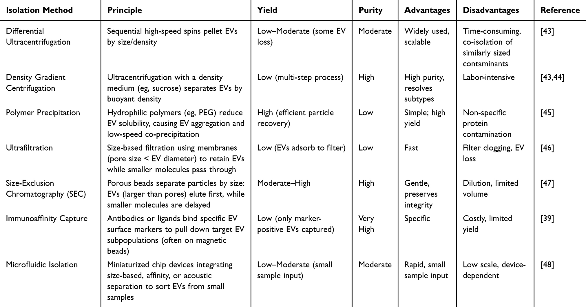 |
Table 2 Summary of EV Isolation Techniques |
Cellular Uptake and Trafficking of EVs
Cells internalize EVs via diverse endocytic pathways, including clathrin-mediated endocytosis, caveolin/lipid raft-dependent uptake, macropinocytosis, phagocytosis, and, occasionally, direct plasma membrane fusion.49,50 Multiple uptake mechanisms can operate simultaneously within a given EV population.49 Following endocytosis, EVs typically traffic to endosomal compartments and often accumulate in late endosomes or lysosomes. Many internalized EVs are degraded in lysosomes or recycled to the cell surface; however, a fraction can escape the endolysosomal system to deliver functional cargo into the cytosol.51 For instance, some EVs fuse with endosomal membranes in an acidification-dependent manner and release their RNA and protein cargo into the cytoplasm.51 Understanding these pathways is crucial for designing therapeutic EVs. Surface modifications can considerably alter EV biodistribution. For example, precoating EVs with albumin forms a “protein corona” that shields them from phagocytes and redirects uptake toward target liver cells, thereby prolonging circulation and enhancing delivery efficacy.52
Unique Features of EVs as Nanocarriers
Enhanced Circulation and Immune System Compatibility
As cell-derived vesicles, EVs demonstrate extended circulation and low immunogenicity in vivo.53 Moreover, their plasma membrane–like composition (eg, displaying the “self” marker CD47) enables them to evade phagocytic clearance by monocytes, thereby prolonging their half-life in the bloodstream.54 Unlike conventional nanocarriers, which frequently trigger an immune reaction or require PEGylation to avoid rapid uptake by the mononuclear phagocyte system,55,56 EVs inherently “fly under the radar” of the immune system, allowing more drugs to reach the tumor site.57 This immune compatibility also translates to decreased risks of adverse immune reactions compared with synthetic nanoparticles.
Advanced Cell-Specific Targeting
EVs have innate tissue-homing ability mediated by parent cell–derived adhesion molecules and cognate receptor–ligand pairs.33,58 Notably, integrin profiles on tumor-derived EVs direct accumulation toward metastasis-prone organ sites, whereas immune cell–derived EVs preferentially target lymphoid tissues.23 This endogenous targeting capacity enables efficient delivery of therapeutic cargo to specific cell types, often surpassing that of synthetic nanocarriers, which require exogenous ligands, stimuli-responsive designs, or external fields to achieve similar specificity.59 For example, a previous study reported that EVs delivered RNA interference molecules to pancreatic cancer cells (PCCs) more effectively than liposomes.54 Such natural tropism enables EV-based nanocarriers to achieve high uptake in target tumor cells while sparing normal tissues, surpassing the targeting selectivity of liposomes, polymeric nanoparticles, and micelles.60
Superior Biological Barrier Penetration
Compared with many synthetic nanocarriers, EV-based carriers can traverse physiological barriers, including the blood–brain barrier (BBB), because of their nanoscale dimensions and native membrane protein composition.61,62 These properties enable EVs to deliver therapeutics to sanctuary sites (eg, brain tumors or metastases) where liposomes and polymeric micelles show limited accumulation.63 Moreover, EV membranes are fluid and dynamic, facilitating fusion or uptake across cell–cell junctions. Thus, EVs frequently achieve comparable or superior tissue penetration and deep tumor infiltration than conventional nanocarriers, facilitating drug delivery to otherwise inaccessible microenvironments.
Intrinsic Biocompatibility and Versatile Payload Capacity
EVs exhibit excellent biocompatibility, high stability, and minimal toxicity, as they are derived from natural cell membranes.64 Moreover, they are well tolerated by the body and can protect therapeutic cargos from degradation. Compared with conventional nanocarriers, EVs can accommodate various payloads, from small-molecule drugs to large macromolecules (eg, small interfering RNA [siRNA], miRNA, proteins, and even CRISPR complexes), without compromising the cargo’s bioactivity.65–67 The lipid bilayer of EVs shields therapeutics from enzymatic degradation and premature clearance, enabling stable delivery of diverse treatments.55 This versatility and favorable safety profile provide EVs a distinct advantage in delivering complex combination therapies that challenge synthetic nanoparticle systems. Figure 2 summarizes these features, highlighting the key properties of EVs that enhance their effectiveness as therapeutic nanocarriers.
 |
Figure 2 Key biological advantages of EVs as carriers for therapeutic cargos. (a) Surface molecules such as CD47 and MHC enhance circulation half-life and immune compatibility, reducing immune clearance. (b) EVs achieve specific cellular targeting through integrin-mediated organ tropism and precise receptor–ligand interactions, facilitating selective therapeutic delivery. (c) Their small size and native surface proteins enable effective penetration across biological barriers, including the blood-brain barrier. (d) EVs possess a lipid bilayer membrane, providing inherent biocompatibility, low toxicity, and versatile cargo-loading capabilities. (e) Immune cell–derived EVs convey antigenic and immunostimulatory cues that modulate and remodel the tumor microenvironment (TME). Symbols: red upward arrow (↑) indicates increase; blue downward arrow (↓) indicates decrease. |
Integrated Immunomodulation and Immune Activation
In addition to passive drug delivery, EVs are unique bioactive carriers that can actively regulate immune responses. Different EV subtypes elicit distinct immunological effects. M1 macrophage–derived EVs carry proinflammatory cytokines and miRNAs that activate innate immunity and enhance antitumor responses,68 whereas M2 macrophage–derived EVs convey anti-inflammatory signals suited that can reprogram inflamed milieus.69 For instance, macrophage–derived EVs can function as natural adjuvants, with preserved macrophage-lineage receptors on their surface engaging antigen-presenting cells (APCs) and enhancing their activation.70 Dendritic cell–derived EVs serve as potent antigen-presenting vesicles, displaying peptide–MHC complexes and costimulatory ligands that strongly activate T cells and natural killer (NK) cells;71 when engineered to co-display IL-12 and anti-CTLA-4, they further drive Th1 polarization while lifting checkpoint restraint.72 T cell–derived EVs likewise carry immunoregulatory miRNAs and cytokines that can dampen or steer immune reactions. In aggregate, EVs’ endogenous interactions with specific cell types, together with configurable surface and cargo features, enable synchronous, multidimensional immunomodulation in the tumor microenvironment (TME), which is rarely achieved by conventional nanocarriers.
EV-Based Cargo Loading Strategies
Successful incorporation of pharmaceutical agents into these nanoscale vesicles determines the therapeutic efficacy of EV-based drug delivery systems. Contemporary loading methods can be systematically categorized into endogenous and exogenous approaches (Figure 3), with comparative details tabulated in Table 3.
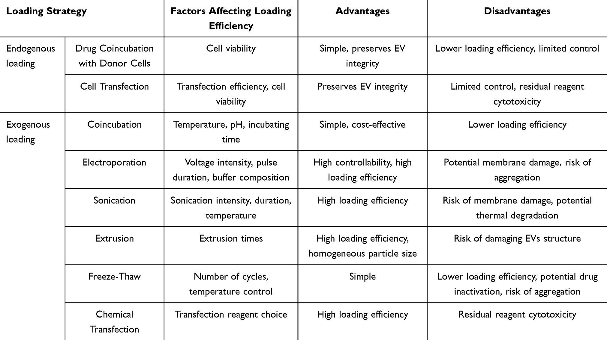 |
Table 3 EVs-Based Cargo Loading Strategies |
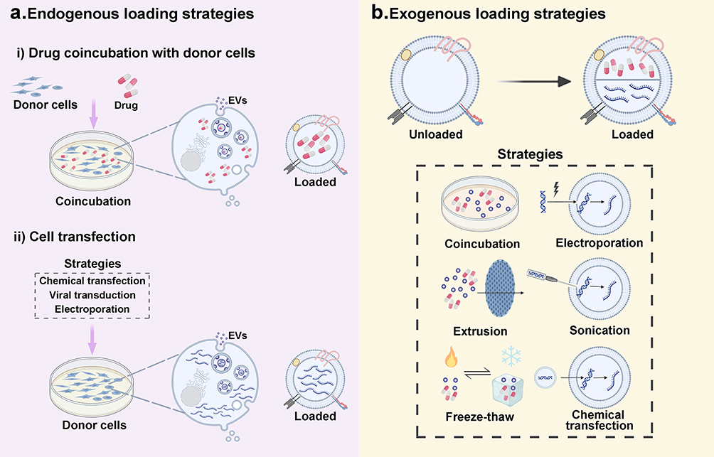 |
Figure 3 EV cargo-loading strategies. (a) Endogenous: donor cells are drug-exposed or genetically manipulated (chemical transfection, viral transduction, electroporation) so cargos are packaged into secreted EVs. (b) Exogenous: cargos are introduced into isolated EVs via coincubation, electroporation, sonication, extrusion, freeze–thaw, or chemical transfection. |
Endogenous Loading Strategies
Endogenous loading incorporates therapeutic cargo into EVs by modulating the intracellular concentrations of target molecules prior to EV biogenesis.73 This strategy exploits cells’ natural sorting mechanisms, whereby elevated cytoplasmic levels of specific therapeutic agents (eg, small-molecule drugs, proteins, and nucleic acids) result in their preferential enrichment during vesicle formation.74 This physiological loading approach preserves the inherent structure and functional properties of EVs by avoiding post-isolation modifications that can compromise therapeutic efficacy.
Drug Coincubation with Donor Cells
In this approach, target drug molecules are cultured with EV-producing donor cells, allowing spontaneous incorporation of therapeutics during EV biogenesis.75 Pessina et al demonstrated that MSCs internalized paclitaxel (PTX) via passive uptake and secreted PTX-loaded EVs without genetic modification.76 The protocol exposed MSCs to high PTX concentrations, yielding efficient drug encapsulation. The PTX-loaded EVs demonstrated significant antitumor activity, making them promising biomimetic drug delivery vehicles.76 In subsequent studies, this approach was expanded to various small-molecule therapeutics, including doxorubicin (DOX) and dexamethasone, using analogous coculture protocols.77,78
However, despite its simplicity, this approach exhibits limited loading efficiency and low process control. Loading outcomes are mainly determined by the physicochemical properties of the drug and donor cell attributes.79 A systematic comparison of DOX loading across PCCs, macrophages (MØs), and pancreatic stellate cells showed distinct capacities.80 Intriguingly, although PCC-derived EVs demonstrated superior drug loading efficiency, MØ-derived EVs exhibited enhanced antitumor efficacy despite having a lower drug content, highlighting the importance of donor cell selection in endogenous loading strategies.80 In addition, cytotoxicity profiles should be carefully considered when selecting therapeutic agents to maintain donor cell viability and ensure sustainable EV production.81
Cell Transfection
Cell transfection is an effective endogenous loading method for introducing therapeutic cargo into EVs. This strategy encompasses chemical transfection, electroporation, and viral transduction to deliver plasmid DNA or functional nucleic acids into donor cells, inducing the expression of specific therapeutic biomolecules.82 Subsequently, the transfected cells package the synthesized molecules into EVs through inherent biogenesis pathways. Zhang et al used lentiviral transduction to generate induced neural stem cells that express tumor-necrosis-factor-related apoptosis-inducing ligand (TRAIL), which produced TRAIL-functionalized EVs.83 These synthesized EVs exhibited preferential membrane localization of TRAIL and enhanced loading efficiency compared with fibroblast-derived EVs (351 vs 187 pg TRAIL/μg EVs), demonstrating superior antineoplastic efficacy against cerebral malignancies.83 Similarly, Kimura et al engineered bone marrow–derived mesenchymal stem cells to express microRNA-29b (miR-29b) using lentiviral vectors. The resultant miR-29b-enriched small EVs (sEVs), which were isolated through ultracentrifugation, demonstrated selective targeting to peritoneal mesothelial cells and effectively suppressed gastric cancer metastasis.84
Although cell transfection demonstrates considerable loading efficiency, the method has several technical challenges, including variable transfection rates, potential genotoxicity, and difficulties in accurately controlling the cargo-loading capacity, which depend on the cell viability and transfection outcomes.85 Furthermore, transfection reagents have been reported to accumulate in late endosomes and be released into the extracellular space via exocytosis. These transfection complexes share similar physical properties with EVs and may co-sediment during purification, potentially contaminating EV preparations and altering their characteristics.86,87
Exogenous Loading Strategies
Exogenous loading strategies, in contrast to endogenous approaches, use physicochemical methods to incorporate therapeutic cargo into pre-isolated EVs. This post-isolation modification strategy offers distinct advantages over endogenous approaches, particularly in terms of process standardization and manufacturing scalability.73
Coincubation
This method offers a gentle and straightforward approach for loading therapeutic agents into EVs. It relies on concentration-gradient-driven diffusion under physiologically relevant conditions to facilitate drug translocation across EV membranes.88 The EV phospholipid bilayer exhibits a preferential affinity for hydrophobic compounds because of its amphipathic nature, enabling the efficient encapsulation of lipophilic therapeutic agents.89 Yang et al achieved approximately 8% drug loading efficiency by simply mixing EVs with DOX and incubating the mixture at 37°C for 30 min.90
Despite its simplicity and cost-effectiveness, conventional coincubation has inherent limitations, particularly for hydrophilic drugs, proteins, and nucleic acids, because of their low membrane permeability.15 To overcome these limitations, several strategic modifications have been developed. One approach involves the use of membrane-active agents, particularly saponins, to induce transient membrane permeabilization. However, surfactant exposure must be carefully optimized to prevent compromising vesicular integrity.91 Another approach focuses on cargo modification to enhance membrane penetration. Zhang et al developed an novel approach using cell-penetrating peptides (CPPs) conjugated to DOX. They achieved a loading efficiency of 37.18% via the coincubation of CPP–DOX conjugates with urinary EVs under optimized conditions (pH 9.0, 37°C, 2 h), indicating a 2.5-fold enhancement compared with conventional methods. These functionalized EVs demonstrated superior antineoplastic efficacy.92 The drug loading efficiency can also be improved by optimizing the incubation conditions. Several factors, including the drug concentration, incubation temperature, duration, and pH, can significantly affect the loading process.93,94 Therefore, these parameters must be carefully adjusted based on the specific characteristics of the cargo and EV source.
Electroporation
This advanced method uses high-intensity electric fields to induce the transient membrane permeabilization of EVs. This method involves suspending EVs and therapeutic cargo in an optimized electroporation buffer and then applying calibrated electric pulses to generate temporary nanopores, which facilitate cargo translocation.73 This approach is particularly versatile for hydrophilic cargos, such as small molecules and nucleic acids, which typically have poor membrane permeability. Gong et al engineered uPA-sEVs-siSrc, an advanced drug delivery system that incorporates Src-siRNA into urokinase plasminogen activator (uPA)-functionalized sEVs.95 This system achieved a 45% loading efficiency under optimized electroporation parameters (400 mV, 125 μF) while preserving vesicular integrity and functionality, resulting in robust antitumor effects. Similarly, Tian et al successfully encapsulated DOX into immature dendritic cell (DC)-derived exosomes with 20% efficiency via electroporation.96 The DOX-loaded exosomes effectively suppressed breast tumor growth and markedly reduced systemic toxicity and cardiotoxicity in MDA-MB-231 tumor–bearing mice.
The primary advantage of electroporation is its high controllability, enabling precise adjustment of experimental conditions for optimal loading. However, several technical challenges must be considered. In particular, excessive electric field strength can compromise membrane integrity, and RNA cargo aggregation during electroporation remains a significant challenge.97,98 To mitigate these issues, key parameters, including voltage intensity and pulse duration, must be systematically optimized based on specific EV populations and cargo characteristics.99 RNA aggregation can be mitigated by incorporating stabilizing agents (eg, trehalose, citric acid, and ethylenediaminetetraacetic acid) into the electroporation buffers, thereby enhancing the loading efficiency and cargo stability.31
Sonication
This method employs acoustic energy and mechanical shear forces to transiently permeabilize EV membranes, facilitating the internalization of therapeutic molecules.82 Subsequently, membrane integrity is restored through a standardized postsonication incubation protocol (37°C, 1 h).100 Studies have shown that sonication-induced membrane perturbation significantly reduces the microviscosity of the EV membrane, thereby enhancing membrane fluidity and drug permeation.101 Accordingly, ultrasound-assisted loading often outperforms simple incubation. For example, Kim et al achieved approximately 28.29% loading efficiency was achieved when using sonication to load PTX into RAW 264.7 MØ-derived EVs, which was higher than that achieved via incubation (4.10%).101 Similarly, in another study on 5-fluorouracil encapsulation, sonication demonstrated superior loading capacity compared to incubation (6.79% vs 4.10%).102
Although sonication shows promising potential for efficient EV drug loading, sonication parameters should be optimized to maintain EV functionality. For example, the intensity and duration of acoustic energy exposure must be precisely controlled to maintain membrane integrity and biological activity. Sun et al reported higher PTX loading efficiency in sEVs via sonication than via electroporation and incubation methods.103 Paradoxically, PTX loaded via incubation exhibited enhanced cytotoxicity against pancreatic ductal adenocarcinoma (PDAC) cells, indicating that sonication-induced membrane alterations may impair cellular uptake mechanisms.103 Furthermore, sonication requires rigorous temperature monitoring to prevent the thermal degradation of vesicular structures and therapeutic cargo.
Extrusion
This technique uses calibrated nanoporous membranes (100–400 nm) and standardized liposome extruders to load drugs into EVs.94 In this technique, transmembrane pressure gradients are used to induce controlled membrane destabilization, thereby facilitating drug translocation into the vesicular lumen.31 Membrane elasticity and drug–membrane interactions predominantly govern the loading efficiency. Subsequently, unencapsulated drugs are eliminated through density gradient ultracentrifugation or dialysis to ensure homogeneity and purity of the preparation, yielding highly purified drug-loaded EVs. Bi et al successfully loaded PTX into H22 hepatocarcinoma-cell-derived EVs through serial extrusion across 200-nm porous membranes, achieving an encapsulation efficiency of 92.33%.104 Compared with free PTX, the engineered EVPA exhibited enhanced antitumor efficacy against hepatocarcinoma in vitro and in vivo.104
Although extrusion demonstrates superior drug loading efficiency and enhances EV size homogeneity, repeated extrusion cycles can compromise the structural integrity of EVs and alter the composition of endogenous cargo through mechanical stress–induced membrane perturbation.94,105
Freeze–Thaw
This method increases drug loading in EVs by inducing membrane microfractures through repeated freeze–thaw cycles. This technique involves rapidly freezing an EV–cargo mixture at −80°C or in liquid nitrogen, followed by controlled thawing at room temperature.82 Multiple cycles (≥3) are used to maximize the drug encapsulation efficiency.82 Ebrahimian et al used a freeze–thaw method combined with coincubation and surfactants to encapsulate thymoquinone into exosomes, effectively targeting MCF7 cancer cells and inducing apoptosis.106
Despite its simplicity, this method generally results in lower drug loading efficiency than sonication or extrusion methods.107 Moreover, the use of this method in drug loading investigations is limited because of the potential for EV aggregation and the risk of drug inactivation from frequent freeze–thaw processes.79
Chemical Transfection
Chemical transfection-mediated drug loading is a promising strategy for incorporating therapeutic molecules into EVs. Because various transfection reagents are commercially available, several EV-based drug delivery systems have been developed. Recent studies have demonstrated the efficacy of chemical transfection. For example, Xing Pei et al used the Exo-Fect reagent to load siFGL1 and siTGF-β1 into cRGD-functionalized EVs, achieving a loading efficiency of 97%.108 The resulting EVs effectively downregulated FGL1 and TGF-β1 expression, thereby inhibiting colorectal cancer (CRC) progression. In a parallel study, Peng et al109 achieved 70% loading efficiency by transfecting siSTAT3 into tumor-derived EVs (T-EVs) isolated from 4T1 triple-negative breast cancer (TNBC) cells. A dual-therapeutic delivery system with enhanced tumor targeting and improved efficacy in TNBC models was generated by coincubating DOX with siSTAT3-loaded T-EVs.109
The potential cytotoxicity associated with the residual transfection reagents should be carefully considered. Moreover, comprehensive characterization studies are needed to fully elucidate the spatial distribution of the loaded therapeutic cargo (whether encapsulated within the EV lumen or surface-associated).110
Engineered EVs for Enhanced Targeted Delivery
Although native EVs have desirable properties, they are being actively engineered to enhance their targeting ability and therapeutic efficacy in cancer treatment. In this section, several major engineering approaches are discussed and examples of how each method enhances EV-mediated drug delivery to tumors are provided. Representative EV engineering strategies are summarized in Figure 4.
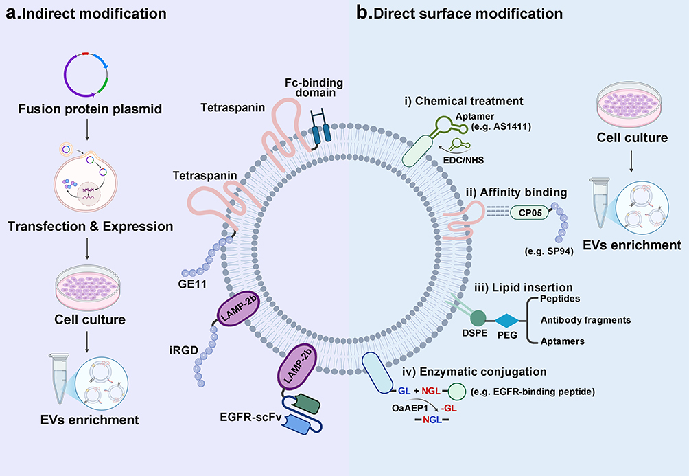 |
Figure 4 Surface modification strategies for targeted EV delivery. (a) Indirect Modification: Genetic modification of parent cells by plasmid transfection to express targeting molecules incorporated into EV membranes. (b) Direct Surface Modification: Surface functionalization of isolated EVs. Methods include i) chemical conjugation, using EDC/NHS to covalently attach ligands such as the AS1411 aptamer; ii) affinity binding, CP05 recognizes CD63 on EVs and anchors CP05–ligand conjugates for noncovalent surface display; iii) lipid insertion, incorporating DSPE-PEG–linked peptides, antibody fragments, or aptamers into the outer leaflet; and iv) enzymatic conjugation, using OaAEP1 to ligate GL/NGL-tagged peptides for site-specific display. |
Indirect Modification: Cell-Based Engineering
Targeted EV engineering can be achieved by genetically modifying donor cells via transfection by introducing engineered expression constructs. These constructs encode fusion proteins comprising targeting moieties that are genetically linked to EV-enriched transmembrane proteins, which function as membrane anchors.111,112 Among the available membrane-anchoring candidates, lysosome-associated membrane protein 2b (LAMP-2b) and tetraspanin family members (eg, CD9, CD63, and CD81) are frequently used because of their abundant expression and stable incorporation within the EV membrane.113,114
LAMP-2b is an optimal scaffold for EV surface modification as its N-terminal domain (29-amino-acid signal peptide) can tolerate genetic fusion with diverse targeting moieties (eg, peptides and antibody fragments) while maintaining efficient protein trafficking and membrane integration, thereby ensuring the stable surface display of the targeting molecules.79 LAMP-2b-based modification has been extensively validated for its versatility. For example, the LAMP-2b-fused αv integrin–targeting iRGD peptide has been successfully expressed on HEK 293FT cell–derived EVs, which were subsequently loaded with DOX via electroporation.115 These iRGD-modified EVs demonstrate a 40% enhancement in cellular uptake by glioblastoma multiforme (GBM) cells in vitro and exhibit significantly improved antitumor efficacy in vivo compared with their unmodified counterparts.115 In another study, an epidermal growth factor receptor (EGFR)-specific single-chain variable fragment (scFv), which targeted the overexpressed EGFR on cancer cells, was successfully fused with LAMP-2b and expressed on HEK 293T cell-derived EVs.116 The targeting ability of these engineered EVs, loaded with LPCAT1-specific siRNA, was evaluated in lung cancer brain metastasis (BM). In vivo biodistribution studies have demonstrated that the scFv-modified exosomes have significantly greater retention than unmodified variants, showing two-fold higher residual fluorescence in the BM region at 96 h postinjection. Moreover, these functionalized exosomes efficiently traversed the BBB, resulting in the significant downregulation of LPCAT1 and subsequent inhibition of tumor cell proliferation in the BM model.116
Tetraspanins, which are a family of abundant EV transmembrane proteins, have also demonstrated efficacy in genetic surface engineering. Yang et al engineered HEK 293T cell–derived EVs by fusing the EGFR-targeting GE11 peptide with CD63.117 These GE11-modified EVs, loaded with DOX, exhibited enhanced cellular uptake in EGFR-positive CRC and GBM cells, along with improved antitumor efficacy and reduced systemic toxicity in vivo. Wiklander et al engineered EVs by fusing the Fc-binding domains to CD63, CD9, and CD81, with the CD63 C-terminal fusion displaying optimal expression and targeting efficiency.118 These Fc-EVs demonstrated versatile antibody decoration capabilities, achieving broad tissue targeting. In another study, trastuzumab (a HER2-targeting antibody)-decorated Fc-EVs exhibited a 339-fold increase in cellular uptake by HER2-positive breast cancer cells (SKBR-3), and Fc-EVs functionalized with PD-L1 antibodies demonstrated comparable targeting enhancement toward PD-1-expressing cells.118
Direct Surface Modification of Isolated EVs
In direct modification strategies, isolated EVs are functionalized by attaching targeting molecules or ligands onto the EV surface using diverse conjugation approaches.
Chemical Treatment
Chemical modification strategies use reactive chemical groups to covalently attach targeting molecules to the EV surface.119 Functional groups on EV membranes—such as amines on surface proteins or lipids (eg, phosphatidylethanolamine), carboxyl groups, and thiols—can be exploited for bioconjugation.120,121 Targeting ligands can be permanently grafted onto EVs using linkers or via click chemistry. Generally, chemical ligation methods are more robust and stable than noncovalent affinity binding, although with a greater risk of altering the native structure or functions of the vesicles.122,123 Current bioconjugation approaches frequently use 1-(3-dimethylaminopropyl)-3-ethylcarbodiimide (EDC)/N-hydroxysuccinimide (NHS)-mediated amide bond formation and azide–alkyne cycloaddition reactions.124 For example, Hosseini et al demonstrated the selective conjugation of the AS1411 aptamer to exosomal surface amines via EDC/NHS chemistry, enabling the specific recognition of nucleolin, which exhibits increased expression on CRC cells.125 This surface engineering strategy markedly enhanced the targeting specificity and efficiency of therapeutic cargo delivery of functionalized exosomes.
Affinity Binding
Affinity-based EV targeting relies on the selective recognition and binding of specific affinity molecules to membrane proteins, receptors, or distinct lipid molecules on the EV surface.126 This noncovalent approach requires only mild incubation conditions for surface functionalization, thereby preserving the structural integrity of EVs while offering operational simplicity compared with chemical conjugation strategies. However, the stability of affinity-based modifications may be compromised under physiological conditions. Thus, environmental factors must be carefully considered.
CD63 functions as a canonical EV marker while being a highly enriched tetraspanin protein in EV membranes.127 Gao et al identified CP05, an affinity peptide that specifically targets the second extracellular loop of CD63, providing a versatile platform for EV modification.128 Notably, CP05 demonstrates a universal binding capability across EVs from diverse sources while preserving their structural and functional integrity. The versatility of CP05 as a modifiable targeting peptide facilitates the straightforward surface functionalization of exosomes. For example, exosome surface decoration using CP05 conjugated to SP94, a hepatocellular carcinoma (HCC)-targeting peptide, markedly enhanced tumor accumulation compared with unmodified exosomes in murine HCC models.128
In addition to peptides, other affinity pairs have been exploited. For example, reticulocyte-derived EVs inherently express transferrin receptors (TfR) on their surface, thereby enabling targeted functionalization through specific transferrin (Tf)–TfR interactions.129 Qi et al functionalized reticulocyte-derived EVs were functionalized with Tf-conjugated superparamagnetic nanoparticles via selective Tf–TfR binding interactions, which conferred enhanced magnetic responsiveness to the EV carriers and facilitated targeted delivery to tumor sites.129
Lipid Insertion
The lipid bilayer membrane structure of EVs facilitates the incorporation of hydrophobic modifying molecules. The biocompatible amphiphilic polymer 1,2-distearoyl-sn-glycero-3-phosphoethanolamine-poly(ethylene glycol) (DSPE-PEG), approved by the Food and Drug Administration (FDA), serves as a prevalent surface modification agent for EVs.130 DSPE-PEG conjugates feature a hydrophobic DSPE tail that inserts into the membrane and a PEG spacer that presents the ligand and enhances stealth by reducing protein adsorption.126,131 The versatile PEG terminus enables the conjugation with various targeting moieties, including peptides, antibody fragments, or aptamers, facilitating diverse targeting strategies.95,132,133 Recently, Zhou et al successfully incorporated integrin α5-targeting peptide (CRYYRITY) into EVs through DSPE-PEG modification, leveraging the spontaneous insertion of DSPE into the lipid bilayer.134 This targeted modification substantially enhanced EV specificity toward cancer-associated fibroblasts (CAFs) in pancreatic cancer, resulting in improved CAF-specific uptake and increased tumor site accumulation compared with unmodified EVs.134
Enzymatic Conjugation
Enzymatic surface functionalization is an emerging EV modification strategy that uses specific ligases to precisely conjugate targeting molecules to the EV membrane. For example, protein ligases, such as Sortase A or OaAEP1 (an asparaginyl endopeptidase), can recognize short peptide motifs on EV surface proteins and covalently attach a ligand peptide at that site. This strategy allows precise EV functionalization with stable, high-density targeting ligands while maintaining vesicle integrity through gentle, site-specific modifications. Pham et al employed OaAEP1 ligase to display an EGFR-targeting (ET) peptide on red blood cell–derived EVs. The ligase catalyzed the covalent attachment of a biotinylated ET-NGL peptide to GL-containing EVs by recognizing the C-terminal NGL motif of the peptide and the N-terminal GL motif on the EV surface. This process facilitated the conjugation of 380 EGFR-binding peptide copies per vesicle to generate multivalent targeted delivery vehicles.135 These EGFR-targeted EVs, loaded with PTX and administered to EGFR-positive lung-tumor-bearing mice, exhibited significantly enhanced tumor accumulation and superior antineoplastic efficacy compared with their nontargeted counterparts.135
Applications of Engineered EVs in Cancer Therapy
Chemotherapeutic Drug Delivery
Chemotherapy remains a cornerstone of cancer treatment; however, conventional chemotherapeutics are limited by broad cytotoxicity, poor solubility, and low bioavailability, which reduce their efficacy.88,136 Engineered EVs offer a biocompatible delivery platform that can improve drug targeting and mitigate systemic toxicity.137 Various anticancer agents have been loaded into EVs with promising preclinical results.
Paclitaxel
PTX has been successfully delivered using EVs to overcome multidrug resistance (MDR) in tumors. The efficacy of PTX is often limited by P-glycoprotein efflux in resistant cancer cells.138,139 Encapsulating PTX in macrophage-derived exosomes (termed exoPTX) can bypass these pumps, leading to higher intratumoral drug retention and potency. Kim et al demonstrated that exoPTX induced over 50-fold greater cytotoxicity in drug-resistant cancer cells in vitro than free PTX and markedly suppressed MDR tumor growth in mice.101 Similarly, PTX-loaded exosomes from M1-polarized macrophages delivered the drug and reprogramed the TME by activating NF-κB signaling in recipient cells, yielding greater tumor apoptosis and an inflammatory immune shift that synergized with chemotherapy.140 Notably, recent studies have reported that EVs enhance intratumoral PTX penetration. Jafarpour et al demonstrated that mesenchymal stromal cell–derived exosomes loaded with PTX penetrated deeply into 3D breast cancer spheroids and induced significantly more cancer cell apoptosis than free PTX.141 These findings indicate that EV carriers can overcome molecular resistance mechanisms and physical transport barriers in solid tumors. In addition, the surface functionalization of EVs can enhance PTX targeting. For example, PANC-1 tumor cell–derived exosomes were engineered with an RGD peptide and magnetic nanoparticles and loaded with PTX (rmExo-PTX). In a pancreatic cancer model, rmExo-PTX showed enhanced tumor accumulation and significantly reduced tumor size compared with those of free PTX.142 These advances underscore the ability of engineered EVs to enhance PTX efficacy by evading MDR, enhancing tumor specificity, and modulating the TME.
Doxorubicin
Although DOX is a potent anthracycline, its clinical use is limited by dose-dependent cardiotoxicity.143 EV-based delivery can concentrate DOX in tumors while minimizing cardiac exposure, thereby enhancing the therapeutic index. Moradi et al loaded DOX into exosomes conjugated with the SP5-52 peptide, which induced greater tumor inhibition than free DOX in vitro and in vivo, while markedly reducing cardiotoxicity in treated mice.144 Xia et al developed iRGD-modified exosomes as carriers for DOX delivery, which effectively penetrated the BBB and targeted central nervous system lymphoma, achieving strong antitumor activity with minimal systemic toxicity.132 These examples illustrate that EV surface engineering (eg, with tumor-homing peptides) can enhance DOX targeting to difficult-to-reach sites. Notably, the biocompatibility and innate homing ability of EVs allow repeated DOX dosing with less off-target accumulation in the heart. Overall, delivering DOX via engineered EVs has consistently achieved equal or superior antitumor efficacy compared with free DOX while markedly alleviating cardiomyopathy and other side effects.145
Other Chemotherapeutics
Other chemotherapeutics, including gemcitabine, methotrexate, and plant-derived drugs (eg, curcumin), have also been incorporated into EVs in experimental settings, often with improved therapeutic indexes.146–148 For example, methotrexate-loaded sEVs showed enhanced tumor accumulation, immune activation, and superior antitumor efficacy in a murine lung cancer model.148 Cisplatin encapsulated in umbilical cord blood–derived M1 macrophage exosomes targeted ovarian tumors in vivo and reversed platinum resistance.149 NK-cell–derived exosomes loaded with oxaliplatin exhibited selective tumor uptake and enhanced antitumor activity in murine CRC, consistent with FasL-mediated apoptosis and sustained release.150 Autologous exosomes loaded with gemcitabine were selectively taken up by pancreatic tumors and reduced systemic toxicity, highlighting the potential of EVs to improve chemotherapy for pancreatic cancer.151 Table 4 summarizes these and related studies.
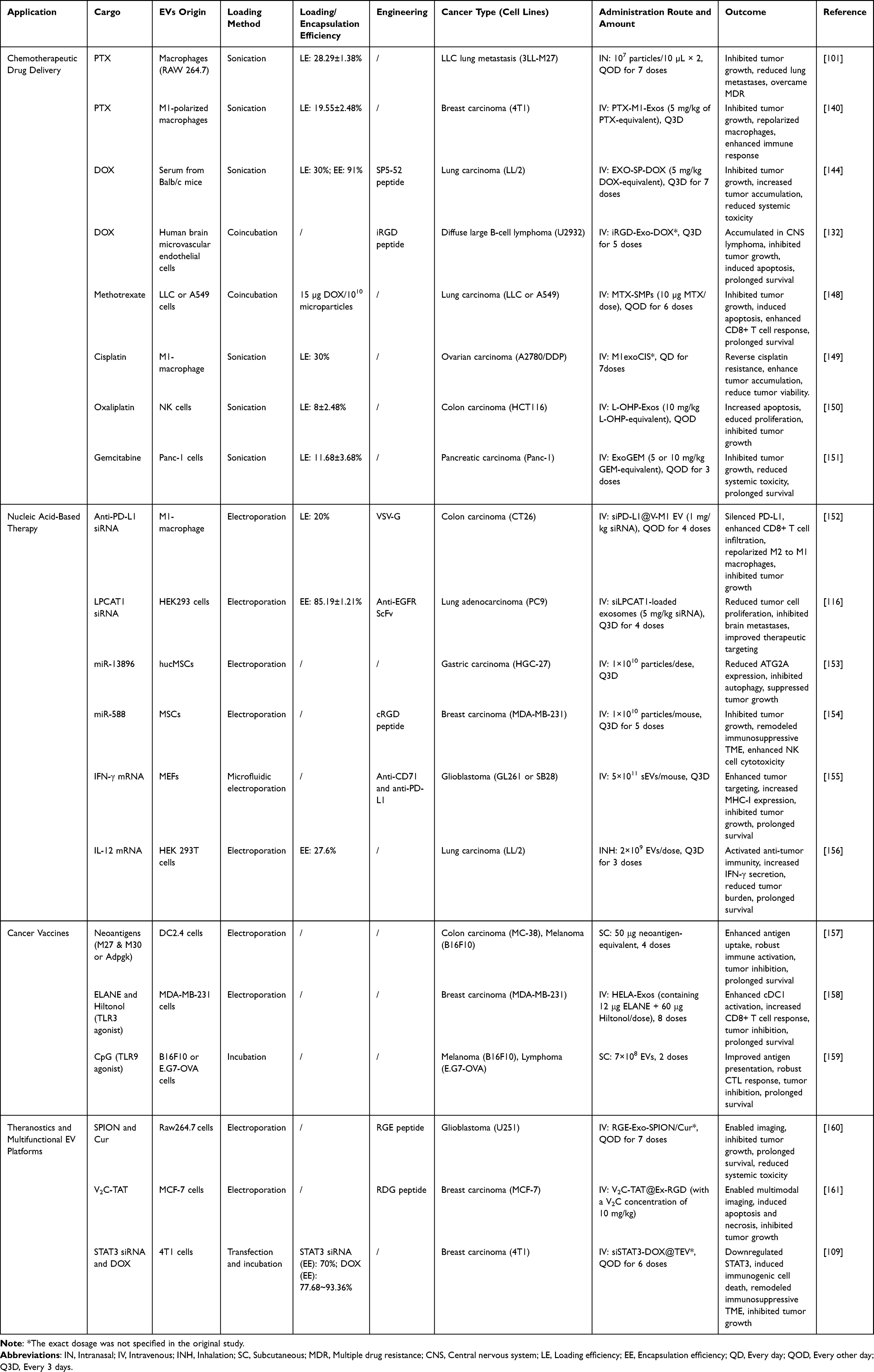 |
Table 4 Application of Engineered EVs in Cancer Therapy |
Nucleic Acid–Based Therapy
Delivering therapeutic nucleic acids to tumors remains challenging because of their instability and poor cellular uptake.162 Free nucleic acids are rapidly degraded by nucleases and have difficulty crossing cell membranes.163 These challenges can be addressed using EVs, which possess a protective lipid bilayer shell and innate targeting capabilities. EV encapsulation shields nucleic acids from enzymatic degradation in the bloodstream, prolonging their circulation and preserving bioactivity until delivery to target cells.164,165 Moreover, EV membranes naturally fuse with recipient cells, facilitating efficient cytosolic delivery of the cargo. In the following subsections, we discuss engineered EV strategies for delivering various classes of nucleic acid therapeutics.
siRNA
siRNA selectively silences disease-associated genes by promoting target mRNA degradation.166 The primary challenge is delivering siRNA intact into tumor cells. EVs have emerged as effective siRNA carriers capable of crossing biological barriers and selectively targeting tumors. For example, Liu et al loaded anti-PD-L1 siRNA (siPD-L1) into M1 macrophage–derived EVs alongside a vesicular stomatitis virus G protein “pseudotype” to enhance EV uptake.152 These engineered EVs successfully delivered siPD-L1 to colon carcinoma tumors, silencing PD-L1 expression in cancer and immune cells. The result was a repolarization of tumor-associated macrophages from an M2 (immunosuppressive) to an M1 (proinflammatory) phenotype and increased CD8+ T-cell infiltration, resulting in potent antitumor immune responses and tumor growth inhibition. In a recent study, Jiang et al targeted a metastasis-associated gene in lung cancer.116 They electroporated an siRNA against LPCAT1 (an oncogenic factor promoting NSCLC progression) into HEK293T-derived exosomes decorated with an anti-EGFR nanobody for tumor targeting. Systemic administration of these exosomes in an orthotopic lung cancer model knocked down LPCAT1 in vivo, thereby suppressing tumor proliferation and substantially reducing brain metastatic lesions.116 These examples showcase the precision of EV–siRNA strategies. By combining cell type–specific EVs with surface ligands and therapeutic siRNAs, gene silencing has been achieved in tumors that could not be reached with free siRNA.
miRNA
miRNAs are short noncoding RNA molecules that participate in posttranscriptional gene regulation through mRNA cleavage or translational repression.167 Recently, EV-delivered miRNAs have been found to exhibit significant antitumor effects by modulating the TME or directly inhibiting neoplastic cell proliferation. In one study, human umbilical cord MSC-derived EVs were loaded with miR-138-5p (a tumor suppressor) via electroporation.153 When injected into gastric cancer models, these EVs transferred miR-138 to the tumor cells, resulting in the downregulation of ATG2A (a gene involved in autophagy) and inhibition of tumor growth and metastasis. Moreover, the treated tumors exhibited increased apoptosis and reduced prosurvival autophagy signaling, indicating that the miRNA successfully reprogrammed tumor cell fate.153 In another example, Zhang et al identified miR-588 as an miRNA that can overcome immune evasion in TNBC.154 They loaded miR-588 into MSC-derived exosomes and decorated the exosome surface with cRGD peptides. Systemic administration of these exosomes resulted in preferential uptake by tumors and significant remodeling of the tumor immune microenvironment. The miR-588 cargo downregulated the immunosuppressive chemokine CCL5 in tumors, leading to enhanced recruitment and activation of NK cells and cytotoxic T lymphocytes (CTL). Tumor growth and metastasis were reduced in treated mice, indicating that EV-mediated miRNA delivery can facilitate direct tumor cell attack and boost antitumor immunity.154 Collectively, these findings demonstrate the versatility of EVs in delivering therapeutic miRNAs. EVs shield miRNAs from degradation, improve their pharmacokinetics, and enable targeted tumor accumulation, achieving functional gene modulation in vivo that is challenging with naked miRNA.
mRNA
mRNA therapy has gained momentum, and EVs are being explored as natural mRNA delivery vehicles. The large size and fragility of mRNA limit its therapeutic application. However, EVs can protect mRNA and facilitate cytoplasmic uptake in target cells.168,169 Dong et al engineered EVs to deliver mRNA encoding interferon-γ (IFN-γ) for cancer immunotherapy.155 They developed a recombinant protein anchor that displayed tumor-specific antibodies on the EV surface, thereby directing the mRNA-loaded EVs to glioblastoma cells overexpressing a particular antigen. Upon administration to tumor-bearing mice, these targeted EVs successfully translated IFN-γ within the tumor tissue, reprogramming the TME toward an immune-active state and significantly sensitizing the tumors to checkpoint inhibitor therapy.155 Liu et al developed an inhalable EV formulation delivering IL-12 mRNA for lung cancer treatment. The IL-12 EV therapy induced a robust local and systemic immune response characterized by elevated IFN-γ levels and activation of cytotoxic lymphocytes, leading to tumor regression and prolonged survival in mice.156 This approach minimized systemic exposure and toxicity, underscoring the advantage of tissue-targeted EV delivery. Overall, EV-based mRNA delivery is emerging as a promising alternative to synthetic lipid nanoparticles, with early successes in expressing cytokines, tumor suppressors, or suicide genes directly in tumors. The ability to target EVs to specific tissues via surface modification, along with their natural membrane fusion that avoids endosomal trapping, gives EVs an advantage in achieving functional mRNA protein expression at the tumor site.
Cancer Vaccines
EVs have emerged as promising delivery platforms of cancer vaccines because of their intrinsic lymphotropism and immunomodulatory capabilities.170 Previous studies have shown that EV-based vaccine formulations selectively accumulate in draining lymphoid tissues and undergo efficient internalization by APCs, significantly enhancing the antigen presentation efficiency.171,172 Moreover, EVs inherently incorporate immunostimulatory molecular constituents derived from their parental cells, effectively promoting DC maturation and providing essential costimulatory signals required for T lymphocyte activation.173 This intrinsic adjuvant functionality amplifies APC activation and subsequent proinflammatory cytokine secretion, thereby optimizing antigen presentation mechanisms and facilitating the generation of tumor-specific T-cell responses with enhanced immunological efficacy. To date, research has primarily focused on DC-derived EVs (DC-EVs) and tumor cell–derived EVs (T-EVs).174
DC-EVs for Cancer Vaccination
Traditional DC vaccines are limited by their complex preparation and high production costs as well as challenges in long-term live-cell preservation.18 In contrast, DC-EVs offer greater storage stability and manufacturing control while retaining key immunostimulatory components of their parent DCs.175 Notably, DC-EVs naturally carry functional major histocompatibility complex class I and II molecules and costimulatory proteins (eg, CD80 and CD86) on their surface.175 These properties enable EVs to directly present antigens and activate T cells in a manner similar to intact DCs. Li et al used DC-EVs as tumor neoantigen carriers for personalized cancer immunotherapy, efficiently delivering tumor-specific antigens to lymph nodes while inducing strong T-cell and B-cell immune responses.157 Moreover, this nanovaccine significantly inhibited tumor growth and prolonged survival in B16F10 melanoma and MC-38 CRC mouse models, exhibiting superior antitumor, antimetastatic, and antirecurrence effects with enhanced immunogenicity compared with liposomal formulations.157 Early-phase clinical trials have also been conducted with DC-derived exosomes. In patients with metastatic melanoma and non-small cell lung cancer, autologous DC-EVs pulsed with tumor-associated antigens were administered as a maintenance immunotherapy.176–178 These trials demonstrated that DC-EV vaccines are safe, well tolerated, and capable of inducing antigen-specific immune responses with no serious adverse events. However, they showed limited clinical tumor regression, suggesting the need for further enhancements.
T-EVs for Cancer Vaccination
T-EVs are an attractive vaccine platform because they carry a natural cargo of tumor antigens that reflect the antigenic repertoire of the source cancer.179 A single T-EV can present multiple tumor-associated antigens simultaneously, offering a broader immune target and potentially addressing intratumoral heterogeneity better than a single-peptide vaccine.180 Moreover, T-EVs often include endogenous adjuvant molecules (eg, Hsp70) from stressed tumor cells, enhancing their inherent immunogenicity. Early studies have revealed that unmodified T-EVs can naturally promote antitumor immunity. For instance, EVs isolated from lung cancer cells were efficiently captured by host dendritic cells in vivo, leading to the activation of both CD4+ and CD8+ T cells and a measurable antitumor effect.181 Building on this, T-EVs are currently being engineered to further enhance their vaccine efficacy. One approach involves loading additional immunostimulatory agents into T-EVs. Huang et al engineered breast cancer–derived EVs (HELA-Exos) by genetically overexpressing α-lactalbumin and incorporating the immunogenic cell death (ICD)-inducing agents ELANE and Hiltonol (a TLR3 agonist) into the vesicles.158 HELA-Exos demonstrated breast cancer cell–specific targeting and induced potent ICD coupled with enhanced cDC1 activation. This engineered system strongly primed tumor-specific CD8+ T cells, achieving significant tumor suppression in mouse TNBC models and patient-derived tumor organoids.158 Bhatta et al loaded T-EVs with the TLR9 agonist CpG, which significantly enhanced DC activation, antigen presentation, and cytotoxic T lymphocyte responses, yielding 80% and 33% tumor-free survival against E.G7 lymphoma and B16F10 melanoma, respectively.159
Theranostics and Multifunctional EV Platforms
Developing theranostic EVs, which combine therapeutic and diagnostic functions in a single platform, is an exciting frontier in EV research.182 Because of their versatile cargo capacity and modifiable surface, engineered EVs can serve as multifunctional nanoparticles for cancer therapy while enabling treatment monitoring or providing imaging contrast. In cancer therapy, this indicates that an EV can deliver a drug while simultaneously carrying a reporter (eg, a fluorescent tag or radionuclide) for imaging or it can transport therapeutic and diagnostic probes to the tumor.182–184
Several proof-of-concept studies have demonstrated the theranostic potential of EVs. For example, superparamagnetic iron oxide nanoparticles (SPIONs) and therapeutic agents have been introduced into exosomal structures to generate vesicles that can be traced via magnetic resonance imaging (MRI) or photoacoustic imaging during drug delivery.185,186 In a previous study, exosomes were engineered to carry curcumin and SPIONS. In addition to curcumin-mediated chemotherapy, these constructs enabled the MRI of glioma sites and induced magnetic fluid hyperthermia under an alternating magnetic field, producing a synergistic tumor-killing effect.160 Another novel approach involved loading exosomes with nucleus-targeting TAT-modified vanadium carbide quantum dots. These constructs enabled multimodal tumor imaging (eg, fluorescence imaging, photoacoustic imaging, and MRI) and provided low-temperature photothermal therapy when exposed to near-infrared laser light, thereby achieving a synergistic tumor-killing effect through nucleus-targeted thermal ablation.161 This all-in-one system highlights the potential of theranostics: enabling tumor imaging, therapy, and real-time response monitoring with a same agent.
In addition, multifunctional EVs can carry different combinations of therapeutic agents for multimodal therapy. An EV can simultaneously deliver a chemotherapeutic drug and an siRNA, targeting cancer on two fronts. Peng et al developed a bifunctional therapeutic by loading STAT3 siRNA and DOX into T-EVs. In this system, siRNA-mediated STAT3 silencing reversed immunosuppression and promoted immune activation, whereas chemotherapy via DOX synergistically amplified the antitumor efficacy.109
Clinical Translation and Challenges
Current Trials and Translational Status
The therapeutic potential of engineered EVs has advanced from preclinical models to early-stage clinical trials (Table 5). Several trials have explored EV-based cancer therapies, establishing their initial safety and feasibility. In one notable Phase I study, autologous tumor-derived exosomes combined with granulocyte macrophage colony-stimulating factor (GM-CSF) were used as a vaccine for CRC.187 In this trial, exosomes isolated from malignant ascites of patients, which are rich in tumor antigens, were administered along with an immune adjuvant (GM-CSF). This approach was well tolerated with no serious adverse effects. Moreover, patients who received exosomes + GM-CSF exhibited tumor-specific T-cell responses, whereas those who received exosomes alone did not.187 This provided a clinical proof-of-concept that exosome-based immunotherapy can safely induce antitumor immunity in humans. Similarly, early trials of DC-derived exosomes pulsed with tumor antigens in melanoma and non-small-cell lung cancer demonstrated favorable safety profiles.177,178 In a phase I study of advanced lung cancer, patients were vaccinated with autologous DC-derived exosomes loaded with MAGE tumor antigens. The treatment was well tolerated (only mild grade 1–2 reactions) and resulted in detectable immune responses in some patients.177 These pioneering trials confirmed that EV-based therapies are safe and provided preliminary evidence of their immune efficacy, albeit with modest clinical outcomes, laying the groundwork for interventions using next-generation engineered EVs.
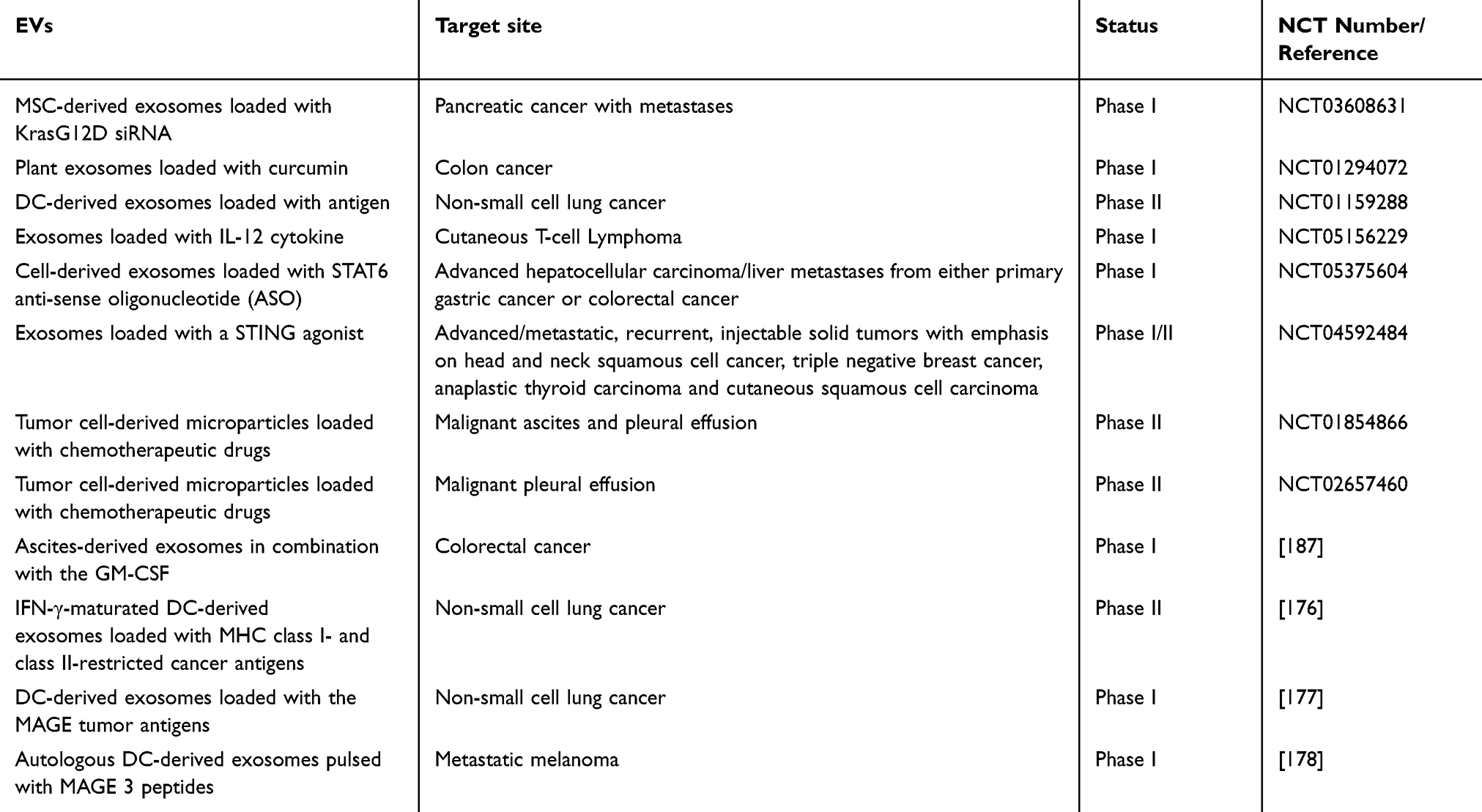 |
Table 5 Clinical Trials of EV-Based Therapeutics (Data from ClinicalTrials.gov and Published References) |
Building on these early efforts, more advanced engineered EV therapeutics have recently entered the clinical pipeline. The development of iExosomes (exosomes engineered to carry siRNA against oncogenic KRASG12D) for the treatment of metastatic pancreatic cancer is a prominent example.54 Preclinical studies in PDAC mouse models have shown that iExosomes can specifically target KRASG12D-mutant cancer cells and significantly suppress tumor growth while prolonging survival.54 These promising results led to a phase I clinical trial (NCT03608631) evaluating iExosomes derived from MSCs in individuals with pancreatic cancer harboring KRASG12D mutations. Interim reports from this trial indicate that iExosome therapy is feasible, safe, and well tolerated in humans, with some treated patients showing reduced circulating KRASG12D DNA and stable disease.188 Although detailed efficacy results are pending, this first-in-human trial targeting a mutant oncoprotein with exosomal siRNA is a milestone for EV-based gene therapy in cancer.
In addition to academic efforts, biotech companies have been actively translating engineered EVs to clinical applications. For example, Codiak BioSciences has developed a surface-engineered exosome platform for cancer immunotherapy. Their product exoIL-12, which displays the IL-12 cytokine on the exosome surface, was evaluated in a phase I trial for cutaneous T-cell lymphoma, aiming to concentrate immunostimulatory IL-12 within tumors while minimizing systemic exposure.189 Another candidate, exoSTING—which comprises exosomes loaded with a STING agonist to activate immune cells in the TME—has completed initial clinical testing in patients with solid tumors.190 Moreover, a first-in-human trial of exoASO-STAT6—exosomes that deliver an antisense oligonucleotide to inhibit STAT6 in tumors—was conducted on patients with advanced HCC.191 These industry-driven trials underscore the growing clinical interest in EV-based therapeutics. Although no EV product has yet received regulatory approval, the initiation and completion of multiple phase I studies indicate rapid progress in translating EV research into potential cancer treatments. The insights gained from these trials, particularly regarding safety, dosing, and manufacturing, will be invaluable for guiding subsequent larger efficacy studies.
Overall, EV-based cancer therapeutics remain in early-stage development; however, their progress is accelerating. First-in-human studies have demonstrated their feasibility, acceptable safety, and early biological activity. Emerging regulatory engagement was demonstrated by the FDA’s clearance of the first investigational new drug application for an unmodified neural EV therapy (AB126) in early 2024.192 These developments indicate the growing acceptance of EVs as a viable therapeutic class. However, translating this momentum into reliable patient benefit will require overcoming persistent challenges to achieve consistent efficacy and support broad clinical use.
Challenges and Potential Solutions
Despite encouraging early findings, translating engineered EVs into the clinic still requires addressing a series of interconnected challenges along the bench-to-bedside path. Transforming research prototypes into patient-ready products requires stricter standards, reproducible workflows, and robust evidence reflecting real-world use. The following subsections provide this context and prospective solutions.
Delivery Efficiency and Targeting
A fundamental challenge is ensuring that EVs efficiently deliver their therapeutic payload to tumor cells in vivo. Natural EVs do not inherently target tumors. After systemic administration, a significant fraction of EVs can be sequestered by the mononuclear phagocyte system or taken up by nontarget cells.82,193–196 This can limit the fraction of the injected EV dose that reaches the tumor. To overcome this issue, targeting moieties can be engineered onto EV surfaces. Several approaches have been shown to enhance uptake by cancer cells while reducing off-target clearance, including displaying tumor-homing peptides or antibody fragments on EV membranes.197 Similarly, decorating EVs with CD47—the “don’t eat me” signal—can help them evade macrophage-mediated phagocytic clearance, prolonging their circulation time. These modifications, along with optimized dosing routes (eg, intratumoral or regional administration when feasible), are being explored to improve delivery efficiency.31,75
In vivo Biodistribution and Safety
Controlling EV biodistribution and assessing safety are challenges closely associated with effective targeting. Studies tracking administered EVs in animal models have shown that EVs tend to accumulate in the liver, spleen, lungs, and kidneys, irrespective of their cellular origin.195,196 Although some sequestration is inevitable, excessive deposition in healthy tissues increases off-target risk, highlighting the need for comprehensive biodistribution and toxicology assessments. Practical mitigation strategies include minimizing vesicle load via high payload-per-EV formulations, selecting administration routes that limit systemic exposure, “humanizing” membranes to reduce innate clearance, and employing stimuli-responsive architectures that release cargo within the TME (eg, pH- or enzyme-triggered), thereby sparing normal tissues.
For universal (allogeneic) EVs, immunogenicity is a critical factor. Clinical experience shows that autologous EVs are generally well tolerated, and early allogeneic platforms (eg, MSC-derived products, iExosomes) demonstrate acceptable short-term safety; however, rigorous repeated-dose immunogenicity assessment remains essential for regulatory approval.177,187,188 Allogeneic vesicles present donor antigens that can trigger host recognition, accelerate clearance, and elicit anti-EV antibodies upon repeated dosing.198 To mitigate immunosurveillance, developers have combined source selection (eg, low-immunogenic or immunomodulatory MSCs), genome engineering (downregulating HLA or other epitopes to yield hypoimmunogenic producers), and surface engineering (displaying CD47 “don’t eat me” cues).198 When integrated judiciously, these approaches are expected to enable off-the-shelf allogeneic EV therapeutics with safety profiles approaching those of autologous products while maintaining on-target efficacy.
Manufacturing Processes and Scalable Production
One of the most significant translational challenges in the use of EVs is the production of therapeutic EVs at scale and with consistent quality. As cells naturally release EVs in relatively small quantities, it may be difficult to isolate sufficient EVs for human dosing. Moreover, conventional ultracentrifugation methods are labor-intensive and difficult to standardize, potentially resulting in batch-to-batch variability.31,199 To address this challenge, new biomanufacturing techniques (eg, culturing cells in bioreactors with high yield outputs, using tangential flow filtration or size-exclusion chromatography for scalable purification, and even inducing cells to secrete more EVs) are under development.200–203 Ensuring scalability also involves developing stringent quality control measures (ie, quantifying the EV particle concentration, size distribution, cargo load, and functional potency for each batch). Collaborations among academia, industry, and regulatory bodies are beginning to establish standardized manufacturing protocols for EVs, which will be crucial for successful commercialization.
In addition to scale-up, long-term preservation is critical to maintain EV stability and potency during storage and distribution. Ultra-low temperatures (−80°C or liquid nitrogen) better preserve particle number, size, and bioactivity than −20 or 4°C; however, repeated freeze–thaw cycles induce aggregation/fusion and functional loss.204 To address these limitations, lyophilization converts EVs into a dry powder and, with appropriate lyoprotectants (eg, trehalose), allows reconstitution while preserving vesicle structure and function.205 In parallel, optimized storage buffers are essential. Geng et al demonstrated that supplementing phosphate-buffered saline with human serum albumin and trehalose substantially improved the short- and long-term stability of EVs at −80°C, facilitating much higher particle recovery after storage and fewer changes in vesicle size or uptake capacity.206 Incorporating optimized buffers, careful cryopreservation, and lyophilization into manufacturing process will extend shelf life and ensure consistent performance.
Regulatory and Standardization Hurdles
The regulatory landscape for EV-based therapeutics continues to evolve, posing a challenge for clinical translation. EV products do not clearly fall into the conventional categories of drugs, biologics, or cell therapies. Moreover, their complex composition makes them difficult to standardize.207 The variations in EV isolation and engineering techniques across research groups result in heterogeneous vesicle preparations, which differ in purity, subtype (eg, exosomes vs microvesicles), and molecular cargo. This fragmentation complicates the development of clear regulatory guidelines. Compared with small-molecule drugs, demonstrating how an EV delivers its cargo and is cleared by the body is more challenging. To advance the field, consensus standards—such as those from the International Society for Extracellular Vesicles—are being developed for characterizing EVs, including defining identity, purity, potency, and safety assays.208,209 In addition, robust assays to measure the critical quality attributes of EVs—such as size, charge, membrane protein markers, and nucleic acid content—are being developed. Moreover, regulatory agencies will likely require consistency in manufacturing processes to maintain reproducibility between batches. An ongoing dialogue between researchers and regulators is essential to address these issues.
Conclusions and Prospects
The Promise and Challenge of EV-Based Therapeutics
Engineered EVs have rapidly become one of the most innovative and versatile nanocarrier systems for cancer therapy. Based on the literature reviewed herein, EV-based delivery systems exhibit innate biocompatibility and intercellular communication while integrating the design flexibility and functional tunability of advanced nanomedicine platforms. The key findings show that EVs can outperform conventional synthetic nanoparticles in targeting tumors, penetrating biological barriers, and delivering complex therapeutic cargos (eg, RNAs and proteins) with minimal toxicity when appropriately engineered. Major applications are already demonstrating success in preclinical models. EVs loaded with chemotherapeutics achieve higher tumor cell eradication with reduced side effects. EV shuttles can deliver gene silencers deep into tumor tissue, and EV-based vaccines can effectively mobilize the immune system against cancer. These achievements underscore the distinct advantages of engineered EVs: they are inherently stealthy in the bloodstream, can be equipped with targeting ligands for precise homing, and can carry multimodal payloads that conventional vectors have difficulty accommodating. Therefore, establishing an EV-specific, screening-focused discovery pipeline is both feasible and timely. Using standardized, scalable EV workflows and identity/potency assays, a minimal, reproducible process can directly compare encapsulated versus free-form performance while capturing essential pharmacokinetic, pharmacodynamic, and safety readouts. This shared framework converts heterogeneous preclinical results into comparable, regulator-relevant evidence and supports scale-up decisions and regulatory interactions.
The clinical translation of engineered EVs remains incomplete, with several challenges necessitating continued innovation and standardization. These include ensuring batch-to-batch consistency, scaling production processes, and meeting regulatory standards. The forthcoming clinical trial data will be pivotal in determining the pharmacological behavior of engineered EVs in humans and assessing whether the antitumor efficacy observed in animal models is reproducible in patients. These uncertainties highlight the need for disciplined standardization and generation of comparative evidence to support reliable clinical development.
Future Outlook
Looking forward, continued advances in EV engineering are anticipated to expand the therapeutic potential of these vesicles in cancer therapy. Bioengineering strategies are advancing on multiple fronts. Surface functionalization enables EV to display tumor-targeting peptides, antibodies, or aptamers, enhancing targeting specificity. In parallel, hybrid delivery systems are gaining momentum. EV–liposome constructs, formed by fusing native EV membranes with synthetic liposomal cores, combine biological interface of EVs with tunable physicochemical properties and stability of liposomes.210,211 For example, one study engineered cancer cell–derived EVs fused with liposomes, achieving markedly higher drug loading and tumor-targeting efficiency than either EVs or liposomes alone.212 There is also increasing interest in EV mimetics (cell-derived nanovesicles produced using extrusion or other methods), which could provide higher yields while retaining their key biofunctional properties.213 In addition, alternative sources of EVs (eg, plant-derived vesicles) are being explored to overcome scalability limitations.214,215 Multifunctional EV platforms capable of codelivering drugs, genes, and immunostimulators are expected to expand, enabling synergistic targeting of tumors through multiple mechanisms.
Artificial intelligence (AI) and automated manufacturing are transforming EV engineering. Machine learning algorithms can analyze large EV datasets to predict biodistribution and guide the rational design of EV surface and cargo properties.216 Future workflows may employ AI-driven models to optimize EV design parameters and tailor therapies to individual patients. On the manufacturing side, scalable, GMP-compliant systems are advancing. For example, perfusion bioreactors and closed microfluidic reactors now allow high-yield EV production under controlled conditions.207 Concurrently, regulatory frameworks are evolving to accommodate EV therapeutics, with global regulatory agencies aligning exosome regulations with established biologics standards, including requirements for rigorous characterization, quality control, and safety testing.207 Collectively, these developments are poised to shape the next era of EV-based oncology therapeutics.
Funding
This work was supported by the Natural Science Foundation of Fujian Province (Grant number: 2023J011260), Joint Funds for the Innovation of Science and Technology, Fujian Province of China (Grant number: 2023Y9396) and Scientific Research Foundation of Fujian Cancer Hospital (Grant number: 2023YN09).
Disclosure
The authors report no conflicts of interest in this work.
References
1. Bray F, Laversanne M, Sung HYA, et al. Global cancer statistics 2022: GLOBOCAN estimates of incidence and mortality worldwide for 36 cancers in 185 countries. CA Cancer J Clin. 2024;74(3):229–263. doi:10.3322/caac.21834
2. Sonkin D, Thomas A, Teicher BA. Cancer treatments: past, present, and future. Cancer Genet. 2024;286:18–24. doi:10.1016/j.cancergen.2024.06.002
3. Peer D, Karp JM, Hong S, Farokhzad OC, Margalit R, Langer R. Nanocarriers as an emerging platform for cancer therapy. Nat Nanotechnol. 2007;2(12):751–760. doi:10.1038/nnano.2007.387
4. Yao YH, Zhou YX, Liu LH, et al. Nanoparticle-based drug delivery in cancer therapy and its role in overcoming drug resistance. Front Mol Biosci. 2020;7:193. doi:10.3389/fmolb.2020.00193
5. Mitchell MJ, Billingsley MM, Haley RM, Wechsler ME, Peppas NA, Langer R. Engineering precision nanoparticles for drug delivery. Nat Rev Drug Discov. 2021;20(2):101–124. doi:10.1038/s41573-020-0090-8
6. Huang XZ, He T, Liang XQ, et al. Advances and applications of nanoparticles in cancer therapy. Medcomm-Oncology. 2024;3(1):e67. doi:10.1002/mog2.67
7. Wu C, Almuaalemi HYM. Synthesis of MOFs and characterization and drug loading efficiency. ChemEngineering. 2025;9(2):24. doi:10.3390/chemengineering9020024
8. Demissie GG, Chen YC, Ciou SY, et al. Hypoxia-Targeted-Therapy: mussel-inspired hollow polydopamine nanocarrier containing MoS2 nanozyme and tirapazamine with anti-angiogenesis property for synergistic tumor therapy. J Colloid Interf Sci. 2025;685:396–414. doi:10.1016/j.jcis.2025.01.149
9. Dinarvand R, Sepehri N, Manoochehri S, Rouhani H, Atyabi F. Polylactide-co-glycolide nanoparticles for controlled delivery of anticancer agents. Int J Nanomed. 2011;6:877–895. doi:10.2147/IJN.S18905
10. Blanco E, Shen H, Ferrari M. Principles of nanoparticle design for overcoming biological barriers to drug delivery. Nat Biotechnol. 2015;33(9):941–951. doi:10.1038/nbt.3330
11. Zelepukin IV, Shevchenko KG, Deyev SM. Rediscovery of mononuclear phagocyte system blockade for nanoparticle drug delivery. Nat Commun. 2024;15(1):4366. doi:10.1038/s41467-024-48838-5
12. Aljabali AA, Obeid MA, Bashatwah RM, et al. Nanomaterials and their impact on the immune system. Int J Mol Sci. 2023;24(3):2008. doi:10.3390/ijms24032008
13. Rennick JJ, Johnston APR, Parton RG. Key principles and methods for studying the endocytosis of biological and nanoparticle therapeutics. Nat Nanotechnol. 2021;16(3):266–276. doi:10.1038/s41565-021-00858-8
14. Luo RH, Liu MM, Tan TT, et al. Emerging significance and therapeutic potential of extracellular vesicles. Int J Biol Sci. 2021;17(10):2476–2486. doi:10.7150/ijbs.59296
15. Peng H, Ji WH, Zhao RC, et al. Exosome: a significant nano-scale drug delivery carrier. J Mater Chem B. 2020;8(34):7591–7608. doi:10.1039/d0tb01499k
16. Chen HR, Yao HB, Chi JX, et al. Engineered exosomes as drug and RNA co-delivery system: new hope for enhanced therapeutics? Front Bioeng Biotech. 2023;11:1254356. doi:10.3389/fbioe.2023.1254356
17. Song G, Zeng CL, Li JR, et al. Exosome-based nanomedicines for digestive system tumors therapy. Nanomedicine. 2025;20(10):1167–1180. doi:10.1080/17435889.2025.2493037
18. Yong TY, Wei ZH, Gan L, Yang XL. Extracellular-vesicle-based drug delivery systems for enhanced antitumor therapies through modulating the cancer-immunity cycle. Adv Mater. 2022;34(52):e2201054. doi:10.1002/adma.202201054
19. Viaud S, Théry C, Ploix S, et al. Dendritic cell-derived exosomes for cancer immunotherapy: what’s next? Cancer Res. 2010;70(4):1281–1285. doi:10.1158/0008-5472.CAN-09-3276
20. Kim H, Park HJ, Chang HW, et al. Exosome-guided direct reprogramming of tumor-associated macrophages from protumorigenic to antitumorigenic to fight cancer. Bioact Mater. 2023;25:527–540. doi:10.1016/j.bioactmat.2022.07.021
21. Jin XY, Zhang J, Zhang YF, et al. Different origin-derived exosomes and their clinical advantages in cancer therapy. Front Immunol. 2024;15:1401852. doi:10.3389/fimmu.2024.1401852
22. Weng ZJ, Zhang BW, Wu CZ, et al. Therapeutic roles of mesenchymal stem cell-derived extracellular vesicles in cancer. J Hematol Oncol. 2021;14(1):136. doi:10.1186/s13045-021-01141-y
23. Liu Y, Cao XT. Organotropic metastasis: role of tumor exosomes. Cell Res. 2016;26(2):149–150. doi:10.1038/cr.2015.153
24. Chiangjong W, Netsirisawan P, Hongeng S, Chutipongtanate S. Red blood cell extracellular vesicle-based drug delivery: challenges and opportunities. Front Med. 2021;8:761362. doi:10.3389/fmed.2021.761362
25. Wang CC, Li N, Li YT, et al. Engineering a HEK-293T exosome-based delivery platform for efficient tumor-targeting chemotherapy/internal irradiation combination therapy. J Nanobiotechnol. 2022;20(1):247. doi:10.1186/s12951-022-01462-1
26. Arya SB, Collie SP, Parent CA. The ins-and-outs of exosome biogenesis, secretion, and internalization. Trends Cell Biol. 2024;34(2):90–108. doi:10.1016/j.tcb.2023.06.006
27. van Niel G, D’Angelo G, Raposo G. Shedding light on the cell biology of extracellular vesicles. Nat Rev Mol Cell Bio. 2018;19(4):213–228. doi:10.1038/nrm.2017.125
28. Rädler J, Gupta D, Zickler A, El Andaloussi S. Exploiting the biogenesis of extracellular vesicles for bioengineering and therapeutic cargo loading. Mol Ther. 2023;31(5):1231–1250. doi:10.1016/j.ymthe.2023.02.013
29. Villarroya-Beltri C, Baixauli F, Gutiérrez-Vázquez C, Sánchez-Madrid F, Mittelbrunn M. Sorting it out: regulation of exosome loading. Semin Cancer Biol. 2014;28:3–13. doi:10.1016/j.semcancer.2014.04.009
30. Pitt JM, Kroemer G, Zitvogel L. Extracellular vesicles: masters of intercellular communication and potential clinical interventions. J Clin Invest. 2016;126(4):1139–1143. doi:10.1172/JCI87316
31. Palakurthi SS, Shah BJ, Kapre S, et al. A comprehensive review of challenges and advances in exosome-based drug delivery systems. Nanoscale Adv. 2024;6(23):5803–5826. doi:10.1039/d4na00501e
32. Huang R, Zhu J, Fan R, et al. Extracellular vesicle-based drug delivery systems in cancer. Extracell Vesicle. 2024;4:100053. doi:10.1016/j.vesic.2024.100053
33. Liu YJ, Wang C. A review of the regulatory mechanisms of extracellular vesicles-mediated intercellular communication. Cell Commun Signal. 2023;21(1):77. doi:10.1186/s12964-023-01103-6
34. Hoshino A, Costa-Silva B, Shen TL, et al. Tumour exosome integrins determine organotropic metastasis. Nature. 2015;527(7578):329–335. doi:10.1038/nature15756
35. Hu MY, Kenific CM, Boudreau N, Lyden D. Tumor-derived nanoseeds condition the soil for metastatic organotropism. Semin Cancer Biol. 2023;93:70–82. doi:10.1016/j.semcancer.2023.05.003
36. Kalluri R, LeBleu VS. The biology, function, and biomedical applications of exosomes. Science. 2020;367(6478):eaau6977. doi:10.1126/science.aau6977
37. Han QF, Li WJ, Hu KS, et al. Exosome biogenesis: machinery, regulation, and therapeutic implications in cancer. Mol Cancer. 2022;21(1):207. doi:10.1186/s12943-022-01671-0
38. de Jong OG, Kooijmans SAA, Murphy DE, et al. Drug delivery with extracellular vesicles: from imagination to innovation. Acc Chem Res. 2019;52(7):1761–1770. doi:10.1021/acs.accounts.9b00109
39. Shami-shah A, Travis BG, Walt DR. Advances in extracellular vesicle isolation methods: a path towards cell-type specific EV isolation. Extracell Vesicles Circ Nucleic Acids. 2023;4(3):447–460. doi:10.20517/evcna.2023.14
40. De Sousa KP, Rossi I, Abdullahi M, Ramirez MI, Stratton D, Inal JM. Isolation and characterization of extracellular vesicles and future directions in diagnosis and therapy. Wiley Interdiscip Rev. 2023;15(1):e1835. doi:10.1002/wnan.1835
41. Jiang Y, Wang FB, Wang K, et al. Engineered exosomes: a promising drug delivery strategy for brain diseases. Curr Med Chem. 2022;29(17):3111–3124. doi:10.2174/0929867328666210902142015
42. Bagci C, Sever-Bahcekapili M, Belder N, Bennett APS, Erdener SE, Dalkara T. Overview of extracellular vesicle characterization techniques and introduction to combined reflectance and fluorescence confocal microscopy to distinguish extracellular vesicle subpopulations. Neurophotonics. 2022;9(2):021903. doi:10.1117/1.NPh.9.2.021903
43. Brennan K, Martin K, FitzGerald SP, et al. A comparison of methods for the isolation and separation of extracellular vesicles from protein and lipid particles in human serum. Sci Rep. 2020;10(1):1039. doi:10.1038/s41598-020-57497-7
44. Lobb RJ, Becker M, Wen SW, et al. Optimized exosome isolation protocol for cell culture supernatant and human plasma. J Extracell Vesicles. 2015;4:27031. doi:10.3402/jev.v4.27031
45. Rider MA, Hurwitz SN, Meckes DG. ExtraPEG: a polyethylene glycol-based method for enrichment of extracellular vesicles. Sci Rep. 2016;6:23978. doi:10.1038/srep23978
46. Chen TG, Chen D, Su WT, Liang JL, Liu XN, Cai MX. Extracellular vesicles as vital players in drug delivery: a focus on clinical disease treatment. Front Bioeng Biotech. 2025;13:1600227. doi:10.3389/fbioe.2025.1600227
47. Monguió-Tortajada M, Gálvez-Montón C, Bayes-Genis A, Roura S, Borràs FE. Extracellular vesicle isolation methods: rising impact of size-exclusion chromatography. Cell Mol Life Sci. 2019;76(12):2369–2382. doi:10.1007/s00018-019-03071-y
48. Bonner SE, van de Wakker SI, Phillips W, et al. Scalable purification of extracellular vesicles with high yield and purity using multimodal flowthrough chromatography. J Extracell Biol. 2024;3(2):e138. doi:10.1002/jex2.138
49. Mulcahy LA, Pink RC, Carter DRF. Routes and mechanisms of extracellular vesicle uptake. J Extracell Vesicles. 2014;3:24641. doi:10.3402/jev.v3.24641
50. Bader J, Brigger F, Leroux JC. Extracellular vesicles versus lipid nanoparticles for the delivery of nucleic acids. Adv Drug Deliv Rev. 2024;215:115461. doi:10.1016/j.addr.2024.115461
51. Joshi BS, de Beer MA, Giepmans BG, Zuhorn IS. Endocytosis of extracellular vesicles and release of their cargo from endosomes. ACS Nano. 2020;14(4):4444–4455. doi:10.1021/acsnano.9b10033
52. Ragni E, Taiana M. Mesenchymal stromal cells-extracellular vesicles: protein Corona as a camouflage mechanism? Extracell Vesicles Circ Nucleic Acids. 2024;5(4):722–727. doi:10.20517/evcna.2024.78
53. Wen ZW, Zhang W, Wu W. The latest applications of exosome-mediated drug delivery in anticancer therapies. Colloids Surf B Biointerfaces. 2025;249:114500. doi:10.1016/j.colsurfb.2025.114500
54. Kamerkar S, LeBleu VS, Sugimoto H, et al. Exosomes facilitate therapeutic targeting of oncogenic KRAS in pancreatic cancer. Nature. 2017;546(7659):498–503. doi:10.1038/nature22341
55. Escudé Martinez de Castilla P, Tong L, Huang C, et al. Extracellular vesicles as a drug delivery system: a systematic review of preclinical studies. Adv Drug Deliv Rev. 2021;175:113801. doi:10.1016/j.addr.2021.05.011
56. Miao GF, He YJ, Lai KR, et al. Accelerated blood clearance of PEGylated nanoparticles induced by PEG-based pharmaceutical excipients. J Control Release. 2023;363:12–26. doi:10.1016/j.jconrel.2023.09.003
57. van der Koog L, Gandek TB, Nagelkerke A. Liposomes and extracellular vesicles as drug delivery systems: a comparison of composition, pharmacokinetics, and functionalization. Adv Healthc Mater. 2022;11(5):e2100639. doi:10.1002/adhm.202100639
58. Cha M, Jeong SH, Jung JH, et al. Quantitative imaging of vesicle-protein interactions reveals close cooperation among proteins. J Extracell Vesicles. 2023;12(5):e12322. doi:10.1002/jev2.12322
59. Sun W, Jang MS, Zhan SQ, et al. Tumor-targeting and redox-responsive photo-cross-linked nanogel derived from multifunctional hyaluronic acid-lipoic acid conjugates for enhanced in vivo protein delivery. Int J Biol Macromol. 2025;314:144444. doi:10.1016/j.ijbiomac.2025.144444
60. Zhang X, Zhang HB, Gu JM, et al. Engineered extracellular vesicles for cancer therapy. Adv Mater. 2021;33(14):e2005709. doi:10.1002/adma.202005709
61. Zhang X, Long ZX, Qin ZX, et al. A detailed evaluation of the advantages among extracellular vesicles from three cell origins for targeting delivery of celastrol and treatment of glioblastoma. Int J Pharm. 2024;667:125005. doi:10.1016/j.ijpharm.2024.125005
62. Ramos-Zaldívar HM, Polakovicova I, Salas-Huenuleo E, et al. Extracellular vesicles through the blood-brain barrier: a review. Fluids Barriers Cns. 2022;19(1):60. doi:10.1186/s12987-022-00359-3
63. Wang YT, Huo YM, Zhao CY, et al. Engineered exosomes with enhanced stability and delivery efficiency for glioblastoma therapy. J Control Release. 2024;368:170–183. doi:10.1016/j.jconrel.2024.02.015
64. Wang J, Yin BH, Lian JB, Wang X. Extracellular vesicles as drug delivery system for cancer therapy. Pharmaceutics. 2024;16(8):1029. doi:10.3390/pharmaceutics16081029
65. Kim H, Jang H, Cho H, et al. Recent advances in exosome-based drug delivery for cancer therapy. Cancers. 2021;13(17):4435. doi:10.3390/cancers13174435
66. Balaraman AK, Babu MA, Moglad E, et al. Exosome-mediated delivery of CRISPR-Cas9: a revolutionary approach to cancer gene editing. Pathol Res Pract. 2025;266:155785. doi:10.1016/j.prp.2024.155785
67. Muskan M, Abeysinghe P, Cecchin R, Branscome H, Morris K, Kashanchi F. Therapeutic potential of RNA-enriched extracellular vesicles: the next generation in RNA delivery via biogenic nanoparticles. Mol Ther. 2024;32(9):2939–2949. doi:10.1016/j.ymthe.2024.02.025
68. Gharavi AT, Hanjani NA, Movahed E, Doroudian M. The role of macrophage subtypes and exosomes in immunomodulation. Cell Mol Biol Lett. 2022;27(1):83. doi:10.1186/s11658-022-00384-y
69. Pei WY, Zhang YY, Zhu XL, et al. Multitargeted immunomodulatory therapy for viral myocarditis by engineered extracellular vesicles. ACS Nano. 2024;18(4):2782–2799. doi:10.1021/acsnano.3c05847
70. Lin WQ, Shen CG, Li MJ, et al. Programmable macrophage vesicle based bionic self-adjuvanting vaccine for immunization against monkeypox virus. Adv Sci. 2025;12(1):e2408608. doi:10.1002/advs.202408608
71. Redkin T, Turubanova V. Dendritic cell-derived exosomes as anti-cancer cell-free agents: new insights into enhancing immunogenic effects. Front Immunol. 2025;16:1586892. doi:10.3389/fimmu.2025.1586892
72. Chen JB, Tan Q, Yang ZM, et al. Dendritic cell derived-extracellular vesicles engineered to express interleukin-12 and anti-CTLA-4 on their surface for combinational cancer immunotherapy. J Extracell Vesicles. 2025;14(4):e70068. doi:10.1002/jev2.70068
73. Tian JQ, Han ZP, Song DD, et al. Engineered exosome for drug delivery: recent development and clinical applications. Int J Nanomed. 2023;18:7923–7940. doi:10.2147/IJN.S444582
74. Liao W, Du Y, Zhang C, et al. Exosomes: the next generation of endogenous nanomaterials for advanced drug delivery and therapy. Acta Biomater. 2019;86:1–14. doi:10.1016/j.actbio.2018.12.045
75. Butreddy A, Kommineni N, Dudhipala N. Exosomes as naturally occurring vehicles for delivery of biopharmaceuticals: insights from drug delivery to clinical perspectives. Nanomaterials. 2021;11(6):1481. doi:10.3390/nano11061481
76. Pascucci L, Coccè V, Bonomi A, et al. Paclitaxel is incorporated by mesenchymal stromal cells and released in exosomes that inhibit in vitro tumor growth: a new approach for drug delivery. J Control Release. 2014;192:262–270. doi:10.1016/j.jconrel.2014.07.042
77. Mirsanei Z, Jamshidi-Adegani F, Vakilian S, et al. Synergistic effects of mesenchymal stem cell-derived extracellular vesicles and dexamethasone on macrophage polarization under inflammatory conditions. Inflammopharmacology. 2024;32(2):1317–1332. doi:10.1007/s10787-024-01438-7
78. Draguet F, Dubois N, Bouland C, et al. Extracellular vesicles derived from human umbilical cord mesenchymal stromal cells as an efficient nanocarrier to deliver siRNA or drug to pancreatic cancer cells. Cancers. 2023;15(11):2901. doi:10.3390/cancers15112901
79. Dilsiz N. Exosomes as new generation vehicles for drug delivery systems. J Drug Deliv Sci Tec. 2024;95:105562. doi:10.1016/j.jddst.2024.105562
80. Kanchanapally R, Deshmukh SK, Chavva SR, et al. Drug-loaded exosomal preparations from different cell types exhibit distinctive loading capability, yield, and antitumor efficacies: a comparative analysis. Int J Nanomed. 2019;14:531–541. doi:10.2147/IJN.S191313
81. Colja S, Jovčevska I, Šamec N, Romih R, Zottel A. Sonication is a suitable method for loading nanobody into glioblastoma small extracellular vesicles. Heliyon. 2023;9(5):e15674. doi:10.1016/j.heliyon.2023.e15674
82. Baruah H, Sarma A, Basak D, Das M. Exosome: from biology to drug delivery. Drug Deliv Transl Re. 2024;14(6):1480–1516. doi:10.1007/s13346-024-01515-y
83. Zhang XP, Taylor H, Valdivia A, et al. Auto-loaded TRAIL-exosomes derived from induced neural stem cells for brain cancer therapy. J Control Release. 2024;372:433–445. doi:10.1016/j.jconrel.2024.06.048
84. Kimura Y, Ohzawa H, Miyato H, et al. Intraperitoneal transfer of microRNA‐29b‐containing small extracellular vesicles can suppress peritoneal metastases of gastric cancer. Cancer Sci. 2023;114(7):2939–2950. doi:10.1111/cas.15793
85. Walker S, Busatto S, Pham A, et al. Extracellular vesicle-based drug delivery systems for cancer treatment. Theranostics. 2019;9(26):8001–8017. doi:10.7150/thno.37097
86. Nordin JZ. Transfection reagents affect Extracellular Vesicle cargo transfer to recipient cells: the importance of appropriate controls in EV research. J Extracell Vesicles. 2022;11(10):e12227. doi:10.1002/jev2.12227
87. McCann J, Sosa-Miranda CD, Guo H, et al. Contaminating transfection complexes can masquerade as small extracellular vesicles and impair their delivery of RNA. J Extracell Vesicles. 2022;11(10):e12220. doi:10.1002/jev2.12220
88. Fu W, Li T, Chen H, Zhu S, Zhou C. Research progress in exosome-based nanoscale drug carriers in tumor therapies. Front Oncol. 2022;12:919279. doi:10.3389/fonc.2022.919279
89. Araujo-Abad S, Manresa-Manresa A, Rodríguez-Cañas E, et al. Glioblastoma-derived small extracellular vesicles: nanoparticles for glioma treatment. Int J Mol Sci. 2023;24(6):5910. doi:10.3390/ijms24065910
90. Yang C, Guan Z, Pang X, et al. Desialylated mesenchymal stem cells-derived extracellular vesicles loaded with doxorubicin for targeted inhibition of hepatocellular carcinoma. Cells. 2022;11(17):2642. doi:10.3390/cells11172642
91. Lin S-W, Tsai J-C, Shyong Y-J. Drug delivery of extracellular vesicles: preparation, delivery strategies and applications. Int J Pharm. 2023;642:123185. doi:10.1016/j.ijpharm.2023.123185
92. Zhang J, Su N, Liu W, et al. An effective cell-penetrating peptide-based loading method to extracellular vesicles and enhancement in cellular delivery of drugs. Anal Bioanal Chem. 2025;417(8):1449–1459. doi:10.1007/s00216-025-05742-1
93. Wang S, Qiao C, Kong X, et al. Adhesion between EVs and tumor cells facilitated EV-encapsulated doxorubicin delivery via ICAM1. Pharmacol Res. 2024;205:107244. doi:10.1016/j.phrs.2024.107244
94. Koh HB, Kim HJ, Kang S-W, Yoo T-H. Exosome-based drug delivery: translation from bench to clinic. Pharmaceutics. 2023;15(8):2042. doi:10.3390/pharmaceutics15082042
95. Gong L, Chen Z, Feng K, et al. A versatile engineered extracellular vesicle platform simultaneously targeting and eliminating senescent stromal cells and tumor cells to promote tumor regression. J Nanobiotechnology. 2024;22(1):105. doi:10.1186/s12951-024-02361-3
96. Tian YH, Li SP, Song J, et al. A doxorubicin delivery platform using engineered natural membrane vesicle exosomes for targeted tumor therapy. Biomaterials. 2014;35(7):2383–2390. doi:10.1016/j.biomaterials.2013.11.083
97. Chen H, Wang L, Zeng X, et al. Exosomes, a New Star for Targeted Delivery. Front Cell Dev Biol. 2021;9:751079. doi:10.3389/fcell.2021.751079
98. Ducrot C, Loiseau S, Wong C, Madec E, Volatron J, Piffoux M. Hybrid extracellular vesicles for drug delivery. Cancer Lett. 2023;558:216107. doi:10.1016/j.canlet.2023.216107
99. Lennaárd AJ, Mamand DR, Wiklander RJ, El Andaloussi S, Wiklander OPB. Optimised electroporation for loading of extracellular vesicles with doxorubicin. Pharmaceutics. 2021;14(1):38. doi:10.3390/pharmaceutics14010038
100. Zeng W, Wen Z, Chen H, Duan Y. Exosomes as carriers for drug delivery in cancer therapy. Pharm Res. 2022;40(4):873–887. doi:10.1007/s11095-022-03224-y
101. Kim MS, Haney MJ, Zhao Y, et al. Development of exosome-encapsulated paclitaxel to overcome MDR in cancer cells. Nanomedicine. 2016;12(3):655–664. doi:10.1016/j.nano.2015.10.012
102. Chen M, Li Y, Ma N, Zang J. Mesenchymal stem cell‑derived exosomes loaded with 5‑Fu against cholangiocarcinoma in vitro. Mol Med Rep. 2022;25(6):213. doi:10.3892/mmr.2022.12729
103. Sun H, Bhandari K, Burrola S, Wu J, Ding W-Q. Pancreatic ductal cell-derived extracellular vesicles are effective drug carriers to enhance Paclitaxel’s efficacy in pancreatic cancer cells through clathrin-mediated endocytosis. Int J Mol Sci. 2022;23(9):4773. doi:10.3390/ijms23094773
104. Bi Y, Chen J, Li Y, et al. The chemo-immunotherapeutic roles of tumor-derived extracellular vesicle-based paclitaxel delivery system in hepatocarcinoma. Mol Pharm. 2024;21(10):5126–5137. doi:10.1021/acs.molpharmaceut.4c00514
105. Fuhrmann G, Serio A, Mazo M, Nair R, Stevens MM. Active loading into extracellular vesicles significantly improves the cellular uptake and photodynamic effect of porphyrins. J Control Release. 2015;205:35–44. doi:10.1016/j.jconrel.2014.11.029
106. Ebrahimian M, Hashemi M, Etemad L, Salmasi Z. Thymoquinone-loaded mesenchymal stem cell-derived exosome as an efficient nano-system against breast cancer cells. Iran J Basic Med Sci. 2022;25(6):723–731. doi:10.22038/IJBMS.2022.64092.14116
107. Kar R, Dhar R, Mukherjee S, et al. Exosome-based smart drug delivery tool for cancer theranostics. Acs Biomater Sci Eng. 2023;9(2):577–594. doi:10.1021/acsbiomaterials.2c01329
108. Pei X, Zhang X, Zhang L, et al. Targeted exosomes for co-delivery of siFGL1 and siTGF-β1 trigger combined cancer immunotherapy by remodeling immunosuppressive tumor microenvironment. Chem Eng J. 2021;421:129774. doi:10.1016/j.cej.2021.129774
109. Peng Z, Zhao T, Gao P, et al. Tumor-derived extracellular vesicles enable tumor tropism chemo-genetherapy for local immune activation in triple-negative breast cancer. ACS Nano. 2024;18(45):30943–30956. doi:10.1021/acsnano.3c12967
110. Yin Y, Han X, Li C, et al. The status of industrialization and development of exosomes as a drug delivery system: a review. Front Pharmacol. 2022;13:961127. doi:10.3389/fphar.2022.961127
111. Frolova L, Li ITS. Targeting capabilities of native and bioengineered extracellular vesicles for drug delivery. Bioengineering. 2022;9(10):496. doi:10.3390/bioengineering9100496
112. Ren E, Liu C, Lv P, Wang JQ, Liu G. Genetically engineered cellular membrane vesicles as tailorable shells for therapeutics. Adv Sci. 2021;8(21):e2100460. doi:10.1002/advs.202100460
113. Song HY, Chen XH, Hao YJ, Wang J, Xie QP, Wang X. Nanoengineering facilitating the target mission: targeted extracellular vesicles delivery systems design. J Nanobiotechnology. 2022;20(1):431. doi:10.1186/s12951-022-01638-9
114. Johnson V, Vasu S, Kumar US, Kumar M. Surface-engineered extracellular vesicles in cancer immunotherapy. Cancers. 2023;15(10):2838. doi:10.3390/cancers15102838
115. Gečys D, Kazlauskas A, Gečytė E, et al. Internalisation of RGD-engineered extracellular vesicles by glioblastoma cells. Biology. 2022;11(10):1483. doi:10.3390/biology11101483
116. Jiang J, Lu Y, Chu J, et al. Anti-EGFR ScFv functionalized exosomes delivering LPCAT1 specific siRNAs for inhibition of lung cancer brain metastases. J Nanobiotechnology. 2024;22(1):159. doi:10.1186/s12951-024-02414-7
117. Yang Y, Wang F, Li Y, et al. Engineered extracellular vesicles with polypeptide for targeted delivery of doxorubicin against EGFR‑positive tumors. Oncol Rep. 2024;52(5):154. doi:10.3892/or.2024.8813
118. Wiklander OPB, Mamand DR, Mohammad DK, et al. Antibody-displaying extracellular vesicles for targeted cancer therapy. Nat Biomed Eng. 2024;8(11):1453–1468. doi:10.1038/s41551-024-01214-6
119. Mohammadi AH, Ghazvinian Z, Bagheri F, Harada M, Baghaei K. Modification of extracellular vesicle surfaces: an approach for targeted drug delivery. Biodrugs. 2023;37(3):353–374. doi:10.1007/s40259-023-00595-5
120. Richardson JJ, Ejima H. Surface engineering of extracellular vesicles through chemical and biological strategies. Chem Mater. 2019;31(7):2191–2201. doi:10.1021/acs.chemmater.9b00050
121. Sadeghi S, Tehrani FR, Tahmasebi S, Shafiee A, Hashemi SM. Exosome engineering in cell therapy and drug delivery. Inflammopharmacology. 2023;31(1):145–169. doi:10.1007/s10787-022-01115-7
122. Yang Q, Li SS, Ou HB, et al. Exosome-based delivery strategies for tumor therapy: an update on modification, loading, and clinical application. J Nanobiotechnol. 2024;22(1):41. doi:10.1186/s12951-024-02298-7
123. Choi H, Choi Y, Yim HY, Mirzaaghasi A, Yoo JK, Choi C. Biodistribution of exosomes and engineering strategies for targeted delivery of therapeutic exosomes. Tissue Eng Regen Med. 2021;18(4):499–511. doi:10.1007/s13770-021-00361-0
124. Bahadorani M, Nasiri M, Dellinger K, Aravamudhan S, Zadegan R. Engineering exosomes for therapeutic applications: decoding biogenesis, content modification, and cargo loading strategies. Int J Nanomed. 2024;19:7137–7164. doi:10.2147/IJN.S464249
125. Hosseini NF, Amini R, Ramezani M, Saidijam M, Hashemi SM, Najafi R. AS1411 aptamer-functionalized exosomes in the targeted delivery of doxorubicin in fighting colorectal cancer. Biomed Pharmacother. 2022;155:113690. doi:10.1016/j.biopha.2022.113690
126. Liu QS, Li DF, Pan XH, Liang YJ. Targeted therapy using engineered extracellular vesicles: principles and strategies for membrane modification. J Nanobiotechnology. 2023;21(1):334. doi:10.1186/s12951-023-02081-0
127. Andreu Z, Yáñez-Mó M. Tetraspanins in extracellular vesicle formation and function. Front Immunol. 2014;5:442. doi:10.3389/fimmu.2014.00442
128. Gao X, Ran N, Dong X, et al. Anchor peptide captures, targets, and loads exosomes of diverse origins for diagnostics and therapy. Sci Transl Med. 2018;10(469):eaaw0534. doi:10.1126/scitranslmed.aat0195
129. Qi H, Liu C, Long L, et al. Blood exosomes endowed with magnetic and targeting properties for cancer therapy. ACS Nano. 2016;10(3):3323–3333. doi:10.1021/acsnano.5b06939
130. Liang Y, Duan L, Lu J, Xia J. Engineering exosomes for targeted drug delivery. Theranostics. 2021;11(7):3183–3195. doi:10.7150/thno.52570
131. Kim MS, Haney MJ, Zhao Y, et al. Engineering macrophage-derived exosomes for targeted paclitaxel delivery to pulmonary metastases: in vitro and in vivo evaluations. Nanomed. 2018;14(1):195–204. doi:10.1016/j.nano.2017.09.011
132. Xia T, Liu Z, Du Y, et al. Bifunctional iRGD-Exo-DOX crosses the blood-brain barrier to target central nervous system lymphoma. Biochem Pharmacol. 2024;223:116138. doi:10.1016/j.bcp.2024.116138
133. Wan Y, Wang LX, Zhu CD, et al. Aptamer-conjugated extracellular nanovesicles for targeted drug delivery. Cancer Res. 2018;78(3):798–808. doi:10.1158/0008-5472.CAN-17-2880
134. Zhou P, Du X, Jia W, Feng K, Zhang Y. Engineered extracellular vesicles for targeted reprogramming of cancer-associated fibroblasts to potentiate therapy of pancreatic cancer. Signal Transduct Target Ther. 2024;9(1):151. doi:10.1038/s41392-024-01872-7
135. Pham TC, Jayasinghe MK, Pham TT, et al. Covalent conjugation of extracellular vesicles with peptides and nanobodies for targeted therapeutic delivery. J Extracell Vesicles. 2021;10(4):e12057. doi:10.1002/jev2.12057
136. Li T, Li X, Han G, et al. The therapeutic potential and clinical significance of exosomes as carriers of drug delivery system. Pharmaceutics. 2022;15(1):21. doi:10.3390/pharmaceutics15010021
137. Ansari MA, Thiruvengadam M, Venkidasamy B, et al. Exosome-based nanomedicine for cancer treatment by targeting inflammatory pathways: current status and future perspectives. Semin Cancer Biol. 2022;86:678–696. doi:10.1016/j.semcancer.2022.04.005
138. Alemzadeh F, Allahqoli L, Mazidimoradi A, et al. Deciphering resistance mechanisms and novel strategies to overcome drug resistance in ovarian cancer: a comprehensive review. Oncol Res. 2024;32(5):831–847. doi:10.32604/or.2024.031006
139. Zhang H, Xu HW, Ashby CR, Assaraf YG, Chen ZS, Liu HM. Chemical molecular-based approach to overcome multidrug resistance in cancer by targeting P-glycoprotein (P-gp). Med Res Rev. 2021;41(1):525–555. doi:10.1002/med.21739
140. Wang P, Wang H, Huang Q, et al. Exosomes from M1-polarized macrophages enhance paclitaxel antitumor activity by activating macrophages-mediated inflammation. Theranostics. 2019;9(6):1714–1727. doi:10.7150/thno.30716
141. Jafarpour S, Ahmadi S, Mokarian F, et al. MSC-derived exosomes enhance the anticancer activity of drugs in 3D spheroid of breast cancer cells. J Drug Deliv Sci Technol. 2024;92:105375. doi:10.1016/j.jddst.2024.105375
142. Al Faruque H, Choi ES, Kim JH, Kim E. Enhanced effect of autologous EVs delivering paclitaxel in pancreatic cancer. J Control Release. 2022;347:330–346. doi:10.1016/j.jconrel.2022.05.012
143. Tian C, Yang YY, Bai BC, et al. Potential of exosomes as diagnostic biomarkers and therapeutic carriers for doxorubicin-induced. Int J Biol Sci. 2021;17(5):1328–1338. doi:10.7150/ijbs.58786
144. Moradi A, Shirangi A, Asadi M, et al. Targeted delivery of doxorubicin by SP5-52 peptide conjugated exosome nanoparticles into lung tumor: an in vitro and in vivo study. J Drug Deliv Sci Tec. 2024;92:105313. doi:10.1016/j.jddst.2023.105313
145. Li J, Wang J, Chen Z. Emerging role of exosomes in cancer therapy: progress and challenges. Mol Cancer. 2025;24(1):13. doi:10.1186/s12943-024-02215-4
146. Mohammadi AH, Bagheri F, Baghaei K. Chondroitin sulfate-tocopherol succinate modified exosomes for targeted drug delivery to CD44-positive cancer cells. Int J Biol Macromol. 2024;275:133625. doi:10.1016/j.ijbiomac.2024.133625
147. Tang K, Zhang Y, Zhang HF, et al. Delivery of chemotherapeutic drugs in tumour cell-derived microparticles. Nat Commun. 2012;3:1282. doi:10.1038/ncomms2282
148. Jiang Z, Li K, Luo YZ, et al. Small tumour microparticle enhances drug delivery efficiency and therapeutic antitumour efficacy. Cancer Nanotechnol. 2022;13(1):19. doi:10.1186/s12645-022-00125-y
149. Zhang XH, Wang JP, Liu N, et al. Umbilical cord blood-derived M1 macrophage exosomes loaded with cisplatin target ovarian cancer in vivo and reverse cisplatin resistance. Mol Pharm. 2023;20(11):5440–5453. doi:10.1021/acs.molpharmaceut.3c00132
150. Han Y, Zheng WC, Zhang YY, et al. Oxaliplatin-loaded natural killer cell-derived exosomes for a safe and efficient chemoimmunotherapy of colorectal cancer. J Pharm Sci. 2025;114(6):103783. doi:10.1016/j.xphs.2025.103783
151. Li YJ, Wu JY, Wang JM, Hu XB, Cai JX, Xiang DX. Gemcitabine loaded autologous exosomes for effective and safe chemotherapy of pancreatic cancer. Acta Biomater. 2020;101:519–530. doi:10.1016/j.actbio.2019.10.022
152. Liu HL, Huang LL, Mao MC, et al. Viral protein-pseudotyped and siRNA-electroporated extracellular vesicles for cancer immunotherapy. Adv Funct Mater. 2020;30(52):2006515. doi:10.1002/adfm.202006515
153. Wu P, Wang M, Jin C, et al. Highly efficient delivery of novel MiR-13896 by human umbilical cord mesenchymal stem cell-derived small extracellular vesicles inhibits gastric cancer progression by targeting ATG2A-mediated autophagy. Biomater Res. 2024;28:0199. doi:10.34133/bmr.0119
154. Zhang Z, Luo X, Xue X, et al. Engineered exosomes carrying miR-588 for treatment of triple negative breast cancer through remodeling the immunosuppressive microenvironment. Int J Nanomed. 2024;19:743–758. doi:10.2147/IJN.S440619
155. Dong SY, Liu X, Bi Y, et al. Adaptive design of mRNA-loaded extracellular vesicles for targeted immunotherapy of cancer. Nat Commun. 2023;14(1):6610. doi:10.1038/s41467-023-42365-5
156. Liu M, Hu S, Yan N, Popowski KD, Cheng K. Inhalable extracellular vesicle delivery of IL-12 mRNA to treat lung cancer and promote systemic immunity. Nat Nanotechnol. 2024;19(4):565–575. doi:10.1038/s41565-023-01580-3
157. Li J, Li J, Peng Y, Du Y, Yang Z, Qi X. Dendritic cell derived exosomes loaded neoantigens for personalized cancer immunotherapies. J Control Release. 2023;353:423–433. doi:10.1016/j.jconrel.2022.11.053
158. Huang L, Rong Y, Tang X, et al. Engineered exosomes as an in situ DC-primed vaccine to boost antitumor immunity in breast cancer. Mol Cancer. 2022;21(1):45. doi:10.1186/s12943-022-01515-x
159. Bhatta R, Han J, Liu Y, et al. Metabolic tagging of extracellular vesicles and development of enhanced extracellular vesicle based cancer vaccines. Nat Commun. 2023;14(1):8047. doi:10.1038/s41467-023-43914-8
160. Jia G, Han Y, An YL, et al. NRP-1 targeted and cargo-loaded exosomes facilitate simultaneous imaging and therapy of glioma in vitro and in vivo. Biomaterials. 2018;178:302–316. doi:10.1016/j.biomaterials.2018.06.029
161. Cao Y, Wu TT, Zhang K, et al. Engineered exosome-mediated near-infrared-II region V2C quantum dot delivery for nucleus-target low-temperature photothermal therapy. ACS Nano. 2019;13(2):1499–1510. doi:10.1021/acsnano.8b07224
162. Silva AC, Lopes CM, Lobo JMS, Amaral MH. Nucleic acids delivery systems: a challenge for pharmaceutical technologists. Curr Drug Metab. 2015;16(1):3–16. doi:10.2174/1389200216666150401110211
163. Zhang HY, Vandesompele J, Braeckmans K, De Smedt SC, Remaut K. Nucleic acid degradation as barrier to gene delivery: a guide to understand and overcome nuclease activity. Chem Soc Rev. 2024;53(1):317–360. doi:10.1039/d3cs00194f
164. Jurgielewicz B, Stice S, Yao Y. Therapeutic potential of nucleic acids when combined with extracellular vesicles. Aging Dis. 2021;12(6):1476–1493. doi:10.14336/AD.2021.0708
165. Du R, Wang C, Zhu L, Yang YL. Extracellular vesicles as delivery vehicles for therapeutic nucleic acids in cancer gene therapy: progress and challenges. Pharmaceutics. 2022;14(10):2236. doi:10.3390/pharmaceutics14102236
166. Ali Zaidi SS, Fatima F, Ali Zaidi SA, Zhou D, Deng W, Liu S. Engineering siRNA therapeutics: challenges and strategies. J Nanobiotechnology. 2023;21(1):381. doi:10.1186/s12951-023-02147-z
167. Yang F, Wang M, Guan X. Exosomes and mimics as novel delivery platform for cancer therapy. Front Pharmacol. 2022;13:1001417. doi:10.3389/fphar.2022.1001417
168. Li MY, Li Y, Li SQ, et al. The nano delivery systems and applications of mRNA. Eur J Med Chem. 2022;227:113910. doi:10.1016/j.ejmech.2021.113910
169. Iqbal Z, Rehman K, Mahmood A, et al. Exosome for mRNA delivery: strategies and therapeutic applications. J Nanobiotechnology. 2024;22(1):395. doi:10.1186/s12951-024-02634-x
170. Negahdaripour M, Vakili B, Nezafat N. Exosome-based vaccines and their position in next generation vaccines. Int Immunopharmacol. 2022;113:109265. doi:10.1016/j.intimp.2022.109265
171. Liang KF, Sun YZ, Xie LZ, et al. Biologically self-assembled tumor cell-derived cancer nanovaccines as an all-in-one platform for cancer immunotherapy. ACS Nano. 2024;18(8):6702–6717. doi:10.1021/acsnano.4c01050
172. Wang Z, Chen H, Ming R, et al. A self-adjuvant multiantigenic nanovaccines simultaneously activate the antiviral and antitumor immunity for the treatment of cancers. J Nanobiotechnology. 2025;23(1):150. doi:10.1186/s12951-025-03208-1
173. Han J, Kim S, Hwang YH, et al. Novel personalized cancer vaccine using tumor extracellular vesicles with attenuated tumorigenicity and enhanced immunogenicity. Adv Sci. 2024;11(25):e2308662. doi:10.1002/advs.202308662
174. Zhao G, Wang YN, Xing SJ, et al. Exosome-based anticancer vaccines: from Bench to bedside. Cancer Lett. 2024;595:216989. doi:10.1016/j.canlet.2024.216989
175. Ghorbaninezhad F, Alemohammad H, Najafzadeh B, et al. Dendritic cell-derived exosomes: a new horizon in personalized cancer immunotherapy? Cancer Lett. 2023;562:216168. doi:10.1016/j.canlet.2023.216168
176. Besse B, Charrier M, Lapierre V, et al. Dendritic cell-derived exosomes as maintenance immunotherapy after first line chemotherapy in NSCLC. Oncoimmunology. 2015;5(4):e1071008. doi:10.1080/2162402X.2015.1071008
177. Morse MA, Garst J, Osada T, et al. A phase I study of dexosome immunotherapy in patients with advanced non-small cell lung cancer. J Transl Med. 2005;3(1):9. doi:10.1186/1479-5876-3-9
178. Escudier B, Dorval T, Chaput N, et al. Vaccination of metastatic melanoma patients with autologous dendritic cell (DC) derived-exosomes: results of the first phase I clinical trial. J Transl Med. 2005;3(1):10. doi:10.1186/1479-5876-3-10
179. Najaflou M, Shahgolzari M, Khosroushahi AY, Fiering S. Tumor-derived extracellular vesicles in cancer immunoediting and their potential as oncoimmunotherapeutics. Cancers. 2023;15(1):82. doi:10.3390/cancers15010082
180. Sheikhhossein HH, Iommelli F, Di Pietro N, et al. Exosome-like systems: from therapies to vaccination for cancer treatment and prevention-exploring the state of the art. Vaccines. 2024;12(5):519. doi:10.3390/vaccines12050519
181. Wang C, Huang X, Wu YJ, Wang JB, Li FR, Guo GQ. Tumor cell-associated exosomes robustly elicit anti-tumor immune responses through modulating dendritic cell vaccines in lung tumor. Int J Biol Sci. 2020;16(4):633–643. doi:10.7150/ijbs.38414
182. Ailuno G, Baldassari S, Lai F, Florio T, Caviglioli G. Exosomes and extracellular vesicles as emerging theranostic platforms in cancer research. Cells. 2020;9(12):2569. doi:10.3390/cells9122569
183. Ashique S, Anand K. Radiolabelled extracellular vesicles as imaging modalities for precise targeted drug delivery. Pharmaceutics. 2023;15(5):1426. doi:10.3390/pharmaceutics15051426
184. Gangadaran P, Hong CM, Ahn BC. An update on in vivo imaging of extracellular vesicles as drug delivery vehicles. Front Pharmacol. 2018;9:169. doi:10.3389/fphar.2018.00169
185. Zhang D, Qin XY, Wu TT, Qiao Q, Song QL, Zhang ZP. Extracellular vesicles based self-grown gold nanopopcorn for combinatorial chemo-photothermal therapy. Biomaterials. 2019;197:220–228. doi:10.1016/j.biomaterials.2019.01.024
186. Wang YB, Wang WZ, Kong FG, et al. Tango of dual nanoparticles: interplays between exosomes and nanomedicine. Bioeng Transl Med. 2022;7(2):e10269. doi:10.1002/btm2.10269
187. Dai S, Wei D, Wu Z, et al. Phase I clinical trial of autologous ascites-derived exosomes combined with GM-CSF for colorectal cancer. Mol Ther. 2008;16(4):782–790. doi:10.1038/mt.2008.1
188. Smaglo BG, LeBleu VS, Lee JJ, et al. iExplore: a phase I study of mesenchymal stem cell derived exosomes with KrasG12D siRNA for metastatic pancreas cancer patients harboring the KrasG12D mutation. Cancer Res. 2024;84(2):C084. doi:10.1158/1538-7445.PANCA2023-C084
189. Lewis ND, Sia CL, Kirwin K, et al. Exosome surface display of IL12 results in tumor-retained pharmacology with superior potency and limited systemic exposure compared with recombinant IL12. Mol Cancer Ther. 2021;20(3):523–534. doi:10.1158/1535-7163.MCT-20-0484
190. ClinicalTrials.gov [homepage on the Internet]. Bethesda (MD): National Library of Medicine (US); 2020. Available from: https://clinicaltrials.gov/study/NCT04592484.
191. ClinicalTrials.gov [homepage on the Internet]. Bethesda (MD): National Library of Medicine (US); 2022. Available from: https://clinicaltrials.gov/study/NCT05375604.
192. Xu G, Jin J, Fu Z, et al. Extracellular vesicle-based drug overview: research landscape, quality control and nonclinical evaluation strategies. Signal Transduction Targeted Ther. 2025;10(1):255. doi:10.1038/s41392-025-02312-w
193. Wang J, Chen D, Ho EA. Challenges in the development and establishment of exosome-based drug delivery systems. J Control Release. 2021;329:894–906. doi:10.1016/j.jconrel.2020.10.020
194. Zhang G, Huang X, Xiu H, et al. Extracellular vesicles: natural liver‐accumulating drug delivery vehicles for the treatment of liver diseases. J Extracell Vesicles. 2020;10(2):e12030. doi:10.1002/jev2.12030
195. Kang M, Jordan V, Blenkiron C, Chamley LW. Biodistribution of extracellular vesicles following administration into animals: a systematic review. J Extracell Vesicles. 2021;10(8):e12085. doi:10.1002/jev2.12085
196. Imai T, Takahashi Y, Nishikawa M, et al. Macrophage‐dependent clearance of systemically administered B16BL6‐derived exosomes from the blood circulation in mice. J Extracell Vesicles. 2015;4:26238. doi:10.3402/jev.v4.26238
197. Shao JT, Zaro J, Shen YX. Advances in exosome-based drug delivery and tumor targeting: from tissue distribution to intracellular fate. Int J Nanomed. 2020;15:9355–9371. doi:10.2147/IJN.S281890
198. Xia YT, Zhang JZ, Liu G, Wolfram J. Immunogenicity of extracellular vesicles. Adv Mater. 2024;36(33):2403199. doi:10.1002/adma.202403199
199. Li P, Kaslan M, Lee SH, Yao J, Gao ZQ. Progress in exosome isolation techniques. Theranostics. 2017;7(3):789–804. doi:10.7150/thno.18133
200. Ozcelik A, Demirbolat GM, Erdogan O, et al. Lab-on-a-chip system for small extracellular vesicle isolation and drug loading for small extracellular vesicle-mediated drug delivery for treatment of cancer patients. Emergent Mater. 2024;8(2):1153–1169. doi:10.1007/s42247-024-00666-5
201. Ge K, Ren Y, Hong Z, et al. Microchip based isolation and drug delivery of patient‐derived extracellular vesicles against their homologous tumor. Adv Healthc Mater. 2024;13(30):e2401990. doi:10.1002/adhm.202401990
202. Wu J, Wu D, Wu G, et al. Scale-out production of extracellular vesicles derived from natural killer cells via mechanical stimulation in a seesaw-motion bioreactor for cancer therapy. Biofabrication. 2022;14(4):045004. doi:10.1088/1758-5090/ac7eeb
203. Chen Y, Zhu Q, Cheng L, et al. Exosome detection via the ultrafast-isolation system: EXODUS. Nat Methods. 2021;18(2):212–218. doi:10.1038/s41592-020-01034-x
204. Wright A, Snyder OL, Christenson LK, He H, Weiss ML. Effect of pre-processing storage condition of cell culture-conditioned medium on extracellular vesicles derived from human umbilical cord-derived mesenchymal stromal cells. Int J Mol Sci. 2022;23(14):7716. doi:10.3390/ijms23147716
205. Ahmadian S, Jafari N, Tamadon A, Ghaffarzadeh A, Rahbarghazi R, Mahdipour M. Different storage and freezing protocols for extracellular vesicles: a systematic review. Stem Cell Res Ther. 2024;15(1):453. doi:10.1186/s13287-024-04005-7
206. Geng TJ, Ding L, Liu MY, et al. Preservation of extracellular vesicles for drug delivery: a comparative evaluation of storage buffers. J Drug Delivery Sci Technol. 2025;107:106850. doi:10.1016/j.jddst.2025.106850
207. Wang CK, Tsai TH, Lee CH. Regulation of exosomes as biologic medicines: regulatory challenges faced in exosome development and manufacturing processes. Clin Transl Sci. 2024;17(8):e13904. doi:10.1111/cts.13904
208. Welsh JA, Goberdhan DCI, O’Driscoll L, et al. Minimal information for studies of extracellular vesicles (MISEV2023): from basic to advanced approaches. J Extracell Vesicles. 2024;13(2):e12404. doi:10.1002/jev2.12404
209. Théry C, Witwer KW, Aikawa E, et al. Minimal information for studies of extracellular vesicles 2018 (MISEV2018): a position statement of the International Society for Extracellular Vesicles and update of the MISEV2014 guidelines. J Extracell Vesicles. 2018;7(1):1535750. doi:10.1080/20013078.2018.1535750
210. Moholkar DN, Kandimalla R, Gupta RC, Aqil F. Advances in lipid-based carriers for cancer therapeutics: liposomes, exosomes and hybrid exosomes. Cancer Lett. 2023;565:216220. doi:10.1016/j.canlet.2023.216220
211. Zheng LL, Hu B, Zhao DY, et al. Recent progresses of exosome-liposome fusions in drug delivery. Chinese Chem Lett. 2024;35(2):108647. doi:10.1016/j.cclet.2023.108647
212. Zhang W, Ngo L, Tsao SCH, Liu DB, Wang YL. Engineered cancer-derived small extracellular vesicle-liposome hybrid delivery system for targeted treatment of breast cancer. ACS Appl Mater Interfaces. 2023;15(13):16420–16433. doi:10.1021/acsami.2c22749
213. Kim H, Kim D, Nam H, Moon S, Kwon YJ, Lee JB. Engineered extracellular vesicles and their mimetics for clinical translation. Methods. 2020;177:80–94. doi:10.1016/j.ymeth.2019.10.005
214. Kürtösi B, Kazsoki A, Zelkó R. A systematic review on plant-derived extracellular vesicles as drug delivery systems. Int J Mol Sci. 2024;25(14):7559. doi:10.3390/ijms25147559
215. Jo HY, Kang SJ, Kim G, Gwak S, Baek G, Rhee WJ. Plant-derived extracellular vesicles: current status and challenges for developing a new paradigm in therapeutics development. View. 2025;6(1):20240115. doi:10.1002/VIW.20240115
216. Greenberg ZF, Graim KS, He M. Towards artificial intelligence-enabled extracellular vesicle precision drug delivery. Adv Drug Deliv Rev. 2023;199:114974. doi:10.1016/j.addr.2023.114974
 © 2025 The Author(s). This work is published and licensed by Dove Medical Press Limited. The
full terms of this license are available at https://www.dovepress.com/terms
and incorporate the Creative Commons Attribution
- Non Commercial (unported, 4.0) License.
By accessing the work you hereby accept the Terms. Non-commercial uses of the work are permitted
without any further permission from Dove Medical Press Limited, provided the work is properly
attributed. For permission for commercial use of this work, please see paragraphs 4.2 and 5 of our Terms.
© 2025 The Author(s). This work is published and licensed by Dove Medical Press Limited. The
full terms of this license are available at https://www.dovepress.com/terms
and incorporate the Creative Commons Attribution
- Non Commercial (unported, 4.0) License.
By accessing the work you hereby accept the Terms. Non-commercial uses of the work are permitted
without any further permission from Dove Medical Press Limited, provided the work is properly
attributed. For permission for commercial use of this work, please see paragraphs 4.2 and 5 of our Terms.
Recommended articles
Nanoparticles for Chemoimmunotherapy Against Triple-Negative Breast Cancer
Liu S, Li J, Gu L, Wu K, Xing H
International Journal of Nanomedicine 2022, 17:5209-5227
Published Date: 7 November 2022
Advances of Drug-Loaded Microsphere Technology for Targeted Immunotherapy Against Prostate Cancer
Feng W
International Journal of Nanomedicine 2025, 20:11479-11489
Published Date: 20 September 2025
SmartLipids in Drug Delivery: Redefining the Third Generation of Lipid Nanocarriers for Clinical and Industrial Translation
Hashmi AR, Zahra F, Sekar M, Al Hamod M, Al Hamood N, Begum MY, Molugulu N, Wong LS, Kumarasamy V
International Journal of Nanomedicine 2026, 21:578218
Published Date: 19 March 2026
Research Progress and Preclinical Prospects of Plant-Derived Extracellular Vesicles in Targeted Delivery of Antitumor Drugs
Huang D, Chen J, Shen H, Jin Q, Chen T, Xie Y, Rao D, Liu M
International Journal of Nanomedicine 2026, 21:596279
Published Date: 13 April 2026
Nanotechnology-Assisted Co-Delivery of Immunotherapeutic Agents for Targeting Ovarian Cancer: Prospects and Challenges
Jiao L, Li M, Tan M, Zheng L, Liu Y, Cao Y
International Journal of Nanomedicine 2026, 21:603950
Published Date: 23 April 2026