Back to Journals » International Journal of General Medicine » Volume 18
Exploring the Diagnostic Potential of Video-Based Breathing Control Training: A Sonographic Evaluation of Abdominal Organ Measures
Authors Hassan MG ![]() , AlOtaibi AM, Altuwaym RH, Alnuwaysir RH, Alharbi RB, Alnajashi LM, Alsaber MK, Aldakan HF, Hawesa HM
, AlOtaibi AM, Altuwaym RH, Alnuwaysir RH, Alharbi RB, Alnajashi LM, Alsaber MK, Aldakan HF, Hawesa HM ![]() , Abdelgabar ZA, Aldahes AS, Yaqob MG
, Abdelgabar ZA, Aldahes AS, Yaqob MG
Received 22 September 2024
Accepted for publication 25 December 2024
Published 21 February 2025 Volume 2025:18 Pages 947—953
DOI https://doi.org/10.2147/IJGM.S483995
Checked for plagiarism Yes
Review by Single anonymous peer review
Peer reviewer comments 2
Editor who approved publication: Dr Woon-Man Kung
Mahasin G Hassan,1 Afnan M AlOtaibi,1 Raghad H Altuwaym,1 Reem H Alnuwaysir,1 Renad B Alharbi,1 Lama M Alnajashi,1 Mayer K Alsaber,1 Hetaf F Aldakan,1 Halima M Hawesa,1 Zohida A Abdelgabar,2 Asma S Aldahes,1 Mohammed G Yaqob3
1Department of Radiological Sciences, College of Health and Rehabilitation Sciences, Princess Nourah bint Abdulrahman University, Riyadh, Saudi Arabia; 2Department of Radiological Sciences, Al-Ghad International Colleges for Applied Sciences, Madina, Saudi Arabia; 3Software Development Department, N24 Company Limited, Riyadh, Saudi Arabia
Correspondence: Mahasin G Hassan, Department of Radiological Sciences, College of Health and Rehabilitation Sciences, Princess Nourah bint Abdulrahman University, PO Box 84428, Riyadh, 11472, Saudi Arabia, Tel +966553661276, Email [email protected]
Background: Breathing control training programs have gained attention due to their potential to influence diagnostic procedures. However, the role of video-based tools to demonstrate breathing maneuvers during sonographic quantification of abdominal organs needs to be studied more. This study explores the impact of a video-based breathing training program on sonographic examination of abdominal organs.
Methodology: This quasi-experimental study recruited 49 healthy volunteers at the ultrasound lab of College of Health and Rehabilitation Sciences in Princess Nourah bint Abdul Rahman University. The study employed a video-based breathing control training program that includes diaphragmatic breathing exercises. Sonographic assessments of abdominal organs were conducted before and after the intervention period using standardized protocols. Data was analyzed using SPSS V 28.
Results: The right renal length (p-value < 0.001), thickness (p-value 0.025), and volume measured (p-value < 0.001) were significantly improved following the video training. Similarly, splenic length (p-value 0.015), width (p-value < 0.001), and volume (p-value 0.001). Additionally, the operator time demonstrated a notable decrease from 13.14 ± 3.4 to 8.1 ± 3.5 minutes after the video training intervention (p-value < 0.001).
Conclusion: The study provides evidence for the beneficial effects of a video-based breathing control training program on sonographic quantification of abdominal organs. These findings suggest potential clinical utility for integrating video-based breathing interventions into practice, although study limitations require further validation and generalization. Nonetheless, the study highlights the promising role of breathing control training in enhancing the diagnostic accuracy of abdominal sonography.
Keywords: breathing, training, kidney, spleen, sonography
Introduction
The utilization of breathing maneuvers has garnered considerable attention in several diagnostic and therapeutic contexts.1,2 They have been utilized in influencing various health conditions such as chronic obstructive pulmonary diseases (COPD),3–5 asthma,6,7 cardiovascular function,8 psychological stress,9 and back pain.10,11 These techniques aim to optimize respiratory mechanics, enhance gas exchange, and modulate autonomic nervous system activity.12
Recent literature indicates that the practice of breathing exercises has the potential to influence intra-abdominal dynamics through modifications in diaphragmatic excursion and the activation of abdominal muscles.13 Studies have demonstrated differences in the return of blood through the inferior vena cava (IVC) and hepatic veins, as observed on duplex sonography, during various breathing techniques, including free breathing, full inspiration and expiration and holding, Valsalva maneuver, and resistance-induced suction.1,14–19 Furthermore, a recent study has unveiled disparities in hepatic rigidity as assessed using magnetic resonance elastography (MRE) during the latter stages of inhalation and exhalation.20
Abdominal ultrasound is a diagnostic technique of great value in the assessment of patients who exhibit abdominal symptoms.21 The noninvasive nature of this imaging technique, the capability to estimate organ size in real-time, and the fact that it does not require the use of radiographic contrast material are the primary attributes that contribute to its advantages.22
During abdominal ultrasonography, breathing methods are currently being utilized in order to improve the clarity of visualization. On a continuous basis, shadows are cast on the top poles of the kidneys by the lower rib structure. During the course of normal respiration and the holding of breath, the entire kidney becomes visible as it aligns with the diaphragm and adjusts its position accordingly.23 A patient may be instructed by the operator to take a deep breath and hold it for a period of time in order to increase the size of the intercostal spaces and improve the visibility of the kidneys of the patient.24 Nevertheless, the effectiveness of breathing techniques in enhancing the sonography experience is constrained by the patient’s understanding and adherence to the prescribed task.
Video-based interventions showed effectiveness in achieving educational and illustrative purposes in the clinical setting.25–28 This study has suggested the implementation of a video training program as a means to effectively illustrate the intended maneuver with precision. The objective of this study is to investigate the effects of a breathing control training program on the sonographic quantification of abdominal organs.
Methodology
This study employed a quasi-experimental design to examine the impact of a video-based breathing control training program on sonographic quantification of abdominal organs. It included young, healthy volunteers between 18 and 25 years old with BMIs up to 29.9 kg/m2. Volunteers with chronic illness (eg, irritable bowel syndrome) and volunteers with BMIs equal to 30 kg/m2 and above were excluded.
The data collection process took place between January 2023 and November 2023 at the College of Health and Rehabilitation Sciences ultrasound lab at Princess Nourah bint Abdulrahman University (PNU).
The breathing control training program was designed through video of different breathing maneuvers, including diaphragmatic breathing and holding. The diaphragmatic breathing is also known as deep inspiration breathing. Each volunteer had to watch this video with an Arabic voiceover and an English subtitle. The instructions were 1) lie down while keeping your shoulders relaxed; 2) inhale slowly, allowing the lower abdomen and chest to expand; 3) hold your breath for as long as possible; and 4) now exhale slowly through your mouth or nose. The volunteer had to practice this maneuver (deep inspiration) for five days before the exam, five times per day, for five minutes.
Sonographic assessment of abdominal organs, including the right (RT) kidney and spleen, was conducted at baseline (standard abdominal scanning before watching video) and immediately following the intervention period. Both required the volunteer to be instructed to take a deep breath and hold it during scanning. Trained sonographers with two years of experience in abdominal scanning performed the scan. All exams were performed under the supervision of one skilled and certified ultrasound imaging specialist.
Participants in the volunteer program were instructed to refrain from consuming any meals for six to eight hours. The RT kidney and spleen were scanned according to AIUM practice guidelines for conducting an ultrasound examination of the abdomen and/or retro-peritoneum.29 When the volunteer was supine, the probe was placed in the right lower intercostal space in the midaxillary line to scan the RT kidney and obtain length and thickness. Then, the probe was rotated for a transverse section to obtain width. The volunteer was in a supine or RT lateral position for the spleen. The spleen was scanned in the longitudinal plane to obtain splenic length. Then, the probe was rotated 90° to view the spleen in the transverse plane to obtain thickness and width. All measurements were taken during deep inspiration and holding. These measurements are RT renal length, width, thickness, volume, and splenic length, width, thickness, and volume. Scanning time was measured as well before and after the intervention. The kidney was considered normal when measured 9–13 cm long, 4–6 cm wide, and 3–5 cm thick. The splenic length was considered normal when measured <13 cm in length, 6–7 cm in width, and 5 to 6cm in thickness. Figure 1 demonstrates renal measurements (length and thickness) with deep inspiration for the same volunteer before and after the training program.
 |
Figure 1 RT renal measurements (length and thickness) with deep inspiration for the same volunteer before (a) and after (b) breathing control training. The kidney measured 7.64 cm in length and 3.12 cm in thickness before the training program, 9.79 cm in length, and 3.49 cm in thickness after the program. |
Data analysis involved comparing pre- and post-intervention sonographic measurements using a paired samples t-test. A significant difference was assumed if the p-value was less than 0.05. The data was analyzed using SPSS V 28 software.
Results
The study included 49 volunteers with a mean age of 21.3 ± 0.9 years and an average body mass index (BMI) of 21.4 ± 3.3 (Table 1).
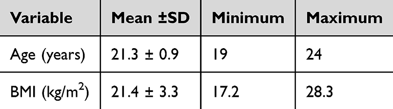 |
Table 1 Demographic Characteristics of Volunteers |
Before the implementation of the video training intervention, the measurements of the renal length, width, thickness, and volume were as follows: 9.60 ± 0.99 cm, 5.32 ± 0.93 cm, 3.90 ± 0.62 cm, and 104.95 ± 29.67 ml. The measurements after the video training were found to have increased to 10.01 ± 0.92 cm, 5.21 ± 0.83 cm, 4.11 ± 0.76 cm, and 167.65 ± 42.63 ml, respectively. These increases indicate considerable improvements in renal measures, except renal width (Table 2).
 |
Table 2 Pre- and Post-Intervention Sonographic Measurements of the RT Kidney |
Furthermore, concerning the spleen, the first measurements of its length, width, thickness, and volume were 8.95 ± 0.93 cm, 7.74 ± 0.72 cm, 4.05 ± 0.84 cm, and 147.72 ± 42.21 ml, respectively. Upon completion of the video training, the aforementioned parameters exhibited a notable rise, reaching 9.27 ± 1.10 cm, 8.06 ± 0.64 cm, 4.23 ± 0.63 cm, and 166.69 ± 42.39 ml, respectively. These measurements, with the exception of splenic thickness, demonstrated considerable enhancements in splenic measurements (Table 3).
 |
Table 3 Pre- and Post-Intervention Sonographic Measurements of the Spleen |
Additionally, the operator time demonstrated a notable decrease from 13.14 ± 3.4 minutes to 8.1 ± 3.5 minutes after the video training intervention (P-value <0.001) with a mean difference of 5.02 minutes (Figure 2).
 |
Figure 2 Pre- and post-intervention measurements of operator time. |
Discussion
The diagnostic implications of our study are significant, as they shed light on the potential role of video-based breathing control training in improving the diagnostic accuracy of sonographic assessments of abdominal organs.
The role of breathing training programs for various breathing maneuvers is well-known in clinical practice to aid both the diagnosis and the treatment of various health conditions.13 Nevertheless, while carrying out a diagnostic test such as a sonographic examination, it may be difficult to ensure that the patient is aware of how to carry out the breathing maneuver.
In this study, we assessed the implementation of a video-based training program as a useful tool to aid sonographic examination. Sonographic abdominal examination showed that both RT kidney and spleen measurements before and after the implementation of the video-based training program were within the normal ranges.30,31 Otherwise, a statistically significant improvement in the measurements was reported.
The observed improvements in organ dimensions following the training program suggest that video-based breathing interventions are superior to the traditional instruction given to the patient; thus, they further enhance the quality and reliability of sonographic images and aid in the diagnosis of various abdominal conditions. This is consistent with the recent literature assessing the utility of video-based training programs in clinical practice; a meta-analysis by Deshpande et al found that video-based tools are effective interventions to improve patient’s knowledge in clinical settings.28 Another meta-analysis by Monteiro et al showed that video-based programs effectively reduce anxiety and the sense of unfamiliarity that challenges the diagnostic procedures.26 For example, video-based educational materials resulted in a significant reduction in the levels of anxiety among patients undergoing coronary angiography and were associated with patient satisfaction.27
Furthermore, the reduction in the amount of time that the operator spent after the intervention indicates the possibility that the training program may expedite the sonographic evaluation process, which could result in diagnoses that are made more quickly and effectively. This efficiency improvement could be especially useful in hectic clinical environments where time limits are common. It would enable medical professionals to do more tests within a specified amount of time without sacrificing the quality of the examinations. Ultimately, this could lead to faster treatment decisions and improved patient outcomes. Overall, the implementation of this training program has the potential to significantly enhance the efficiency and effectiveness of sonographic evaluations in clinical settings.
In general, the results of our research indicate that including breathing control training in sonographic imaging techniques has the potential to enhance diagnostic accuracy and efficiency, which will ultimately be beneficial to patient care. However, additional study is required to validate these findings in patient groups that are larger and more diverse, as well as to investigate the long-term diagnostic consequences of breathing therapies in clinical practice. Further research is needed to fully understand the impact of breathing control training on sonographic evaluations across different patient populations. This will help to determine the broader implications and benefits of incorporating these techniques into clinical settings.
When analyzing the results of the study, it is important to consider the fact that the study has a number of limitations. Because of these constraints, the results may not be applicable to a wider range of situations because the sample size was relatively small and the research was carried out at a single center. The fact that the study only used one arm for the experience and did not include a control group is another factor that undermines the trustworthiness of the conclusion. When conducting future research endeavors, it is essential to address these limitations to improve the reliability and usefulness of the findings in this respective field.
Conclusion
In conclusion, the findings of our research demonstrate that video-based breathing control training is an effective method for enhancing sonographic quantification of abdominal organs. This is demonstrated by the fact that there were significant improvements in renal and splenic measurements, as well as a notable reduction in the amount of time that the operator wasted after the intervention. According to the results of our research, the incorporation of video-based breathing treatments into sonographic imaging procedures has the potential to improve diagnostic accuracy and efficiency, which would be beneficial to clinical decision-making and patient care in the field of abdominal health. There is a need for additional studies to validate these findings in larger patient groups, which will ultimately optimize the use of video-based breathing therapies in clinical settings for the purpose of improving diagnostic outcomes and patient care.
Abbreviations
BMI, Body Mass Index; COPD, Chronic Obstructive Pulmonary Disease; IVC, Inferior Vena Cava; MRE, Magnetic Resonance Elastography; RT, Right; SPSS, Statistical Package for the Social Sciences.
Ethical Considerations
This study was approved by the Institutional Review Board at Princess Nourah bint Abdul Rahman University (PNU), IRB no. 22-1009. This study adheres to the ethical principles outlined in the Declaration of Helsinki. The researchers explained the study’s procedures to the volunteers, covering the protocols, potential risks, benefits of participation, and the measures implemented to protect their personal information. Informed consent was obtained from all volunteers before the procedure. To ensure confidentiality, volunteers’ data were carefully coded and securely stored.
Funding
This research was funded by the Deanship of Scientific Research at Princess Nourah bint Abdulrahman University, through the Research Funding Program, Grant No. (FRP-1444-31).
Disclosure
The authors declare no conflicts of interest in this work.
References
1. Altinkaya N, Koc Z, Ulusan S, Demir S, Gurel K. Effects of respiratory manoeuvres on hepatic vein Doppler waveform and flow velocities in a healthy population. Eur J Radiol. 2011;79(1):60–63. doi:10.1016/j.ejrad.2010.01.011
2. Cazorla S, Busegnies Y, D’Ans P, Héritier M, Poncin W, Breathing control exercises delivered in a group setting for patients with chronic obstructive pulmonary disease: a randomized controlled trial. Healthcare. 2023;11(6).
3. Bendstrup KE, Ingemann Jensen J, Holm S, Bengtsson B. Out-patient rehabilitation improves activities of daily living, quality of life and exercise tolerance in chronic obstructive pulmonary disease. Eur Respir J. 1997;10(12):2801–2806. doi:10.1183/09031936.97.10122801
4. Vaes AW, Delbressine JML, Mesquita R, et al. Impact of pulmonary rehabilitation on activities of daily living in patients with chronic obstructive pulmonary disease. J Appl Physiol. 2018;126(3):607–615. doi:10.1152/japplphysiol.00790.2018
5. Finch L, Frankel D, Gallant B, et al. Occupational therapy in pulmonary rehabilitation programs: a scoping review. Respir Med. 2022;199:106881. doi:10.1016/j.rmed.2022.106881
6. Thomas M, McKinley RK, Freeman E, Foy C, Prodger P, Price D. Breathing retraining for dysfunctional breathing in asthma: a randomised controlled trial. Thorax. 2003;58(2):110–115. doi:10.1136/thorax.58.2.110
7. Thomas M, McKinley RK, Mellor S, et al. Breathing exercises for asthma: a randomised controlled trial. Thorax. 2009;64(1):55–61. doi:10.1136/thx.2008.100867
8. Ait Ali L, Pingitore A, Piaggi P, et al. Respiratory training late after Fontan intervention: impact on cardiorespiratory performance. Pediatr Cardiol. 2018;39(4):695–704. doi:10.1007/s00246-018-1808-9
9. Banushi B, Brendle M, Ragnhildstveit A, et al. Breathwork interventions for adults with clinically diagnosed anxiety disorders: a scoping review. Brain Sciences. 2023;13(2):256. doi:10.3390/brainsci13020256
10. Ahmadnezhad L, Yalfani A, Borujeni BG. Inspiratory muscle training in rehabilitation of low back pain: a randomized controlled trial. J Sport Rehabil. 2020;29(8):1151–1158. doi:10.1123/jsr.2019-0231
11. Kang J Il, Jeong DK, Choi H. Effect of exhalation exercise on trunk muscle activity and Oswestry disability index of patients with chronic low back pain. J Phys Ther Sci. 2016;28(6):1738–1742. doi:10.1589/jpts.28.1738
12. Zaccaro A, Piarulli A, Laurino M, et al. How breath-control can change your life: a systematic review on psycho-physiological correlates of slow breathing. Front Human Neurosci. 2018;12. doi:10.3389/fnhum.2018.00353
13. Hamasaki H. Effects of diaphragmatic breathing on health: a narrative review. Medicines. 2020;7(10):65. doi:10.3390/medicines7100065
14. Seung SL, Kyoung WK, Beom JP, et al. Effect of respiration on the spectral Doppler wave of the right hepatic vein in right lobe living donor liver transplant recipients. J Ultrasound Med. 2007;26(12):1723–1733. doi:10.7863/jum.2007.26.12.1723
15. Abu-Yousef MM. Normal and respiratory variations of the hepatic and portal venous duplex Doppler waveforms with simultaneous electrocardiographic correlation. J Ultrasound Med. 1992;11(6):263–268. doi:10.7863/jum.1992.11.6.263
16. Bang DH, Son Y, Lee YH, Yoon KH. Doppler ultrasonography measurement of hepatic hemodynamics during Valsalva maneuver: healthy volunteer study. Ultrasonography. 2014;34(1):32–38. doi:10.14366/usg.14029
17. Byeon K, Choi JO, Yang JH, et al. The response of the vena cava to abdominal breathing. J AlternComplementary Med. 2012;18(2):153–157. doi:10.1089/acm.2010.0656
18. Kimura BJ, Dalugdugan R, Gilcrease GW, Phan JN, Showalter BK, Wolfson T. The effect of breathing manner on inferior vena caval diameter. Eur J Echocardiography. 2011;12(2):120–123. doi:10.1093/ejechocard/jeq157
19. Gutzeit A, Roos JE, Hergan K, et al. Suction against resistance: a new breathing technique to significantly improve the blood flow ratio of the superior and inferior vena cava. Eur Radiol. 2014;24(12):3034–3041. doi:10.1007/s00330-014-3328-1
20. Ren H, Yang D, Xu H, et al. Effect of breath holding at the end of the inspiration and expiration phases on liver stiffness measured by 2D-MR elastography. Abdom Radiol. 2021;46(6):2516–2526. doi:10.1007/s00261-020-02893-w
21. Wells PNT. Abdominal ultrasound diagnosis. Br Med Bull. 1980;36(3):257–260. doi:10.1093/oxfordjournals.bmb.a071650
22. Sternbach G. Abdominal ultrasound. Ann Emerg Med. 1986;15(3):295–299. doi:10.1016/S0196-0644(86)80568-6
23. Rumack C, Levine D. Diagnostic ultrasound. 2023. Available from: https://books.google.com/books?hl=en&lr=&id=ixzcEAAAQBAJ&oi=fnd&pg=PP1&ots=RLsczuxBIF&sig=72LG-fA3WbLZXsmXltF1lErlfRw.
24. Ultrasound of the urinary tract - statpearls - NCBI bookshelf [Internet]. [
25. Larkins K, Khan M, Mohan H, Warrier S, Heriot A. A systematic review of video-based educational interventions in robotic surgical training. J Robotic Sur Sprinr Nat. 2023;17(4):1329–1339. doi:10.1007/s11701-023-01605-y
26. Monteiro Grilo A, Ferreira AC, Pedro Ramos M, Carolino E, Filipa Pires A, Vieira L. Effectiveness of educational videos on patient’s preparation for diagnostic procedures: systematic review and meta-analysis. Prev Med Rep. 2022;28:101895. doi:10.1016/j.pmedr.2022.101895
27. Jamshidi N, Abbaszadeh A, Kalyani MN, Sharif F. Effectiveness of video information on coronary angiography patients’ outcomes. Collegian. 2013;20(3):153–159. doi:10.1016/j.colegn.2012.06.001
28. Deshpande N, Wu M, Kelly C, et al. Video-based educational interventions for patients with chronic illnesses: systematic review. J Med Inter Res. 2023;25:e41092. doi:10.2196/41092
29. The AIUM practice parameter for the performance of an ultrasound examination of the abdomen and/or retroperitoneum. J Ultrasound Med. 2022;41(4):E1–E8. doi:10.1002/jum.15874
30. Chiorean L, Zdrenghea M, Badea R. Ultrasonography of the spleen. Picl Essay Med Ultrason. 2014;16(1):48–59. doi:10.11152/mu.2014.2066.161.lc1mz2
31. Koratala A, Bhattacharya D, Kazory A. Point of care renal ultrasonography for the busy nephrologist: a pictorial review. World J Nephrol. 2019;8(3):44. doi:10.5527/wjcc.v8.i3.44
 © 2025 The Author(s). This work is published and licensed by Dove Medical Press Limited. The
full terms of this license are available at https://www.dovepress.com/terms
and incorporate the Creative Commons Attribution
- Non Commercial (unported, 3.0) License.
By accessing the work you hereby accept the Terms. Non-commercial uses of the work are permitted
without any further permission from Dove Medical Press Limited, provided the work is properly
attributed. For permission for commercial use of this work, please see paragraphs 4.2 and 5 of our Terms.
© 2025 The Author(s). This work is published and licensed by Dove Medical Press Limited. The
full terms of this license are available at https://www.dovepress.com/terms
and incorporate the Creative Commons Attribution
- Non Commercial (unported, 3.0) License.
By accessing the work you hereby accept the Terms. Non-commercial uses of the work are permitted
without any further permission from Dove Medical Press Limited, provided the work is properly
attributed. For permission for commercial use of this work, please see paragraphs 4.2 and 5 of our Terms.
Recommended articles
Complicated Giant Splenic Hydatid Cyst: Case Report and Literature Review
Hakimi T, Zarif SK, Rezavi F, Aslamzai M, Halimi SA, Aslamy MA, Jawed MA
International Medical Case Reports Journal 2025, 18:1099-1103
Published Date: 23 August 2025