
Back to Journals » Clinical Ophthalmology » Volume 20

Epidemiology, Microbiology, Management and Outcomes of Endophthalmitis: An 18 Year Retrospective Observational Study at a Tertiary Referral Center in Australia
Authors Maher C  , Guo B, Sim B, Loewenthal MR, Gillies D, Hall AB
Received 23 November 2025
Accepted for publication 27 January 2026
Published 6 February 2026 Volume 2026:20 583832
DOI https://doi.org/10.2147/OPTH.S583832
Checked for plagiarism Yes
Review by Single anonymous peer review
Peer reviewer comments 2
Editor who approved publication: Dr Scott Fraser
Clare Maher,1 Brad Guo,1 Benjamin Sim,1 Mark R Loewenthal,1,2 Donna Gillies,1 Anthony Bennett Hall1
1Ophthalmology Department, John Hunter Hospital, Newcastle, NSW, 2305, Australia; 2School of Medicine and Public Health, The University of Newcastle, Newcastle, NSW, 2305, Australia
Correspondence: Clare Maher, Ophthalmology Department, John Hunter Hospital, Lookout Road, New Lambton Heights, Newcastle, NSW, 2305, Australia, Tel +61448866450, Email [email protected]
Purpose: This study characterizes the epidemiology, microbiological profile, management and outcomes of endophthalmitis over 18 years at a tertiary referral center in regional New South Wales (NSW), Australia.
Methods: A retrospective observational study of all endophthalmitis cases at John Hunter Hospital (November 2006 to December 2024) identified via ICD-10 codes and confirmed via record review. Data included demographics, clinical presentation, etiology, microbiology, management and surgical interventions.
Results: In total, 232 cases (227 patients) were identified (median age 75.6 years). Annual incidence declined significantly (slope = − 0.17 cases/year, 95% CI − 0.29 to − 0.052); p = 0.005). Exogenous endophthalmitis accounted for 87.1% of cases, most commonly following cataract surgery or intravitreal injection (38.1% each). Endogenous cases (12.9%) were mainly associated with bacteremia (56.7%) and immunosuppression (20%). Culture positivity was 56%, with 97.7% of isolates being bacterial. Gram positive cocci were identified in 84% of culture-proven cases, predominantly coagulase-negative staphylococci (42%), while gram-negative organisms comprised 9.4%. Intravitreal antibiotics were administered in 97.3% of cases and pars plana vitrectomy (PPV) performed in 41.5%. Overall, 52.5% of eyes improved in visual acuity. The Win Ratio for improvement with vitrectomy versus antibiotics alone was 1.31 in favor of vitrectomy (95% CI 0.77– 2.22; p = 0.31). In eyes presenting with light perception vision, the Win Ratio was 2.28 in favor of vitrectomy (95% CI 0.79– 6.55; p = 0.13).
Conclusion: Exogenous endophthalmitis remains the leading cause of endophthalmitis at a major regional tertiary referral center. Findings align with the Endophthalmitis Vitrectomy Study, suggesting vitrectomy benefits mainly eyes presenting with light perception only vision.
Plain Language Summary: What is Already Known on this Topic
Endophthalmitis is a rare but serious infection inside the eye that can occur after surgery, intravitreal injections, trauma, or from infections elsewhere in the body. Although uncommon, it can cause permanent vision loss if not treated quickly. Earlier research shows that the causes and bacteria involved can change over time, particularly with the rapid growth of intravitreal injections. However, long-term Australian data, especially from regional centres, are limited.
What This Study Adds
This study presents an 18-year review of all endophthalmitis cases managed at a major regional hospital in New South Wales, Australia. It describes trends in how often cases occurred, the reasons they developed, the types of bacteria involved, and how patients were treated. It also compares visual outcomes between patients treated with intravitreal antibiotics alone and those who underwent vitrectomy surgery, and identifies which patients are most likely to benefit from surgery.
How this Study Might Affect Research, Practice, or Policy
The findings help clinicians recognise risk factors for poor outcomes and support decision-making around vitrectomy in severe cases. They also reinforce the importance of infection-prevention strategies in cataract surgery and intravitreal injections.
Keywords: endophthalmitis, intravitreal injection, cataract surgery
Introduction
Endophthalmitis is a rare but potentially devastating intraocular infection.1 It typically presents with reduced vision, ocular pain, redness and periocular swelling, and if not promptly treated, can lead to severe vision loss, panophthalmitis, corneal infiltration and perforation and ultimately phthisis bulbi.2 Intravitreal antibiotics and vitrectomy are established components of endophthalmitis management; however, the optimal timing of vitrectomy remains contentious.2,3
Endophthalmitis may be classified as exogenous, originating from external sources, such as intraocular surgery, trauma, or adjacent infections, or endogenous, resulting from hematogenous spread from systemic infections.4 Exogenous causes remain more common, though endogenous causes are increasingly recognized in immunocompromised and hospitalized patients.
Given the rise in intraocular procedures, particularly intravitreal injections (IVI), understanding epidemiological patterns and microbiological profiles is essential for guiding prevention and treatment.5 While previous Australian studies have characterized endophthalmitis in metropolitan populations, few have examined long-term trends in regional referral centers. Moreover, long-term regional trends in endophthalmitis incidence, microbiological spectrum, and clinical outcomes in the anti-VEGF era remain poorly defined.
Methods
This retrospective study was conducted at John Hunter Hospital, a major tertiary referral center in Newcastle, New South Wales, covering the Hunter New England Local Health District (HNELHD). The region spans approximately 131,785 square kilometers and serves a population of nearly 1 million.6 As a tertiary referral center for the district, it receives both regional and extra-regional endophthalmitis cases, including patients from the private sector in instances where vitreoretinal surgeons are unavailable locally. Patients referred from John Hunter Hospital to Sydney Eye Hospital for further management (pars plana vitrectomy) were also included in this study.
Ethics approval for this retrospective study was granted by the Hunter New England Local Health District Ethics Committee (20250525–014). The requirement for individual patient consent was waived by the Ethics Committee as the project involved retrospective review of existing clinical records, posed no identifiable risk to participants, and met the criteria for waiver of consent under New South Wales Health guidelines. This study was conducted in accordance with the principles of the Declaration of Helsinki.
Case Identification
All patients diagnosed with endophthalmitis between November 2006 and December 2024 were identified via International Statistical Classification of Diseases and Related Health Problems (ICD)-10 codes (including endogenous and exogenous endophthalmitis). Each case was verified by detailed medical record review. Recurrent episodes were counted separately if they occurred more than six months apart and had a distinct etiology (for example, a new intravitreal injection or surgical procedure).
Data Collection
Patient demographics, etiology, microbiology, treatment modality, and visual acuity (VA) at presentation and follow-up were extracted by detailed medical record review and entered directly into a study-specific REDCap database.7 VA was assessed using Snellen or logMAR charts where applicable and recorded in meters (eg 6/6), or categorized as counting fingers, hand movements, light perception, or no light perception. Final VA was variable in timeframe but was defined as any measurement taken at least two months after treatment. For exogenous cases, the time interval between the inciting ocular procedure and presentation with endophthalmitis was recorded and summarized by intervention type.
Statistical Analysis
Categorical data was tabulated and, where appropriate, P-values were calculated with 2-sided Fishers exact tests. Numeric data was summarized by medians, quartiles, and ranges. P-values for comparison of paired data were calculated using Wilcoxon’s sign-rank test. Because visual acuity (VA) is an ordinal variable, outcomes were compared between treatment groups using the Win Ratio method, a method widely adopted in cardiovascular trials.8 When comparing an intervention to a control treatment, a Win Ratio greater than one indicates that the intervention is better than the control. A Win Ratio less than one means that the intervention is worse than the control.
The Win-Ratio: More Detail
Motivation: VA measured on an ordinal scale only allows qualitative comparisons between any two eyes: better, worse, or same. The Win Ratio produces numerical (quantitative) estimates of effect size for an intervention when the outcome is based on this ordinal classification.
The VA for an eye which received an intervention can be compared with the VA for a “control” eye that did not receive the intervention. A “win” means the intervention eye has better VA than the control eye. A “loss” is the reverse, control is better than intervention. A tie is self-explanatory.
This comparison is made for every possible pairwise comparison between each eye in the intervention group and each eye in the control group. The  , is then calculated. For example, if 20 eyes receive an intervention (eg vitrectomy) and 40 eyes do not receive this intervention, there will be 20×40 = 800 pairwise comparisons possible. If 500 of these are “wins”, 250 are “losses”, and 50 are ties, the Win Ratio is then
  A corresponding confidence interval is calculated.9
Statistical calculations were made with Stata statistical software10 including the use of “winratiotest” user written module.11
Results
Patient Demographics
A total of 232 cases of endophthalmitis were identified in 227 patients from November 2006 to December 2024. Two individuals had two separate cases while two patients experienced three. The median age at diagnosis was 75.6 years (range 13–98 years), with a left-skewed distribution (Figure 1). Exogenous cases occurred in older patients (median 76.8 years) compared with endogenous cases (median 60.5 years; p < 0.001).
 |
Figure 1 Age distribution of endophthalmitis cases. A left skewed distribution is observed, with median age of 75.6 years and range from 13 to 98 years. |
Females comprised 53.9% of all cases and males 46.1%. There was a slight preponderance of males in the endogenous group (60%) and females in the exogenous group (56%) (p = 0.12).
Temporal Trends
The median number of cases per year was 12 (range 5–22). As depicted in Figure 2, there is a significant downward trend in the total annual incidence of endophthalmitis over the 18 year period, with a slope of −0.170 per year (95% CI: −0.29 to −0.052; P = 0.005).
 |
Figure 2 Endophthalmitis cases per year from 2005 to 2024 The red line demonstrates reduction in cases over time. |
When stratified by cause, post-cataract surgery causes showed a significant reduction over time, with a slope of −0.53 (95% CI −0.76 to −0.29; P < 0.001 t-test) (Figure 3). In contrast, post-intravitreal injection (IVI) cases demonstrated a relative increase over the same period, as evident in Figure 4.
 |
Figure 3 Endophthalmitis cases post cataract from 2006 to 2024, with red line demonstrating reduction in cases over time. |
 |
Figure 4 Endophthalmitis cases from 2006 to 2024. Blue line is post-intravitreal injection, red line is post-cataract and green line is post-vitrectomy. |
Among exogenous endophthalmitis cases, the median time to presentation from the inciting ocular procedure was 5 days (IQR 3–11; range 1–68). Post–cataract surgery cases presented at a median of 6 days (IQR 4–14; range 1–68), post–intravitreal injection at 4 days (IQR 2–6; range 1–26), and post–vitrectomy at 4 days (IQR 2–5; range 1–23). Corneal procedures were associated with a longer median time to presentation of 13 days (IQR 5–21; range 2–35), while post-trabeculectomy cases demonstrated the most delayed presentation, with a median of 28 days (IQR 27–29; range 25–30).
Etiology
Of the 232 cases, majority (202, 87.1%) were exogenous in origin, while the remaining 30 eyes (12.9%) were endogenous. As demonstrated in Table 1, among exogenous causes, most common causes were post-cataract surgery (38%) and post-intravitreal injection (38%), followed by post-vitrectomy (7.9%). Less frequent sources included post-corneal procedures such as graft of suture removal (4%) and post-trabeculectomy (3.5%). Endogenous cases were primarily associated with bacteremia (56.7%), followed by immunosuppression (20%).
 |
Table 1 Causes of Exogenous and Endogenous Endophthalmitis, by Number and Percentage |
Micro-Organism
A causative organism was identified in 56% of cases, with 97.7% of these being bacterial in origin and only three fungal case (2.3%) identified. The majority of organisms (90%) were gram positive, as depicted in Table 2. Coagulase negative staphylococci were the most common culture-proven organisms, accounting for 43% of identified pathogens. About 72% of those being Staphylococcus epidermidis. Of the streptococci (20 cases), 8 were viridans (40%), while 5 were Str. pneumoniae (25%). Of the enterococci 5/6 cases (83.3%) was due to Enterococcus faecalis.
 |
Table 2 Causative Bacterial Organisms in Culture-Positive Endophthalmitis Cases (127) |
Gram negative organisms comprised 9% of positive cultures, with Pseudomonas aeruginosa responsible for 50% of these gram-negative infections.
The fungal species isolated were Scedosporium apiospermum (endogenous cause), Candida albicans (endogenous cause), and Aspergillus fumigatus (exogenous cause).
Treatment
Nearly all patients (97.3%) received intravitreal antibiotics. The few patients who did not were primarily elderly patients with dementia or behavioral disturbance that made safe administration impossible, as well as critically unwell patients in intensive care. About 41.5% of eyes underwent pars plana vitrectomy (PPV) (43% exogenous, 31% endogenous). Surgical removal of the eye was rarely required, with evisceration performed in 3.1% of cases and enucleation in 0.4%.
All patients with fungal endophthalmitis received systemic and intravitreal antifungal treatment, yet outcomes were poor, comprising light perception vision, enucleation, and death. The case of Scedosporium apiospermum endogenous endophthalmitis progressed despite therapy, necessitating enucleation for pain and visual loss.
Visual Acuity Outcomes
Final visual acuity was available for 62% of cases (90.3% exogenous, 9.7 endogenous). Across the cohort, 52.5% of eyes improved, 39% remained unchanged and 8.5% deteriorated. Visual acuity was classified on an ordinal scale (numerical acuity, count fingers, hand movement, light perception and no light perception), with Figure 5 depicting presenting and final visual acuity. The median improvement was +1 visual category (Figure 6), representing a significant improvement (p < 0.001). Significant gains were observed following post injection and post cataract cases (p < 0.001), while other subgroups demonstrated stable or minimal improvement.
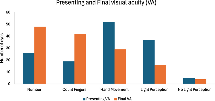 |
Figure 5 Bar chart depicting presenting and final visual acuity (VA). Presenting VA values are displayed only for eyes with documented final VA to ensure accurate population matching. |
 |
Figure 6 Distribution of change in visual acuity (presentation minus final VA). Positive values correspond to improved vision and negative values to visual deterioration. |
Effect of Vitrectomy
Across all cases with outcome data (n = 141), 34.8% underwent vitrectomy (49 eyes). The overall Win Ratio for visual improvement with vitrectomy versus intravitreal antibiotics alone was 1.31 (95% CI 0.77 to 2.22; p = 0.31).
Of the 141 eyes, 36 presented with light perception (LP) vision. Within this LP subgroup, 20 eyes underwent vitrectomy, yielding a Win Ratio of 2.28 (95% CI 0.79–6.55; p = 0.13), indicating a directionally favorable trend consistent with the Endophthalmitis Vitrectomy Study (EVS) findings.12
The remaining 105 eyes presented with vision better than LP; among these, 29 underwent vitrectomy, resulting in a Win Ratio of 0.99 (95% CI 0.52–1.89), indicating neither benefit nor harm.
Discussion
Over the past two decades, the epidemiology of endophthalmitis has shifted significantly, shaped by evolving surgical techniques, infection-control measures and the widespread use of intravitreal anti-VEGF (vascular endothelial growth factor) therapy.13 In this regional Australian cohort, exogenous endophthalmitis remained the predominant form, accounting for 87% of cases, with post-cataract and post intravitreal injection representing the major causes.
Comparable Australian studies, including earlier work from the Endophthalmitis Population Study of Western Australia (EPSWA) (2005), reported post-cataract surgery as leading cause of endophthalmitis in Western Australia, followed by post-trauma.14 The shift post-surgical to post-injection predominance observed in this study was similarly reported in the 2013 state-wide analysis from the Royal Victorian Eye and Ear Hospital.15 This evolution underscores the profound impact of anti-VEGF therapy on contemporary endophthalmitis etiology. However, few studies have specifically examined regional populations, where limited access to tertiary ophthalmic services and delayed presentations may influence disease characteristics and outcomes. To our knowledge, this represents one of the first regional Australian cohorts to characterize endophthalmitis patterns in the anti-VEGF era.
The decline in post-cataract endophthalmitis observed in this study mirrors previous Australian findings15 and likely reflects improvements in peri-operative prophylaxis, particularly the routine use of intracameral antibiotics, smaller incision techniques and stricter sterile protocols. Following the landmark ESCRS multicenter study,16 governing bodies such as the Royal Australian and New Zealand College of Ophthalmologists17 and national therapeutic guidelines18 recommend the use of intracameral antibiotics for endophthalmitis prophylaxis.19
Conversely, the rise in post-intravitreal cases parallels the exponential increase in anti-VEGF therapy. It is estimated that more than 108,000 Australians received eye injection treatment in 2023.5 Although the risk of infection per injection remains extremely low, the cumulative risk from repeated procedures contributes to a growing overall burden of cases. A 2024 study by Israilevich et al reported an overall endophthalmitis risk of 0.035% (1 in 2857 injections), with a progressively higher cumulative risk of endophthalmitis with each additional injection.20 This trend reinforces the importance of maintaining strict aseptic techniques during injections and ongoing surveillance as treatment volumes continue to expand.
Most postoperative endophthalmitis presented within the first week; however, delayed-onset cases were observed, consistent with the recognized spectrum of acute and delayed postoperative endophthalmitis reported in the literature.4
The microbiological profile aligns with global findings, where gram-positive bacteria, especially coagulase negative staphylococci dominate.21 The emergence of Pseudomonas aeruginosa in a subset of cases highlights the need for vigilance regarding virulent gram-negative organisms. Nearly all patients in this study received intravitreal antibiotics and approximately 40% underwent vitrectomy. Overall, 52.5% of eyes improved, with the greatest visual gains seen in post-cataract and post-intravitreal injection cases compared to post-trabeculectomy and in endogenous causes.
The role of early vitrectomy in endophthalmitis remains debated. The Endophthalmitis Vitrectomy Study (EVS) established that early vitrectomy provides significant visual benefit only in patients presenting with light perception (LP) vision or worse, while showing no added advantage in those with better presenting visual acuity.12 Our findings align with this conclusion. While vitrectomy was not associated with a statistically significant overall improvement, a favorable trend was observed in eyes presenting with LP vision. This suggests that, in contemporary practice, vitrectomy continues to offer the greatest benefit for severe presentation, consistent with EVS and subsequent studies.
In endogenous endophthalmitis, a similar but imprecise trend toward improved outcomes with vitrectomy was noted, supporting prior evidence that early surgical clearance of infected material may be beneficial in selected cases, particularly in immunocompromised patients or those with virulent organisms.22
The strengths of this study include its long timeframe, large catchment area and comprehensive microbiological and clinical dataset. The application of the win ratio allowed for nuanced comparison of ordinal visual outcomes. However, the retrospective design, incomplete follow-up VA data and inability to adjust for evolving surgical practices, such as the introduction of intracameral antibiotics, represent important limitations. The setting in which intravitreal injections were administered was not formally documented; however, in our region, the vast majority of intravitreal injections are performed in outpatient clinic settings in both public and private practices rather than in operating theatres. Consequently, our results should be interpreted as representative of outpatient injection practice. Additionally, the single-center nature of this study and absence of denominator procedural data precluded estimation of true incidence rates and limited the generalizability of these findings.
Final VA was defined as measurements taken at least two months after treatment. In contrast, the Endophthalmitis Vitrectomy Study (EVS)12 defined final VA at 6 months. Consequently, our study may have underestimated final visual outcomes, as some patients may have experienced further improvement in vision but were discharged to local or private ophthalmology care.
Loss to follow up further limited the availability of final VA data, as many patients continued care elsewhere. This introduces potential selection bias, since the demographic and clinical characteristics of those who returned for follow-up may differ from the broader study population.
Future work on this dataset will further characterize subgroups of patients with differing visual outcomes, particularly those who experience deterioration despite treatment. Detailed analysis of clinical, microbiological and management factors in these cases may help identify predictors of poor prognosis and refine treatment algorithms.
Conclusion
In summary, exogenous endophthalmitis remains the predominant form of the disease, most often related to cataract surgery and intravitreal injections. The decline in post cataract cases likely reflects advances in intraoperative prophylaxis, while the increase in injection related cases reflects the rise in anti-VEGF therapy. Intravitreal antibiotics remain the cornerstone of treatment, and vitrectomy may offer benefit primarily in cases presenting with light perception vision, consistent with EVS findings. Ongoing surveillance, adherence to sterile techniques, and multicenter prospective studies are essential to further refine strategies for prevention and management. In contemporary practice, early referral to vitreoretinal services and timely consideration of vitrectomy remain key factors in optimizing outcomes, particularly in severe presentations.
Abbreviations
ICD-10, International Classification of Diseases, Tenth Revision; NSW, New South Wales; PPV, pars plana vitrectomy; CI, confidence interval; HNELHD, Hunter New England Local Health District; VA, visual acuity; logMAR, logarithm of the minimum angle of resolution; IVI, intravitreal injection; LP, light perception; NLP, no light perception; CNS, coagulase-negative Staphylococcus; HSV-2, herpes simplex virus type 2; VEGF, vascular endothelial growth factor; EPSWA, Endophthalmitis Population Study of Western Australia; ESCRS, European Society of Cataract and Refractive Surgeons; RANZCO, Royal Australian and New Zealand College of Ophthalmologists; EVS, Endophthalmitis Vitrectomy Study; REDCap, Research Electronic Data Capture.
Data Sharing Statement
Data supporting the findings of this study are available from the corresponding author upon reasonable request.
Ethics Approval and Informed Consent
This study was approved by the Hunter New England Local Health District Ethics Committee (20250525-014). Informed consent was waived due to the retrospective design and use of de-identified data. This study was conducted in accordance with the principles of the Declaration of Helsinki.
Author Contributions
All authors meet ICMJE authorship criteria. All authors made a significant contribution to the work reported, whether that is in the conception, study design, execution, acquisition of data, analysis and interpretation, or in all these areas; took part in drafting, revising or critically reviewing the article; gave final approval of the version to be published; have agreed on the journal to which the article has been submitted; and agree to be accountable for all aspects of the work.
Disclosure
The authors report no conflicts of interest in this work.
References
1. Shao EH, Yates WB, Ho IV, Chang AA, Simunovic MP. Endophthalmitis: changes in presentation, management and the role of early vitrectomy. Ophthalmol Ther. 2021;10(4):877–11. doi:10.1007/s40123-021-00406-6
2. Durand ML. Bacterial and Fungal Endophthalmitis. Clin Microbiol Rev. 2017;30(3):597–613. doi:10.1128/CMR.00113-16
3. Sromicki JW, Stahel M, Blum RA, Rudolph KA, Barthelmes D. Early vitrectomy in endophthalmitis: visual outcomes and complication rates. Ophthalmol Ther. 2025;14(8):2031–2042. doi:10.1007/s40123-025-01196-x
4. Safneck JR. Endophthalmitis: a review of recent trends. Saudi J Ophthalmol. 2012;26(2):181–189. doi:10.1016/j.sjopt.2012.02.011
5. Australia MDF. Call to Federal Government to bulk bill eye injections for pensioners; 2025. Available from: https://www.mdfoundation.com.au/news/call-to-federal-government-to-bulk-bill-eye-injections-for-pensioners/.
6. England NH-HN. Hunter New England. NSW Health; 2025. Available from: https://www.health.nsw.gov.au/lhd/Pages/hnelhd.aspx.2025.
7. Harris PA, Taylor R, Minor BL, et al. The REDCap consortium: building an international community of software platform partners. J Biomed Inform. 2019;95:103208. doi:10.1016/j.jbi.2019.103208
8. Redfors B, Gregson J, Crowley A, et al. The win ratio approach for composite endpoints: practical guidance based on previous experience. Eur Heart J. 2020;41(46):4391–4399. doi:10.1093/eurheartj/ehaa665
9. Finkelstein DM, Schoenfeld DA. Combining mortality and longitudinal measures in clinical trials. Stat Med. 1999;18(11):1341–1354. doi:10.1002/(SICI)1097-0258(19990615)18:11<1341::AID-SIM129>3.0.CO;2-7
10. Stata Statistical Software: Release 19 [computer program]; 2025.
11. Gregson J, Ferreira JP, Collier T. winratiotest: a command for implementing the win ratio and stratified win ratio in Stata. Stata J. 2023;23(3):835–850. doi:10.1177/1536867X231196480
12. Forster RK. The endophthalmitis vitrectomy study. Arch Ophthalmol. 1995;113(12):1555–1557. doi:10.1001/archopht.1995.01100120085015
13. Malmin A, Syre H, Ushakova A, Utheim TP, Forsaa VA. Twenty years of endophthalmitis: incidence, aetiology and clinical outcome. Acta Ophthalmol. 2021;99(1):e62–e69. doi:10.1111/aos.14511
14. Ng JQ, Morlet N, Pearman JW, et al. Management and outcomes of postoperative endophthalmitis since the endophthalmitis vitrectomy study: the Endophthalmitis Population Study of Western Australia (EPSWA)’s fifth report. Ophthalmology. 2005;112(7):1199–1206. doi:10.1016/j.ophtha.2005.01.050
15. Kam J, Dawkins R, Buck D, Sandhu S, Allen P. Changing patterns of endophthalmitis at a state-wide service in Australia over a 14 year period. Invest Ophthalmol Visual Sci. 2013;54(15):3174.
16. Group EES. Prophylaxis of postoperative endophthalmitis following cataract surgery: results of the ESCRS multicenter study and identification of risk factors. J Cataract Refract Surg. 2007;33(6):978–988. doi:10.1016/j.jcrs.2007.02.032
17. Ophthalmologists RAa NZCo. Preferred practice patterns: cataract and intraocular lens surgery; 2021.
18. Guidelines T. Surgical antibiotic prophylaxis for specific procedures; 2025. Available from: https://tgldcdp.tg.org.au/viewTopic?etgAccess=true&guidelinePage=Antibiotic&topicfile=surgical-antibiotic-prophylaxis-procedures&guidelinename=Antibiotic§ionId=toc_d1e2646#toc_d1e2646.
19. Excellence NIfHa C. Cataracts in adults: management; 2017.
20. Israilevich RN, Mansour H, Patel SN, et al. Risk of endophthalmitis based on cumulative number of anti-VEGF intravitreal injections. Ophthalmology. 2024;131(6):667–673. doi:10.1016/j.ophtha.2023.12.033
21. Tan CL, Harsha S, Allen PJ, Dawkins RCH. Endophthalmitis: microbiology and organism identification using current and emerging techniques. Ocul Immunol Inflamm. 2023;31(2):393–401. doi:10.1080/09273948.2022.2027468
22. Zhang YQ, Wang WJ. Treatment outcomes after pars plana vitrectomy for endogenous endophthalmitis. Retina. 2005;25(6):746–750. doi:10.1097/00006982-200509000-00010
 © 2026 The Author(s). This work is published and licensed by Dove Medical Press Limited. The
full terms of this license are available at https://www.dovepress.com/terms
and incorporate the Creative Commons Attribution
- Non Commercial (unported, 4.0) License.
By accessing the work you hereby accept the Terms. Non-commercial uses of the work are permitted
without any further permission from Dove Medical Press Limited, provided the work is properly
attributed. For permission for commercial use of this work, please see paragraphs 4.2 and 5 of our Terms.
© 2026 The Author(s). This work is published and licensed by Dove Medical Press Limited. The
full terms of this license are available at https://www.dovepress.com/terms
and incorporate the Creative Commons Attribution
- Non Commercial (unported, 4.0) License.
By accessing the work you hereby accept the Terms. Non-commercial uses of the work are permitted
without any further permission from Dove Medical Press Limited, provided the work is properly
attributed. For permission for commercial use of this work, please see paragraphs 4.2 and 5 of our Terms.
Recommended articles
Changes in Aqueous Humor Cytokine Profile Following Intravitreal Brolucizumab Injection
Ruamviboonsuk V, Kongwattananon W, Chuaypen N
Clinical Ophthalmology 2025, 19:427-437
Published Date: 11 February 2025
Cohort Validation of the Endophthalmitis Vitrectomy Study for Generalized Etiologies
Carpenter E, Zhou Y, Wilson MK, Gilberstadt N, Bowman L, Hu JK
Clinical Ophthalmology 2026, 20:542667
Published Date: 20 January 2026