Back to Journals » International Journal of Nanomedicine » Volume 21
Enhancing Therapeutic Efficacy Through Tailored Pharmacokinetics: A Review of Mesoporous Silica Nanoparticle-Based Delivery Systems
Authors Aulifa DL ![]() , Maharani A, Purnomo KG, Salsabilla RA, Fahlevi ZA, Pramudita FW, Wibisono TTDW, Amaliah S
, Maharani A, Purnomo KG, Salsabilla RA, Fahlevi ZA, Pramudita FW, Wibisono TTDW, Amaliah S ![]() , Subra L, Budiman A
, Subra L, Budiman A
Received 19 February 2026
Accepted for publication 1 May 2026
Published 19 May 2026 Volume 2026:21 604271
DOI https://doi.org/10.2147/IJN.S604271
Checked for plagiarism Yes
Review by Single anonymous peer review
Peer reviewer comments 3
Editor who approved publication: Prof. Dr. RDK Misra
Diah Lia Aulifa,1 Anisa Maharani,2 Keisya Ghaliyah Purnomo,2 Rifa Alya Salsabilla,2 Zakia Aurora Fahlevi,2 Fransisca Widi Pramudita,1 Tribuana Tungga Dewi Wansha Wibisono,1 Salma Amaliah,2 Laila Subra,3 Arif Budiman2
1Department of Pharmaceutical Analysis and Medicinal Chemistry, Faculty of Pharmacy, Universitas Padjadjaran, Bandung, West Java, Indonesia; 2Department of Pharmaceutics and Pharmaceutical Technology, Faculty of Pharmacy, Universitas Padjadjaran, Bandung, West Java, Indonesia; 3Faculty of Bioeconomic and Health Sciences, Geomatika University College, Kuala Lumpur, 54200, Malaysia
Correspondence: Arif Budiman, Department of Pharmaceutics and Pharmaceutical Technology, Faculty of Pharmacy, Universitas Padjadjaran, Bandung, West Java, Indonesia, Email [email protected] Diah Lia Aulifa, Department of Pharmaceutical Analysis and Medicinal Chemistry, Faculty of Pharmacy, Universitas Padjadjaran, Bandung, West Java, Indonesia, Email [email protected]
Abstract: Low solubility and bioavailability represent major challenges in drug development, resulting in sub-optimal pharmacokinetic profiles, including poor gastrointestinal absorption and limited systemic exposure. Mesoporous silica nanoparticles (MSNs) have emerged as promising drug delivery systems due to their high surface area, tunable pore size, and high pore volume, which enable enhanced drug loading and stabilization of drugs in an amorphous state. These properties are critical for improving dissolution behavior. Numerous studies confirm that drug encapsulation into MSNs significantly enhances the dissolution rate, leading to substantial pharmacokinetic improvements—specifically increased Area Under the Curve (AUC) and higher Maximum Plasma Concentration (Cmax)—which are directly correlated with enhanced systemic exposure and the potential for improved therapeutic efficacy for poorly soluble drugs. These comprehensive findings indicate that MSN-based delivery systems offer a powerful strategy to overcome the intrinsic pharmacokinetic limitations of poorly soluble compounds and warrant further investigation into their long-term safety and clinical application.
Keywords: bioavailability, pharmacokinetics, mesoporous silica, in vivo studies, poorly water soluble-drugs
Introduction
Drug solubility is a major obstacle in the development of new drugs. Approximately 40% of commercialized products and 70–90% of drug candidates in development have poor solubility, resulting in low bioavailability, reduced therapeutic effects, and increased dosages.1 BCS classifies drugs based on their solubility in the physiological pH range and permeability, to identify compounds that are low risk in terms of drug absorption.2,3 For BCS Class I compounds (high solubility, high permeability),4 BCS Class II compounds (low solubility, high permeability),5 BCS Class III compounds (high solubility, low permeability),6 and BCS Class IV compounds (low solubility, low permeability).7 Among these, BCS class II and class IV drugs present significant formulation challenges due to their limited dissolution and absorption in the gastrointestinal tract. These physicochemical limitations often result in poor and variable pharmacokinetic performance, ultimately reducing bioavailability and therapeutic effectiveness, due to reduced systemic absorption and altered gastrointestinal drug deposition, where poorly soluble drugs may precipitate or remain localized in the intestinal lumen, leading to incomplete dissolution and inconsistent absorption.8 This underscores that drug efficacy is governed not only by solubility and permeability but also by gastrointestinal deposition behavior, which plays a critical role in determining in vivo dissolution and subsequent absorption outcomes. Therefore, improving drug solubility is a critical focus in pharmaceutical development to enhance absorption, optimize therapeutic outcomes, and minimize dose-related issues.9
Drug Delivery Systems (DDS) are a method of drug formulation that accelerate the delivery of drugs to specific target locations within the body.10,11 This continue to be developed to overcome the limitations of conventional systems, such as through controlled-release formulations, aiming to maximize therapeutic efficacy and minimize accumulation outside the target site.12 Nanotechnology is fundamentally revolutionizing pharmaceutical drug delivery for poorly water-soluble drugs by enabling precise control over particle size, surface characteristics, and drug encapsulation at the nanoscale.13 These properties significantly enhance drug dissolution through increased surface area, improve cellular uptake via endocytosis, and enable protection of drug molecules from enzymatic degradation and premature clearance.14–16 In addition, nanoscale systems can modulate drug transport pathways, including enhanced permeation across biological membranes and potential lymphatic uptake, thereby improving bioavailability and therapeutic efficiency.17 Simultaneously, nanotechnology offers the ability to target diseased tissues more precisely, thereby minimizing off-target toxicity of active molecules, improving therapeutic selectivity, and potentially reducing healthcare costs.18–21 Mesoporous silica (MS) is one critical type of DDS that capitalizes on nano technology, enabling the efficient loading of hydrophobic drugs into the MSN pores, often using non-aqueous solvents, which is essential for improving solubility, bioavailability, and achieving controlled drug release.22 Mesoporous silica nanoparticles (MSNs) are porous nanomaterials characterized by highly ordered pore structures, large surface area, and tunable pore size. These structural properties facilitate the confinement of poorly water-soluble drugs within nanoscale pores, promoting their dispersion in an amorphous or semi-crystalline state, which enhances apparent solubility and dissolution rate. Additionally, MSNs possess abundant silanol groups that enable surface interactions with drug molecules and allow further functionalization for controlled and targeted drug delivery.23,24 These features collectively contribute to improved drug solubility, enhanced bioavailability, and optimized therapeutic performance. Furthermore, the ability of MSNs to be surface-functionalized enables site-specific drug delivery, including applications in vascular-targeted therapies such as atherosclerosis, where drug release can be concentrated at sites of vascular injury or plaque formation.25
Pharmacokinetics (PK) plays an important role in drug development because it determines how drugs are absorbed, distributed, metabolized, and excreted, as well as determining therapeutic doses, potential toxicity, and the risk of drug interactions.26,27 Key PK parameters such as AUC, Cmax, t½, Vd, and clearance are benchmarks for bioavailability and therapeutic efficacy.28 However, many hydrophobic drugs face additional challenges, such as low solubility, poor absorption, and rapid elimination, resulting in low bioavailability.29 These limitations ultimately lead to a decrease in AUC and Cmax, as only a small fraction of the administered dose reaches systemic circulation.30 To address these issues, drug delivery systems (DDS), including nanoparticle, are widely used to improve solubility and enhance absorption.31 The small and uniformly distributed pore size of mesoporous silica results in an extremely large specific surface area, which facilitates the efficient loading and release of substances (eg, drugs).32 Additionally, it increases membrane permeability, thereby enhancing drug penetration across biological barriers. Nanoparticle-based delivery systems can also partially circumvent first-pass hepatic metabolism by protecting drug molecules from enzymatic degradation in the gastrointestinal tract and by facilitating alternative uptake pathways, such as lymphatic transport and intracellular protection within nanocarriers. This protection reduces premature metabolic breakdown and increases the fraction of drug reaching systemic circulation. Furthermore, these systems provide better formulation stability and enable more controlled drug release, which collectively improves pharmacokinetic performance.33–35 Through this mechanism, nanoparticle strategies can significantly modify pharmacokinetic behavior by increasing AUC and Cmax, prolonging half-life (t½), and reducing clearance, ultimately improving drug exposure and therapeutic outcomes.36,37
A number of studies have reported on the effect of Mesoporous Silica Nanoparticles (MSNs) on drug pharmacokinetics, highlighting their crucial role in overcoming various physical and chemical challenges in the development of new drug formulations, such as low solubility or poor stability.38 However, although several reviews have discussed nanotechnology-based drug delivery systems and mesoporous materials, most of them primarily focus on formulation development, material design, or general drug delivery.39–42 In contrast, a comprehensive analysis specifically integrating the pharmacokinetic outcomes of MSNs, particularly in terms of ADME (Absorption, Distribution, Metabolism, and Excretion) modulation and quantitative in vivo pharmacokinetic parameters, remains limited. Therefore, this review aims to address this gap by summarizing and critically evaluating the pharmacokinetic effects of MSNs on drug absorption, distribution, metabolism, and excretion, with particular emphasis on in vivo performance and mechanistic insights a molecular pharmacy perspective.
Methods
This study is presented as a narrative review that explores and analyzes recent advancements pharmacokinetic (PK) studies mesoporous silica. Relevant studies were collected from major scientific databases, including PubMed, Scopus, ScienceDirect, and Google Scholar. The search strategy utilized combinations of keywords such as “nanoparticle,” “mesoporous silica,” “pharmacokinetic studies,” “pharmacological activities,” “dissolution,” “toxicity,” encompassing publications from 2000 to 2025. The review only incorporated articles from peer-reviewed journals that were published in English, with priority directed towards research that particularly explored pharmacokinetic studies in mesoporous silica. The included studies comprised original research articles and experimental reports focusing on in vitro and in vivo pharmacokinetic and biopharmaceutical evaluations of mesoporous silica nanoparticles, while review articles were used solely to support background and conceptual framework development. To ensure analytical rigor, only studies that reported quantitative in vivo pharmacokinetic parameters (eg, AUC, Cmax, t½) with appropriate statistical measures were selected for the core analysis presented in Table 1; studies lacking complete quantitative PK data or focusing exclusively on in vitro characterization were excluded. Studies were also excluded if they are unrelated to nanoparticle study of mesoporous silica, or did not mention pharmacokinetic study, dissolution rate, toxicity, and pharmacological study. Information obtained from the chosen studies was gathered and extracted thematically emphasizing the dissolution rate, in vitro and in vivo performance, and comparative advantages over pharmacokinetic study in mesoporous silica. This focused, quality-driven selection process, while yielding a relatively modest number of core studies (n=17), was deliberately adopted to enable a thorough and critical examination of representative MSN formulations rather than a superficial survey of a larger but less homogeneous body of literature. The narrative review approach was considered suitable for encompassing the varying results of pharmacokinetic study in mesoporous silica investigated in the literature, enabling a thorough and comprehensive examination of their mechanisms and therapeutic implications concerning the enhancement of drug solubility, stability, and safety. The methodology’s flowchart is shown in Figure 1.
 |
Table 1 Previous Studies of Drug-Loaded MSN |
 |
Figure 1 Methodology’s Chart. |
Mesoporous Silica Nanoparticles (MSNs)
Mesoporous silica nanoparticles (MSNs) have been widely utilized across various industries and scientific disciplines, including heterogeneous catalysis, electrochemistry, analytical chemistry, molecular biology, and several other areas.24,57 Their defining characteristic is a moderately sized pore range in diameter, which is vital for high-surface-area applications like drug delivery and catalysis,58 and a substantial surface area, making them highly appealing to scientists involved in drug delivery research, given their ability to act as a carrier with minimal influence on the therapeutic process, alongside their capacity to be altered using diverse reagents to incorporate specific attributes or fine-tune their characteristics, eg, pore size, controlling the drug release, design it to target a specific organ or tissue, etc.59
Physicochemical Properties of MSNs
Mesoporous silica nanoparticles (MSNs) possess unique physicochemical properties that make them highly suitable for drug delivery applications. A key feature is their exceptionally high specific surface area, typically ranging from several hundred to over one thousand m2/g, which provides abundant active sites for drug adsorption and enables high drug loading capacity.60,61 MSNs also exhibit a well-defined and tunable pore diameter, generally within the mesoporous range of 2–50 nm, allowing efficient encapsulation of a wide variety of drug molecules with different sizes. In addition, their relatively large pore volume further enhances drug storage capacity and supports controlled release behavior.62,63 The particle size of MSNs, typically between 50 and 300 nm, plays a critical role in determining cellular uptake, biodistribution, and overall pharmacokinetic performance.64 Collectively, these physicochemical characteristics enable MSNs to function as efficient nanocarriers that improve drug solubility, stability, and therapeutic efficacy through controlled loading and release mechanisms.
Classification of MSNs
Based on their mesoporous material categories, MSNs are generally classified into the M41S series, SBA series, FDU series, KIT series, and others.65 MCM-41 features a highly ordered hexagonal pore architecture with comparatively small pore diameters, while SBA-15 possesses larger pore dimensions and thicker pore walls, resulting in enhanced hydrothermal stability.66 KIT and FDU-type MSNs exhibit distinct three-dimensional pore networks with tunable mesostructures depending on synthesis conditions.67,68 These structural differences significantly influence drug loading capacity, release kinetics, and the stability of encapsulated compounds, where materials with larger pore volume and more robust interconnected frameworks generally demonstrate higher drug encapsulation efficiency and more sustained release behavior due to improved molecular accessibility and diffusion pathways within the mesoporous channels.57 For instance, a comparative study on ibuprofen-loaded mesoporous silicas showed that SBA-15 exhibited the highest drug loading capacity, exceeding 100 wt% (>1:1 drug-to-carrier ratio), attributed to its larger pore diameter and higher pore volume, which facilitated efficient diffusion and near-complete pore filling. MCM-41 showed a much lower loading capacity (~11.7 wt% under low-concentration loading) and only approached maximal pore filling under high-concentration conditions, but remained limited compared to SBA-15 due to pore confinement and channel blockage. In contrast, TUD-1 displayed the lowest loading capacity despite its 3D interconnected pore network, primarily due to its lower total pore volume. TCPSi retained approximately 40% free mesopore volume after loading, indicating incomplete pore filling caused by pore-size heterogeneity and partial drug loss during washing. Collectively, these results confirm that drug loading capacity is mainly governed by mesopore volume and pore architecture rather than surface area alone.69
Preparation of MSNs
MSNs are commonly synthesized using several primary approaches, including sol–gel synthesis, templating techniques, and surfactant-assisted synthesis, which enable precise control over particle morphology, pore architecture, and surface properties (Figure 2). The sol–gel method serves as the fundamental chemical route for most MSN preparations, involving the hydrolysis and condensation of silica precursors to form an amorphous silica network.70 In addition, templating strategies both soft and hard templating are widely employed to generate well-ordered mesoporous structures through the use of structure-directing agents that guide pore formation.71 Surfactant-assisted synthesis is also extensively utilized, in which amphiphilic molecules self-assemble into micelles that act as templates for silica deposition.72
 |
Figure 2 Schematic illustration of mesoporous silica nanoparticle (MSN) synthesis. Formation of MSNs starting from spherical micelles, self-assembly into ordered phases, deposition of silica precursors (eg, TEOS), and surfactant removal to yield porous MSNs. Surface modifications (eg, PEGylation, ligand conjugation) are optional and can enhance targeting, biocompatibility, and pharmacokinetic performance. Adapted from Hoffman F, Cornelius M, Morell J, Froba M. Silica-based mesoporous organic-inorganic hybrid materials. Angew Chem Int Ed 2006; 45: 3216–51. |
In this context, the formation of MSNs is based on silica contains a tetrahedral network in which one silicon atom is covalently linked to four oxygen atoms, forming the structural foundation of mesoporous silica with pore sizes ranging from 2 to 50 nm.73 In the synthesis process, soluble silica precursors organize into liquid-crystalline mesophases through interactions with block copolymers or amphiphilic surfactants acting as structure-directing agents.74,75 Following this, silane condensation occurs, and the surfactant templates are removed by solvent extraction or calcination, producing amorphous mesoporous silica with diverse pore architectures, including hexagonal, cubic, lamellar, and disordered forms.76 MCM-41 was first introduced as a new mesoporous material featuring a highly ordered hexagonal arrangement of uniform pores, with tunable channel sizes ranging from 1.5 to 10 nm.77 Typically, it is synthesized using cetyltrimethylammonium bromide (CTAB) as the surfactant and tetraethyl orthosilicate (TEOS) as the silica precursor. Under strongly alkaline conditions, CTAB molecules form rod-shaped micelles that assemble into hexagonal structures.78,79 When TEOS is added, the resulting silicate species deposit around these micellar arrays, driven by electrostatic attraction between negatively charged Si–O groups and the positively charged N+(CH3)3 headgroups, promoting silane hydrolysis and condensation.80 Subsequent calcination removes the surfactant template, yielding the final mesoporous material. Several years later, SBA-15 was introduced to have a similar feature as MCM-41 and shown to be a highly promising nanomaterial for biomedical applications.81,82 In 2001, the first biomedical application of mesoporous silica for drug delivery was documented, showing that MCM-41 could successfully load and release the anti-inflammatory drug ibuprofen at a loading capacity of 30% by weight.83,84
For MSNs synthesis, numerous parameters can be modified, such as pH, the type of surfactant, and the silica precursor.85,86 They can be fabricated using several approaches, including soft-templating techniques (such as single-vessel, vesicle, and microemulsion templating), hard-templating strategies, polymer-latex templating, the Stöber process, sol–gel methods, and various others.87 MSNs are typically produced using a surfactant-templated sol–gel method. Their structural and morphological characteristics are shaped by various factors, including the choice of surfactant, the silica precursor,88 the catalyst used, as well as external conditions like pH and temperature.89 Three primary types of structure-directing agents are commonly used in MSNs synthesis for example cationic surfactants (CTAB and CTAC), anionic surfactants (including phosphoric acid, sodium dodecyl sulfate, and alkyl carboxylic acids),90 and non-ionic surfactants (Pluronic F123, F127, PEO, and PPO).91
Characterization of MSNs
A thorough characterization of mesoporous silica nanoparticles (MSNs) is fundamental to verifying their structural integrity, surface properties, and overall suitability for advanced drug delivery. Scanning Electron Microscopy (SEM) and Transmission Electron Microscopy (TEM) are the primary techniques employed to evaluate particle morphology, size distribution, and pore architecture. SEM analysis typically reveals monodisperse spherical particles with uniform size distributions, often exhibiting distinct hexagonal symmetry on the surface—a hallmark of a highly ordered mesoporous framework.57 Meanwhile, TEM provides superior resolution for probing the internal matrix, validating the presence of ordered hexagonal channels and enabling the precise quantification of both particle diameter and pore dimensions. For instance, Huang et al (2014) identified MSNs with a mean particle size of ~105 nm and pore widths of ~2.1 nm, confirming a well-defined architecture optimized for molecular encapsulation.92 Furthermore, evidence suggests that tuning synthesis parameters, such as surfactant-to-silica ratios, can yield diverse morphologies ranging from lamellar outer shells to dendritic pore networks, underscoring the indispensable role of TEM in refining formulation design.76,93
The specific surface area and pore volume of MSNs are conventionally quantified using Brunauer–Emmett–Teller (BET) analysis via nitrogen adsorption–desorption isotherms. Most MSNs demonstrate characteristic Type IV isotherms with pronounced hysteresis loops, which definitively confirm their mesoporous nature.57 Reported BET surface areas vary substantially depending on the synthesis conditions, ranging from approximately 380 m2/g to over 1190 m2/g, with corresponding pore volumes between 0.7 and 1.0 cm3/g.93,94 These exceptional surface-to-volume ratios are directly responsible for the high drug-loading capacities and the amorphous stabilization effects observed in MSN-based systems—both of which are critical drivers for enhancing dissolution kinetics and subsequent systemic absorption.
Complementary insights into the hydrodynamic behavior and colloidal stability of MSNs in suspension are obtained through Dynamic Light Scattering (DLS) and Zeta Potential (ZP) analyses. DLS measurements typically indicate narrow polydispersity indices, with hydrodynamic diameters ranging from 65 to 100 nm for well-dispersed formulations, thereby confirming minimal particle aggregation.95,96 Simultaneously, Zeta potential evaluates the surface charge, a key parameter influencing interactions with biological membranes and plasma proteins. Unmodified MSNs generally exhibit negative zeta potential values (approximately –17 to –25 mV at physiological pH) due to the deprotonation of surface silanol groups, which provides sufficient electrostatic repulsion to maintain colloidal stability in aqueous media.94 These surface characteristics can be further tailored through functionalization to optimize biodistribution and cellular uptake. Ultimately, the precise control and characterization of these parameters guarantee a stable and predictable pharmacokinetic profile by preventing premature clearance and ensuring consistent, controlled drug release.
Drug Loading Mechanisms of MSNs
Drug loading into MSNs is primarily achieved through physical adsorption within their high-surface-area mesoporous architecture (2–50 nm). In this process, drug molecules diffuse into the pore channels and interact with the silica surface via hydrogen bonding, electrostatic interactions, and van der Waals forces.97 Common loading approaches include adsorption from concentrated drug solutions and solvent evaporation techniques, where solvent removal facilitates the deposition of drug molecules within the mesopores.98 In addition to these conventional methods, alternative strategies such as covalent conjugation, hybrid matrix encapsulation, and diffusion-supported loading have been explored to further enhance loading efficiency and modulate drug release behavior, particularly for complex or high-molecular-weight compounds.99,100 The efficiency of drug loading is strongly influenced by several physicochemical properties of MSNs, including surface area, pore volume, pore size, and surface functionalization. Materials with larger pore volumes and tunable surface chemistry generally exhibit higher loading capacity due to improved molecular accessibility and stronger drug–carrier interactions.101
Following incorporation into the mesoporous framework, MSNs also play a crucial role in stabilizing drugs in an amorphous state (Figure 3). The spatial confinement of drug molecules within nanoscale pores restricts molecular mobility and inhibits nucleation and crystal growth, thereby preventing recrystallization.102 Moreover, interactions between drug molecules and surface silanol groups further contribute to the stabilization of the amorphous phase.103–105 This effect is particularly significant for poorly water-soluble drugs, where confinement within mesoporous carriers promotes disordered molecular arrangements, resulting in enhanced apparent solubility and dissolution rates. Numerous studies have demonstrated the significant impact of mesoporous confinement on poorly water-soluble drug performance. For instance, fenofibrate-loaded SBA-15 exhibited an approximately 3–5-fold increase in dissolution rate compared to its crystalline counterpart, attributed to its amorphous dispersion within large mesopores.106 Similarly, curcumin-loaded MSNs showed a 10–20-fold enhancement in solubility and a 2–3-fold improvement in oral bioavailability.106 In another study, indomethacin confined within MCM-41 achieved a ~4–6-fold increase in dissolution efficiency, with X-ray diffraction (XRD) and nuclear magnetic resonance (NMR) analysis confirming complete suppression of crystallinity.107 Collectively, these findings highlight that nanoscale confinement within MSNs effectively stabilizes drugs in an amorphous state, leading to substantial improvements in solubility, dissolution behavior, and pharmacokinetic performance.
 |
Figure 3 Schematic illustration of (A) drug loading and amorphous stabilization within mesoporous silica nanoparticles (MSNs), and (B) drug release mechanisms from MSNs. |
Drug Release Behavior of MSNs
The therapeutic effectiveness of MSN-based drug delivery systems is primarily governed by the kinetics of drug release from their mesoporous structure. Typically, the release process follows a diffusion-controlled mechanism (Figure 3), where the release rate is determined by the pore geometry and the physicochemical interactions between the drug and the silanol groups on the MSN surface.107 This mechanism operates through concentration gradients, with drug molecules migrating from regions of higher concentration within the nanopores to areas of lower concentration in the surrounding environment.108 The sustained release enabled by diffusion-controlled mechanisms is particularly advantageous in optimizing therapeutic outcomes, as it extends the duration of drug action, stabilizes plasma concentrations, and reduces dosing frequency, thus improving patient adherence to treatment.109
In addition to passive diffusion, stimulus-responsive nanoformulations have been engineered to offer more precise control over drug release.22,110,111 These advanced systems incorporate “gatekeepers” or molecular valves that respond to internal physiological signals (eg, pH, redox potential, and enzymes) or external stimuli (eg, light and magnetic fields), ensuring that the drug is released exclusively at the intended site of action.112–115 This targeted approach allows for both spatial and temporal control over drug delivery, reducing off-target effects and enhancing the precision of treatment. Research has demonstrated the effectiveness of these systems in improving site-specific drug delivery, particularly for treating localized diseases such as cancer, where the acidic tumor microenvironment or other disease-specific markers can trigger controlled drug release.116
Numerous recent studies highlight how different release mechanisms from MSNs impact drug delivery efficacy. For instance, in systems governed by passive diffusion, Mahajan and Rajput (2018) demonstrated that ritonavir (RTV) encapsulated in MCM-48 MSNs significantly enhanced pharmacokinetics compared to the free drug. Specifically, the Cmax and AUC0‑t of RTV‑MCM‑48NPs increased by approximately 2.48-fold and 1.94-fold, respectively, relative to unencapsulated ritonavir, indicating a marked improvement in systemic exposure due to sustained drug release from the mesoporous structure.117 Another example of diffusion-controlled release is seen with MSNs enhancing the dissolution profiles of hydrophobic anticancer agents like paclitaxel and docetaxel. In these cases, MSN formulations achieved significantly higher cumulative release percentages over 8 hours compared to free drug suspensions, with approximately 72.6–85.1% cumulative release versus negligible release from the unencapsulated drug, illustrating how the porous structure of MSNs facilitates controlled passive release.118 In contrast, stimulus-responsive MSNs are engineered to respond to specific environmental triggers. For instance, Ortiz et al (2024) developed pH-sensitive MSN systems functionalized with transferrin gatekeepers, which remain closed at physiological pH but open in acidic conditions, enabling on-demand drug release at tumor sites and enhancing site-specific delivery.119 Similarly, histidine-modified poly-L-lysine (PLL-His) serves as a pH-responsive gatekeeper on MSNs, enabling controlled release of anti-inflammatory drugs in acidic microenvironments, demonstrating how stimulus-responsive modifications allow for drug release exclusively in targeted physiological conditions.120
Surface Modifications of MSNs
Surface functionalization of MSNs plays a pivotal role in improving targeting, biocompatibility, and stability, all of which are essential for optimizing pharmacokinetic performance. The abundant silanol (Si–OH) groups on the MSN surface offer a flexible platform for both covalent and non-covalent modifications, allowing for precise adjustments in the physicochemical and biological properties of MSNs.121,122 Functionalization strategies include grafting targeting ligands (eg, folic acid, peptides, antibodies) to enhance receptor-mediated uptake, applying polymer coatings (eg, PEGylation) to improve colloidal stability and reduce opsonization, and introducing stimulus-responsive moieties that enable controlled release in response to specific microenvironmental cues.123,124 These modifications not only allow for targeted drug delivery to disease sites but also optimize the pharmacokinetics by extending circulation time, reducing premature drug release, and improving the therapeutic index of the encapsulated drugs.125
Recent studies highlight the significant impact of surface functionalization on MSN pharmacokinetics. For example, pH-responsive MSNs functionalized with folic acid (FA) and chitosan have been designed for targeted delivery of epirubicin in breast cancer models. These MSNs exhibited a high drug loading capacity (~79.5%) and improved in vitro and in vivo pharmacokinetics, showing superior tumor targeting and enhanced therapeutic efficacy with minimal off-target toxicity, resulting in improved systemic exposure.126 Similarly, redox- and pH-triggered surface modifications have been used to enable dual stimuli-responsive release, enhancing the ability of MSNs to deliver drugs specifically to diseased microenvironments while maintaining stability in systemic circulation, thus improving bioavailability and reducing off-target effects.127 Additionally, surface functionalization of MSNs with polymers or ligands has also been shown to significantly improve cellular uptake and biocompatibility, which directly contributes to enhancing pharmacokinetics in imaging, theranostics, and drug delivery applications.128
Pharmacokinetic Improvements of MSNs-Based Formulations
Pharmacokinetics Consideration in MSN-Based Drug Delivery
Pharmacokinetics is a term that describes how the concentrations of drugs and their metabolites in body fluids and tissues change over time.129,130 It also explains the journey of a drug within the body through four main processes, namely absorption, distribution, metabolism, and excretion, which are collectively referred to as ADME.131 Pharmacokinetics models the concentration–time profile using key parameters such as volume of distribution, area under the curve (AUC), clearance (CL), half-life (t½), maximum concentration (Cmax), and bioavailability (F).132 These parameters provide essential information on how a drug is distributed within the body. Pharmacokinetic parameters depend on the physicochemical properties of the drug, such as solubility, membrane permeability, plasma protein binding, and the ability to cross biological barriers.133 In addition, physiological factors of the body also play a role, including blood flow, liver function, and kidney function, which are involved in the processes of metabolism and excretion.134 Individual variability, such as age, sex, genetics, disease conditions, and drug–drug interactions, can lead to differences in pharmacokinetic profiles among patients.135 A thorough understanding of pharmacokinetic parameters is therefore crucial in drug development and in determining the optimal dosing regimen, to ensure the achievement of therapeutic concentrations that are both effective and safe for patients.136
Among these parameters, the area under the concentration–time curve (AUC), clearance (CL), maximum concentration (Cmax), and half-life (t½) are particularly important for evaluating drug exposure and elimination. AUC represents the total systemic exposure of a drug, determined by both the extent of absorption and the rate of elimination.137 A higher AUC indicates greater absorption, which may enhance therapeutic efficacy but also raise the risk of toxicity, whereas a lower AUC suggests poor absorption, potentially leading to subtherapeutic drug levels.138,139 Clearance describes the body’s efficiency in eliminating a drug from systemic circulation: high clearance results in rapid removal and lower exposure,140 while low clearance slows elimination and prolongs systemic exposure.141 Under first-order kinetics, clearance remains constant even though the elimination rate varies with drug concentration. Physiologically, total body clearance is the sum of clearance via multiple elimination routes, including hepatic, renal, and other pathways.142 In the context of nanomaterial-based formulations such as mesoporous silica nanoparticles (MSNs), the concept of clearance remains relevant but involves more complex interactions due to the distinct biological behavior of nanoparticles compared to small-molecule drugs.143
Cmax reflects the peak plasma concentration, where excessively high levels can trigger concentration-dependent toxicities,144 while low values may lead to insufficient therapeutic effect.145 Half-life (t½) indicates how long a drug persists in the body; a prolonged t½ extends therapeutic effects but may cause accumulation and adverse outcomes,146 whereas a short t½ requires more frequent dosing to maintain efficacy.28
Pharmacokinetics is a branch of pharmaceutical science that studies the path of drugs in the body, encompassing four main processes: absorption, distribution, metabolism, and excretion (ADME).147 After administration, drugs undergo an absorption process from the site of administration into systemic circulation, which can occur through passive diffusion, active transport, or endocytosis, depending on the drug’s physicochemical properties.148 After entering the bloodstream, drugs are distributed to various tissues and organs, influenced by blood flow, membrane permeability, and drug binding to plasma proteins.149 Next, drugs undergo metabolism, primarily in the liver through cytochrome P450 enzymes,150 to be converted into more polar metabolites for easier excretion, this process can produce active or inactive metabolites.151 The final stage is excretion, which is the removal of drugs or their metabolites from the body through the kidneys (urine), bile (feces), sweat, or respiration.152 Overall, pharmacokinetic mechanisms determine drug levels in the blood, the duration of therapeutic effects, and the potential for drug interactions and toxicity.153
Research conducted by Huynh et al (2022) discusses the pharmacokinetic mechanisms of ACT-1004-1239, a CXCR7 receptor antagonist being developed for the treatment of inflammatory diseases and cancer. A Phase I study in six healthy subjects showed that the drug was well absorbed through the gastrointestinal tract and rapidly detected in plasma, indicating efficient absorption. After entering the circulation, ACT-1004-1239 undergoes primary metabolism in the liver by the CYP3A4 enzyme through oxidation and amide hydrolysis, resulting in two main metabolites, M1 and M23. Elimination of the drug occurs primarily through feces (69.6%) and a small portion through urine (14.5%), indicating that the dominant excretion route is the biliary pathway. Overall, the ADME mechanism of ACT-1004-1239 reflects the characteristics of a lipophilic drug with good absorption, extensive hepatic metabolism, and excretion primarily through the biliary system without the formation of toxic metabolites.154
Pharmacokinetics plays a crucial role in determining the success of therapy and disease recovery. Pharmacokinetic variability, whether influenced by the physicochemical properties of the drug or the patient’s physiological condition, can affect the attainment of therapeutic concentrations in the body. Imbalances in the pharmacokinetic profile, such as poor absorption, uneven distribution, excessively rapid or slow metabolism, and impaired excretion, may result in drug levels falling below or exceeding the therapeutic range.155,156 These conditions can potentially reduce therapeutic effectiveness or increase the risk of toxicity. Several studies have demonstrated that monitoring pharmacokinetic parameters, such as in antiretroviral therapy for HIV patients.157 Antibiotic therapy in critically ill patients,158 and personalized therapy in hemophilia A.159
Pharmacokinetics Performance of MSN-Based Formulations
Prior studies have shown that drug-loaded MSNs can improve the pharmacokinetic performance, as summarized in Table 1.
Analysis of Pharmacokinetics Parameters of Drug Loaded-MSN
Table 2 summarizes the quantitative pharmacokinetic improvements achieved by the representative MSN formulations discussed in this section. The selected studies highlight significant enhancements in pharmacokinetic parameters, including increases in AUC (ranging from 1.47-fold to 21.7-fold), Cmax (up to 15.7-fold), and half-life/MRT (up to 24-fold) across various drug classes.
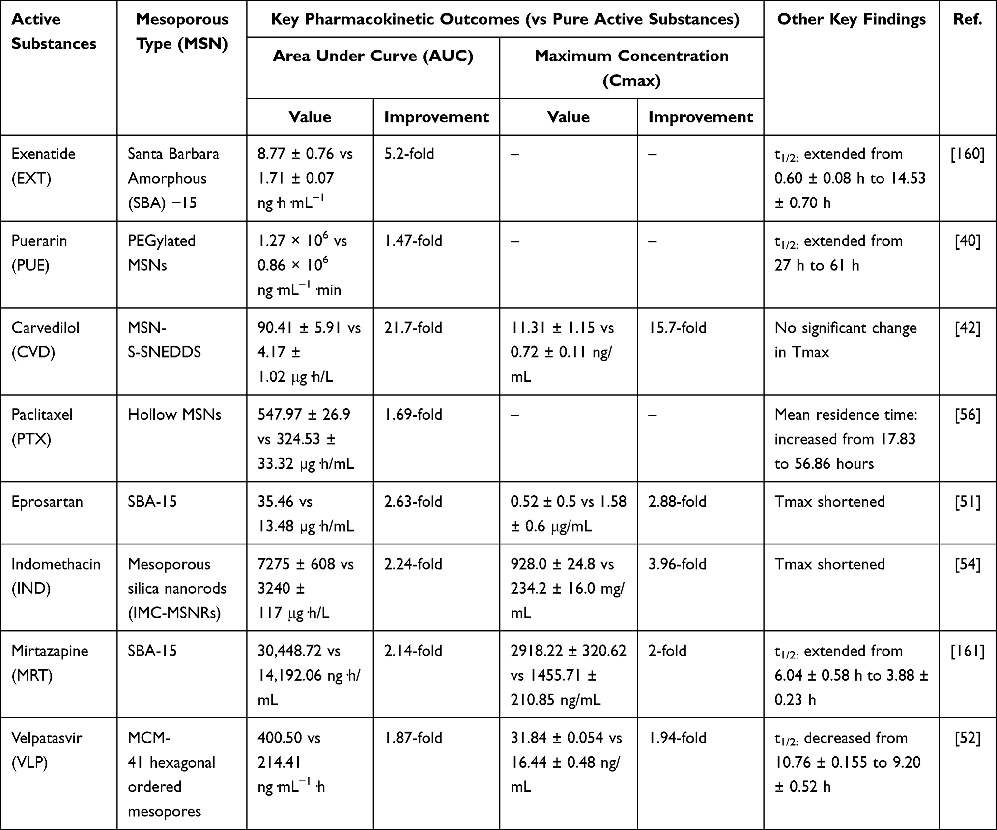 |
Table 2 Representative Pharmacokinetic Improvements Achieved with MSN-Based Formulations |
Area Under Curve
Pharmacokinetic evaluation of three selected studies showed that mesoporous silica nanoparticles (MSNs) increased the systemic bioavailability of drugs, as evidenced by a significant increase in the area under the plasma concentration-time curve (AUC). Chen et al (2017) evaluated the pharmacokinetic profile of exenatide (EXT) loaded into SBA-15 mesoporous silica (EXT-SBA-15) compared with an aqueous solution of EXT (EXT-Sol). The results indicated a pronounced enhancement of drug exposure, where the AUC0–∞ of EXT-SBA-15 was significantly higher (8.77 ± 0.76 ng·h·mL−1) than that of EXT-Sol (1.71 ± 0.07 ng·h·mL−1), accompanied by a remarkable extension of half-life from 0.60 ± 0.08 h to 14.53 ± 0.70 h. These findings highlight the ability of SBA-15 carriers to prolong systemic circulation and achieve sustained release of peptide drugs.40 In another example, Liu et al (2016) assessed the pharmacokinetic profile of puerarin encapsulated in PEGylated MSNs. Compared with puerarin injection, PUE-PEG-MSNs achieved a 1.47-fold increase (from 0.86 × 10⁶ to 1.27 × 10⁶ ng·mL−1·min) in AUC and a 2.3-fold prolongation of half-life (from 27 to 61 min), demonstrating the dual benefits of enhanced drug exposure and extended circulation time. The PEG surface modification was particularly important in reducing rapid clearance and improving systemic retention.42
A study by Jang et al (2015) on a solid self-nanoemulsifying drug delivery system (S-SNEDDS) based on MSN for carvedilol found that this combination was able to form an emulsion with very small droplet sizes (~130 nm) when it came into contact with gastrointestinal fluids. This result is consistent with the high surface-area-to-volume ratio of MSNs, which promotes rapid supersaturation and maximizes the concentration gradient for intestinal absorption. As a result, carvedilol in the 500 MSN formulation showed a 21.7-fold increase in AUC (4.17 ± 1.02 to 90.41 ± 5.91 μg·h/L) and a 15.7-fold increase in Cmax (from 0.72 ± 0.11 to 11.31 ± 1.15 µg/mL) compared to the pure drug, with no significant change in Tmax. Collectively, the observed enhancements in AUC can be mechanistically attributed to the intrinsic physicochemical properties of MSNs. The high specific surface area and ordered mesoporous architecture of carriers such as SBA-15 and PEG-MSNs promote efficient drug loading and stabilize the encapsulated payload in a high-energy amorphous state. This nanoscale confinement enhances apparent solubility and dissolution rate while enabling sustained, diffusion-controlled release from the pore channels, thereby prolonging therapeutic plasma concentrations and substantially increasing total systemic exposure.
Clearance
Clearance describes the body’s efficiency in eliminating a drug from systemic circulation: high clearance results in rapid removal and lower exposure, while low clearance slows elimination and prolongs systemic exposure.162 A study by Fu et al (2016) demonstrated that incorporating paclitaxel into mesoporous silica nanoparticles (MSNs) markedly altered the disposition of the drug following intraperitoneal (IP) administration. The MSN formulation produced a sustained drug-release profile within the peritoneal cavity, resulting in a substantial prolongation of drug retention compared to free paclitaxel. Pharmacokinetic evaluation showed that the MSN-based system increased the peritoneal AUC from 324.53 ± 33.32 to 547.97 ± 26.9 µg·h/mL, representing a 1.69-fold enhancement in exposure. Because clearance is inversely proportional to AUC (CL ∝ 1/AUC), this increase in peritoneal exposure corresponds to an estimated 41% reduction in apparent peritoneal clearance relative to the control formulation. The mean residence time (MRT) similarly increased from 17.83 to 56.86 hours, indicating prolonged local drug persistence. Together, these findings highlight that MSNs significantly slow the elimination of paclitaxel from the peritoneal cavity, promoting sustained locoregional drug availability and enhancing tumor accumulation by 6.5-fold.51 Mechanistically, this reduction in clearance is a direct consequence of the sustained, diffusion-controlled release enabled by the ordered mesoporous architecture of the hollow MSNs employed in the study. The high pore volume and tortuous diffusion pathways inherent to mesoporous carriers retard drug efflux from the administration site, effectively decoupling local drug disposition from rapid systemic elimination. The reduction in clearance, combined with improved retention and targeted delivery, underscores the potential of MSN-based carriers for optimizing paclitaxel pharmacokinetics.
Maximum Concentration
Cmax reflects the peak plasma concentration, where excessively high levels can trigger concentration-dependent toxicities, while low values may lead to insufficient therapeutic effect. Abdellatif and Gebreel (2025) the pharmacokinetic profile of eprosartan loaded into spheroidal mesoporous silica SBA-15 and aminofunctionalized mesoporous silica SBA-15 was evaluated to assess the improvement in drug exposure. The results demonstrated a significant enhancement in systemic availability, where the Cmax of the MSNs film increased by 2.88-fold (Cmax, 4.52 ± 0.5 μg/mL) compared to the oral dispersion of eprosartan (Cmax, 1.58 ± 0.6 μg/mL), with a statistically significant difference (unpaired t-test, p = 0.029). This elevation in Cmax can be mechanistically linked to the amino-functionalization of the SBA-15 surface. The introduction of amine groups enhances mucoadhesion and membrane permeability, promoting faster drug absorption across the buccal mucosa and consequently elevating peak plasma concentrations. These findings indicate that incorporating eprosartan into a mesoporous silica nanosystem notably enhances the absorption rate and the extent of drug exposure, suggesting an improvement in bioavailability attributed to better dissolution and permeability characteristics provided by the MSN-based delivery system.163 In addition, differences in the shape of mesoporous silica can also affect Cmax. Indomethacin as a drug is loaded in mesoporous silica with different structures, namely mesoporous silica nanospheres (IMC-MSNs) and mesoporous silica nanorods (IMC-MSNRs). In the Cmax evaluation, IMC-MSNRs had a higher concentration in blood plasma (2.22-fold) with a value of 928.0 ± 24.8 mg/mL compared to IMC-MSNs which had a Cmax of 417.4 ± 36.3 mg/mL. While in free IMC the Cmax value only showed 234.2 ± 16.0 mg/mL. The superior performance of IMC-MSNRs is directly attributable to particle morphology. The elongated nanorod geometry provides a higher surface area-to-volume ratio and a unique helical pore structure. These features accelerate dissolution kinetics and prolong systemic circulation time compared to isotropic nanospheres, thereby maximizing the peak plasma concentration. Furthermore, in the dissolution test, IMC-MSNRs had a faster dissolution profile. This is because IMC-MSNRs have a helical pore structure and a larger surface area ratio.50 These examples underscore that rational manipulation of MSN size, shape, and surface chemistry can be strategically employed to fine-tune Cmax and optimize the onset of therapeutic action.
Half-Life
Half-life (t½) indicates how long a drug persists in the body; a prolonged t½ extends therapeutic effects but may cause accumulation and adverse outcomes, whereas a short t½ requires more frequent dosing to maintain efficacy.28 Mussalam et al (2022) the pharmacokinetic study comparing MRT oral suspension and optimized MRT-SBA-15, the t½ of the optimized formulation (604 ± 0.58 hr) was significantly longer than that of the oral suspension (3.88 ± 0.23 hr). This 1.56-fold increase in elimination half-life suggests that the mesoporous silica-based delivery system slowed down the drug’s elimination rate, thereby maintaining therapeutic concentrations for a longer duration. This prolongation of t½ is mechanistically linked to the ordered mesoporous architecture of SBA-15, which functions as a drug reservoir enabling sustained, diffusion-controlled release. The gradual liberation of mirtazapine from the pore channels prolongs the absorption phase and delays the onset of elimination, thereby extending the apparent terminal half-life. Such an extended t½ may contribute to improved patient compliance and enhanced therapeutic efficacy, supporting the potential of MRT-SBA-15 as a sustained-release formulation.54 Furthermore, a study conducted by Mehmood et al (2020) evaluated the pharmacokinetic profiles of free velpatasvir namely an antivirus that has low solubility in water and velpatasvir loaded in mesoporous silica (VLP-MSN) which is expected to increase the bioavailability of velpatasvir. One of the parameters evaluated was the half-life (t1/2) to find out how long it takes for a drug to reach half of its initial concentration in the body. The test results reported that the t1/2 for VLP-MSN was 9.20 ± 0.52, indicating a decrease compared to free MSN, which had a t1/2 value of 10.76 ± 0.155. This apparent shortening of half-life paradoxically reflects improved bioavailability. By stabilizing velpatasvir in an amorphous state within the mesoporous matrix, the MSN carrier dramatically accelerates dissolution, shifting the rate-limiting step from absorption to elimination. The drug is absorbed more rapidly and completely, leading to faster systemic clearance and a shorter observed t½, yet with achieving an overall systemic exposure.49
Pharmacological Activity of MSN-Based Formulations
In addition to pharmacokinetic improvements, the three selected studies also showed significant increases in pharmacological activity after MSN-based drug delivery. Chen et al (2017) reported that exenatide encapsulated in SBA-15 (EXT-SBA-15) produced a markedly prolonged hypoglycemic effect in diabetic mice, with blood glucose levels significantly reduced for up to 25 days after a single administration, in contrast to the rapid decline observed with EXT solution. This sustained pharmacodynamic effect closely aligned with the extended half-life and increased AUC reported in the pharmacokinetic study, thereby confirming the therapeutic advantage of MSN encapsulation for peptide drugs.40 Similarly, Alhowyan et al (2023) demonstrated that ocular delivery of 5-fluorouracil using carboxymethyl chitosan-coated MSNs (AMSN-CMC-FU) not only enhanced ocular bioavailability but also translated into superior anticancer efficacy in vitro, while histopathological examination in rabbits confirmed good ocular tolerance without signs of irritation. These results suggest that pharmacokinetic gains achieved through MSN formulations directly support improved therapeutic activity and safety in ophthalmic drug delivery.41 Furthermore, Liu et al (2016) showed that puerarin loaded into PEGylated MSNs (PUE@PEG-MSNs) conferred superior cardioprotective effects in a rat model of myocardial ischemia compared with puerarin injection. The enhanced activity was attributed to increased bioavailability and extended systemic circulation, which allowed the antioxidant and vasodilatory properties of puerarin to be sustained at therapeutically relevant levels.42
Safety and Toxicity of MSN-Based Formulations
The translational potential of MSNs is fundamentally tied to their biocompatibility and long-term fate in vivo. Although amorphous silica is generally regarded as safe due to its degradation into orthosilicic acid (Si(OH)4) a naturally excretable metabolite the biological behavior of MSNs is governed by multiple physicochemical parameters, including particle size, surface charge, porosity, and coating chemistry.52 Unmodified MSNs, particularly those containing residual surfactants, have been shown to induce oxidative stress and membrane damage at elevated concentrations (>200 μg/mL) in vitro.164 However, surface functionalization with polymers such as PEG, chitosan, or phospholipids substantially reduces nonspecific protein adsorption and improves hemocompatibility, often maintaining hemolysis rates below 5% under physiological conditions.165 While cationic surface moieties enhance cellular uptake, they may also trigger platelet activation and hemolysis, underscoring the importance of charge-neutral or zwitterionic coatings for systemic administration.166 With respect to long-term biodistribution, systemically administered MSNs are predominantly sequestered by the mononuclear phagocyte system (MPS) within the liver and spleen. Particle size critically influences clearance kinetics: smaller particles (<50 nm) exhibit partial renal excretion, whereas larger particles (>150 nm) undergo slower degradation via hydrolysis of siloxane (Si–O–Si) bonds, followed by urinary elimination of orthosilicic acid.167,168 Chronic exposure studies in rodent models have revealed minimal organ accumulation and no significant histopathological abnormalities at therapeutic doses (<50 mg/kg).169 Additionally, the formation of a dynamic protein corona upon contact with biological fluids modulates opsonization and immune recognition. PEGylated or zwitterionic surfaces preferentially adsorb dysopsonins such as albumin and clusterin, thereby prolonging systemic circulation.170 Collectively, while well-engineered MSNs exhibit a favorable acute safety profile, comprehensive evaluation of chronic exposure, immunotoxicity, and organ-specific retention remains essential for clinical advancement.171
Despite robust preclinical efficacy, the clinical translation of MSN-based pharmaceutical products is hindered by the absence of standardized regulatory pathways and the inherent complexity of hybrid nanomaterials. Unlike conventional small-molecule drugs, MSNs reside in a regulatory grey zone, necessitating case-by-case evaluation by agencies such as the FDA and EMA. Key translational challenges include ensuring batch-to-batch consistency in particle size, pore architecture, and surface chemistry—parameters that directly influence pharmacokinetic performance and immunogenicity.127 Establishing quality control protocols aligned with international standards (eg, ISO 10993 for biocompatibility and ICH Q8–Q10 for pharmaceutical development) is a prerequisite for Good Manufacturing Practice (GMP) production.172–175 Furthermore, regulatory approval requires comprehensive ADME profiling, including definitive data on silica degradation products and acceptable thresholds for residual surfactants or solvents.176,177 Although silica-based platforms such as “Cornell dots” have advanced to early-phase clinical trials for imaging applications the development pathway for drug-loaded MSNs remains in its infancy. Accelerating clinical translation will require harmonized pharmacological endpoints, standardized immunotoxicity assays, and long-term toxicological data generated under Good Laboratory Practice (GLP) conditions.178
Bridging the divide between laboratory-scale innovation and industrial application demands a fundamental shift toward scalable and reproducible manufacturing processes. The conventional sol–gel synthesis of MSNs is highly sensitive to minor fluctuations in pH, temperature, and stirring rate, often resulting in significant batch-to-batch variability in pore morphology and drug loading efficiency. Recent advances in continuous-flow microfluidic reactors offer a promising solution, enabling precise control over reactant mixing and residence time to achieve uniform particle size distributions on a multi-kilogram scale.129 Concurrently, the adoption of “green synthesis” strategies—employing biotemplates, ionic liquids, or supercritical fluids—addresses environmental concerns associated with traditional surfactant removal and organic solvent use. From a formulation perspective, maintaining long-term colloidal stability necessitates optimized lyophilization protocols incorporating cryoprotectants such as trehalose or mannitol.179 Post-synthetic modifications, including polymer grafting and ligand conjugation, must also be standardized to ensure consistent functionalization density, a factor that directly impacts pharmacokinetic behavior and therapeutic efficacy. Looking ahead, the integration of artificial intelligence (AI)-driven quality control and high-throughput screening will be instrumental in de-risking the manufacturing pipeline. By establishing robust, GMP-compliant supply chains and adhering to evolving regulatory guidelines. MSN-based delivery systems can transition from promising preclinical tools to clinically viable, next-generation therapeutics.
Discussion
A critical synthesis of the reviewed studies reveals a consistent yet highly tunable pharmacokinetic enhancement conferred by MSN-based delivery. The magnitude of improvement in AUC and Cmax is not uniform but varies substantially depending on the interplay between drug properties, MSN architecture, and surface functionalization strategies. Each drug integrated into MSNs demonstrates unique pharmacokinetic characteristics shaped by its physicochemical attributes, therapeutic category, and the particular alterations made to the MSN carrier. Numerous pharmaceuticals characterized by inherently inadequate oral absorption—such as peptides, natural compounds, and anticancer agents—encounter significant constraints, including low solubility, rapid enzymatic degradation, and brief systemic residence durations. These problems often lead to diminished bioavailability and inadequate therapeutic results. Although dose escalation may mitigate inadequate absorption, this strategy frequently increases the likelihood of systemic toxicity and treatment-related side effects. In this context, MSNs present a viable approach to surmount pharmacokinetic limitations by improving solubility, stabilizing unstable molecules, and regulating drug-release profiles via their distinctive structural and chemical characteristics. Their extensive surface area, adjustable pore structure, and tailored surface chemistry provide substantial drug loading, enhanced absorption, and extended systemic circulation, hence improving therapeutic effectiveness.
Numerous studies have demonstrated the ability of MSN formulations to significantly enhance pharmacokinetic performance. Quantitatively, the reviewed studies demonstrate substantial variability in pharmacokinetic enhancement, with reported AUC improvement ranging from 1.47-fold (puerarin-loaded MSNs) to 21.7-fold (carvedilol-loaded MSNs), and Cmax enhancements reaching up to 15.7-fold, underscoring the high tenability of MSN carrier. These quantitative findings are consistent with the pharmacokinetic data summarized in Table 1, where multiple MSN-based formulations demonstrate measurable improvements in systemic exposure. As illustrated in Figure 4, improvements in key pharmacokinetic parameters—including area under the curve (AUC), maximum plasma concentration (Cmax), and elimination half-life (t½)—arise from synergistic mechanisms intrinsic to the MSN platform. Confinement within mesoporous channels promotes drug amorphization or nanodispersion, reducing crystallinity and increasing the surface-area-to-volume ratio. At the molecular level, the spatial confinement of drug molecules within nanoscale pores restricts molecular mobility and prevents the organization required for nucleation and crystal growth. In addition, interactions between drug molecules and the silanol groups on the silica surface – such as hydrogen bonding and electrostatic interactions – further inhibit recrystallization and stabilize the drug in a high-energy amorphous state. This dual effect of physical confinement and interfacial interaction plays a critical role in maintaining amorphous stability during storage and dissolution. As a result, this structural transformation enhances apparent solubility and accelerates dissolution kinetics, thereby increasing the fraction of drug available for absorption. Importantly, elevated AUC values often reflect an improved extent of absorption rather than merely accelerated absorption rates.
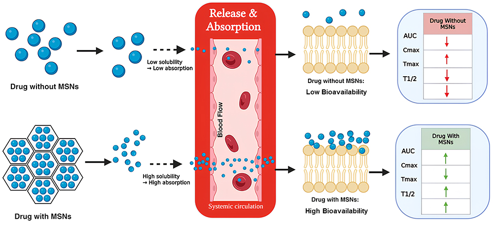 |
Figure 4 Comparative pharmacokinetic profiles of Drug without MSNs and Drug with MSNs. Upward arrows (↑) indicate an increase in pharmacokinetic parameters (eg, Cmax, AUC), while downward arrows (↓) indicate a decrease. Rightward arrows (→) represent a shift in the absorption or elimination profile. |
The ordered porous architecture further enables controlled and sustained release, minimizing burst effects while prolonging systemic exposure. Such release modulation can extend plasma residence time and, in some cases, increase the apparent half-life without fundamentally altering intrinsic elimination pathways. Surface functionalization amplifies these effects: modifications such as PEGylation or ligand conjugation reduce opsonization, limit recognition by the reticuloendothelial system, and decrease nonspecific plasma protein interactions. Collectively, these strategies prolong circulation time and enhance the probability of drug accumulation at target tissues.
The enhanced systemic exposure noted in Table 1 and Table 2 is a direct result of the interplay between diffusion-controlled and stimulus-responsive release mechanisms intrinsic to the MSN platform. In diffusion-controlled systems, the mesoporous channels act as a reservoir, where drug release is primarily governed by Fickian diffusion. For instance, Fu et al (2016) utilized hollow MSNs to achieve sustained, diffusion-mediated release of paclitaxel, leading to extended retention in the peritoneal cavity (mean residence time [MRT] increased from 17.8 to 56.9 hours) and improved tumor accumulation.180 Similarly, Liu et al (2016) demonstrated that PEGylation of MSNs for puerarin encapsulation altered the diffusion characteristics by creating a hydrophilic barrier, which mitigated the initial burst release and enhanced the AUC by 1.47-fold. Beyond passive diffusion, surface functionalization facilitates stimulus-responsive activity, which further optimizes pharmacokinetic profile.51 For example, Jang et al (2015) incorporated carvedilol into an MSN-based self-nanoemulsifying drug delivery system (S-SNEDDS), creating a supersaturated condition upon interaction with gastrointestinal fluids. This stimulus-responsive process promoted rapid and complete absorption, resulting in a 21.7-fold increase in AUC.42 Similarly, Alhowyan et al (2023) employed carboxymethyl chitosan-coated MSNs for ocular delivery of 5-fluorouracil, where the coating enabled pH-triggered controlled release in the eye, maintaining therapeutic levels and resulting in a 6.14-fold enhancement in ocular AUC.56 These examples emphasize the critical role that controlling the timing and location of drug release plays in optimizing pharmacokinetics, whether through manufactured diffusion barriers or environmental triggers.
The magnitude of pharmacokinetic enhancement is highly dependent on formulation-specific parameters, including pore size, particle size, surface chemistry, and drug-to-carrier ratio. Suboptimal design may result in insufficient loading, premature leakage, or excessively slow release. Therefore, pharmacokinetic modulation by MSNs should be viewed as a tunable process requiring rational optimization to balance solubility enhancement, release kinetics, biodistribution, and systemic stability. Through careful engineering of mesoporous architecture and surface properties, MSNs can translate physicochemical modifications into meaningful alterations in systemic drug exposure. These pharmacokinetic improvements are closely linked to therapeutic performance. Increased systemic exposure and prolonged retention can enhance pharmacodynamic response, reduce dosing frequency, and potentially mitigate peak-related toxicity by smoothing plasma concentration fluctuations. Thus, the principal contribution of MSNs extends beyond bioavailability enhancement to the strategic reshaping of drug exposure profiles in order to optimize therapeutic outcomes.
Despite the promising pharmacokinetic advancements, the clinical application of MSN-based drug delivery systems faces several significant challenges. Aggregation remains a major concern, particularly in aqueous environments, where MSNs can cluster, negatively impacting their stability, drug-loading capacity, and controlled release properties. Additionally, premature drug release can occur when MSNs are exposed to physiological conditions that compromise their structural integrity, leading to reduced therapeutic efficacy and potentially increased toxicity. Another challenge is the biodegradability of MSNs, as their slow degradation rate may result in accumulation over time, causing long-term toxicity or triggering immune responses. Notably, The toxicity and tolerability of MSNs are significantly dependent on the formulation. According to the Safety and Toxicity section, unmodified MSNs can cause oxidative stress at high doses (>200 μg/mL), but surface-functionalized MSNs demonstrate much enhanced hemocompatibility, with hemolysis rates around 5%. Particle size determines clearance routes, with diminutive particles promoting renal excretion and bigger particles collecting in the liver and spleen. This heterogeneity highlights the necessity for rigorous, formulation-specific safety assessments.
To overcome these challenges, surface functionalization plays a crucial role in improving stability, targeting, and biocompatibility of MSNs. Modifications such as PEGylation or grafting targeting ligands (eg, folic acid) enhance colloidal stability, extend circulation time, and enable targeted drug delivery, which optimizes pharmacokinetics. For example, Liu et al (2016) demonstrated that PEGylation of MSNs for puerarin encapsulation altered diffusion characteristics, reducing the initial burst release and improving AUC by 1.47-fold. These functionalizations help mitigate premature drug release, enhance therapeutic efficacy, and improve drug retention at the target site, ultimately boosting the therapeutic index.41 The integration of MSN with other targeting ligands or hybrid systems, as seen in recent nanomedicine developments, could further refine the precision of drug delivery.42,181,182 Moreover, while many studies show favorable short-term biocompatibility, long-term safety data remain limited. Concerns such as chronic accumulation, organ-specific retention, immunological modulation, and prolonged degradation require further investigation. The absence of standardized toxicological evaluation frameworks complicates regulatory assessments and inter-study comparability. Addressing these issues requires ongoing innovations in surface functionalization, encapsulation techniques, and release rate control to ensure that MSNs are biodegradable or can be safely eliminated after completing their therapeutic role. Future advancements in MSN-based systems will need to integrate pharmacokinetic optimization with clearance pathway investigations, biodegradation kinetics, and chronic safety profiles to ensure that enhanced systemic exposure is met with consistent and reliable safety assurance.
Conclusion
In conclusion, mesoporous silica nanoparticles (MSNs) show strong potential as drug delivery systems to improve the solubility, bioavailability, and pharmacokinetic profiles of poorly soluble drugs. Their high surface area, tunable pore size, and versatile surface modification enable efficient drug loading, stabilization of drugs in an amorphous form, and controlled release, leading to enhanced absorption and systemic exposure. However, despite promising preclinical results, the clinical translation of MSN-based formulations remains challenged by concerns regarding long-term safety, clearance mechanisms, large-scale manufacturing, and the lack of standardized regulatory frameworks. Future research should focus on chronic toxicity studies in large animal models to enhance predictions of human biodistribution and organ retention, the development of physiologically based pharmacokinetic (PBPK) models to simulate the in vivo fate of MSNs, and the development of standardized characterization protocols to guarantee batch-to-batch consistency under Good Manufacturing Practice (GMP) conditions. Moreover, meticulously designed early-phase clinical trials would be crucial to confirm the safety and pharmacokinetic benefits demonstrated in preclinical studies. By methodically addressing these translational limitations, MSN-based delivery systems can progress from promising laboratory instruments to clinically viable platforms for next-generation medicines.
Author’s Perspective
Current evidence positions mesoporous silica nanoparticles (MSNs) as one of the most adaptable nanoplatforms for overcoming fundamental pharmacokinetic barriers across diverse therapeutic classes. Improvements in solubility, molecular stability, and systemic exposure have been demonstrated repeatedly, suggesting that the advantages of MSNs extend far beyond any single drug category. Yet, despite this breadth of potential, most advances remain confined to preclinical exploration. A pronounced gap still separates mechanistic promise from clinically deployable technology. Bridging this divide requires a shift from predominantly empirical optimization toward predictive, design-oriented development frameworks capable of translating physicochemical enhancements into reproducible therapeutic benefit.
Several entrenched challenges continue to limit the progression of MSNs toward clinical maturity. Formulation-dependent variability in loading efficiency, release kinetics, and systemic stability remains substantial, undermining reproducibility both within and across research groups. The absence of standardized characterization protocols further complicates cross-study interpretation, while long-term human data on biodistribution, clearance, biodegradation, and chronic exposure remain markedly insufficient to establish regulatory confidence. Manufacturing constraints compound these issues: achieving batch-to-batch control over pore architecture and surface chemistry—parameters central to performance—remains technically demanding and difficult to scale. Without decisive solutions to these interconnected obstacles, MSNs risk remaining confined to proof-of-concept demonstrations rather than advancing toward meaningful clinical implementation.
Realizing the clinical promise of MSNs demands a decisive evolution in research strategy. Progress will rely on rational design principles supported by computational modeling, machine-learning–based prediction of drug–carrier interactions, and quantitative structure–property frameworks that enable anticipatory formulation design. Equally critical is comprehensive evaluation of long-term safety, biodegradation pathways, and in vivo fate under clinically relevant dosing paradigms to define realistic translational boundaries. Parallel development of scalable, quality-assured manufacturing pipelines is essential to ensure physicochemical uniformity across production sites and regulatory jurisdictions. Momentum in these areas would reposition MSNs not merely as experimental enhancers of pharmacokinetics, but as robust, clinically credible platforms with the capacity to support next-generation therapeutics—particularly those constrained by poor solubility, structural fragility, or rapid systemic clearance.
Acknowledgments
This research was funded by Universitas Padjadjaran through the Indonesian Endowment Fund for Education (LPDP) on behalf of the Indonesian Ministry of Higher Education, Science and Technology and managed under the EQUITY Program (Contract No. 4303/83/DT.03.08/2025 and 3927/UN6. RKT/HK.07.00/2025).
Funding
This research was funded by Universitas Padjadjaran through the Indonesia Endowment Funds for Education (LPDP) to Diah Lia Aulifa (Hibah EQUITY-WCU Kemdiktisaintek) No: 3907/UN6.3.1/PT.00/2025).
Disclosure
The authors report no conflicts of interest in this work.
References
1. Kumari L, Choudhari Y, Patel P, et al. Advancement in solubilization approaches: a step towards bioavailability enhancement of poorly soluble drugs. Life. 2023;13(5). doi:10.3390/life13051099
2. Wu CY, Benet LZ. Predicting drug disposition via application of BCS: transport/absorption/elimination interplay and development of a biopharmaceutics drug disposition classification system. Pharm Res. 2005;22(1):11–30. doi:10.1007/s11095-004-9004-4
3. Camenisch GP. Drug disposition classification systems in discovery and development: a comparative review of the BDDCS, ECCS and ECCCS concepts. Pharm Res. 2016;33(11):2583–2593. doi:10.1007/s11095-016-2001-6
4. Dahan A, Miller JM, Amidon GL. Prediction of solubility and permeability class membership: provisional BCS classification of the world’s top oral drugs. AAPS J. 2009;11(4):740–746. doi:10.1208/s12248-009-9144-x
5. Kumar S, Bhargava D, Thakkar A, Arora S. Drug carrier systems for solubility enhancement of BCS class II drugs: a critical review. Crit Rev Ther Drug Carrier Syst. 2013;30(3):217–256. doi:10.1615/CritRevTherDrugCarrierSyst.2013005964
6. Parr A, Hidalgo IJ, Bode C, et al. The effect of excipients on the permeability of BCS class III compounds and implications for biowaivers. Pharm Res. 2016;33(1):167–176. doi:10.1007/s11095-015-1773-4
7. Flanagan T. Potential for pharmaceutical excipients to impact absorption: a mechanistic review for BCS class 1 and 3 drugs. Eur J Pharm Biopharm. 2019;141:130–138. doi:10.1016/j.ejpb.2019.05.020
8. Kesisoglou F, Wu Y. Understanding the effect of API properties on bioavailability through absorption modeling. AAPS J. 2008;10(4):516–525. doi:10.1208/s12248-008-9061-4
9. Savjani KT, Gajjar AK, Savjani JK. Drug solubility: importance and enhancement techniques. ISRN Pharm. 2012;2012:1–10. doi:10.5402/2012/195727
10. Amalia E, Afinasari A. Nanoparticle drug delivery system. Indonesia J Pharmaceut. 2022;4(2):347–359. doi:10.24198/idjp.v4i1.35312
11. Huang P, Lian D, Ma H, et al. New advances in gated materials of mesoporous silica for drug controlled release. Chin Chem Lett. 2021;32(12):3696–3704. doi:10.1016/j.cclet.2021.06.034
12. Ezike TC, Okpala US, Onoja UL, et al. Advances in drug delivery systems, challenges and future directions. Heliyon. 2023;9(6):e17488. doi:10.1016/j.heliyon.2023.e17488
13. Fan Y, Marioli M, Zhang K. Analytical characterization of liposomes and other lipid nanoparticles for drug delivery. J Pharm Biomed Anal. 2021;192:113642. doi:10.1016/j.jpba.2020.113642
14. Thakur S, Godela R, Mandava K, Kolure R. Advances in nanocarrier technology for drug encapsulation: a comprehensive overview. Discov Mater. 2025;5(1):124. doi:10.1007/s43939-025-00271-1
15. Kurul F, Turkmen H, Cetin AE, Topkaya SN. Nanomedicine: how nanomaterials are transforming drug delivery, bio-imaging, and diagnosis. Next Nanotechnol. 2025;7:100129. doi:10.1016/j.nxnano.2024.100129
16. Mashweu AR, Azov VA. Nanotechnology in drug delivery: anatomy and molecular insight into the self-assembly of peptide-based hydrogels. Molecules. 2024;29(23):5654. doi:10.3390/molecules29235654
17. Wang D, Jiang Q, Dong Z, et al. Nanocarriers transport across the gastrointestinal barriers: the contribution to oral bioavailability via blood circulation and lymphatic pathway. Adv Drug Deliv Rev. 2023;203:115130. doi:10.1016/j.addr.2023.115130
18. Laracuente ML, Yu MH, McHugh KJ. Zero-order drug delivery: state of the art and future prospects. J Control Release. 2020;327:834–856. doi:10.1016/j.jconrel.2020.09.020
19. Yuan H, Chen Y, Hu Y, et al. Disulfide bond-driven nanoassembly of lipophilic epirubicin prodrugs for breast cancer therapy. J Pharm Investig. 2025;55(6):889–902. doi:10.1007/s40005-025-00731-z
20. Feng C, Wang Y, Xu J, et al. Precisely tailoring molecular structure of doxorubicin prodrugs to enable stable nanoassembly, rapid activation, and potent antitumor effect. Pharmaceutics. 2024;16(12):1582. doi:10.3390/pharmaceutics16121582
21. Nie Y, Li D, Peng Y, et al. Metal organic framework coated MnO2 nanosheets delivering doxorubicin and self-activated DNAzyme for chemo-gene combinatorial treatment of cancer. Int J Pharm. 2020;585:119513. doi:10.1016/j.ijpharm.2020.119513
22. Zhang W, Liu H, Qiu X, Zuo F, Wang B. Mesoporous silica nanoparticles as a drug delivery mechanism. Open Life Sci. 2024;19(1):20220867. doi:10.1515/biol-2022-0867
23. Ali NS, Harharah HN, Salih IK, Cata Saady NM, Zendehboudi S, Albayati TM. Applying MCM-48 mesoporous material, equilibrium, isotherm, and mechanism for the effective adsorption of 4-nitroaniline from wastewater. Sci Rep. 2023;13(1):9837. doi:10.1038/s41598-023-37090-4
24. Djayanti K, Maharjan P, Cho KH, et al. Mesoporous silica nanoparticles as a potential nanoplatform: therapeutic applications and considerations. Int J Mol Sci. 2023;24(7):6349. doi:10.3390/ijms24076349
25. Budiman A, Mutmainah L, Anjelina M, et al. The application of mesoporous silica nanoparticles in enhancing the efficacy of anti-atherosclerosis therapies: a review. Int J Nanomed. 2025;20:9825–9856. doi:10.2147/IJN.S538100
26. Asiri Y. The role of pharmacokinetics in pharma ceutical toxicology. J Pharmaceut Toxicol. 2023;6(2):54–56. doi:10.37532/jpt
27. Dewangan T. Pharmacokinetic considerations in drug development: a review no title. Southafrican Gastroenterol Rev. 2024;22(3):169–186.
28. Yamazaki S. Relationships of changes in pharmacokinetic parameters of substrate drugs in drug–drug interactions on metabolizing enzymes and transporters. J Clin Pharmacol. 2018;58(8):1053–1060. doi:10.1002/jcph.1104
29. Nyamba I, Sombié CB, Yabré M, et al. Pharmaceutical approaches for enhancing solubility and oral bioavailability of poorly soluble drugs. Eur J Pharm Biopharm. 2024;204:114513. doi:10.1016/j.ejpb.2024.114513
30. Boyd BJ, Bergström CAS, Vinarov Z, et al. Successful oral delivery of poorly water-soluble drugs both depends on the intraluminal behavior of drugs and of appropriate advanced drug delivery systems. Eur J Pharm Sci. 2019;137:104967. doi:10.1016/j.ejps.2019.104967
31. Shegokar R, Müller RH. Nanocrystals: industrially feasible multifunctional formulation technology for poorly soluble actives. Int J Pharm. 2010;399(1–2):129–139. doi:10.1016/j.ijpharm.2010.07.044
32. Suib SL. A review of recent developments of mesoporous materials. Chem Rec. 2017;17(12):1169–1183. doi:10.1002/tcr.201700025
33. Homayun B, Lin X, Choi HJ. Challenges and recent progress in oral drug delivery systems for biopharmaceuticals. Pharmaceutics. 2019;11(3):129. doi:10.3390/pharmaceutics11030129
34. Liu Y, Liang Y, Yuhong J, et al. Advances in nanotechnology for enhancing the solubility and bioavailability of poorly soluble drugs. Drug Des Devel Ther. 2024;18:1469–1495. doi:10.2147/DDDT.S447496
35. Preeti, Sambhakar S, Malik R, et al. Nanoemulsion: an emerging novel technology for improving the bioavailability of drugs. Scientifica. 2023;2023:1–25. doi:10.1155/2023/6640103
36. Sabjan KB, Munawar SM, Rajendiran D, Vinoji SK, Kasinathan K. Nanoemulsion as oral drug delivery - a review. Curr Drug Res Rev. 2020;12(1):4–15. doi:10.2174/2589977511666191024173508
37. Haripriyaa M, Suthindhiran K. Pharmacokinetics of nanoparticles: current knowledge, future directions and its implications in drug delivery. Futur J Pharm Sci. 2023;9(1):113. doi:10.1186/s43094-023-00569-y
38. Florek J, Caillard R, Kleitz F. Evaluation of mesoporous silica nanoparticles for oral drug delivery – current status and perspective of MSNs drug carriers. Nanoscale. 2017;9(40):15252–15277. doi:10.1039/C7NR05762H
39. Dogra P, Adolphi NL, Wang Z, et al. Establishing the effects of mesoporous silica nanoparticle properties on in vivo disposition using imaging-based pharmacokinetics. Nat Commun. 2018;9(1):4551. doi:10.1038/s41467-018-06730-z
40. Chen C, Zheng H, Xu J, Shi X, Li F, Wang X. Sustained-release study on exenatide loaded into mesoporous silica nanoparticles: in vitro characterization and in vivo evaluation. DARU J Pharma Sci. 2017;25(1):20. doi:10.1186/s40199-017-0186-9
41. Alhowyan AA, Kalam MA, Iqbal M, et al. Mesoporous silica nanoparticles coated with carboxymethyl chitosan for 5-fluorouracil ocular delivery: characterization, in vitro and in vivo studies. Molecules. 2023;28(3):1260. doi:10.3390/molecules28031260
42. Liu X, Ding Y, Zhao B, et al. In vitro and in vivo evaluation of puerarin-loaded PEGylated mesoporous silica nanoparticles. Drug Dev Ind Pharm. 2016;42(12):2031–2037. doi:10.1080/03639045.2016.1190742
43. Kanwar S, Vijayavenkataraman S. 3D-printed polycaprolactone-magnetic nanoparticles composite multifunctional scaffolds for bone tissue regeneration and hyperthermia treatment. Int J Bioprint. 2024;4538. doi:10.36922/ijb.4538
44. Zhou J, Zhu F, Li J, Wang Y. Concealed body mesoporous silica nanoparticles for orally delivering indometacin with chiral recognition function. Mater Sci Eng C. 2018;90:314–324. doi:10.1016/j.msec.2018.04.071
45. Ke J, Wang Y, Wang L, et al. Synthesis and characterization of core-shell mesoporous silica nanoparticles with various shell thickness as indomethacin carriers: in vitro and in vivo evaluation. Microporous Mesoporous Mater. 2020;297:110043. doi:10.1016/j.micromeso.2020.110043
46. Popovici R, Alexa I, Novac O, et al. Pharmacokinetics study on mesoporous silica-captopril controlled release systems. Dig J Nanomater Biostruct. 2011;6:1619–1630.
47. Pandita D, Munjal A, Poonia N, Awasthi R, Kalonia H, Lather V. Albumin-coated mesoporous silica nanoparticles of docetaxel: preparation, characterization, and pharmacokinetic evaluation. Assay Drug Dev Technol. 2021;19(4):226–236. doi:10.1089/adt.2020.1039
48. Jia L, Shen J, Li Z, et al. In vitro and in vivo evaluation of paclitaxel-loaded mesoporous silica nanoparticles with three pore sizes. Int J Pharm. 2013;445(1–2):12–19. doi:10.1016/j.ijpharm.2013.01.058
49. Zhang J, Han R, Chen W, et al. Analysis of the literature and patents on solid dispersions from 1980 to 2015. Molecules. 2018;23(7):1697. doi:10.3390/molecules23071697
50. Korzekwa K, Nagar S. Process and system clearances in pharmacokinetic models: our basic clearance concepts are correct. Drug Metab Dispos. 2023;51(4):532–542. doi:10.1124/dmd.122.001060
51. Fu Q, Hargrove D, Lu X. Improving paclitaxel pharmacokinetics by using tumor-specific mesoporous silica nanoparticles with intraperitoneal delivery. Nanomedicine. 2016;12(7):1951–1959. doi:10.1016/j.nano.2016.04.013
52. Musallam AA, Mahdy MA, Elnahas HM, Aldeeb RA. Optimization of mirtazapine loaded into mesoporous silica nanostructures via Box-Behnken design: in-vitro characterization and in-vivo assessment. Drug Deliv. 2022;29(1):1582–1594. doi:10.1080/10717544.2022.2075985
53. Sun JG, Jiang Q, Zhang XP, et al. Mesoporous silica nanoparticles as a delivery system for improving antiangiogenic therapy. Int J Nanomed. 2019;14:1489–1501. doi:10.2147/IJN.S195504
54. Abdellatif MM, Gebreel RM. Eprosartan loaded mesoporous silica nanoparticles embedded in mucoadhesive buccal films: a strategy for improved bioavailability. J Drug Deliv Sci Technol. 2025;112:107248. doi:10.1016/j.jddst.2025.107248
55. Mahajan RR, Ravi PR. Mesoporous silica-based amorphous solid dispersions to enhance the oral bioavailability of Neratinib maleate. J Drug Deliv Sci Technol. 2024;101:106157. doi:10.1016/j.jddst.2024.106157
56. Jang H, Kim N, Jin SG. Development of a carvedilol-loaded solid self-nanoemulsifying system with increased solubility and bioavailability using mesoporous silica nanoparticles. Int J Mol Sci. 2025;26(4):1592. doi:10.3390/ijms26041592
57. Trzeciak K, Chotera-Ouda A, Bak-Sypien II, Potrzebowski MJ. Mesoporous silica particles as drug delivery systems—the state of the art in loading methods and the recent progress in analytical techniques for monitoring these processes. Pharmaceutics. 2021;13(7):950. doi:10.3390/pharmaceutics13070950
58. Nasrollahzadeh M, Issaabadi Z, Sajjadi M, Sajadi SM, Atarod M. Types of nanostructures. Interf Sci Technol. 2019;28:29–80. doi:10.1016/B978-0-12-813586-0.00002-X
59. Shahbaz S, Esmaeili M, Fathian Nasab MH, et al. PEGylated mesoporous silica core–shell redox-responsive nanoparticles for delivering paclitaxel to breast cancer cells. Int J Pharm. 2024;655:124024. doi:10.1016/j.ijpharm.2024.124024
60. Chen SY, Kwek KK, Lee ZH, Lin HM. Synthesis and characterization of multifunctional mesoporous silica nanoparticles with dual targeting and dual-mode imaging for cancer therapy. J Photochem Photobiol B. 2025;273:113290. doi:10.1016/j.jphotobiol.2025.113290
61. Saputra OA, Lestari WA, Safitriono WN, Handayani M, Lestari WW, Wibowo FR. β-Amino alcohol-based organosilane tailored magnetite embedded mesoporous silica nanoparticles exhibit controlled-release of curcumin triggered by pH. Mater Lett. 2021;305:130804. doi:10.1016/j.matlet.2021.130804
62. Sábio RM, Meneguin AB, Martins Dos Santos A, Monteiro AS, Chorilli M. Exploiting mesoporous silica nanoparticles as versatile drug carriers for several routes of administration. Microporous Mesoporous Mater. 2021;312:110774. doi:10.1016/j.micromeso.2020.110774
63. Li J, Shen S, Kong F, Jiang T, Tang C, Yin C. Effects of pore size on in vitro and in vivo anticancer efficacies of mesoporous silica nanoparticles. RSC Adv. 2018;8(43):24633–24640. doi:10.1039/C8RA03914C
64. Niroumand U, Firouzabadi N, Goshtasbi G, Hassani B, Ghasemiyeh P, Mohammadi-Samani S. The effect of size, morphology and surface properties of mesoporous silica nanoparticles on pharmacokinetic aspects and potential toxicity concerns. Front Mater. 2023;10. doi:10.3389/fmats.2023.1189463
65. Kankala RK, Han Y, Na J, et al. Nanoarchitectured structure and surface biofunctionality of mesoporous silica nanoparticles. Adv Mater. 2020;32(23). doi:10.1002/adma.201907035
66. Karczmarska A, Laskowska W, Stróż D, Pawlik K. Inside the framework: structural exploration of mesoporous silicas MCM-41, SBA-15, and SBA-16. Materials. 2025;18(15):3597. doi:10.3390/ma18153597
67. Seljak KB, Kocbek P, Gašperlin M. Mesoporous silica nanoparticles as delivery carriers: an overview of drug loading techniques. J Drug Deliv Sci Technol. 2020;59:101906. doi:10.1016/j.jddst.2020.101906
68. Tsoncheva T, Ivanova L, Rosenholm J, Linden M. Cobalt oxide species supported on SBA-15, KIT-5 and KIT-6 mesoporous silicas for ethyl acetate total oxidation. Appl Catal B. 2009;89(3–4):365–374. doi:10.1016/j.apcatb.2008.12.015
69. Heikkilä T, Salonen J, Tuura J, et al. Evaluation of mesoporous TCPSi, MCM-41, SBA-15, and TUD-1 materials as API carriers for oral drug delivery. Drug Deliv. 2007;14(6):337–347. doi:10.1080/10717540601098823
70. Gonçalves MC. Sol-gel silica nanoparticles in medicine: a natural choice. Design, synthesis and products. Molecules. 2018;23(8):2021. doi:10.3390/molecules23082021
71. Robertson M, Guillen-Obando A, Barbour A, Smith P, Griffin A, Qiang Z. Direct synthesis of ordered mesoporous materials from thermoplastic elastomers. Nat Commun. 2023;14(1):639. doi:10.1038/s41467-023-36362-x
72. Rajotia M, Yadav A, Saroj VK, Panda S. Engineering silica nanoparticles for precision nanomedicine: synthesis & functionalization – a review. RSC Sustainability. 2026;4(1):142–156. doi:10.1039/D5SU00741K
73. Narayan R, Nayak UY, Raichur AM, Garg S. Mesoporous silica nanoparticles: a comprehensive review on synthesis and recent advances. Pharmaceutics. 2018;10(3):118. doi:10.3390/pharmaceutics10030118
74. Steinbach JC, Fait F, Mayer HA, Kandelbauer A. Sol–gel-controlled size and morphology of mesoporous silica microspheres using hard templates. ACS Omega. 2023;8(33):30273–30284. doi:10.1021/acsomega.3c03098
75. Han Y, Zhang L, Yang W. Synthesis of mesoporous silica using the sol–gel approach: adjusting architecture and composition for novel applications. Nanomaterials. 2024;14(11):903. doi:10.3390/nano14110903
76. Arcos D, López-Noriega A, Ruiz-Hernández E, Ruiz L, González-Calbet JM, Vallet-Regí M. Syntesis of mesoporous microparticles for biomedical applications. Key Eng Mater. 2008;377:181–194. doi:10.4028/www.scientific.net/KEM.377.181
77. Zhang F, Liu G, He W, et al. Mesoporous silica with multiple catalytic functionalities. Adv Funct Mater. 2008;18(22):3590–3597. doi:10.1002/adfm.200800458
78. Bae JY, Jang SG, Cho J, Kang M. Amine-functionalized mesoporous silica for efficient CO2 capture: stability, performance, and industrial feasibility. Int J Mol Sci. 2025;26(9):4313. doi:10.3390/ijms26094313
79. Fujii Y, Zhou S, Shimada M, Kubo M. Synthesis of monodispersed hollow mesoporous organosilica and silica nanoparticles with controllable shell thickness using soft and hard templates. Langmuir. 2023;39(13):4571–4582. doi:10.1021/acs.langmuir.2c03121
80. Barberena-Fernández AM, Carmona-Quiroga PM, Blanco-Varela MT. Interaction of TEOS with cementitious materials: chemical and physical effects. Cem Concr Compos. 2015;55:145–152. doi:10.1016/j.cemconcomp.2014.09.010
81. Rathinavel S, Ekambaram S, Korrapati PS, Sangeetha D. Retracted: design and fabrication of electrospun SBA-15-incorporated PVA with curcumin: a biomimetic nanoscaffold for skin tissue engineering. Biomed Mater. 2020;15(3):035009. doi:10.1088/1748-605X/ab6b2f
82. Porras M, Adrover ME, Pedernera M, Bucalá V, Gallo L. Novel techniques for drug loading quantification in mesoporous SBA-15 using chemometric-assisted UV and FT-IR data. J Pharm Biomed Anal. 2022;216:114830. doi:10.1016/j.jpba.2022.114830
83. Xu B, Li S, Shi R, Liu H. Multifunctional mesoporous silica nanoparticles for biomedical applications. Signal Transduct Target Ther. 2023;8(1):435. doi:10.1038/s41392-023-01654-7
84. Vallet-Regi M, Rámila A, Del Real RP, Pérez-Pariente J. A new property of MCM-41: drug delivery system. Chem Mater. 2001;13(2):308–311. doi:10.1021/cm0011559
85. Ghaferi M, Koohi Moftakhari Esfahani M, Raza A, Al Harthi S, Ebrahimi Shahmabadi H, Alavi SE. Mesoporous silica nanoparticles: synthesis methods and their therapeutic use-recent advances. J Drug Target. 2021;29(2):131–154. doi:10.1080/1061186X.2020.1812614
86. El-Dakroury WA, El-Tokhy FS, Asaad GF, et al. Development, characterization, and optimization of folate-chitosan surface-modified PLGA-decorated mesoporous silica nanoparticles for effective delivery of leflunomide for the management of rheumatoid arthritis. Eur J Pharm Biopharm. 2025;214:114808. doi:10.1016/j.ejpb.2025.114808
87. Jyothirmayi P, Anusha A, Sushma V, et al. Mesoporous silica nanoparticles as promising nano carriers- an overview. European J Pharmaceut Med Res. 2024;11(1):121–129.
88. Singh LP, Bhattacharyya SK, Kumar R, et al. Sol-Gel processing of silica nanoparticles and their applications. Adv Colloid Interface Sci. 2014;214:17–37. doi:10.1016/j.cis.2014.10.007
89. Costa JAS, Paranhos CM. Mitigation of silica-rich wastes: an alternative to the synthesis eco-friendly silica-based mesoporous materials. Microporous Mesoporous Mater. 2020;309:110570. doi:10.1016/j.micromeso.2020.110570
90. Carvalho GC, Marena GD, Karnopp JCF, et al. Cetyltrimethylammonium bromide in the synthesis of mesoporous silica nanoparticles: general aspects and in vitro toxicity. Adv Colloid Interface Sci. 2022;307:102746. doi:10.1016/j.cis.2022.102746
91. Khalfallah A. Structure and applications of surfactants. In: Surfactants - Fundamental Concepts and Emerging Perspectives. IntechOpen; 2024. doi10.5772/intechopen.111401
92. Huang X, Young NP, Townley HE. Characterization and comparison of mesoporous silica particles for optimized drug delivery. Nanomater Nanotechnol. 2014;4:2. doi:10.5772/58290
93. AbouAitah K, Hassan HA, Swiderska-Sroda A, et al. Targeted nano-drug delivery of colchicine against colon cancer cells by means of mesoporous silica nanoparticles. Cancers. 2020;12(1):144. doi:10.3390/cancers12010144
94. Kumar B, Kulanthaivel S, Mondal A, et al. Mesoporous silica nanoparticle based enzyme responsive system for colon specific drug delivery through guar gum capping. Colloids Surf B Biointerf. 2017;150:352–361. doi:10.1016/j.colsurfb.2016.10.049
95. Paris JL, Vora LK, Pérez-Moreno AM, et al. Dissolving microneedle array patches containing mesoporous silica nanoparticles of different pore sizes as a tunable sustained release platform. Int J Pharm. 2025;669:125064. doi:10.1016/j.ijpharm.2024.125064
96. Guerrero-Florez V, Barbara A, Kodjikian S, Oukacine F, Trens P, Cattoën X. Dynamic light scattering unveils stochastic degradation in large-pore mesoporous silica nanoparticles. J Colloid Interface Sci. 2024;676:1098–1108. doi:10.1016/j.jcis.2024.07.151
97. Li Z, Zhang Y, Feng N. Mesoporous silica nanoparticles: synthesis, classification, drug loading, pharmacokinetics, biocompatibility, and application in drug delivery. Expert Opin Drug Deliv. 2019;16(3):219–237. doi:10.1080/17425247.2019.1575806
98. Shah IU, Jadhav SA, Belekar VM, Patil PS. Smart polymer grafted silica based drug delivery systems. Polym Adv Technol. 2023;34(1):24–43. doi:10.1002/pat.5890
99. Nair A, Day CM, Garg S, et al. Polymeric functionalization of mesoporous silica nanoparticles: biomedical insights. Int J Pharm. 2024;660:124314. doi:10.1016/j.ijpharm.2024.124314
100. Trzeciak K, Kaźmierski S, Wielgus E, Potrzebowski MJ. DiSupLo - New extremely easy and efficient method for loading of active pharmaceutical ingredients into the pores of MCM-41 mesoporous silica particles. Microporous Mesoporous Mater. 2020;308:110506. doi:10.1016/j.micromeso.2020.110506
101. McCarthy CA, Ahern RJ, Dontireddy R, Ryan KB, Crean AM. Mesoporous silica formulation strategies for drug dissolution enhancement: a review. Expert Opin Drug Deliv. 2016;13(1):93–108. doi:10.1517/17425247.2016.1100165
102. Maleki A, Kettiger H, Schoubben A, Rosenholm JM, Ambrogi V, Hamidi M. Mesoporous silica materials: from physico-chemical properties to enhanced dissolution of poorly water-soluble drugs. J Control Release. 2017;262:329–347. doi:10.1016/j.jconrel.2017.07.047
103. Manzano M, Vallet-Regí M. New developments in ordered mesoporous materials for drug delivery. J Mater Chem. 2010;20(27):5593. doi:10.1039/b922651f
104. Tzankov B, Tzankova V, Aluani D, et al. Development of MCM-41 mesoporous silica nanoparticles as a platform for pramipexole delivery. J Drug Deliv Sci Technol. 2019;51:26–35. doi:10.1016/j.jddst.2019.02.008
105. Xuan H, Hu H, Geng C, et al. Biofunctionalized chondrogenic shape-memory ternary scaffolds for efficient cell-free cartilage regeneration. Acta Biomater. 2020;105:97–110. doi:10.1016/j.actbio.2020.01.015
106. Quan G, Wu Q, Zhang X, et al. Enhancing in vitro dissolution and in vivo bioavailability of fenofibrate by solid self-emulsifying matrix combined with SBA-15 mesoporous silica. Colloids Surf B Biointerf. 2016;141:476–482. doi:10.1016/j.colsurfb.2016.02.013
107. Gangwar RK, Tomar GB, Dhumale VA, Zinjarde S, Sharma RB, Datar S. Curcumin conjugated silica nanoparticles for improving bioavailability and its anticancer applications. J Agric Food Chem. 2013;130926133947000. doi:10.1021/jf402894x
108. Watanabe T, Wakiyama N, Usui F, Ikeda M, Isobe T, Senna M. Stability of amorphous indomethacin compounded with silica. Int J Pharm. 2001;226(1–2):81–91. doi:10.1016/S0378-5173(01)00776-1
109. Benkő F, Kristó K, Sovány T. Mesoporous silica nanoparticles as drug delivery systems. Pharmaceuticals. 2025;18(9):1392. doi:10.3390/ph18091392
110. Khalbas AH, Albayati TM, Ali NS, Salih IK. Drug loading methods and kinetic release models using of mesoporous silica nanoparticles as a drug delivery system: a review. S Afr J Chem Eng. 2024;50:261–280. doi:10.1016/j.sajce.2024.08.013
111. Wang H, Tang C, Xiang Y, et al. Tea polyphenol-derived nanomedicine for targeted photothermal thrombolysis and inflammation suppression. J Nanobiotechnol. 2024;22(1):146. doi:10.1186/s12951-024-02446-z
112. Xie L, Liu L, Xu S, Wang T, Yue X, Li G. An efficient voltammetric sensing platform for trace determination of Norfloxacin based on nanoplate-like α-zirconium phosphate/carboxylated multiwalled carbon nanotube nanocomposites. Microchem J. 2024;206:111451. doi:10.1016/j.microc.2024.111451
113. Yang J, Zhu J, Lu S, Qin H, Zhou W. Transdermal psoriasis treatment inspired by tumor microenvironment-mediated immunomodulation and advanced by exosomal engineering. J Control Release. 2025;382:113664. doi:10.1016/j.jconrel.2025.113664
114. Chen X, Cheng X, Soeriyadi AH, et al. Stimuli-responsive functionalized mesoporous silica nanoparticles for drug release in response to various biological stimuli. Biomater Sci. 2014;2(1):121–130. doi:10.1039/C3BM60148J
115. Cai D, Han C, Liu C, et al. Chitosan-capped enzyme-responsive hollow mesoporous silica nanoplatforms for colon-specific drug delivery. Nanoscale Res Lett. 2020;15(1):123. doi:10.1186/s11671-020-03351-8
116. Wang Y, Cui Y, Huang J, et al. Redox and pH dual-responsive mesoporous silica nanoparticles for site-specific drug delivery. Appl Surf Sci. 2015;356:1282–1288. doi:10.1016/j.apsusc.2015.07.151
117. Li Z, Guo J, Qi G, Zhang M, Hao L. pH-responsive drug delivery and imaging study of hybrid mesoporous silica nanoparticles. Molecules. 2022;27(19):6519. doi:10.3390/molecules27196519
118. Zhu Y, Bai Y, He J, Qiu X. Advances in the stimuli-responsive mesoporous silica nanoparticles as drug delivery system nanotechnology for controlled release and cancer therapy. 3 Biotech. 2023;13(8):274. doi:10.1007/s13205-023-03651-7
119. Mahajan M, Rajput S. Development of mesoporous silica nanoparticles of ritonavir with enhanced bioavailability potential: formulation optimization, in-vitro and in-vivo evaluation. Int J Pharm Sci Res. 2018;9(10):4127. doi:10.13040/IJPSR.0975-8232.9(10).4127-37
120. Farooqi IR, Raza M, Zahid F, Islam Z, Asghar M, Asghar M. Mesoporous silica nanocarriers for enhanced solubility and bioavailability of hydrophobic anticancer drugs. Indus J Biosci Res. 2025;3(8):366–373. doi:10.70749/ijbr.v3i8.2177
121. Ortiz AC, Jackson N, Buelvas N, et al. Release kinetics approach of stimuli-responsive mesoporous silica nanocarriers: pH-sensitive linker versus pH-sensitive framework. J Drug Deliv Sci Technol. 2024;91:105212. doi:10.1016/j.jddst.2023.105212
122. Permana Z, Xeliem JN, Zefrina NF, et al. Enhanced delivery of anti-inflammatory therapeutics using pH-responsive histidine-modified poly-L-lysine on mesoporous silica nanoparticles. Narra J. 2025;5(1):e1815. doi:10.52225/narra.v5i1.1815
123. Yang C, Shi Z, Feng C, et al. An adjustable pH-responsive drug delivery system based on self-assembly polypeptide-modified mesoporous silica. Macromol Biosci. 2020;20(6). doi:10.1002/mabi.202000034
124. Shakeran Z, Keyhanfar M, Varshosaz J, Sutherland DS. Biodegradable nanocarriers based on chitosan-modified mesoporous silica nanoparticles for delivery of methotrexate for application in breast cancer treatment. Mater Sci Eng C. 2021;118:111526. doi:10.1016/j.msec.2020.111526
125. Hosseini M, Homayouni Tabrizi M, Behboodian B, Soltani M. The potential of luteolin-conjugated mesoporous silica nanoparticles functionalized with folic acid in targeted cancer therapy in an in vitro model. Discover Oncol. 2025;16(1):1960. doi:10.1007/s12672-025-03815-2
126. Sun X, Wang N, Yang LY, Ouyang XK, Huang F. Folic acid and PEI modified mesoporous silica for targeted delivery of curcumin. Pharmaceutics. 2019;11(9):430. doi:10.3390/pharmaceutics11090430
127. Almatroudi A. Advances in mesoporous silica and hybrid nanoparticles for drug delivery: synthesis, functionalization, and biomedical applications. Pharmaceutics. 2025;17(12):1602. doi:10.3390/pharmaceutics17121602
128. Farsani NK, Afshari S, Poor AS, et al. pH-responsive mesoporous silica nanoparticles functionalized with folic acid and chitosan for targeted epirubicin delivery: in vitro and in vivo efficacy in breast cancer. Int J Biol Macromol. 2025;309:142558. doi:10.1016/j.ijbiomac.2025.142558
129. Liu Y, Zhao M, Zhang M, Yang B, Qi YK, Fu Q. Mesoporous silica nanoparticle-based nanomedicine: preparation, functional modification, and theranostic applications. Mater Today Bio. 2025;34:102223. doi:10.1016/j.mtbio.2025.102223
130. Kovtareva S, Kusepova L, Tazhkenova G, Mashan T, Bazarbaeva K, Kopishev E. Surface modification of mesoporous silica nanoparticles for application in targeted delivery systems of antitumour drugs. Polymers. 2024;16(8):1105. doi:10.3390/polym16081105
131. Asha, Goyal P, Pooja, Dabas R. Fundamentals of pharmacokinetics and drug delivery. 2025:29–52. doi:10.1007/978-1-0716-4554-3_2
132. Byers JP, Sarver JG. Pharmacokinetic modeling. In: Pharmacology. Elsevier; 2009:201–277. doi:10.1016/B978-0-12-369521-5.00010-5
133. Open Resources for Nursing. Nursing Pharmacology.
134. Tillement JP, Tremblay D. Clinical pharmacokinetic criteria for drug research. In: Comprehensive Medicinal Chemistry II. Elsevier; 2007:11–30. doi:10.1016/B0-08-045044-X/00117-6
135. Rathi PB, Kale M, Soleymani J, Jouyban A. Solubility of etoricoxib in aqueous solutions of glycerin, methanol, polyethylene glycols 200, 400, 600, and propylene glycol at 298.2 K. J Chem Eng Data. 2018;63(2):321–330. doi:10.1021/acs.jced.7b00709
136. Di L, Kerns EH. Pharmacokinetics. In: Drug-Like Properties. Elsevier; 2016:267–281. doi:10.1016/B978-0-12-801076-1.00019-8
137. Ahmed S, Zhou Z, Zhou J, Chen SQ. Pharmacogenomics of drug metabolizing enzymes and transporters: relevance to precision medicine. Genomics Proteomics Bioinf. 2016;14(5):298–313. doi:10.1016/j.gpb.2016.03.008
138. Jamei M, Dickinson GL, Rostami-Hodjegan A. A framework for assessing inter-individual variability in pharmacokinetics using virtual human populations and integrating general knowledge of physical chemistry, biology, anatomy, physiology and genetics: a tale of ‘bottom-up’ vs ‘top-down’ recognition. Drug Metab Pharmacokinet. 2009;24(1):53–75. doi:10.2133/dmpk.24.53
139. Chen ML, Lesko L, Williams RL. Measures of exposure versus measures of rate and extent of absorption. Clin Pharmacokinet. 2001;40(8):565–572. doi:10.2165/00003088-200140080-00001
140. Junyaprasert VB, Morakul B. Nanocrystals for enhancement of oral bioavailability of poorly water-soluble drugs. Asian J Pharm Sci. 2015;10(1):13–23. doi:10.1016/j.ajps.2014.08.005
141. Zhang L, Wang S, Zhang M, Sun J. Nanocarriers for oral drug delivery. J Drug Target. 2013;21(6):515–527. doi:10.3109/1061186X.2013.789033
142. Smith DA, Beaumont K, Maurer TS, Di L. Clearance in drug design. J Med Chem. 2019;62(5):2245–2255. doi:10.1021/acs.jmedchem.8b01263
143. Bhosle VK, Altit G, Autmizguine J, Chemtob S. Basic pharmacologic principles. In: Fetal and Neonatal Physiology. Elsevier; 2017:187–201.e3. doi:10.1016/B978-0-323-35214-7.00018-4
144. Mehvar R. Clearance concepts: fundamentals and application to pharmacokinetic behavior of drugs. J Pharm Pharm Sci. 2018;21(1s):88s–102s. doi:10.18433/jpps29896
145. Zhang Y, Lin X, Chen X, et al. Strategies to regulate the degradation and clearance of mesoporous silica nanoparticles: a review. Int J Nanomed. 2024;19:5859–5878. doi:10.2147/IJN.S451919
146. Simon N, von Fabeck K. Are plasma drug concentrations still necessary? Rethinking the pharmacokinetic link in dose–response relationships. Front Pharmacol. 2025;16. doi:10.3389/fphar.2025.1660323
147. Bardal SK, Waechter JE, Martin DS. Pharmacokinetics. In: Applied Pharmacology. Elsevier; 2011:17–34. doi:10.1016/B978-1-4377-0310-8.00002-6
148. Kristensen JH, Hasselbalch RB, Strandkjær N, et al. Half-life and clearance of cardiac troponin I and troponin T in humans. Circulation. 2024;150(15):1187–1198. doi:10.1161/CIRCULATIONAHA.123.066565
149. Ghazi IM, Cawley MJ. The science of pharmacokinetics: an overview and applications. In: Remington. Elsevier; 2021:207–218. doi:10.1016/B978-0-12-820007-0.00011-8
150. Paul A. Drug absorption and bioavailability. In: Introduction to Basics of Pharmacology and Toxicology. Springer Singapore; 2019:81–88. doi:10.1007/978-981-32-9779-1_5
151. Benet LZ, Kroetz D, Sheiner L, Hardman J, Limbird L. Pharmacokinetics: the dynamics of drug absorption, distribution, metabolism, and elimination. Goodman Gilman’s Pharmacol Basis Therapeut. 1996;3:e27.
152. Zhao M, Ma J, Li M, et al. Cytochrome P450 enzymes and drug metabolism in humans. Int J Mol Sci. 2021;22(23):12808. doi:10.3390/ijms222312808
153. Remmer H. The role of the liver in drug metabolism. Am J Med. 1970;49(5):617–629. doi:10.1016/S0002-9343(70)80129-2
154. Casey G. Metabolism and excretion: eliminating drugs from the body. Kai Tiaki: Nursing New Zealand. 2012;18(7):20.
155. Aslam S. Pharmacodynamics and pharmacokinetic mechanism of a drug. Pharmaceut Analytical Chemistr. 2022;7(6):171.
156. Huynh C, Seeland S, Segrestaa J, et al. Absorption, metabolism, and excretion of ACT-1004-1239, a first-in-class CXCR7 antagonist: in vitro, preclinical, and clinical data. Front Pharmacol. 2022;13. doi:10.3389/fphar.2022.812065
157. Brown D, Tomlin M, Schneider D, Lane SS, Brown D, Vukich J. Pharmacokinetic principles. J Cataract Refractive Surg. 2010;37(1):13–52. doi:10.1007/978-1-84996-146-2_2
158. Patel A, Patel R. Pharmacokinetics and drug disposition: the role of physiological and biochemical factors in drug absorption and elimination. J Appl Optics. 2023;44(1):48–67.
159. Fabbiani M, Di Giambenedetto S, Bracciale L, et al. Pharmacokinetic variability of antiretroviral drugs and correlation with virological outcome: 2 years of experience in routine clinical practice. J Antimicrob Chemother. 2009;64(1):109–117. doi:10.1093/jac/dkp132
160. Huang YH, Zhao XZ, Zu YZ, et al. Enhanced solubility and bioavailability of apigenin via preparation of solid dispersions of mesoporous silica nanoparticles. IJ Pharmaceut Res. 2019;18(1):e126145. doi:10.22037/ijpr.2019.2347
161. Zhang W, Zheng N, Chen L, et al. Effect of shape on mesoporous silica nanoparticles for oral delivery of indomethacin. Pharmaceutics. 2018;11(1):4. doi:10.3390/pharmaceutics11010004
162. Tanaka R. Pharmacokinetic variability and significance of therapeutic drug monitoring for broad-spectrum antimicrobials in critically ill patients. J Pharm Health Care Sci. 2025;11(1):21. doi:10.1186/s40780-025-00425-6
163. Sarmiento Doncel S, Diaz Mosquera GA, Cortes JM, Ramirez Plazas N, Meza FJ, Agudelo Rico C. Impact of pharmacokinetics to reduce bleeding in a cohort of patients with severe hemophilia A in a personalized comprehensive management program. Hematol Rep. 2021;13(4):8904. doi:10.4081/hr.2021.8904
164. Mehmood Y, Khan IU, Shahzad Y, et al. In-vitro and in-vivo evaluation of velpatasvir- loaded mesoporous silica scaffolds. a prospective carrier for drug bioavailability enhancement. Pharmaceutics. 2020;12(4):307. doi:10.3390/pharmaceutics12040307
165. Croissant JG, Fatieiev Y, Almalik A, Khashab NM. Mesoporous silica and organosilica nanoparticles: physical chemistry, biosafety, delivery strategies, and biomedical applications. Adv Healthc Mater. 2018;7(4). doi:10.1002/adhm.201700831
166. Ahmadi A, Sokunbi M, Patel T, Chang MW, Ahmad Z, Singh N. Influence of critical parameters on cytotoxicity induced by mesoporous silica nanoparticles. Nanomaterials. 2022;12(12):2016. doi:10.3390/nano12122016
167. de la Harpe K, Kondiah P, Choonara Y, Marimuthu T, du Toit L, Pillay V. The hemocompatibility of nanoparticles: a review of cell–nanoparticle interactions and hemostasis. Cells. 2019;8(10):1209. doi:10.3390/cells8101209
168. Fröhlich E. The role of surface charge in cellular uptake and cytotoxicity of medical nanoparticles. Int J Nanomed. 2012;5577. doi:10.2147/IJN.S36111
169. Belton DJ, Deschaume O, Perry CC. An overview of the fundamentals of the chemistry of silica with relevance to biosilicification and technological advances. FEBS J. 2012;279(10):1710–1720. doi:10.1111/j.1742-4658.2012.08531.x
170. Anselmo AC, Gupta V, Zern BJ, et al. Delivering nanoparticles to lungs while avoiding liver and spleen through adsorption on red blood cells. ACS Nano. 2013;7(12):11129–11137. doi:10.1021/nn404853z
171. Lérida-Viso A, Estepa-Fernández A, García-Fernández A, Martí-Centelles V, Martínez-Máñez R. Biosafety of mesoporous silica nanoparticles; towards clinical translation. Adv Drug Deliv Rev. 2023;201:115049. doi:10.1016/j.addr.2023.115049
172. Bilardo R, Traldi F, Vdovchenko A, Resmini M. Influence of surface chemistry and morphology of nanoparticles on protein Corona formation. WIREs Nanomed Nanobiotechnol. 2022;14(4). doi:10.1002/wnan.1788
173. Lu J, Liong M, Li Z, Zink JI, Tamanoi F. Biocompatibility, biodistribution, and drug-delivery efficiency of mesoporous silica nanoparticles for cancer therapy in animals. Small. 2010;6(16):1794–1805. doi:10.1002/smll.201000538
174. ICH. Guideline, ICH Harmonised Tripartite. Pharmaceutical Quality System Q10. International conference on harmonisation of technical requirements for registration of pharmaceuticals for human use. 2008.
175. ICH. ICH Harmonised tripartite guideline. Quality Risk Management. Q9. International conference on harmonisation of technical requirements for registration of pharmaceuticals for human use. 2005.
176. Mühlebach S, Borchard G, Yildiz S. Regulatory challenges and approaches to characterize nanomedicines and their follow-on similars. Nanomedicine. 2015;10(4):659–674. doi:10.2217/nnm.14.189
177. ICH. ICH Harmonised Tripartite Guideline. Pharmaceutical Development. Q8 (2R). International Conference on harmonisation of technical requirements for registration of pharmaceuticals for human use. 2009.
178. Thangaraju P, Varthya S. Medical device guidelines and regulations handbook. Biolog Eval Med Devices. 2022.
179. Desai N, Rana D, Patel M, Bajwa N, Prasad R, Vora LK. Nanoparticle therapeutics in clinical perspective: classification, marketed products, and regulatory landscape. Small. 2025;21(29):2502315. doi:10.1002/smll.202502315
180. Andreani T, Kiill CP, de Souza ALR, et al. Effect of cryoprotectants on the reconstitution of silica nanoparticles produced by sol–gel technology. J Therm Anal Calorim. 2015;120(1):1001–1007. doi:10.1007/s10973-014-4275-4
181. Liang Q, Zeng J, Jiang Z, et al. Multivoid magnetic nanoparticles as high-performance magnetic particle imaging tracers for precise glioma detection. Bioconjug Chem. 2026;37(1):118–127. doi:10.1021/acs.bioconjchem.5c00527
182. Wang X, Sun K, Dong J, et al. Carrier-free nanoparticles based on natural products trigger dual “synergy and attenuation” for enhanced phototherapy of liver cancer. Mater Today Bio. 2025;35:102278. doi:10.1016/j.mtbio.2025.102278
 © 2026 The Author(s). This work is published and licensed by Dove Medical Press Limited. The
full terms of this license are available at https://www.dovepress.com/terms
and incorporate the Creative Commons Attribution
- Non Commercial (unported, 4.0) License.
By accessing the work you hereby accept the Terms. Non-commercial uses of the work are permitted
without any further permission from Dove Medical Press Limited, provided the work is properly
attributed. For permission for commercial use of this work, please see paragraphs 4.2 and 5 of our Terms.
© 2026 The Author(s). This work is published and licensed by Dove Medical Press Limited. The
full terms of this license are available at https://www.dovepress.com/terms
and incorporate the Creative Commons Attribution
- Non Commercial (unported, 4.0) License.
By accessing the work you hereby accept the Terms. Non-commercial uses of the work are permitted
without any further permission from Dove Medical Press Limited, provided the work is properly
attributed. For permission for commercial use of this work, please see paragraphs 4.2 and 5 of our Terms.
Recommended articles
The Application of Mesoporous Silica Nanoparticles in Enhancing the Efficacy of Anti-Atherosclerosis Therapies: A Review
Budiman A, Mutmainah L, Anjelina M, Fitriawati MK, Pilihanto EI, Amaliah S, Aulifa DL
International Journal of Nanomedicine 2025, 20:9825-9856
Published Date: 10 August 2025
Pharmacokinetic and Safety Profiles of MIT-001, a Novel Ferroptosis Inhibitor, After Subcutaneous Administration in Healthy Participants
Lee S, Bae S, Yu KS, Lee S
Drug Design, Development and Therapy 2025, 19:7999-8008
Published Date: 9 September 2025
Liposomal Delivery as a Strategy to Improve Berberine Bioavailability: A Double-Blind, Crossover, Randomized Pilot Study in Healthy Males
Purpura M, Jäger R, Godavarthi A, Ceylan Hİ, Chandrappa A, Tinsley GM
Nutrition and Dietary Supplements 2026, 18:582434
Published Date: 20 February 2026