Back to Journals » International Journal of Nanomedicine » Volume 13 » T-NANO 2014 Abstracts
Enhanced bioactivity of GO–Fe3O4 nanocomposite against pathogenic bacterial strains
Authors Singh R ![]() , Smitha MS, Karuppiah S, Singh SP
, Smitha MS, Karuppiah S, Singh SP
Received 18 October 2016
Accepted for publication 31 October 2016
Published 15 March 2018 Volume 2018:13(T-NANO 2014 Abstracts) Pages 63—66
DOI https://doi.org/10.2147/IJN.S125004
Checked for plagiarism Yes
Review by Single anonymous peer review
Peer reviewer comments 2
Editor who approved publication: Professor Lei Yang
Rajni Singh,1 Mony Sreedharan Smitha,1 Stalin Karuppiah,2 Surinder P Singh2
1Amity Institute of Microbial Biotechnology, Amity University, Noida, Uttar Pradesh, 2CSIR-National Physical Laboratory, New Delhi, Delhi, India
Abstract: GO with –OH, –CHO, –CO, –COOH, and epoxide groups is considered more suitable than other commonly used materials for biomedical applications. The presence of –CO, –CHO, and –OH groups renders the easy functionalization of GO by biomolecules and drugs. Therefore, in this study, we designed a multifunctional GO–Fe3O4 nanocomposite and investigated its bactericidal activity against different bacterial strains with an evaluation of percentage inhibition. Our results revealed the potential of GO–Fe3O4 nanocomposites as an effective bactericidal, which can be used for dynamic applications in medical devices and food and other industries.
Keywords: graphene oxide, GO nanocomposites, GO–Fe3O4, pathogenic strains, antibacterial
Introduction
Nanotechnology enables engineering of the materials and tailoring of their physicochemical properties at a nanoscale level. The application of nanotechnology in biomedical fields is one of the major thrust areas that have attracted a great deal of interest. Being abundant in nature, carbon and its allotrope, graphene are considered more environmentally friendly. Graphene, a newly discovered allotrope of carbon is a single, tightly packed layer of pure carbon atoms bonded together in a hexagonal honeycomb lattice. GO, an oxidized form of graphene with –OH, –CHO, –CO, –COOH, and epoxide groups, is considered as more suitable for biomedical applications. The presence of –COOH, –CHO, and –OH groups renders the easy functionalization of GO by biomolecules and drugs. In recent years, multidrug resistance, shown by various microorganism against existing antibiotics, necessitates the search for new antimicrobial agents, or modifications in existing agents, to improve their antimicrobial activity. Different material systems, such as Ag, ZnO, TiO2, CuO, and Fe3O4, have been extensively used as antimicrobial agents.1 Beside these, various investigations have been conducted on the antibacterial activity of graphene-based materials, namely graphene-based hybrids against Escherichia coli, Staphylococcus aureus, Salmonella typhimurium, Bacillus subtilis, and Enterococcus faecalis.2–4 Tang et al5 reported that GO–Ag nanocomposite functions as a bactericide against the Gram-negative E. coli through disrupting the bacterial cell wall integrity, whereas it exhibits bacteriostatic effects on the Gram-positive S. aureus by dramatically inhibiting cell division.
In this study, we designed a multifunctional GO–Fe3O4 nanocomposite and investigated its bactericidal activity against different bacterial strains with an evaluation of percentage inhibition.
Materials and methods
In this study, GO was synthesized by the modified Hummers method.6 GO–Fe3O4 nanocomposites were synthesized by dispersing GO in deionized water (1:1 w/v) and sonicating for ~30 minutes in a conical flask to obtain a homogeneous suspension. FeCl3 (65 mg) and FeCl2·4H2O (40 mg) (Fe2+/Fe3+=1:2) were dissolved in 50 mL of deionized water and were purged with N for 30 minutes while undergoing constant stirring. Fifty milliliters of ammonia solution was then introduced dropwise with vigorous stirring. The pH was controlled between 11 and 12 throughout the reaction. The mixture was refluxed for ~3 hours at 75°C to form a stable suspension of GO–Fe3O4 nanocomposites. These nanocomposites were centrifuged at 10,000 rpm for 20 minutes with water to remove anionic and cationic impurities.
The GO and its nanocomposite were characterized by ultraviolet–visible (UV–Vis)–near-infrared spectroscopy, Fourier transform infrared spectra spectroscopy, and Tecnai G2 transmission electron microscope.
Bacterial toxicities of GO and GO–Fe3O4 at different (0.01%–0.04%) concentrations were tested against four different pathogenic bacteria including two Gram-negative strains, Klebsiella pneumoniae (ATCC 13883) and Proteus hauseri (ATCC 13315), and two Gram-positive strains, S. aureus (ATCC 25923) and Streptococcus pyogenes (ATCC 19615). A turbidity assay was used to measure the bacterial growth at 660 nm using an UV–Vis spectrophotometer, whereas a cell-viability test was used to further measure the bacterial growth by the colony-forming unit method after treatment with GO and GO–Fe3O4. Colonies were counted and compared with control plates (graphene-based materials) to calculate percentage inhibition.
Results and discussion
The synthesis of GO involves the oxidation of graphite by strong oxidizing agents leading to the formation of –COOH, C–O, and epoxide functionalities in the edges and surfaces below and above the plane of the GO nanosheets. Furthermore, high energy sonication provided the effective exfoliation of graphite into few layer GO nanosheets. Fourier transform infrared spectra bands observed at 3,400, 1,720, 1,620, 1,380, and 1,220 cm−1 were attributed to –OH, –CO, aromatic C=C, –COOH, and epoxy groups present on the GO nanosheets. Further, the decrease in the intensities of the infrared band and the absence of band at 1,720 cm−1 for the GO–Fe3O4 nanocomposite are attributed to the chemical deposition of iron ions onto the GO nanosheets, and the presence of IR band at 560 cm−1 related to the Fe–O bond confirms the attachment of Fe3O4 onto the surface of GO (Figure 1A).
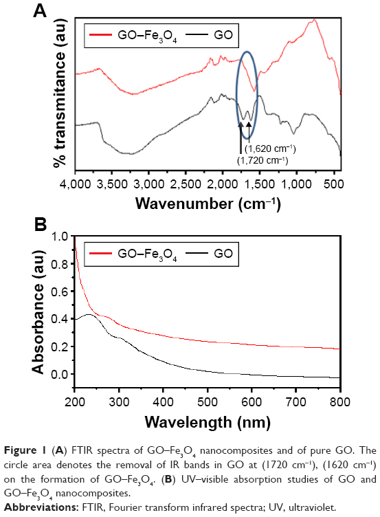 | Figure 1 (A) FTIR spectra of GO–Fe3O4 nanocomposites and of pure GO. The circle area denotes the removal of IR bands in GO at (1720 cm−1), (1620 cm−1) on the formation of GO–Fe3O4. (B) UV–visible absorption studies of GO and GO–Fe3O4 nanocomposites. |
Figure 1B shows the optical absorption spectra (UV–Vis) of the GO nanosheets exhibiting an absorption peak at 230 nm, corresponding to the π→π* transition of aromatic C–C bonds, and a shoulder at ~300 nm, which is attributed to the n→π* transitions of C=O bonds. The UV–Vis absorption of the GO–Fe3O4 nanocomposite has no significant visible peak, indicating possible surface interactions.
Figure 2 shows the structural and size characterization of GO–Fe3O4 using transmission electron microscopy (TEM). TEM micrograph revealed that Fe3O4 nanoparticles of ~3–10 nm in size are uniformly distributed onto GO nanosheets.
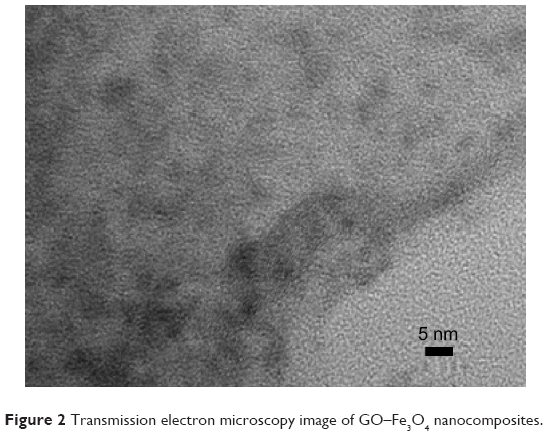 | Figure 2 Transmission electron microscopy image of GO–Fe3O4 nanocomposites. |
The bacterial toxicity experiment revealed a dramatic decrease in the number of bacteria in response to an increase in the concentration of GO–Fe3O4 nanocomposites. Significantly, we found that GO–Fe3O4 nanocomposites almost completely suppressed the growth of P. hauseri, leading to a viability loss of up to 98%, whereas the metabolic activity of K. pneumoniae and S. aureus cells was reduced by up to 87% and 83%, respectively. In a similar manner, the cell viability of S. pyogenes was reduced by up to 90% and 83% with a GO–Fe3O4 nanocomposite of 0.03% and 0.04%, respectively (Figures 3A and B).
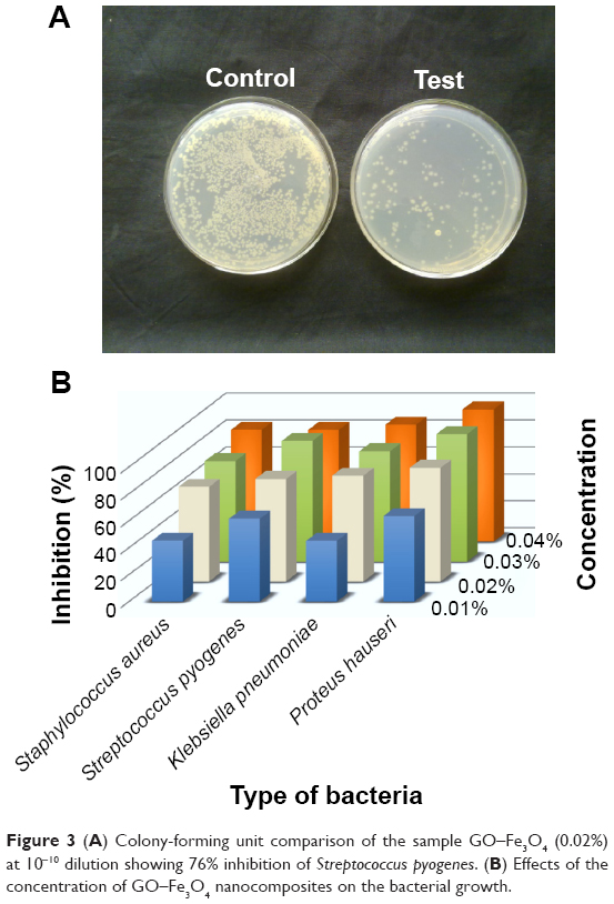 | Figure 3 (A) Colony-forming unit comparison of the sample GO–Fe3O4 (0.02%) at 10−10 dilution showing 76% inhibition of Streptococcus pyogenes. (B) Effects of the concentration of GO–Fe3O4 nanocomposites on the bacterial growth. |
Compared to GO–Fe3O4 nanocomposites, the GO nanosheets displayed weaker antimicrobial activity toward Gram-positive and Gram-negative bacteria. This can be explained on the basis of the negative charge found on the surface of the bacterial cells. The bacterial cells and the GO nanosheets are negatively charged and would repel each other under experimental conditions. However, in the case of Gram-negative bacteria, lipopolysaccharide subunits in the outer membrane contain sugars, phosphates, and lipids, whereas in Gram-positive bacteria, there is the presence of phosphate groups in teichoic acids of the lipid bilayer. These groups facilitate the Hydrogen bonding with the oxygenate group, such as –OH, –COOH, –CO, and epoxide of GO, and assist in wrapping GO nanosheets around the bacterial cells, blocking the cells from taking up nutrition from the growth medium, and eventually leading to cell death.3
In comparison with GO, a significant decrease in the negative charge was observed on the GO–Fe3O4 nanocomposites, which results in a better contact between bacterial cells and the GO–Fe3O4 enhancing its antibacterial activity by exposing the bacterial cell to the Fe3O4 nanoparticles. The close interaction inflicts oxidative stress inside the bacterial cell and consequent damage to proteins, membranes, and DNA, leading to cell death.
Conclusion
Overall, our results demonstrate the potential of GO–Fe3O4 nanocomposites as an effective bactericidal which can be used for dynamic applications in the field of medical devices and food and other industries.
Acknowledgment
R Singh gratefully acknowledges the support from DST-SERB (grant SB/FT/LS-206/2012) and SP Singh acknowledges the support from the CSIR network (grant Nano-SHE; BSC0112) funding.
Disclosure
The authors report no conflicts of interest in this work.
References
Singh R, Smitha MS, Singh SP. The role of nanotechnology in combating multi-drug resistant bacteria. J Nanosci Nanotechnol. 2014;14(7):4745–4756. | ||
Hu W, Peng C, Luo W, et al. Graphene-based antibacterial paper. ACS Nano. 2010;4(7):4317–4323. | ||
Akhavan O, Ghaderi E. Toxicity of graphene and graphene oxide nanowalls against bacteria. ACS Nano. 2010;4(10):5731–5736. | ||
Krishnamoorthy K, Veerapandian M, Zhang LH, Yun K, Kim SJ. Antibacterial efficiency of graphene nanosheets against pathogenic bacteria via lipid peroxidation. J Phys Chem C. 2012;116(32):17280–17287. | ||
Tang J, Chen Q, Xu L, et al. Graphene oxide-silver nanocomposite as a highly effective antibacterial agent with species-specific mechanisms. ACS Appl Mater Interfaces. 2013;5(9):3867–3874. | ||
Park S, Ruoff RS. Chemical methods for the production of graphenes. Nat Nanotehcnol. 2009;4:217–224. |
 © 2018 The Author(s). This work is published and licensed by Dove Medical Press Limited. The
full terms of this license are available at https://www.dovepress.com/terms
and incorporate the Creative Commons Attribution
- Non Commercial (unported, 3.0) License.
By accessing the work you hereby accept the Terms. Non-commercial uses of the work are permitted
without any further permission from Dove Medical Press Limited, provided the work is properly
attributed. For permission for commercial use of this work, please see paragraphs 4.2 and 5 of our Terms.
© 2018 The Author(s). This work is published and licensed by Dove Medical Press Limited. The
full terms of this license are available at https://www.dovepress.com/terms
and incorporate the Creative Commons Attribution
- Non Commercial (unported, 3.0) License.
By accessing the work you hereby accept the Terms. Non-commercial uses of the work are permitted
without any further permission from Dove Medical Press Limited, provided the work is properly
attributed. For permission for commercial use of this work, please see paragraphs 4.2 and 5 of our Terms.