
Back to Journals » International Journal of General Medicine » Volume 18
Endoscopic Ultrasound-Guided Fine-Needle Aspiration/Biopsy for Splenic Lesions: A Highly Accurate and Safe Diagnostic Tool
Authors Shaaban HE  , Hara K, Haba S, Kuwahara T, Okuno N, Koda H, Urata M , Kondo T, Yamamoto Y, Okasha HH
Received 30 January 2025
Accepted for publication 17 May 2025
Published 7 June 2025 Volume 2025:18 Pages 2941—2948
DOI https://doi.org/10.2147/IJGM.S520042
Checked for plagiarism Yes
Review by Single anonymous peer review
Peer reviewer comments 2
Editor who approved publication: Dr Xudong Zhu
Hossam Eldin Shaaban,1 Kazuo Hara,1 Shin Haba,1 Takamichi Kuwahara,1 Nozomi Okuno,1 Hiroki Koda,1 Minako Urata,1 Takashi Kondo,1 Yoshitaro Yamamoto,1 Hussein Hassan Okasha2
1Department of Gastroenterology, Aichi Cancer Center Hospital, Nagoya, Japan; 2Division of Gastroenterology and Hepatology, Department of Internal Medicine, Kasr Al-Aini School of Medicine, Cairo University, Cairo, Egypt
Correspondence: Kazuo Hara, Department of Gastroenterology, Aichi Cancer Center Hospital, 1-1 Kanokoden, Chikusa-ku, Nagoya, Aichi, 464-8681, Japan, Email [email protected]
Backgrounds/Aims: Tissue acquisition from splenic lesions is challenging. The role of endoscopic ultrasound in tissue acquisition for splenic lesions is not fully established due to paucity of published literature. This retrospective study evaluated the effectiveness and safety of endoscopic ultrasound-guided fine-needle aspiration/biopsy for splenic lesions.
Methods: Medical records of 18 patients who underwent EUS-FNA/B of splenic lesions at Aichi Cancer Center Hospital, Nagoya, Japan from February 2012 to June 2023 were retrospectively reviewed.
Results: Eighteen patients with splenic lesions underwent EUS-FNA/B. Among them, 3 were diagnosed with malignant lesions and 15 with benign lesions. The diagnostic performance of EUS-FNA/B was excellent, with a diagnostic accuracy of 100% (18/18; CI, 81.5– 100%), sensitivity of 100% (3/3; CI, 29.2– 100%), and specificity of 100% (15/15; CI, 78.2– 100%). A total of 34 lesions were sampled across all patients, including 24 splenic lesions, 6 lymph nodes, 2 pancreatic masses, and 2 hepatic focal lesions. Minor complications were observed in three patients: one case of post-procedural bleeding, one of self-limiting abdominal pain, and one case of transient fever that resolved within 24 hours with antibiotics.
Conclusion: EUS-FNA/B of splenic lesions demonstrated to be safe and of high diagnostic yield.
Keywords: endoscopic ultrasound (EUS), EUS guided fine needle aspiration/biopsy (EUS-FNA/B), splenic lesion, accuracy, safety
Introduction
Splenic lesions are uncommon and often incidentally detected during imaging by CT, MRI, or ultrasound for unrelated conditions such as trauma or cancer surveillance.1,2 The prevalence of incidental splenic lesions on CT scans performed on trauma patients is reported to be less than 1.5%.3 These lesions can be broadly categorized into benign and malignant types.
Among benign lesions, simple cysts are the most common, accounting for approximately 0.07–0.3% of splenic lesions seen on imaging.4,5 Splenic abscesses, while rare, have an estimated incidence of 0.14–0.7%, and are more commonly seen in immunocompromised patients or in the setting of hematogenous infection.4,6 Other benign conditions include hemangiomas, hamartomas, and granulomatous diseases such as tuberculosis or sarcoidosis. In contrast, malignant lesions typically consist of lymphoma, metastatic disease (eg, from lung or gastrointestinal primaries), and, less commonly, primary splenic neoplasms such as angiosarcoma. Among these, lymphoma and metastases represent the most frequent conditions that necessitate tissue confirmation for diagnosis and management.
Differentiating between benign and malignant splenic lesions is essential, as it directly influences patient care decisions. While benign lesions may only require imaging follow-up or no intervention, malignant lesions often demand systemic therapy, oncologic evaluation, or even surgical consideration. Therefore, obtaining an accurate tissue diagnosis is critical for determining the appropriate clinical pathway.1,7
Traditional diagnostic methods, including percutaneous biopsy, carry significant risks of bleeding due to the spleen’s highly vascular structure. Surgical biopsy, while definitive, is invasive, resource intensive and associated with higher morbidity.8
Reported diagnostic yields for percutaneous splenic biopsies range from 78% to 92%, with bleeding rates up to 13% in some series.7,8 In contrast, emerging data suggest that endoscopic ultrasound-guided fine-needle aspiration or biopsy (EUS-FNA/B) offers a safer, minimally invasive alternative, especially when lesions are deeply located or inaccessible via percutaneous routes.1,2
Endoscopic ultrasound-guided tissue acquisition (EUS-FNA/B) provides a minimally invasive alternative, leveraging real-time imaging to allow safe and effective sampling of splenic lesions.4,5
Despite the growing use of EUS-FNA/B for lymph nodes, liver, pancreas, and adrenal lesions, its application in splenic lesions remains underexplored due to the rarity of such cases and limited published literature.2 Most available studies are limited by small sample sizes, heterogeneous procedural techniques, insufficient long-term follow-up, and limited safety data, which restrict the generalizability of their findings. Additionally, many prior reports have focused on isolated case series or lacked rigorous outcome assessment.1,7
With the widespread adoption of cross-sectional imaging and advancements in endoscopic ultrasound technology, incidental splenic lesions are being identified with increasing frequency.2,3 Improvements in EUS, including higher-resolution imaging, contrast-enhanced techniques, and more efficient needle systems, have enhanced the safety and diagnostic yield of EUS-FNA/B.4,5 These developments have contributed to a growing clinical demand for non-surgical, minimally invasive approaches to splenic tissue diagnosis, particularly in patients who are poor surgical candidates or present with high-risk lesion locations.4,8 In this context, the present study aims to evaluate the diagnostic performance, adequacy, and safety of EUS-FNA/B for splenic lesions. It adds to the literature by analyzing an 11-year experience at a high-volume tertiary cancer center, offering one of the largest single-center cohorts to date focused exclusively on splenic EUS-FNA/B. It provides comprehensive data on diagnostic accuracy, procedure-related complications, and long-term follow-up, thereby addressing key gaps in current evidence. Moreover, the inclusion of both benign and malignant diagnoses in a clinically diverse patient population strengthens the real-world applicability of the results.
Methods
Study Design and Setting
This retrospective study was conducted at a tertiary care center (Aichi Cancer Center), including patients who underwent EUS-FNA/B for splenic lesions between February 2012 and June 2023. This study was approved by the Ethics Committee of Aichi Cancer Center Hospital, Nagoya, Japan. Due to the retrospective nature of the study, the requirement for informed consent was waived by the ethics committee. All patient data were anonymized to ensure confidentiality. The study was conducted in accordance with the ethical standards of the institutional and/or national research committee and with the 1964 helsinki declaration and its later amendments or comparable ethical standards.
All cases with EUS guided FNA are recorded in a dedicated computer in a separate Excel sheet in our center to facilitate their easy tracing and their data retrieval when needed. Retrospective data were extracted from electronic medical records by two dedicated authors (Shaaban HE and Yamamoto Y), who were not involved in performing the procedures or assigning final diagnoses. A standardized data collection sheet in Microsoft Excel was used to gather demographic, procedural, histopathologic, and follow-up data. The accuracy of the extracted data was ensured through repeated manual verification. Each entry was reviewed multiple times to confirm consistency with the original patient records prior to statistical analysis. Given the retrospective nature of the study, blinding of reviewers was not feasible. Data completeness and consistency in reporting were confirmed and they followed a unified institutional protocol. There was no missing data as the electronic system had all the required data for all the study patients.
All EUS-FNA/B procedures were performed by twelve experienced endoscopists, each with over five years of advanced endoscopy training. All operators were also EUS trainers at our tertiary care center. Procedures were performed during their routine clinical lists, without randomization between operators. There was some variability in individual technique, particularly in needle selection, number of passes, and use of adjunctive methods such as ROSE, suction, and contrast enhancement. These technical variations are described in detail in the Procedures and Results sections.
Histopathological evaluation was conducted by board-certified gastrointestinal pathologists at our center. All study cases were discussed in multidisciplinary meetings to minimize inter-observer variability and ensure diagnostic consistency. Tissue adequacy was defined by the presence of sufficient cellularity to allow for definitive histological classification, with or without the aid of immunohistochemistry, as needed. The adequacy of samples was also judged intra-procedurally where possible, using rapid on-site evaluation (ROSE) in 11 of the 24 splenic lesion punctures. Samples of ROSE (cytology) and the FNA/FNB cellblocks were examined with standard Hematoxylin and Eosin stains and using immunohistochemistry and flow cytometry in selected cases when needed.
Patient Selection
Patients with radiologically suspicious splenic lesions identified on cross-sectional imaging (CT or MRI) were included. Exclusion criteria included uncorrected coagulopathy, hemodynamic instability, or contraindications to sedation. At our institution, EUS-FNA/B is the preferred diagnostic approach for splenic focal lesions sampling rather than percutaneous or surgical approaches. This is due to the availability of service and the high skill level of the our endoscopists based on having a high volume of cases and long years of experience.
Procedures
EUS-FNA/B was performed using a linear-array echoendoscope with 22G, 25G, or 20G needles, selected based on lesion characteristics. Suction or slow-pull techniques were employed as needed, and rapid on-site evaluation (ROSE) was utilized in 11 of 24 total punctures. A mean of 2 passes (range: 1–4) per lesion was performed. Additional sites, including lymph nodes, hepatic lesions, and pancreatic masses, were biopsied concomitantly in some cases.
Outcome Measures
Primary outcomes included diagnostic accuracy, sensitivity, specificity, and adequacy of tissue sampling. Secondary outcomes included procedure-related adverse events, classified according to the American Society for Gastrointestinal Endoscopy (ASGE) lexicon.9 We used Microsoft® Excel for Mac Version 16.92 (24120731) for calculation of the descriptive statistics stated in our manuscript. Given the retrospective design and limited sample size, a formal power calculation was not performed prior to data collection. However, confidence intervals were calculated for all diagnostic performance metrics to better reflect the statistical uncertainty and precision of the observed outcomes. These intervals help contextualize the 100% accuracy reported and mitigate overinterpretation of findings from a small cohort.
Follow-Up
All benign lesions were followed for a minimum of 2 years to confirm stability, unless resolution was documented earlier or splenectomy was performed. No malignancies were diagnosed during follow-up, supporting the initial EUS-FNA/B findings. Follow-up protocols were consistent across the study period. Patients with malignant findings received further oncologic evaluation.
Results
We studied 18 patients (11 males and 7 females) with a mean age of 58 years, ranging from 41 to 75 years (Table 1). The lesions varied in size from 6 to 70 mm, with an average size of 21.5 mm. 12 patients had a single lesion, while the remaining 6 presented with multiple lesions. 11 lesions exhibited a hypoechoic pattern on cross-sectional imaging. Across 21 procedures, a total of 34 puncture sites were sampled, ensuring adequate sampling in all cases. Fine needle aspiration (FNA) was performed in 15 cases, while fine needle biopsy (FNB) was executed in 13 cases. Adjunctive techniques included ROSE in 45.8% of cases (11/24) and the use of the contrast agent Sonazoid in 37.5% (9/24). Suction was applied in 15 procedures, while the slow-pull technique was used in 4 cases (Table 2). Additional biopsies included six lymph nodes, two pancreatic masses, and two hepatic masses. The final diagnosis was shown in (Table 3) where 15 benign diagnoses were found vs 3 malignant diagnoses. Among benign lesions, sarcoidosis was the most common diagnosis (22.2%), followed by hemangiomas, inflammatory lesions, SANT (sclerosing angiomatoid nodular transformation), and necrosis. Malignant cases included neuroendocrine tumor, diffuse large B-cell lymphoma, and gastric cancer metastasis (each 5.5%). The diagnostic performance showed a sensitivity of 100% (3/3; 95% CI, 29.2–100%), a specificity of 100% (15/15; 95% CI, 78.2–100%), a positive predictive value of 100% (3/3; 95% CI, 29.2–100%), a negative predictive value of (15/15; 95% CI, 78.2–100%) and a diagnostic accuracy of 100% (18/18; 95% CI, 81.5–100%) (Table 4). Adverse events were observed in 4 cases (19.05%), including mild intra-procedural bleeding (9.52%), post-procedural fever resolving with antibiotics (4.76%), and self-limiting abdominal discomfort (4.76%). There were no late complications in any of the patients on follow up (Table 5). During the follow-up period, two benign lesions resolved spontaneously, and other two patients underwent splenectomy for further evaluation. Their post splenectomy diagnosis matched that of the EUS-FNA/B result. No false positives or false negatives were observed during follow-up. As shown in Figure 1, a CT scan reveals a hypoechoic splenic lesion. Figure 2 illustrates the EUS-guided fine-needle aspiration procedure.
 |
Table 1 Patients Characteristics |
 |
Table 2 Procedural Details of EUS-FNA/B |
 |
Table 3 Final Diagnosis |
 |
Table 4 Diagnostic Yield of EUS-FNA/B for Splenic Lesions |
 |
Table 5 Adverse Events Associated with EUS-FNA/B |
 |
Figure 1 A computed tomography scan showing a splenic focal lesion (black arrow). |
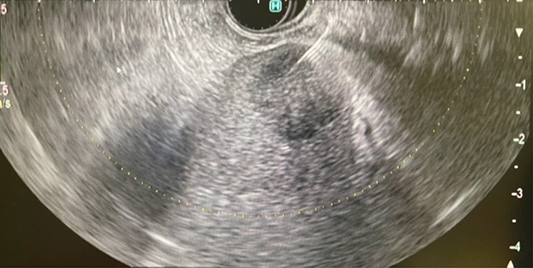 |
Figure 2 Endoscopic ultrasound-guided fine-needle aspiration/biopsy puncture of a splenic focal lesion. |
Discussion
In the recent time, guidelines developed for managing incidental splenic findings, offering a systematic approach to evaluation and follow-up.3
This study demonstrates that EUS-FNA/B achieved 100% diagnostic accuracy for splenic lesions with no major complications. These findings align with previous reports by Heller et al4 and Liu et al8 highlighting the high diagnostic yield of EUS-guided biopsies.
The diagnostic accuracy observed in this study exceeds that of pooled analyses for EUS-guided biopsies in other organs, such as the pancreas (sensitivity ~85%).2,7 The absence of severe complications further underscores the safety profile of EUS-FNA/B for splenic lesions.
EUS-FNA/B provides a minimally invasive alternative to percutaneous biopsy, particularly in high-risk patients. Its utility in diagnosing both benign and malignant splenic lesions expands its role in clinical practice.2
Infectious or cystic splenic lesions represent another important clinical subset. In our study, cystic lesions, abscesses, or hydatid cysts were not included. However, EUS has also been used in such settings, as shown in case reports describing EUS-guided drainage of splenic abscesses or diagnosis of hydatid cysts.6,10 Future prospective studies should explore EUS utility in these subgroups in addition to validation of our findings and establishment of standardized guidelines for EUS-FNA/B in splenic lesions. Additionally, the growing use of EUS in evaluating incidental lesions detected on CT/MRI strengthens its relevance in routine diagnostic algorithms.
Limitations
The study’s retrospective nature and small sample size limit the generalizability of findings. Additionally, the lack of standardized protocols for needle selection or ROSE limits procedural reproducibility. Also the center is a tertiary care endoscopy unit with a high volume and a high skill level of the interventional endoscopists. The pathologists are expert in their fields. This might explain the 100% diagnostic accuracy and low complication rate. Results from low volume centers or community hospitals with less experienced operators might give different results. The study did not compare the EUS FNA/FNB with the other modalities in the same center because the endoscopic approach was the first choice in the center as mentioned above.
Conclusion
EUS-FNA/B is a highly effective and safe diagnostic tool for splenic lesions, achieving 100% diagnostic accuracy and demonstrating an excellent safety profile with minimal complications. However, this result should be interpreted cautiously given the limited sample size. Confidence intervals were included to reflect statistical variability. These findings support its role as a minimally invasive alternative to percutaneous biopsy, particularly in high-risk patients or when other modalities are infeasible. Future studies should focus on validating these results through multicenter, prospective trials and standardizing procedural protocols to optimize outcomes.
Disclosure
The authors report no conflicts of interest in this work.
References
1. Pan X, Zheng Y, Liu J, et al. Endoscopic ultrasound-guided tissue acquisition for splenic lesions: a systematic review and meta-analysis. PLoS One. 2022;17(11):e0276529. doi:10.1371/journal.pone.0276529
2. Bellisario F, Attili F, Campana F, et al. Endoscopic ultrasound-guided fine needle biopsy in the diagnostic work-up of deep-seated lymphadenopathies and spleen lesions: a monocentric experience. Diagnostics. 2023;13(8):2839. doi:10.3390/diagnostics13172839
3. Gore RM, Ecanow JS. Management of splenic ‘incidentalomas’ found on ultrasound and computed tomography. Cancer Imaging. 2015;15(Suppl 1):O11. doi:10.1186/1470-7330-15-S1-O11
4. Heller MT, Harisinghani M, Neitlich JD, et al. Managing incidental findings on abdominal and pelvic CT and MRI, part 3: white paper of the ACR incidental findings committee II on splenic and nodal findings. J Am College Radiol. 2013;10(11):833–839. doi:10.1016/j.jacr.2013.05.020
5. Corvino A, Granata V, Tafuri D, et al. Incidental focal spleen lesions: integrated imaging and pattern recognition approach to the differential diagnosis. Diagnostics. 2023;13(15):2536. doi:10.3390/diagnostics13152536
6. Guo Q, Liu H, Wang G. Ruptured splenic hydatid cyst. Heliyon. 2024;10(13):e33701. doi:10.1016/j.heliyon.2024.e33701
7. Rana SS, Sharma V, Sharma R, et al. Safety and utility of endoscopic ultrasound-guided fine-needle aspiration of focal splenic lesions. Ann Gastroenterol. 2017;30(2):205–210. doi:10.20524/aog.2017.0121
8. Liu HHW, Godfrey EM, Carroll NR. Tissue sampling of splenic lesions – comparison between EUS-FNA and percutaneous ultrasound-guided biopsy. United Eu Gastroenterol J. 2023;11(8):731–738. doi:10.1002/ueg2.12345
9. Cotton PB, Eisen GM, Aabakken L, ASGE Standards of Practice Committee. The lexicon for endoscopic adverse events: report of an ASGE workshop. Gastrointest Endosc. 2010;71(3):446–454. doi:10.1016/j.gie.2009.10.027
10. Canakis A, Baron TH. EUS-guided transmural drainage of a splenic abscess. Gastrointest Endosc. 2024. doi:10.1016/j.gie.2024.10.065
 © 2025 The Author(s). This work is published and licensed by Dove Medical Press Limited. The
full terms of this license are available at https://www.dovepress.com/terms
and incorporate the Creative Commons Attribution
- Non Commercial (unported, 4.0) License.
By accessing the work you hereby accept the Terms. Non-commercial uses of the work are permitted
without any further permission from Dove Medical Press Limited, provided the work is properly
attributed. For permission for commercial use of this work, please see paragraphs 4.2 and 5 of our Terms.
© 2025 The Author(s). This work is published and licensed by Dove Medical Press Limited. The
full terms of this license are available at https://www.dovepress.com/terms
and incorporate the Creative Commons Attribution
- Non Commercial (unported, 4.0) License.
By accessing the work you hereby accept the Terms. Non-commercial uses of the work are permitted
without any further permission from Dove Medical Press Limited, provided the work is properly
attributed. For permission for commercial use of this work, please see paragraphs 4.2 and 5 of our Terms.