
Back to Journals » International Journal of General Medicine » Volume 18
Elevated Expression of STAT6, ERG, and miR-647 Expression as Predictive Biomarkers for Prostate Cancer
Authors Yuan HZ, Jin SM, Yang WD, Du M, Wang L, Xiao L
Received 27 December 2024
Accepted for publication 11 March 2025
Published 10 April 2025 Volume 2025:18 Pages 2067—2075
DOI https://doi.org/10.2147/IJGM.S512606
Checked for plagiarism Yes
Review by Single anonymous peer review
Peer reviewer comments 3
Editor who approved publication: Prof. Dr. Leonardo Reis
Hua-Zun Yuan,1,* Sheng-Ming Jin,2,3,* Wen-Dong Yang,4,* Min Du,1 Lei Wang,1 Li Xiao1
1Department of Pathology, Huadong Hospital, Fudan University, Shanghai, People’s Republic of China; 2Department of Urology, Fudan University Shanghai Cancer Center, Shanghai, People’s Republic of China; 3Department of Oncology, Shanghai Medical College, Fudan University, Shanghai, People’s Republic of China; 4Cancer Institute, Xuzhou Medical University, Xuzhou, Jiangsu Province, People’s Republic of China
*These authors contributed equally to this work
Correspondence: Li Xiao, Department of Pathology, Huadong Hospital, Fudan University, No. 221 West Yan’an Road, Shanghai, People’s Republic of China, Tel +86-18916615822, Email [email protected]
Aim: The aim of this study was to investigate the clinical significance of STAT6, ERG, miR-647 in prostate cancer (PCa).
Methods: This was a retrospective study. There were 210 consecutive patients diagnosed with prostate cancer or benign prostatic hyperplasia in our hospital from July 2020 to July 2023. Among those patients, 108 patients pathologically diagnosed as prostate cancer were divided into the prostate cancer group (PCa group), and 102 patients pathologically diagnosed as having benign prostatic hyperplasia were divided into the benign prostatic hyperplasia group (BPH group).
Results: The levels of STAT6 mRNA, ERG mRNA, and miR-647 expression in prostate cancer tissue were higher than those in BPH tissues, with statistically significant differences (P< 0.05) . The levels of STAT6 mRNA, ERG mRNA, and miR-647 indicators in prostate cancer patients were not significantly different with respect to patient age and tumor size (P > 0.05) but were related to lymph node metastasis, T stage, and Gleason score (P < 0.05). On the other hand, the tPSA/fPSA had significantly different between two groups (P < 0.05). PCa group had smaller MRI sagittal diameter and anteroposterior diameter in comparison to BPH group (P < 0.05), furthermore, PCa group had larger PI-RAD in comparison to BPH group (P < 0.05).
Conclusion: The higher level of STAT6, ERG, and miR-647 in prostate tissue are closely related to the occurrence of prostate cancer and have certain value in predicting the onset of prostate cancer.
Keywords: prostate cancer, signal transducer and activator of transcription 6, ETS-related gene, miR-647, Prostate cancer
Introduction
Prostate cancer (PCa) is a leading cause of cancer-related mortality and the second most common male malignancy worldwide.1 In 2020, approximately 1.41 million new cases were diagnosed, accounting for 7.3% of global cancer cases and 3.8% of cancer-related deaths.2–4 Early-stage prostate cancer often presents without clear symptoms, but as the disease progresses, issues like difficulty urinating may arise. By the time of diagnosis, many patients are in the advanced stages, where treatment outcomes are generally poor, and the mortality rate remains high. This highlights the importance of early diagnosis for better prognosis and treatment outcomes.5 Given the complexity of prostate cancer, recent research has focused on molecular biomarkers such as microRNAs, STAT signaling pathways, and the ERG gene. These factors have shown potential in understanding the molecular mechanisms driving PCa progression and could be critical in developing more effective diagnostic and therapeutic strategies.
MicroRNAs (miRNAs), as an important molecule that has been proven to be closely related to tumorigenesis and development, have potential value in tumor diagnosis, treatment and prognosis, and are currently the focus of tumor research.6 MiR-647 is an important member of the miRNA family and is closely related to the occurrence and development of prostate cancer. It can affect key biological processes such as the proliferation, apoptosis, invasion and metastasis of prostate cancer cells by regulating the expression of a series of downstream target genes.7,8 A study had demonstrated that LncRNA PROX1-AS1 promotes proliferation, invasion, migration in prostate cancer via targeting miR-647.9 Moreover, Chen et al10 found that Circular RNA CircNOLC1, Upregulated by NF-KappaB, Promotes the Progression of Prostate Cancer via miR-647/PAQR4 Axis. The dysregulation of miRNA expression levels runs through the early to late stages of prostate cancer, and the miRNA-based prostate cancer treatment method may become a new technology for prostate cancer gene therapy.11,12 Therefore, more research is needed to prove miR-647 significance in prostate cancer and provide stronger evidence for the precise diagnosis and treatment of prostate cancer.
Signal transduction and activator of transcription (STAT) is a potential cytoplasmic transcription factor that responds to cytokines to activate gene transcription and is widely distributed in tissues and cells.13 STAT6, in particular, plays a crucial role in regulating cell growth, differentiation, and apoptosis, and it is involved in key processes such as tumorigenesis, inflammation, and immune responses.14 While STAT6 is recognized for its significance in cancer, including its involvement in promoting tumor progression through the activation of specific gene expression pathways, its precise mechanisms in prostate cancer (PCa) remain underexplored. Previous studies have indicated that STAT6 may influence the tumor microenvironment in PCa by modulating immune cell infiltration and inflammatory responses, which are critical to cancer progression. Su et al found that STAT6 polymorphism was correlated with gefitinib-induced diarrhea in patients with non-small cell lung cancer,15 but more detailed research is needed to understand how STAT6 specifically contributes to PCa development. Incorporating these insights into the background can add depth and provide a more comprehensive context for the role of STAT6 in prostate cancer.
The ERG gene encodes a protein that belongs to the ETS family of transcription factors.16 These transcription factors are involved in regulating the expression of a large number of genes, thereby influencing cell growth, differentiation, and development.17 Alterations or mutations in the ERG gene can have significant implications. In some cancers, such as certain types of leukemia and prostate cancer, abnormal activation or overexpression of the ERG gene has been observed.18 This dysregulation can contribute to the oncogenic process by promoting cell proliferation, inhibiting apoptosis, and influencing other aspects of tumorigenesis.19 At present, the function of ERG in prostate cancer (PCa) is not fully understood, and much of the research on its role in the initiation and progression of PCa remains in its early stages. Specifically, the mechanisms through which ERG contributes to the malignancy of PCa are still unclear, such as whether it influences the tumor microenvironment or interacts with other signaling pathways to drive tumor growth. This study aims to address these gaps by investigating the molecular interactions of ERG in PCa and exploring how its expression correlates with clinical outcomes, thus providing a clearer understanding of its role in the progression and prognosis of PCa.
MiR-647, STAT6, and ERG have shown promising potential as biomarkers in prostate cancer (PCa), not only for diagnostic purposes but also for their therapeutic and prognostic implications. miR-647, a microRNA, has been implicated in regulating various signaling pathways involved in tumorigenesis, and its expression levels have been linked to tumor progression and metastasis. Therapeutically, modulating miR-647 expression could offer a novel approach to inhibit cancer cell proliferation and metastasis, potentially improving treatment outcomes. Similarly, STAT6, a transcription factor involved in immune regulation and cell survival, has emerged as a key player in PCa progression. Given its role in tumor immune evasion and inflammation, targeting STAT6 may provide a strategy for enhancing anti-tumor immunity or overcoming resistance to therapies. Additionally, ERG, a gene commonly overexpressed in PCa, plays a significant role in gene regulation, and its dysregulation has been associated with cancer cell proliferation and survival. As such, ERG expression could serve as a prognostic marker, providing insight into the aggressiveness of the disease and the likelihood of recurrence. In clinical practice, integrating these markers into routine diagnostic and prognostic evaluations could improve personalized treatment strategies, allowing for more accurate risk stratification and targeted therapies.
This study aims to explore the expression levels of STAT6, ERG, and miR-647 in prostate tissue, with the goal of providing new molecular markers for early diagnosis and treatment of prostate cancer.
Methods
Study Population
In this retrospective study, we collected data of 210 consecutive patients diagnosed with prostate cancer or benign prostatic hyperplasia in our hospital from July 2020 to July 2023. Among those patients, 108 patients pathologically diagnosed as prostate cancer were divided into the prostate cancer group (PCa group), and 102 patients pathologically diagnosed as having benign prostatic hyperplasia were divided into the benign prostatic hyperplasia group (BPH group). Additionally, the study was approved by the Ethics Review Board of Huadong Hospital, Fudan University. Informed consent was obtained from all study participants. All the methods were carried out in accordance with the Declaration of Helsinki.
Inclusion and Exclusion Criteria
Inclusion criteria was as follow: 1) All the patients in the PCa group selected in this institute were diagnosed as PCa after prostate biopsy or surgery after hospitalization to obtain the pathological results.20 All the patients in the BPH group were diagnosed as BPH through prostate biopsy or surgery to obtain the pathological results;21 2) Age ≥60 years old; 3) Patients without heart, liver, kidney, and hematopoietic system disorders or other functional impairments.
Exclusion criteria was as follow: 1) Patients with other systemic tumors and those with prostate metastasis from other tumors; 2) PCa patients who have already been treated with drugs, surgery or other means; 3) Patients with a family history of hereditary diseases.
Immunohistochemistry (IHC)
Briefly, the paraffin-submerged tissues were sliced (5 μm thick), tagged at 4°C with the monoclonal human STAT6 and ERG antibody (dilution 1:1000, Proteintech) overnight, stained via staining kit (Zhongshan Biotechnology, Beijing, China), visualized, and then the staining score was assessed based on color intensity and positive cell rate. An average intensity score of ≥4 indicated high expression, and <4 depicted no or low expression.
Quantitative Polymerase Chain Reaction (qPCR)
Comparison of STAT6 mRNA, ERG mRNA levels, and miR-647 expression levels between prostate cancer tissue and adjacent non-cancerous tissue were conducted using fluorescence quantitative PCR technology. Tissue RNA was extracted using a kit (QIAGEN, Germany), RNA quality was measured using an N50 microspectrophotometer (IMPLEN, Germany), RNA was reverse transcribed into cDNA according to the instructions of the reverse transcription kit (Vazyme, China), diluted to equal concentration, and SYBR quantitative PCR reaction mixture (Applied Biosystems, USA) was prepared for amplification on an Mx3005P QPCR instrument (Agilent, USA). Using GAPDH gene as the reference, the primer sequences were: STAT6 upstream primer: 5′-LAGGAGAGCAGGGGAAAGGAAG-3′, STAT6 downstream primer: 5′-LTGGCAGGTGGTGGAACTCTT-3′; using GAPDH gene as the reference, ERG primer sequences were: ERG upstream primer: ERG-F5′-TTATCGTGCCAGTAGCAGGT-3′, ERG downstream primer: ERG-R5′-GATGTTGACGTCTGGAAGGC-3′; using U6 as the reference, miR-647 upstream primer: 5′-CTCAACTGGTGTCGTGGAGTCGGCAATTCAGTTGAGGAAGGAAG-3′, downstream primer: 5′-ACACTCCAGCTGGGGTGGCTGCACTCACT-3′, miR-647 expression was calculated using the 2-ΔΔCt method, and both reference genes and primers were synthesized and provided by Guangzhou Xingzhi Biotechnology Co., Ltd.
Data Collection
Collect clinical information from each subject in our clinical electronic medical record database, including age, BMI (body mass index), tumor size, Gleason score, serum tPSA (total Prostate Specific Antigen), fPSA (free Prostate Specific Antigen), MRI sagittal diameter, MRI anteroposterior diameter, MRI transverse diameter, PI-RAD and ISUP classification.
Statistical Analysis
In this study, SPSS25.0 statistical software was used for statistical analysis. For measurement data, ANOVA or t-test was conducted for the data that met the normal distribution and homogeneity of variance conditions, and the measured values of each index were expressed as “mean ± standard deviation (Mean ± SD)”; for data that did not meet the normal distribution, non-parametric rank sum test was used, and the measured values of each index were expressed as “median (25% percentile, 75% percentile) (M (P25.P75))”; chi-square test was used for count data. All data were rounded to two decimal places after the decimal point, and P < 0.05 was considered statistically significant.
Results
Comparison of Baseline Data Between Two Groups
In our study, there was no significant difference between two groups in terms of age, BMI, tumor size (p > 0.05)(Table 1).
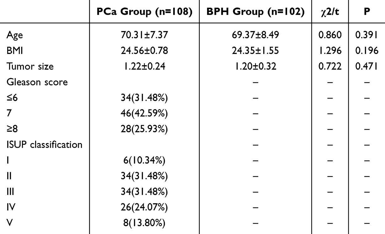 |
Table 1 Comparison of Baseline Data Between Two Groups |
Immunohistochemical Staining of STAT6 and ERG
Membranous and cytoplasmic staining of ERG could be assessed in patients with prostate cancer (Figure 1). Patients with prostate cancer showed positive staining for ERG. ERG is highly expressed in Gleason scores of 7–9 and ISUP grades of III–IV. However, there was no STAT6 nuclear staining in human BPH tissues and Pca tissues (Figure 2).
 |
Figure 1 Representative IHC staining of STAT6 and ERG in human BPH tissues and Pca tissues. |
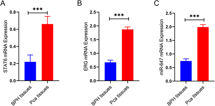 |
Figure 2 Comparison of STAT6 mRNA, ERG mRNA Levels, and miR-647 Expression Levels between BPH tissues and Pca tissues. (A) STAT6, (B) ERG, (C) miR-647. *** P<0.05. |
Comparison of STAT6 mRNA, ERG mRNA Levels, and miR-647 Expression Levels Between BPH Tissues and Pca Tissues
The qPCR results showed that STAT6 mRNA, ERG mRNA, and miR-647 expression in Pca tissues were all higher than those in BPH tissues, with statistically significant differences (P < 0.001, Figure 3).
 |
Figure 3 Compare the levels of serum prostate-specific antigen in the two groups. (A) fPSA, (B) tPSA, (C) tPSA/fPSA. *P<0.001. Abbreviations: fPSA, free Prostate Specific Antigen; tPSA, total Prostate Specific Antigen. |
Comparison of STAT6 mRNA, ERG mRNA, and miR-647 Indicators Among Prostate Cancer Patients with Different Clinical Pathological Features
The levels of STAT6 mRNA, ERG mRNA, and miR-647 indicators in prostate cancer patients were not significantly different with respect to patient age and tumor size (P > 0.05) but were related to lymph node metastasis, T stage, and Gleason score (P < 0.05, Table 2).
 |
Table 2 Comparison of STAT6 mRNA, ERG mRNA, and miR-647 Indicators Among Prostate Cancer Patients with Different Clinical Pathological Features |
Comparison of Serum Prostate-Specific Antigen Between Two Groups
Compared with BPH group, the serum fPSA and tPSA of the PCa group had increased in the PCa group, but there were not significantly different (P > 0.05). On the other hand, the tPSA/fPSA had significantly different between two groups (P < 0.05)(Figure 3).
Comparison of Magnetic Resonance Imaging Parameters Between Two Groups
PCa group had smaller MRI sagittal diameter and anteroposterior diameter in comparison to BPH group (P < 0.05), furthermore, PCa group had larger PI-RAD in comparison to BPH group (P < 0.05) (Table 3).
 |
Table 3 Comparison of Magnetic Resonance Imaging Parameters Between Two Groups |
Discussion
PCa, as one of the common malignant tumors in the male urinary system, ranks the second in the global male cancer incidence rate and the fourth in the cancer mortality rate.22,23 Early patients often do not have specific clinical manifestations and are not easily distinguishable from benign lesions such as BPH and prostatitis.24 Therefore, until PCa presents with urinary tract obstruction symptoms, most patients have already reached the advanced stage. Establish an effective clinical diagnostic tool for PCa and explore more and more effective PCa diagnostic markers to provide a basis for the early identification of PCa patients.
In our study, we found that STAT6 mRNA, ERG mRNA, and miR-647 expression in Pca tissues were all higher than those in BPH tissues. In the tumor microenvironment of prostate cancer, various cytokines and growth factors are secreted. These factors can activate intracellular signaling pathways, including the JAK-STAT pathway.25 Activation of the JAK-STAT pathway may lead to upregulation of STAT6 expression. Genetic alterations or mutations in prostate cancer cells themselves may directly affect the regulation of STAT6, resulting in its increased level.26 Additionally, the interaction between tumor cells and stromal cells in the prostate cancer microenvironment may also influence the expression and activation of STAT6.27 Stromal cells may secrete factors that promote STAT6 activation and accumulation. Moreover, epigenetic modifications, such as DNA methylation and histone acetylation, may also play a role in regulating the expression of STAT6 in prostate cancer, either enhancing or suppressing its expression.28 The complex network of signaling molecules and regulatory factors within the tumor can contribute to the dysregulation of STAT6 and its consequent elevation in prostate cancer patients. Therefore, the level of STAT6 may have potential as a biomarker to assess the aggressiveness and prognosis of prostate cancer, helping in treatment decision-making.
Some studies had found that fusions between the TMPRSS2 gene and the ERG gene are frequently found in prostate cancer.29–31 These genetic fusions can lead to dysregulated expression and overactivation of ERG, resulting in an increased level.32 On the other hand, aberrant activation of certain signaling cascades within the tumor microenvironment may promote the upregulation of ERG.33 For example, alterations in growth factor signaling or hormonal signaling pathways might contribute to enhanced ERG expression.34–36 Therefore, the increase of ERG level can help in the diagnosis and identification of prostate cancer, providing valuable information for clinicians to make more accurate judgments.
In this research, the results showed that miR-647 is highly expressed In prostate cancer tissues, which was consistent with current findings.9,10 Aberrant methylation patterns or changes in histone modifications in the genomic region of miR-647 may contribute to its increased expression.37 Interactions with transcription factors that are abnormally activated or regulated in prostate cancer can also drive the high expression of miR-647.38 These transcription factors may bind to specific sites on the miR-647 promoter and facilitate its transcription. In addition, the tumor microenvironment, including factors secreted by other cells or extracellular matrix components, may provide cues that stimulate miR-647 expression.39 Meanwhile, more evidence is also needed to confirm the role and mechanism of miR-647 in prostate cancer.
Interestingly, there was no STAT6 nuclear staining in human Pca tissues. One possible reason that there are post-translational modifications or regulatory events that prevent STAT6 from translocating to the nucleus or being properly activated in prostate cancer cells. These modifications could include phosphorylation, acetylation, or other modifications that affect its function and localization. On the other hand, due to downregulation of the STAT6 gene or suppression of its expression through various epigenetic or transcriptional mechanisms, the expression level of STAT6 itself might be very low. In addition, differences in sample collection, processing, or staining techniques could potentially contribute to the lack of observed STAT6 nuclear staining. Variations in fixation, antigen retrieval, or antibody specificity might lead to false-negative results. We cannot deny that the biological complexity of prostate cancer means that not all relevant factors or markers may be easily detected or understood. There could be additional undiscovered factors or interactions that influence the absence of STAT6 nuclear staining in this context. Therefore, further research is needed to fully understand the complex mechanisms underlying the STAT6 in prostate cancer patients and its specific roles in the disease process.
Several established biomarkers, such as PCA3 and the TMPRSS2-ERG fusion, have been widely recognized and utilized in the diagnosis and prognosis of prostate cancer (PCa). PCA3, a non-coding RNA, has shown high specificity for prostate cancer detection and is used as a supplementary diagnostic tool, particularly in patients with elevated prostate-specific antigen (PSA) levels who are at risk for PCa.40 The TMPRSS2-ERG fusion gene, another significant biomarker, is commonly observed in PCa and has been linked to disease progression and prognosis, especially in cases of early-stage cancer.41 These biomarkers have provided valuable insights into PCa biology, leading to improved diagnostic strategies and personalized treatment options. In contrast, miR-647, STAT6, and ERG offer novel perspectives that could complement these established biomarkers. While PCA3 and TMPRSS2-ERG fusion are primarily focused on gene expression and fusion events, miR-647, a microRNA, has the potential to regulate multiple signaling pathways involved in cancer progression, offering a new layer of molecular insight. STAT6, a transcription factor, has been implicated in immune regulation and tumor microenvironment modulation, potentially influencing both tumor growth and response to therapy.42 The ERG gene, although known for its role in gene regulation in various cancers, is still under investigation for its specific mechanisms in prostate cancer, with its dysregulation potentially contributing to tumorigenesis by promoting cell proliferation and survival.43 By comparing these established biomarkers to miR-647, STAT6, and ERG, it becomes evident that while the latter trio may not yet be as widely used in clinical practice, their emerging role as predictive biomarkers offers substantial promise. Their involvement in different biological pathways suggests they could provide additional value for early diagnosis, prognosis prediction, and therapeutic decision-making in prostate cancer. Further research is needed to validate their clinical utility and to explore how they could be integrated with existing biomarkers to enhance patient outcomes.
However, there existed some limitations. First, the study is based on a relatively small sample size, which may limit the generalizability of the findings to the broader population. Additionally, while the expression levels of STAT6, ERG, and miR-647 show potential as biomarkers, the lack of longitudinal data restricts the ability to assess their predictive power over time. The cross-sectional design of the study also does not allow for the determination of causal relationships between these biomarkers and prostate cancer progression. Moreover, the research predominantly focuses on molecular markers without considering the clinical heterogeneity of prostate cancer patients, which could influence the biomarker’s effectiveness. Further studies involving larger and more diverse cohorts, as well as a long-term follow-up, are necessary to validate the clinical utility and applicability of these biomarkers.
In conclusion, the higher level of STAT6, ERG, and miR-647 in prostate tissue are closely related to the occurrence of prostate cancer and have certain value in predicting the onset of prostate cancer.
Disclosure
The authors report no conflicts of interest in this work.
References
1. Nadamuni M, D’Amico AV, Donovan JL, Hamdy FC. Decision making in prostate cancer. N Engl J Med. 2023;389(14):1335–1338. doi:10.1056/NEJMclde2307619
2. Guo H, Vuille JA, Wittner BS, et al. DNA hypomethylation silences anti-tumor immune genes in early prostate cancer and CTCs. Cell. 2023;186(13):2765–2782.e28. doi:10.1016/j.cell.2023.05.028
3. Sung H, Ferlay J, Siegel RL, et al. Global cancer statistics 2020: GLOBOCAN estimates of incidence and mortality worldwide for 36 cancers in 185 countries. CA Cancer J Clin. 2021;71(3):209–249. doi:10.3322/caac.21660
4. Siegel RL, Giaquinto AN, Jemal A. Cancer statistics, 2024. CA Cancer J Clin. 2024;74(1):12–49. doi:10.3322/caac.21820
5. Siegel RL, Miller KD, Fuchs HE, Jemal A. Cancer statistics, 2022. CA Cancer J Clin. 2022;72(1):7–33. doi:10.3322/caac.21708
6. Orafidiya F, Deng L, Bevan CL, Fletcher CE. Crosstalk between long non coding RNAs, microRNAs and DNA damage repair in prostate cancer: new therapeutic opportunities? Cancers. 2022;14(3).
7. Powles T, Yuen KC, Gillessen S, et al. Atezolizumab with enzalutamide versus enzalutamide alone in metastatic castration-resistant prostate cancer: a randomized Phase 3 trial. Nat Med. 2022;28(1):144–153. doi:10.1038/s41591-021-01600-6
8. Coman RA, Schitcu VH, Budisan L, et al. Evaluation of miR-148a-3p and miR-106a-5p as biomarkers for prostate cancer: pilot study. Genes. 2024;15(5). doi:10.3390/genes15050584
9. Qian C, Liao C, Tan B, et al. LncRNA PROX1-AS1 promotes proliferation, invasion, and migration in prostate cancer via targeting miR-647. Eur Rev Med Pharmacol Sci. 2020;24(17):8628. doi:10.26355/eurrev_202009_22771
10. Chen W, Cen S, Zhou X, et al. Circular RNA CircNOLC1, upregulated by NF-KappaB, promotes the progression of prostate cancer via miR-647/PAQR4 axis. Front Cell Dev Biol. 2021;8. doi:10.3389/fcell.2020.624764
11. Belderbos BPS, de Wit R, Lolkema MPJ, Mathijssen RHJ, van Soest RJ. Novel treatment options in the management of metastatic castration-naïve prostate cancer; which treatment modality to choose? Ann Oncol. 2019;30(10):1591–1600. doi:10.1093/annonc/mdz210
12. Xu G, Meng Y, Wang L, et al. miRNA-214-5p inhibits prostate cancer cell proliferation by targeting SOX4. World J Surg Oncol. 2021;19(1):338. doi:10.1186/s12957-021-02449-2
13. Song H, Huang W, Jia F, et al. Targeted degradation of signal transduction and activator of transcription 3 by chaperone-mediated autophagy targeting chimeric nanoplatform. ACS Nano. 2024;18(2):1599–1610. doi:10.1021/acsnano.3c09536
14. Arneson-Wissink PC, Mendez H, Pelz K, et al. Hepatic signal transducer and activator of transcription-3 signalling drives early-stage pancreatic cancer cachexia via suppressed ketogenesis. J Cachexia Sarcopenia Muscle. 2024;15(3):975–988. doi:10.1002/jcsm.13466
15. Guan S, Chen X, Chen YH, Huang M, Zhang L, Wang XD. STAT6 polymorphism was correlated with gefitinib-induced diarrhea in patients with non-small cell lung cancer. J Clin Oncol. 2021;39(15_suppl):e21046–e21046. doi:10.1200/JCO.2021.39.15_suppl.e21046
16. Kim TD, Shin S, Janknecht R. ETS transcription factor ERG cooperates with histone demethylase KDM4A. Oncol Rep. 2016;35(6):3679–3688. doi:10.3892/or.2016.4747
17. Mochmann LH, Bock J, Ortiz-Tánchez J, et al. Genome-wide screen reveals WNT11, a non-canonical WNT gene, as a direct target of ETS transcription factor ERG. Oncogene. 2011;30(17):2044–2056. doi:10.1038/onc.2010.582
18. Stephen N, Badhe BA. Diagnostic utility of immunohistochemical markers alpha methyl acyl coA racemase (AMACR) and Ets related gene (ERG) in prostate cancer. Int J Clin Exp Pathol. 2022;15(9):364–372.
19. Khosh Kish E, Choudhry M, Gamallat Y, Buharideen SMDD, Bismar TA, Bismar TA. The Expression of Proto-Oncogene ETS-Related Gene (ERG) plays a central role in the oncogenic mechanism involved in the development and progression of prostate cancer. Int J mol Sci. 2022;23(9). doi:10.3390/ijms23094772
20. Cornford P, van den Bergh RCN, Briers E, et al. EAU-EANM-ESTRO-ESUR-ISUP-SIOG guidelines on prostate cancer-2024 update. part i: screening, diagnosis, and local treatment with curative intent. Eur Urol. 2024.
21. Hugosson J, Månsson M, Wallström J, et al. Prostate Cancer Screening with PSA and MRI Followed by Targeted Biopsy Only. N Engl J Med. 2022;387(23):2126–2137. doi:10.1056/NEJMoa2209454
22. Wang Z, Chao Z, Wang Q, et al. EXO1/P53/SREBP1 axis-regulated lipid metabolism promotes prostate cancer progression. J Transl Med. 2024;22(1):104. doi:10.1186/s12967-023-04822-z
23. Lang J, Zhao X, Qi Y, et al. Reshaping prostate tumor microenvironment to suppress metastasis via cancer-associated fibroblast inactivation with peptide-assembly-based nanosystem. ACS Nano. 2019;13(11):12357–12371. doi:10.1021/acsnano.9b04857
24. Salvi S, Bandini E, Carloni S, et al. Detection and investigation of extracellular vesicles in serum and urine supernatant of prostate cancer patients. Diagnostics. 2021;11(3). doi:10.3390/diagnostics11030466
25. Narayan V, Barber-Rotenberg JS, Jung IY, et al. PSMA-targeting TGFβ-insensitive armored CAR T cells in metastatic castration-resistant prostate cancer: a Phase 1 trial. Nat Med. 2022;28(4):724–734. doi:10.1038/s41591-022-01726-1
26. Jiang L, Zhao XH, Mao YL, Wang JF, Zheng HJ, You QS. Long non-coding RNA RP11-468E2.5 curtails colorectal cancer cell proliferation and stimulates apoptosis via the JAK/STAT signaling pathway by targeting STAT5 and STAT6. J Exp Clin Cancer Res. 2019;38(1):465. doi:10.1186/s13046-019-1428-0
27. Dai E, Zhu Z, Wahed S, Qu Z, Storkus WJ, Guo ZS. Epigenetic modulation of antitumor immunity for improved cancer immunotherapy. mol Cancer. 2021;20(1):171. doi:10.1186/s12943-021-01464-x
28. Michalak EM, Burr ML, Bannister AJ, Dawson MA. The roles of DNA, RNA and histone methylation in ageing and cancer. Nat Rev mol Cell Biol. 2019;20(10):573–589. doi:10.1038/s41580-019-0143-1
29. Murphy SJ, Kosari F, Karnes RJ, et al. Retention of interstitial genes between TMPRSS2 and ERG is associated with low-risk prostate cancer. Cancer Res. 2017;77(22):6157–6167. doi:10.1158/0008-5472.CAN-17-0529
30. Ma T, Jin L, Bai S, et al. Loss of feedback regulation between FAM3B and androgen receptor driving prostate cancer progression. J Natl Cancer Inst. 2024;116(3):421–433. doi:10.1093/jnci/djad215
31. Shrestha E, Coulter JB, Guzman W, et al. Oncogenic gene fusions in nonneoplastic precursors as evidence that bacterial infection can initiate prostate cancer. Proc Natl Acad Sci U S A. 2021;118(32). doi:10.1073/pnas.2018976118
32. Pakula H, Linn DE, Schmidt DR, Van Gorsel M, Vander Heiden MG, Li Z. Protocols for studies on TMPRSS2/ERG in prostate cancer. Methods mol Biol. 2018;1786:131–151.
33. Neves M, Marolda V, Mayor F, Penela P. Crosstalk between CXCR4/ACKR3 and EGFR signaling in breast cancer cells. Int J mol Sci. 2022;23(19). doi:10.3390/ijms231911887
34. Lähde M, Heino S, Högström J, et al. Expression of R-Spondin 1 in Apc Min/+ mice suppresses growth of intestinal adenomas by altering wnt and transforming growth factor beta signaling. Gastroenterology. 2021;160(1):245–259. doi:10.1053/j.gastro.2020.09.011
35. Su YS, Kuo MZ, Kuo YT, et al. Diterpenoid anthraquinones as chemopreventive agents altered microRNA and transcriptome expressions in cancer cells. Biomed Pharmacother. 2021;136:111260. doi:10.1016/j.biopha.2021.111260
36. Chen J, Zaidi S, Rao S, et al. Analysis of genomes and transcriptomes of hepatocellular carcinomas identifies mutations and gene expression changes in the transforming growth factor-β pathway. Gastroenterology. 2018;154(1):195–210. doi:10.1053/j.gastro.2017.09.007
37. Aschner M, Skalny AV, Santamaria A, et al. Epigenetic mechanisms of aluminum-induced neurotoxicity and alzheimer’s disease: a focus on non-coding RNAs. Neurochem Res. 2024;49(11):2988–3005. doi:10.1007/s11064-024-04214-9
38. Deshpande AS, Ulahannan N, Pendleton M, et al. Identifying synergistic high-order 3D chromatin conformations from genome-scale nanopore concatemer sequencing. Nat Biotechnol. 2022;40(10):1488–1499. doi:10.1038/s41587-022-01289-z
39. Wang S, Zhu C, Zhang B, et al. BMSC-derived extracellular matrix better optimizes the microenvironment to support nerve regeneration. Biomaterials. 2022;280:121251. doi:10.1016/j.biomaterials.2021.121251
40. Smit KN, Boers R, Vaarwater J, et al. Genome-wide aberrant methylation in primary metastatic UM and their matched metastases. Sci Rep. 2022;12(1):42. doi:10.1038/s41598-021-03964-8
41. Petrillo F, Iervolino A, Angrisano T, et al. Dysregulation of principal cell miRNAs facilitates epigenetic regulation of AQP2 and results in nephrogenic diabetes insipidus. J Am Soc Nephrol. 2021;32(6):1339–1354. doi:10.1681/ASN.2020010031
42. Zhang X, Sai B, Wang F, et al. Hypoxic BMSC-derived exosomal miRNAs promote metastasis of lung cancer cells via STAT3-induced EMT. mol Cancer. 2019;18(1):40. doi:10.1186/s12943-019-0959-5
43. Cao W, Chen HD, Yu YW, Li N, Chen WQ. Changing profiles of cancer burden worldwide and in China: a secondary analysis of the global cancer statistics 2020. Chin Med J. 2021;134(7):783–791. doi:10.1097/CM9.0000000000001474
 © 2025 The Author(s). This work is published and licensed by Dove Medical Press Limited. The
full terms of this license are available at https://www.dovepress.com/terms
and incorporate the Creative Commons Attribution
- Non Commercial (unported, 4.0) License.
By accessing the work you hereby accept the Terms. Non-commercial uses of the work are permitted
without any further permission from Dove Medical Press Limited, provided the work is properly
attributed. For permission for commercial use of this work, please see paragraphs 4.2 and 5 of our Terms.
© 2025 The Author(s). This work is published and licensed by Dove Medical Press Limited. The
full terms of this license are available at https://www.dovepress.com/terms
and incorporate the Creative Commons Attribution
- Non Commercial (unported, 4.0) License.
By accessing the work you hereby accept the Terms. Non-commercial uses of the work are permitted
without any further permission from Dove Medical Press Limited, provided the work is properly
attributed. For permission for commercial use of this work, please see paragraphs 4.2 and 5 of our Terms.