
Back to Journals » Diabetes, Metabolic Syndrome and Obesity » Volume 18
Electroacupuncture Exerts Anti-Obesity Effects by Affecting the Gut Microbiota and Upregulating Acetic Acid and 5-HT Levels in Obese Rats
Authors Zhang J, Yang C, Xiong J  , Qian H, Xie Y, Xie S, Luo Y, Yu J, Yu H, Xu J
Received 5 March 2025
Accepted for publication 5 August 2025
Published 24 August 2025 Volume 2025:18 Pages 2995—3013
DOI https://doi.org/10.2147/DMSO.S514613
Checked for plagiarism Yes
Review by Single anonymous peer review
Peer reviewer comments 2
Editor who approved publication: Dr Rebecca Baqiyyah Conway
Jie Zhang, Chun Yang, Jian Xiong, Hongyu Qian, Yuanyuan Xie, Shunkai Xie, Yi Luo, Jianhong Yu, Hai Yu, Jing Xu
College of Acupuncture and Tuina, Chengdu University of Traditional Chinese Medicine, Chengdu, 610075, People’s Republic of China
Correspondence: Jing Xu, Email [email protected]
Background: Obesity is a highly prevalent metabolic disorder that significantly increases the risk of cardiovascular and cerebrovascular diseases. Acupuncture has demonstrated remarkable efficacy in treating obesity, offering substantial societal health benefits.
Objective: Electroacupuncture has shown promise in the treatment of obesity (OB). The development of obesity is directly linked to the gut microbiota. Therefore, we aimed to explore the mechanism by which electroacupuncture alleviates obesity induced by a high-fat diet (HFD) by modulating the gut microbiota.
Methods: Fifty specific pathogen-free Sprague–Dawley (male) rats were obtained, and 44 rats were randomly selected and subjected to an HFD. The remaining six rats were used as controls and subjected to a basic diet. The successfully-induced-obese rats were randomly divided into four groups: the obese group (n=6), the obese+electroacupuncture group (n=6), the obese+antibiotic group (n=6), and the obese+antibiotic+electroacupuncture group (n=6). Furthermore, the weight-reducing effect of electroacupuncture was investigated in HFD-induced obese rats by monitoring their body weight, food consumption, Lee’s index, blood fat, and liver steatosis. The levels of 5-hydroxytryptamine (5-HT), gut microbiota composition, and short-chain fatty acid content were measured and characterized.
Results: Electroacupuncture significantly reduced body weight in obese rats and alleviated lipid metabolism disorders. Furthermore, it also improved the number, abundance, and structure of the gut microbiota in obese rats and increased acetic acid and 5-HT content. Moreover, Lactobacillus faecis (L. faecis) and Bacteroides fragilis (B. fragilis) were correlated with obesity-related indicators, acetic acid levels, and 5-HT content.
Conclusion: Electroacupuncture may exert anti-obesity effects by modulating acetic acid levels and 5-HT release by intestinal enterochromaffin cells. L. faecis and B. fragilis in the gut microbiota may be potential targets for the treatment of obesity in the future.
Keywords: obesity, electroacupuncture, gut microbiota, acetic acid, 5-HT
Graphical Abstract:

Introduction
Obesity (OB) is a highly prevalent clinical metabolic disorder characterized by long-term weight gain and an increased body fat percentage.1 The latest epidemiological surveys indicate a rise in the prevalence of obesity across most countries since 1980, with more than 200 million individuals now categorized as obese or overweight, constituting approximately 30% of the global population.2,3 According to relevant statistics, about half of Chinese adults and one-fifth of Chinese children are overweight or obese, establishing China as the country with the highest population of overweight or obese individuals globally.4 Excessive weight, or obesity, is associated with an increased risk of cardiovascular and cerebrovascular diseases, gastroesophageal reflux disease, kidney disease, sleep apnea, cancer, and various other diseases.5 A few observational studies have shown that individuals with obesity may experience a decline in their quality of life and an increase in the utilization and associated costs of healthcare services.6,7 Currently, the World Health Organization identifies obesity as the foremost health issue globally.8 The treatment and management of obesity is a notable medical concern that is currently of great social concern. Modern medicine continues to lack a treatment modality for obesity that is both satisfactory and effective. Reports regarding adverse reactions and side effects associated with weight loss drugs, such as rimonabant, phentermine, topiramate, liraglutide, orlistat, and sermaglutide, have gradually garnered increased attention from the medical community. Therefore, there is an immediate need to explore safe and effective treatment strategies for the treatment of obesity.
In recent years, adjunctive therapies have increasingly gained prominence for the treatment and management of obesity, one of which is acupuncture. Several clinical studies have demonstrated its efficacy in the treatment of obesity, alongside its minimal toxicity, long-lasting effects, and favorable patient compliance and satisfaction.9 The clinical effect of acupuncture on obesity is remarkable. However, its underlying mechanism of action remains unclear, posing a notable barrier to its widespread clinical application. An increasing number of studies have shown that the intestinal flora is closely associated with obesity. Patients with obesity exhibit gut dysfunction and gut ecological dysregulation, while intestinal microbial disorders are associated with increased accumulation of adipose tissue in the host.10,11 Many studies have demonstrated a reduction in the diversity and richness of the gut microbiome in individuals with obesity.12–14 Moreover, short-chain fatty acids (SCFAs), the primary metabolites of dietary fiber and anaerobic fermentation by intestinal bacteria, hold significant therapeutic promise in the management of obesity, as SCFAs are key microbial metabolites that maintain intestinal barrier function, including providing energy to the host epithelium and increasing the expression of tight junction proteins in the intestinal barrier.10,15 Additionally, SCFAs are thought to be the primary signaling molecules that mediate host–microbe communication through enterochromaffin cells (ECC). Ninety-five percent of 5-hydroxytryptamine (5-HT) is stored within ECC and enteric neurons. Gut-derived 5-HT has been shown to play a role in the regulation of gastric emptying and pancreatic secretion, as well as in activating intestinal hormones such as glucagon-like peptide-1 (GLP-1).16,17 Acupuncture targeting the intestinal flora for the treatment of diseases has emerged as a prominent area of research, and a growing body of research on acupuncture has demonstrated the importance of the intestinal flora.18 Therefore, in this study, we investigated the improvement of intestinal flora, SCFA levels, and 5-HT content while providing supporting research on the potential mechanism underlying the anti-obesity effect of electroacupuncture in obese rats.
Materials and Methods
Animals and Groups
Fifty specific pathogen-free Sprague–Dawley (male) rats were obtained from Chengdu Dasuo Experimental Animal Co., Ltd. (License No. SCXK-Chuan-2020-030). The rats were maintained at room temperature (25°C±2°C), a 12-h light-dark cycle, and 40–70% humidity. Throughout the duration of the experiments, rats had ad libitum access to food and drinking water. Following 1 week of adaptive feeding, 44 rats were randomly selected and subjected to a high-fat diet (HFD; 4.43 kcal/g, 20% protein, 35% carbohydrate, and 45% fat). The remaining six rats were used as controls and subjected to a basic diet (1.36 kcal/g, 55% protein, 10% carbohydrate, and 35% fat), with ad libitum access to water. After 6 weeks, obesity was considered induced if the average body weight of rats that were fed an HFD was 20% higher than that of the control group.19 The obese rats were randomly divided into four groups, namely the obese group (DIO; n=6), the obese+electroacupuncture group (DIO+EA; n=6), the obese+antibiotic group (DIO+Ab; n=6), and the obese+antibiotic+electroacupuncture group (DIO+Ab+EA; n=6). Rats in all the groups continued to be fed an HFD. Body weight was measured once a week, and the experiment was concluded after 4 weeks. Samples of fecal matter, liver tissue, blood, colon tissue, and other tissues were obtained from the rats. All animal experiments were approved by the Animal Ethics Committee of Chengdu University of Traditional Chinese Medicine (No. 2019–06). All experiments were treated by the Regulations of Animal Administration issued by the State Committee of Science and Technology of the People’s Republic of China. The experimental procedure is illustrated in Figure 1.
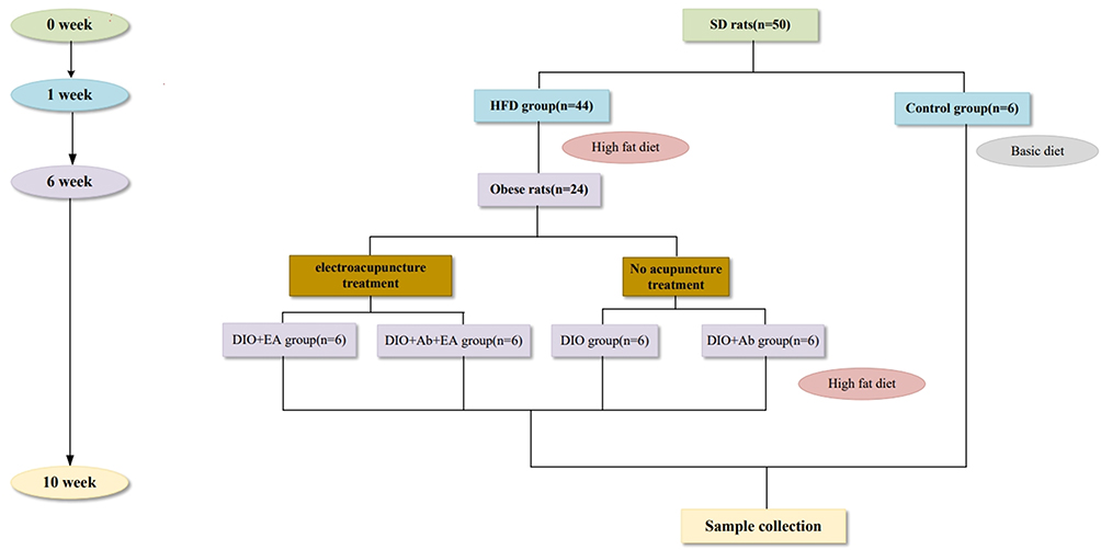 |
Figure 1 Groups and experimental procedure. |
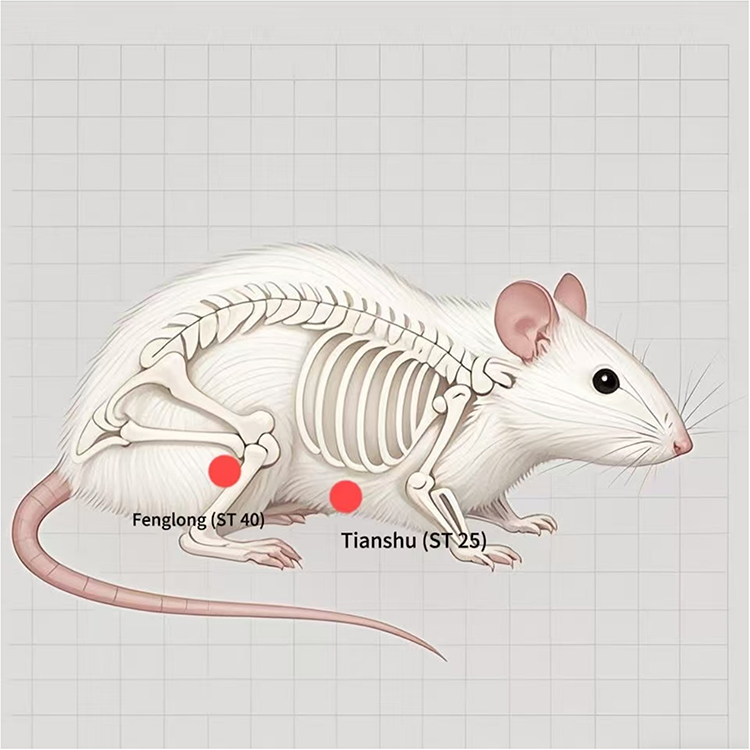 |
Figure 2 Mouse acupoint diagram. |
Electroacupuncture Therapy and Antibiotic Intervention
Rats in the DIO+EA and DIO+Ab+EA groups were immobilized and subjected to electroacupuncture treatment, starting from the 6th week of the animal experiments. Rats in the control, DIO, and DIO+Ab groups were immobilized in a similar manner but did not receive acupuncture. The selected points were bilateral Tianshu and Fenglong (refer to the animal acupuncture point atlas in Chinese Veterinary Acupuncture, Experimental Acupuncture for the positioning of the points). The Fenglong acupoint (ST40) is situated 1 mm below the Housanli acupoint (the Housanli being on the posterior lateral aspect of the knee, approximately 5 mm below the fibular tuberosity) (Figure 2). The Tianshu acupoint (ST25) lies in the transverse section of the navel, 5 mm from the navel (Figure 2). The standard operating procedure (SOP) for the acupuncture treatment was as follows: Rats in each treatment group were immobilized on a rat platform, and their abdomen and hind limbs were exposed. A 2-cm area surrounding the treatment acupoints was shaved, and the skin at the acupoint area was disinfected using an alcohol-soaked cotton ball. The acupuncture depth was approximately 3 mm at ST25 and 6 mm at ST40, and the shank was connected to electricity after the needle was inserted. Intermittent needle treatment was conducted using a frequency of 2/15 Hz, a voltage of 1.5 V, and a retention duration of 30 min. The acupuncture tool was a 0.18 mm×13 mm milli-needle connected to the SDZ-II type Electro Stimulation Device (Suzhou Medical Supplies Factory Co., Ltd., Suzhou, China). Electroacupuncture was performed once daily for 6 days, encompassing a total of four treatment cycles and 1 day of rest between treatments. Rats in the DIO+Ab and DIO+Ab+EA groups were treated with an aqueous solution of antibiotics (1 g/L each of ampicillin, neomycin, and metronidazole).20 The rats were provided unrestricted access to the antibiotic solution instead of regular drinking water, starting in the 6th week of the animal experiments.
Measurements
Body Weight and Lee’s Index
The body hair and activity of the rats were observed daily. Subsequently, the body weight (g) and length (cm; the length from the incisors to the anus) were used to calculate Lee’s index according to the following formula:

Serum Biochemical Testing
Four weeks after intervention, all rats were fasted overnight for 8 h. Subsequently, rats were anesthetized with 2% sodium pentobarbital (50 mg/kg). Blood samples were obtained from the abdominal aorta and centrifuged at 2000 rpm for 10 min. Then, the supernatant was collected and stored at 4°C until biochemical analysis. The concentrations of total cholesterol (TC), triglycerides (TG), high-density lipoprotein (HDL), and low-density lipoprotein (LDL) in the serum were determined using commercial kits purchased from Mindray (No. 141621008, 141,721,007, 142122006, and 142022005). All experiments were performed according to the manufacturer’s instructions.
Enzyme-Linked Immunosorbent Assay (ELISA)
Colon tissue samples were homogenized using 9× homogenate media, followed by centrifugation at 3000–4000 rpm for 10 min. Then, the supernatant was collected to prepare a 10% tissue homogenate. The homogenates of colon tissue were used to perform ELISA to determine the concentration of 5-HT in colon tissues and of inducible nitric oxide synthase (iNOS), tumor necrosis factor-α (TNF-α), interleukin-10 (IL-10), interleukin-6 (IL-6), and interleukin-1β (IL-1β) in the serum. ELISA kits were purchased from Elabscience (No. gxNLCPY8H0, oFdZgbC024, aFdZgbD026, 9hSfs5QL06, 5hUfs8QL07, Jq4Gr5tr40, Hq4Ar8tr49, DRzhBy74HY, DUzjOy76HP, FZAtokjDF2, and TZNtmkjDY9).
Liver and Colon Histopathology
Liver tissues were fixed using 4% paraformaldehyde, trimmed, dehydrated, paraffin-embedded, stained with hematoxylin and eosin (HE), sliced, and subsequently sealed, adhering strictly to the SOP. A blinded pathologist evaluated the liver histological sections under 100× and 200× magnification. Paraffin-embedded sections of colon tissue were dewaxed, stained with hexamidine silver (G1059; Servicebio Co., Ltd), and subjected to tissue acidification, incubation, color development, dehydration, and sealing. A blinded pathologist evaluated the histological tissue sections under 200× magnification.
16S rRNA Metagenomic Sequencing
Total genomic DNA was obtained from each group. Fresh fecal pellets were collected from each group both prior to electroacupuncture treatment and euthanasia. Amplification was performed using specific primers with a barcode (16S V3+V4). Libraries were constructed using the Illumina TruSeq DNA PCR-Free Library Preparation Kit. The constructed libraries were subjected to Qubit quantification, library detection, and sequencing using NovaSeq 6000. The raw sequencing data (raw data) exhibits a certain proportion of interference data (dirty data). To ensure the accuracy and reliability of the analysis, we subjected the raw data to assembly and filtering to obtain valid data (clean data). Subsequently, the valid data was subjected to noise reduction using DADA 2 or deblur (DADA 2 by default), and the sequences with an abundance of <5 were excluded to obtain the final set of amplicon sequence variants (ASVs). For the resultant ASVs, one approach involved annotating the representative sequence of each ASV to acquire the corresponding species information and the abundance distribution of said species. Simultaneously, species relative abundance analysis, alpha diversity computation, principal co-ordinate analysis (PcoA) based on Bray–Curtis distance, Venn diagram, and species abundance clustering heat map analysis were conducted to determine species richness and evenness within individual samples as well as the differences in community structure between different samples or groups.
High-Performance Liquid Chromatography (HPLC)
Fresh fecal pellets were collected from each group before euthanasia. The pellets were resuspended in 0.5% phosphoric acid, followed by centrifugation for 10 min. Subsequently, an equal volume of ethyl acetate was added for extraction. The extracted solution was centrifuged for 10 min, and 4-methyl valeric acid was added as the internal standard, mixed in a sample bottle, and subjected to gas chromatography-mass spectrometry (GC-MS) for detection. The samples were separated using an Agilent DB-WAX capillary column (30 m, 0.25 mm ID, 0.25 μm) in a gas chromatography system (Agilent 7890A/5975C GC-MS). MSD ChemStation software was used to determine the peak area, retention time, curve, and SCFA content (acetic acid, butyric acid, propionic acid, isobutyric acid, isovaleric acid, and capric acid) in the samples.
Statistical Analysis
GraphPad Prism 9.0 software was used for statistical analysis and the generation of statistical maps. Measurements were tested for normality and chi-square, and the normal distribution was expressed as mean ± standard deviation (x ± s). Following the confirmation of normality and homogeneity through variance analysis for each group’s data, analysis of variance (ANOVA) was used for comparison between groups. In cases where homogeneity of variance and the normality assumption were violated, a non-parametric test (Kruskal–Wallis) was employed. The Pearson correlation coefficient was applied for correlation analysis conforming to the normal distribution, and the Spearman correlation coefficient was employed for correlation analysis not conforming to the normal distribution. P<0.05 was considered to indicate statistically significant differences.
Results
Electroacupuncture Treatment Reduces Body Weight, Lipid Accumulation, and Inflammatory Factor Levels in Obese Rats
Electroacupuncture Treatment Reduces Body Weight, Lee’s Index, and Appetite in Obese Rats
Throughout the experiment, rats in all groups displayed similar behavior and engaged in typical grooming activities. To determine the effect of electroacupuncture on obesity, we measured the body weight of the rats before the commencement of the experiments and subsequently on a weekly basis following the initiation of the experiments, amounting to a total of 11 measurements. After 6 weeks, rats subjected to an HFD exhibited significantly higher body weight than rats in the control group (Figure 3A). In detail, 24 rats (46.4%) gained 20% or more weight compared to the mean weight of the rats in the control group. These 24 rats exhibiting high body weight were randomly assigned to the DIO or DIO+EA groups (n=6). After 3 weeks of electroacupuncture treatment, the body weight of the rats in the DIO+EA group was significantly lower than that of rats in the DIO group (P<0.05); however, their body weights were higher than that of rats in the control group (P<0.05). After 4 weeks of electroacupuncture treatment, the body weight of rats in the DIO+EA group exhibited a continued decrease compared with rats in the DIO group (P<0.05), and no statistically significant difference was observed compared with the body weight of rats in the control group (Figure 3A). As shown in Figure 3B, before the commencement of electroacupuncture treatment, rats in the DIO+EA and DIO groups exhibited a higher Lee’s index than the rats in the control group (P<0.05), and no statistically significant difference was observed between the DIO+EA and DIO groups. After 4 weeks of electroacupuncture treatment, Lee’s index of the rats in the DIO+EA group was lower than that of the rats in the DIO group (P<0.05), and no statistically significant difference was observed between the DIO+EA and control groups (Table 1–3). By comparing body weight and Lee’s index at the beginning of electroacupuncture treatment (week 6) and at the end of electroacupuncture treatment (week 10), we found that both the body weight and Lee’s index in the DIO group were higher at week 10 than at week 6 (P<0.01). Furthermore, no statistically significant difference was observed between any of the groups (Figure 3C and D). As shown in Figure 3E, all groups of rats had ad libitum access to food and water. After the commencement of electroacupuncture treatment, rats in the DIO+EA group exhibited reduced food intake compared with the rats in the control group; therefore, the rats in the DIO+EA group exhibited reduced appetite.
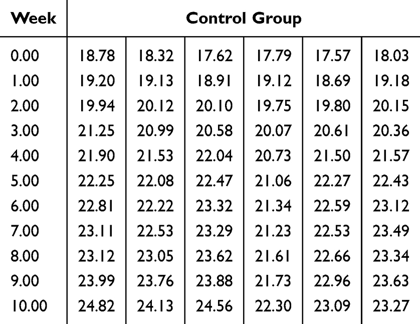 |
Table 1 Lee’s Index of Control Group |
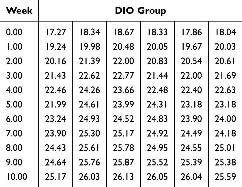 |
Table 2 Lee’s Index of DIO Group |
 |
Table 3 Lee’s Index of DIO+EA Group |
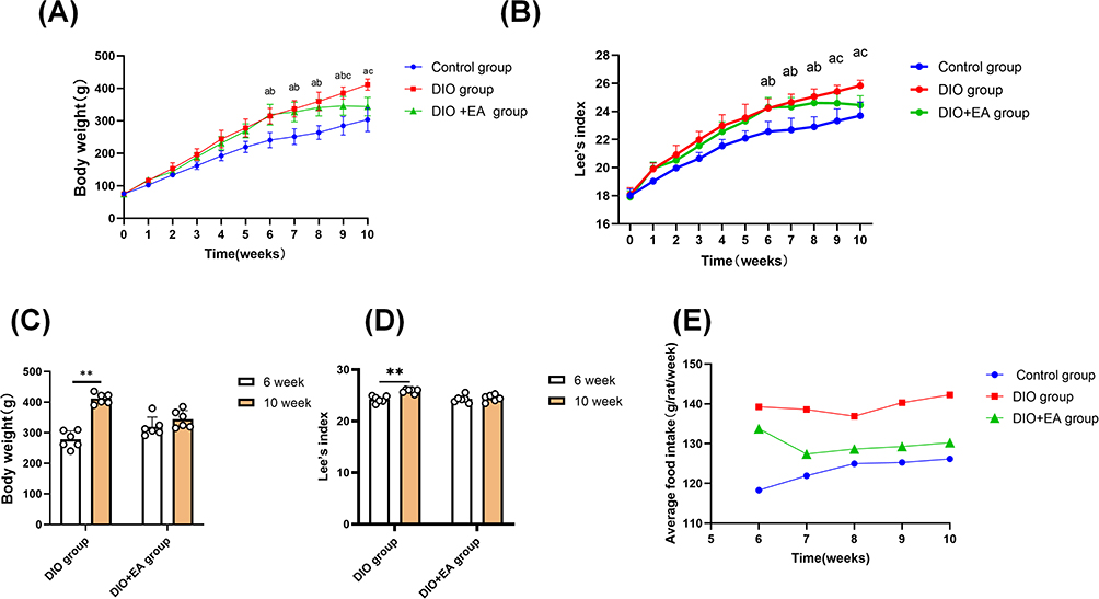 |
Figure 3 Electroacupuncture treatment reduced body weight, Lee’s index, and appetite in obese rats. Notes: (A) Weight change. (B) Lee’s index. (C) Comparison of body weight between the DIO and DIO+EA groups at weeks 6 and 10. (D) Comparison of Lee’s index between the DIO and DIO+EA groups at weeks 6 and 10. (E) The mean feed intake during the experiment. Data are expressed as mean ± standard deviation. “a” indicates control group vs DIO group; “b” indicates control group vs DIO+EA group; and “c” indicates DIO group vs DIO+EA group with statistically significant differences (P<0.05, **P<0.01; n=6). |
Electroacupuncture Treatment Reduces Serum TC and LDL Levels and Hepatic Lipid Accumulation in Obese Rats
The serum total cholesterol (TC) and low-density lipoprotein (LDL) levels were markedly increased in obese rats compared with those in the control group (P < 0.01). After electroacupuncture treatment, the DIO+EA group showed a reduction in both TC and LDL levels; however, these values remained significantly higher than those in the control group (Figure 4B and C). In contrast, high-density lipoprotein (HDL) levels were significantly lower in the control group than in the DIO and DIO+EA groups (P < 0.01). No statistically significant difference in HDL levels was observed between the DIO and DIO+EA groups (Figure 4D). Furthermore, no statistically significant differences in the serum TG levels were observed among the groups (Figure 4A). These results suggest that obesity in rats induced by an HFD results in lipid accumulation in the serum, and electroacupuncture treatment reduces blood lipid levels in obese rats. HE staining of liver tissues showed that the liver structure in the control group was normal with clear sinusoids. However, the liver sections from the DIO group exhibited obvious hepatocyte steatosis and substantial vacuolation in the cytoplasm. Following electroacupuncture treatment, we observed microbubble steatosis in the liver and minimal circular vacuoles in the cytoplasm (Figure 4E). The liver histopathological changes demonstrated that electroacupuncture treatment reduces hepatic lipid accumulation in obese rats.
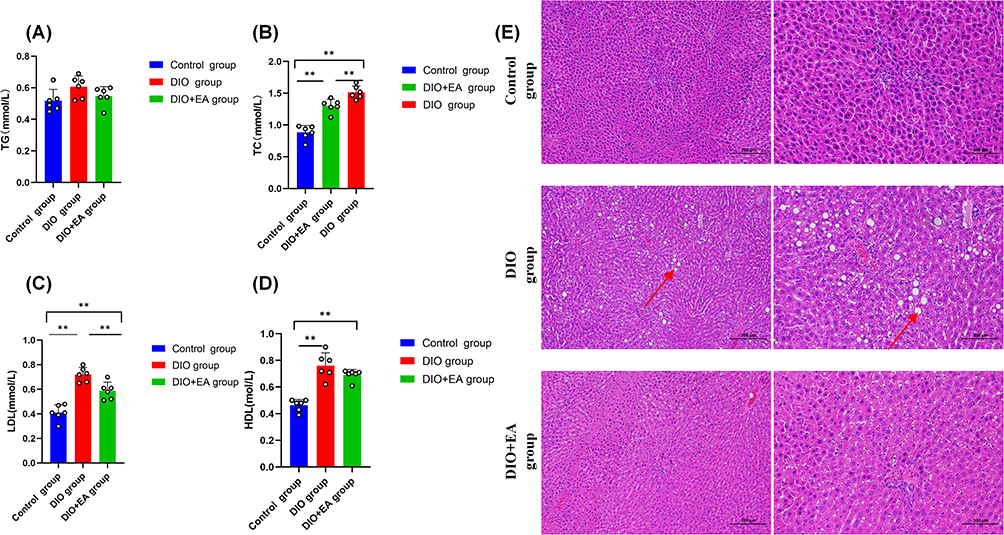 |
Figure 4 Electroacupuncture treatment reduces lipid accumulation in obese rats. Notes: (A) TG. (B) TC. (C) LDL. (D) HDL. (E) HE-stained liver pathological tissues under 100× and 200× magnification. Data are expressed as mean ± standard deviation (**P<0.01; n=6). |
Electroacupuncture Treatment Reduces the Levels of the Serum Inflammatory Factors TNF-α and IL-1β in Obese Rats
Currently, obesity is recognized as a chronic state characterized by low-grade inflammation. Inflammation is one of the key processes in the development of diseases associated with insulin resistance, diabetes, and obesity.21 As shown in Figure 5, an analysis of serum pro-inflammatory factors, namely TNF-α, IL-1β, IL-6, and iNOS, showed that the levels of TNF-α and IL-1β were significantly higher in the DIO group than those in the control group (P<0.01). Conversely, TNF-α levels in the DIO+EA group were lower than those in the DIO group subsequent to electroacupuncture treatment (P<0.05). IL-1β levels in the DIO+EA group were also lower than those in the DIO group (P<0.01). No statistically significant differences were observed in the IL-6 and iNOS levels between the groups (Figure 5A–D). As shown in Figure 5E, the serum levels of the anti-inflammatory factor IL-10 were significantly higher in the control group than those in the DIO group (P<0.05). These results suggest that the levels of certain inflammatory factors are elevated in obese rats and that electroacupuncture reduces their levels.
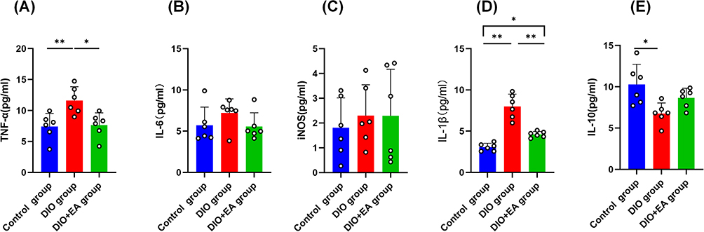 |
Figure 5 Electroacupuncture treatment reduces the serum levels of inflammatory factors in obese rats. Notes: (A) Serum TNF-α. (B) Serum IL-6. (C) Serum iNOS. (D) Serum IL-1 β. (E) Serum IL-10. Data are expressed as mean ± standard deviation (**P<0.01, *P<0.05; n=6). |
Number, Abundance, and Structure of Intestinal Flora are Altered in Obese Rats, and Electroacupuncture Treatment Regulates Intestinal Flora Changes in Obese Rats
As shown in Figure 6D, the Venn diagram comparing the number of ASVs in each group showed 3784 ASVs in the control group, 1624 ASVs in the DIO group, and 3890 ASVs in the DIO+EA group. The number of bacterial gene sequences in the DIO group was significantly reduced. Moreover, as shown in Figure 6A and B, the microbial diversity was reduced in the DIO group. However, the microbial diversity in the DIO+EA group was restored following electroacupuncture treatment, with a statistically significant difference in the Chao1 index of alpha diversity (P<0.05), although no statistically significant difference was observed in the Shannon index. To determine the differences in intestinal flora between the groups, we conducted a PcoA based on Bray–Curtis distance. The microbial community structure in the control and DIO groups, respectively, exhibited obvious clustering, whereas the microbial community structure in the DIO+EA group was similar to that in the control and DIO groups (Figure 6C). The relative abundance of microbial species was analyzed in each group, and the predominant species at the phylum level were determined to be Firmicutes and Bacteroides. The six most prevalent groups in terms of relative abundance were Firmicutes, Bacteroidota, Verrucomicrobiota, Proteobacteria, Euryarchaeota, and Actinobacteriota, and no statistically significant differences were observed between the groups. The top six consortia at the order level in terms of relative abundance were Bacteroidales, Lachnospirales, Lactobacillales, Verrucomicrobiales, Peptostreptococcales-Tissierellales, and Oscillospirales. The six most prevalent families in terms of relative abundance were Lachnospiraceae, Lactobacillaceae, Akkermansiaceae, Peptostreptococcaceae, Succinivibrionaceae, and Bacteroidaceae. The six most prevalent genera in terms of relative abundance were Lactobacillus, Akkermansia, Romboutsia, Anaerobiospirillum, Clostridium_sen_ssu_stricto_1, and Bacteroides. The six most prevalent species-level consortia in terms of relative abundance were Lactobacillus faecis (L. faecis), archaeon_enrichment, Prevotellaceae_bacterium, Prevotella_copri, wallaby_gut, and Bacteroides fragilis (B. fragilis). We further analyzed the changes at each level. The DIO group exhibited a decreased abundance of Lactobacillales, Lactobacillaceae, Lactobacillus, and L. faecis at the order, family, genus, and species levels (P<0.05); however, microbes were increased following electroacupuncture treatment in the DIO+EA group at the family and genus levels. The DIO group exhibited a significantly increased abundance of Bacteroidaceae and Bacteroides (P<0.01). However, their abundance was significantly downregulated after electroacupuncture treatment (P<0.01) (Figure 6E–O).
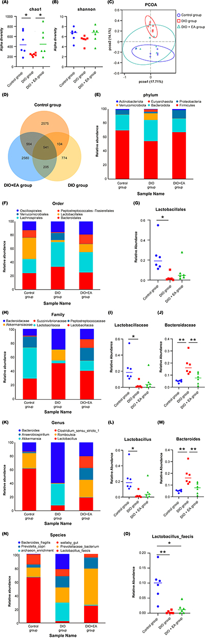 |
Figure 6 Electroacupuncture treatment regulates the number, abundance, and structural changes of the intestinal microflora in obese rats. Notes: (A) Chao1 index of alpha diversity and beta diversity based on Bray–Curtis distance. (B) Shannon index of alpha diversity and beta diversity based on Bray–Curtis distance. (C) PcoA analysis. (D) Number of ASVs in rats. (E) Relative abundance of microbes at the phylum level. (F) Relative abundance of microbes at the order level. (G): Lactobacillales at the order level. (H) Relative abundance of microbes at the family level. (I) Lactobacillaceae at the family level. (J) Bacteroidaceae at the family level. (K) Relative abundance of microbes at the genus level. (L) Lactobacillus at the genus level. (M) Bacteroides at the genus level. (N) Relative abundance of microbes at the species level. (O) L. faecis at the species level. Data are presented as mean ± standard deviation (**P<0.01, *P<0.05; n=6). |
Electroacupuncture Affects L. faecis and B. fragilis and Improves Acetic Acid Levels and 5-HT Secretion by Colonic ECC to Exert Anti-Obesity Effects
Electroacupuncture Affects the Levels of L. faecis, B. fragilis, and Acetic Acid
To analyze the intestinal flora that may be associated with obesity, we performed Spearman analysis to determine the correlation between the percentage of the six most abundant species and obesity-related indicators. The results showed that L. faecis was negatively correlated with TC, LDL, HDL, IL-1β, and body weight (P<0.05). B. fragilis was positively correlated with TC, HDL, IL-1β, and TNF-α (P<0.05) (Figure 7A). L. faecis and B. fragilis were among the top six species in terms of relative abundance in each group. The abundance of L. faecis in the DIO group was lower than that in the control group but was restored in the DIO+EA group. Conversely, the abundance of B. fragilis was increased in the DIO group (Figure 7B). Thus, L. faecis may be beneficial for weight loss, whereas B. fragilis exhibits stable abundance in obese rats. Several studies have validated that the effect of intestinal flora on obesity may be mediated via microflora metabolites, ie, SCFAs.22,23 As shown in Figure 7B, the SCFAs in each group were analyzed and compared with those in the control group. In both the DIO and DIO+EA groups, SCFAs significantly decreased (P<0.01). Following electroacupuncture treatment, SCFAs in the DIO+EA group increased compared with those in the DIO group. As shown in Figure 7C–H, analysis of the levels of the SCFA acetic acid, propionic acid, isobutyric acid, butyric acid, isovaleric acid, and hexanoic acid in each group revealed that the control and the DIO+EA groups both exhibited acetic acid levels significantly higher than those in the DIO group (P<0.01), whereas propionic acid, isobutyric acid, butyric acid, isovaleric acid, and hexanoic acid were not statistically significantly different among the groups. Furthermore, we determined the correlation of acetic acid, propionate, isobutyrate, butyrate, isovaleric acid, and caproate with body weight and found that only acetic acid was negatively correlated with body weight (P<0.05) (Figure 7I). Therefore, the anti-obesity effect of electroacupuncture may be associated with L. faecis, B. fragilis, and the microflora metabolite acetic acid.
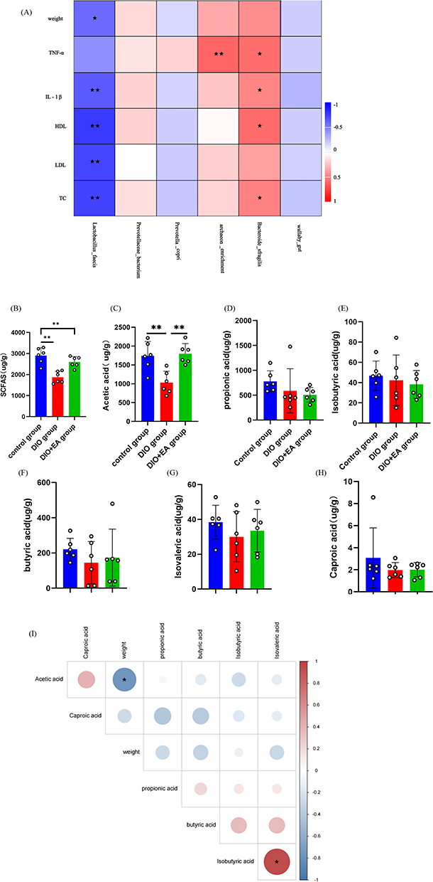 |
Figure 7 Electroacupuncture treatment of OB is associated with L. faecis, (B) fragilis, and acetic acid levels. Notes: (A) The top six species in terms of relative abundance were correlated with OB-related indicators. (B) The levels of SCFAs. (C) The levels of acetic acid, (D) propionic acid, (E) isobutyrate acid, (F) butyric acid, (G) isovaleric acid, and (H) caproic acid. (I) Spearman correlation analysis of acetic acid, isobutyric acid, butyric acid, propionate, isovalerate, and caproic acid with body weight. Data are presented as mean ± standard deviation. (**P<0.01, *P<0.05; n=6). |
Anti-Obesity Effect of Electroacupuncture May Be Closely Associated with L. faecis, B. fragilis, Acetic Acid, and 5-HT Secreted by Colonic ECC
5-HT, a neurotransmitter and hormone, regulates intestinal movement and the intestinal endocrine system and is closely associated with obesity. Colonic ECC primarily secretes 5-HT. In this study, the mean ECC counts in colonic tissue sections in three fields of view at 200× magnification showed no statistically significant differences. However, the ECC count showed a tendency to decrease in the DIO and DIO+EA groups (Figure 8A). Figure 8B depicts ECC in colonic tissue sections of each group. As shown in Figure 8C, the DIO group exhibited lower 5-HT levels in the colon compared to the control group, whereas the DIO+EA group demonstrated significantly higher 5-HT levels compared to the DIO group (P<0.01). According to the findings of relevant studies, the gut microbiota stimulates 5-HT production by ECC through SCFAs.24 Figure 8D illustrates the correlation between the contents of acetic acid, isobutyric acid, butyrate acid, propionic acid, isovaleric acid, caproic acid, and 5-HT. We found that 5-HT was positively correlated with acetic acid (P<0.05). This suggests that the anti-obesity effect of electroacupuncture may be closely associated with 5-HT. We subsequently explored the correlation among the gut microbes, the microflora metabolite acetate acid, and the enteroendocrine hormone 5-HT (Figure 8E). Correlation analysis of the relative abundance of L. faecis and B. fragilis with acetic acid, 5-HT, and body weight showed that body weight was negatively associated with L. faecis, acetic acid, and 5-HT, whereas L. faecis was positively correlated with 5-HT (P<0.05). Moreover, B. fragilis was negatively correlated with 5-HT (P<0.05). These results suggest that electroacupuncture may exert anti-obesity effects by modulating L. faecis and B. fragilis and by upregulating acetic acid and 5-HT levels.
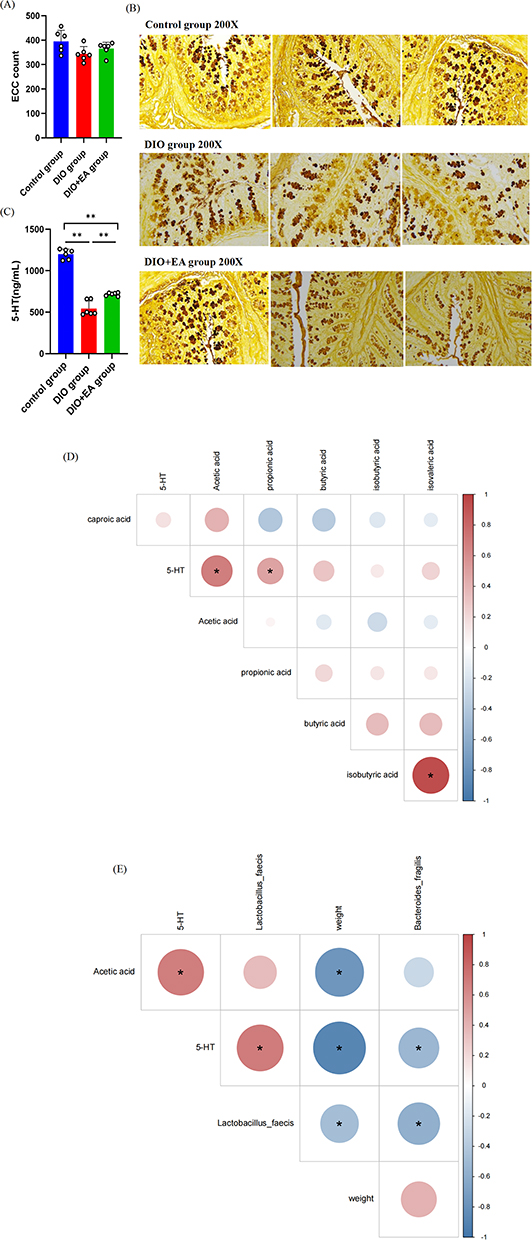 |
Figure 8 Anti-obesity effect of electroacupuncture may be closely associated with L. faecis, (B) fragilis, acetic acid, and 5-HT secreted by colonic ECC. Notes: (A) Colonic ECC counts in each group. (B) Silver amine staining of colon tissue sections under 200× magnification. (C) 5-HT content. (D) Correlation analysis of acetic acid, propionic acid, isobutyric acid, butyric acid, isovaleric acid, and capric acid content with 5-HT. (E) Correlation analysis of L. faecis and B. fragilis with acetic acid, 5-HT, and body weight. Data are presented as mean ± standard deviation (**P<0.01, *P<0.05; n=6). |
Antibiotics Inhibit the Anti-Obesity Effect of Electroacupuncture by Affecting the Intestinal Microflora and Downregulating Acetic Acid and 5-HT
Antibiotics Inhibit the Anti-Obesity Effect of Electroacupuncture
To determine the effect of antibiotics on obese rats, we administered antibiotics to deplete the intestinal microorganisms in obese rats. Changes in body weight and lipid levels were monitored in the DIO, DIO+EA, DIO+Ab, and DIO+Ab+EA groups. No statistically significant differences in body weight and Lee’s index were observed at the beginning (week 6) among the DIO, DIO+Ab, and DIO+Ab+EA groups. However, by the end of the experiment (week 10), body weight and Lee’s index in the DIO+EA group were lower than those in other groups (P<0.05) (Figure 9A and B). Serum levels of TC, HDL, and LDL in the DIO+Ab and DIO+Ab+EA groups were not significantly different from those in the DIO group. Serum levels of LDL in the DIO+Ab group were significantly higher than those in the DIO+EA group (P<0.05) (Figure 8C–E). No statistically significant differences in serum levels of TG were observed among all the groups (Figure 9F). As shown in Figure 9G, hepatocytes in the DIO+Ab and DIO+Ab+EA groups exhibited steatosis, with several vacuolated fat-like cells visible in the cytoplasm, similar to the changes in the liver of rats in the DIO group. As shown in Figure 9H and I, no statistically significant differences in the inflammation indicators TNF-α and IL-1β were observed among the DIO, DIO+Ab, and DIO+Ab+EA groups. However, TNF-α levels in the DIO+EA group were significantly lower than those in the DIO and DIO+Ab groups, and IL-1β levels in the DIO+EA group were significantly lower than those in the other three groups (P<0.05).
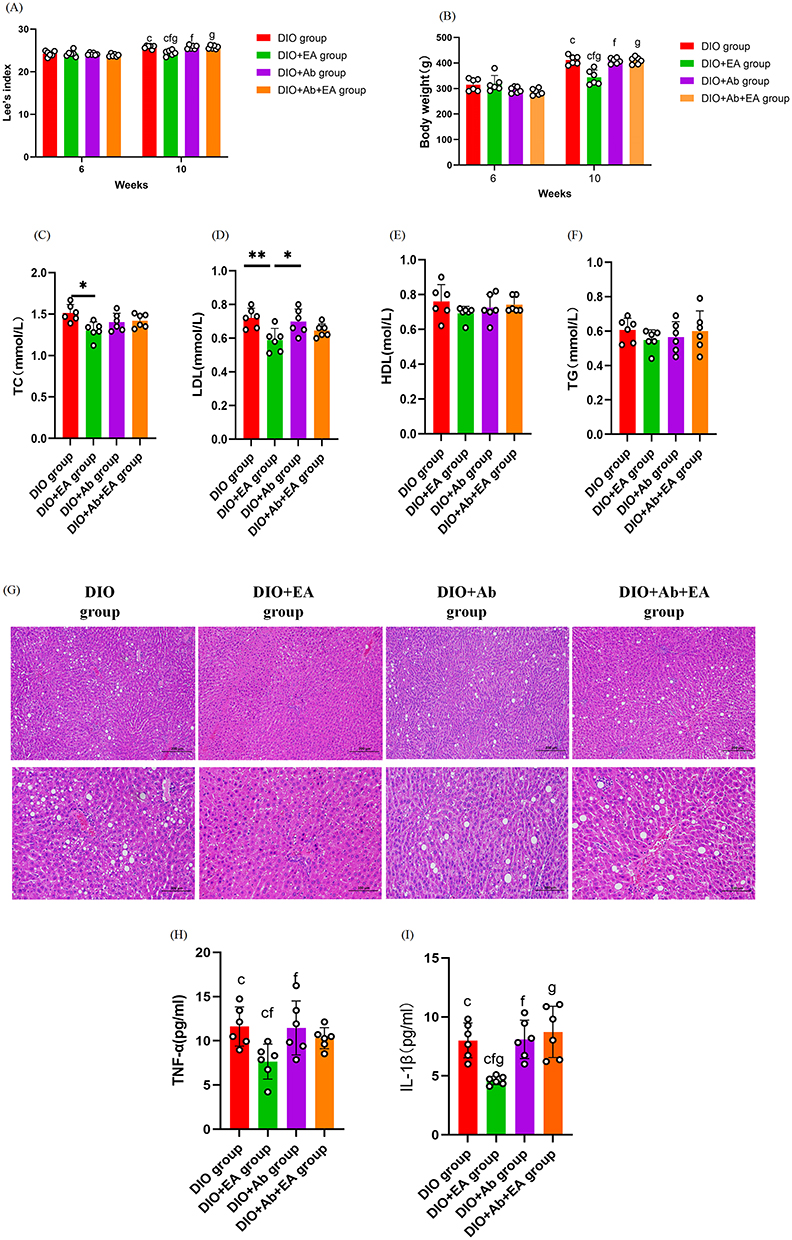 |
Figure 9 Antibiotics inhibit the anti-obesity effect of electroacupuncture. Notes: (A): Body weight of obese rats at weeks 6 and 10. (B) Lee’s index at weeks 6 and 10. (C) The level of TC; (D) LDL’ (E) HDL; (F) and TG. (G) HE-stained liver pathological tissues at 100× and 200× magnifications. (H) The levels of TNF-α (I) and IL-1β. Data are presented as the mean ± standard deviation. “c” indicates DIO group vs DIO+EA group; “d” indicates DIO group vs DIO+Ab group; “e” indicates DIO group vs DIO+Ab+EA group; “f” indicates DIO+EA group vs DIO+Ab group; “g” indicates DIO+EA group vs DIO+Ab+EA group; “h” indicates DIO+Ab group vs DIO+Ab+EA group, with statistically significant differences (**P<0.01, *P<0.05; n=6). |
Antibiotics Inhibit the Anti-Obesity Effect of Electroacupuncture by Affecting the Intestinal Microflora and Downregulating Acetic Acid and 5-HT
As shown in Figure 10, the Wayn diagram of the number of ASVs in the DIO, DIO+EA, DIO+Ab, and DIO+Ab+EA groups showed a total of 1256 ASVs in the DIO group, 3890 ASVs in the DIO+EA group, 573 ASVs in the DIO+Ab group, and 807 ASVs in the DIO+Ab+EA group. The number of gene sequences pertaining to microbial flora was significantly reduced in obese rats following antibiotic intervention. Alpha diversity in both the DIO+Ab and DIO+Ab+EA groups was lower than that in the DIO+EA group, with statistically significant differences in the Chao1 index and Shannon index (P<0.05). Moreover, the Shannon index of alpha diversity in the DIO+Ab and DIO+Ab+EA groups was lower than that in the DIO group (P<0.05) (Figure 10B and C). To assess the differences in intestinal flora structure across the groups, we performed PcoA utilizing the weighted_unifrac distance for beta diversity. The intestinal flora structure in the DIO and DIO+EA groups was similar, as was that in the DIO+Ab and DIO+Ab+EA groups (Figure 10D). The results showed that the administration of antibiotics to obese rats significantly reduced the number, structure, and abundance of the intestinal flora. As shown in Figure 10E–G, at the species level, the relative abundance of L. faecis in the DIO+Ab and DIO+Ab+EA groups was statistically significantly reduced (P<0.05). The analysis of acetic acid and 5-HT content in each group showed that the DIO+Ab and DIO+Ab+EA groups exhibited decreased levels of acetic acid and 5-HT compared with the other groups (P<0.05) (Figure 10H and I). Spearman correlation analysis was performed to compare the correlation of L. faecis and B. fragilis with acetic acid, 5-HT, and body weight across the control, DIO, DIO+EA, DIO+Ab, and DIO+Ab+EA groups. The results showed that L. faecis was positively correlated with acetic acid and 5-HT and negatively correlated with body weight (P<0.05). Furthermore, B. fragilis was negatively correlated with acetic acid and 5-HT while displaying a positive correlation with body weight (P<0.05). Moreover, the results also indicated a positive correlation between acetic acid and 5-HT as well as a negative correlation between acetic acid and body weight (P<0.05) (Figure 10J). The anti-obesity effect of electroacupuncture was inhibited by antibiotic intervention, and changes in intestinal flora and the levels of acetic acid and 5-HT were diminished following antibiotic intervention, suggesting that antibiotics may inhibit the anti-obesity effect of electroacupuncture by affecting the intestinal flora and downregulating acetic acid and colonic 5-HT content in obese rats.
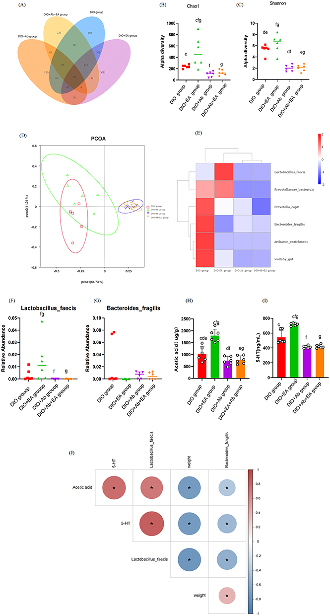 |
Figure 10 Antibiotics inhibit the anti-obesity effect of electroacupuncture by affecting intestinal flora and downregulating acetic acid and 5-HT levels. Notes: (A) Venn diagram of ASVs in obese rats. (B) Chao1 index of alpha diversity. (C) Shannon index of alpha diversity. (D) PcoA of beta diversity based on Bray–Curtis distance. (E) Cluster heat map of the six most abundant microbial species. (F) Abundance of L. faecis. (G) Abundance of B. fragilis. (H) Acetic acid content. (I) 5-HT content. (J) Correlation of L. faecis and B. fragilis with acetic acid, 5-HT, and body weight. Data are expressed as mean ± standard deviation. “c” indicates DIO group vs DIO+EA group; “d” indicates DIO group vs DIO+Ab group; “e” indicates DIO group vs DIO+Ab+EA group; “f” indicates DIO+EA group vs DIO+Ab group; “g” indicates DIO+EA group vs DIO+Ab+EA group; and “h” indicates DIO+Ab group vs DIO+Ab+EA group with statistically significant differences (**P<0.01, *P<0.05; n=6). |
Discussion
In this study, we demonstrated that electroacupuncture treatment affects the food intake of rats and effectively reduces the intake of HFD and liver steatosis. Obesity is considered a chronic condition characterized by low-grade inflammation, potentially associated with intestinal flora dysbiosis and impaired barrier function in obese rats.25 In this study, obese rats exhibited increased levels of TNF-α and IL-1β inflammatory factors alongside decreased levels of the anti-inflammatory factor IL-10, which improved following electroacupuncture treatment. Our findings are consistent with prior research that reported increased levels of IL-6, TNF-α, and IL-1β in insulin-resistant obese rats.26
Several animal experiments and clinical trials have validated that obesity and gut microflora are closely associated.27 Recently, related studies have shown that individuals with obesity exhibit a higher proportion of Firmicutes and that the proportion of Firmicutes/Bacteroides tends to increase as the body mass index increases.28 However, our study showed no significant differences in the abundance of Firmicutes and Bacteroidetes between obese and normal rats. Consistent with the findings of most trials, the number, diversity, and richness of the gut microbiota in obese rats in this study were reduced.22 Most studies suggest that Lactobacillus acidophilus, a member of the Lactobacillus genus, can alleviate obesity, reverse intestinal dysbiosis induced by an HFD, maintain intestinal barrier integrity, and reduce chronic low-grade inflammation associated with obesity.29,30 While limited research has been conducted on L. faecis, the results of this study might provide novel therapeutic targets for subsequent investigations into the effects of this species on obesity.
Analysis of the relative abundance of Bacteroidaceae and Bacteroides at the family and genus levels in this study and relevant clinical studies have shown that B. fragilis is positively correlated with obesity.31 A fixed dietary pattern might lead to a lack of competition for other intestinal symbioses, thereby leading to the overgrowth of Bacteroides.32 Moreover, B. fragilis, a commensal gram-negative obligate anaerobic bacterium, might cause a range of diseases involving a permeable intestinal barrier.33 Disorders associated with intestinal permeability and increased inflammation could contribute to the advancement of obesity.34 We found that the correlations among B. fragilis, body weight, and inflammatory factors were consistent with the findings of the aforementioned study.
Antibiotics are often used to assess the effects of gut microbes on the host.35 In this study, antibiotic intervention inhibited the anti-obesity effect of electroacupuncture in the DIO and DIO+EA groups. Moreover, the anti-obesity effect was validated by the improvement in the structure, quantity, and abundance of microbes in the obese rats, characterized by an increased number of probiotic microbes and decreased colonization. These findings provide support for further exploration into the specific mechanisms by which electroacupuncture exerts its anti-obesity effect via the gut microbiota.
The metabolite SCFAs exerts notable beneficial effects on energy metabolism, intestinal homeostasis, and the regulation of immune responses. In obesity and metabolic diseases, aberrant production of SCFAs has been observed.36 Relevant studies have shown that acetic acid, an SCFA, can mitigate obesity and obesity-related type 2 diabetes.37 Supplementation of exogenous acetate in the diet has been suggested as a promising novel strategy for the prevention and management of obesity.38 A study on obesity elucidated that Bifidobacterium fragilis in the gut might contribute to obesity progression, in part by lowering acetic acid levels.31 In this study, the levels of SCFAs and acetic acid were significantly decreased in obese rats, and electroacupuncture treatment upregulated the levels of SCFAs and acetic acid, indicating that the mechanism underlying the anti-obesity effects of electroacupuncture may involve acetic acid. SCFAs are the primary signaling molecules that mediate microbial communication through ECC. Moreover, the gut microflora metabolite SCFAs maintains a balance between the synthesis and secretion of 5-HT by ECC.16,39
5-HT, a neurotransmitter and hormone, regulates intestinal endocrine secretion and intestinal motility. This intestinal peptide, associated with intestinal motility and the enteroendocrine system, is closely associated with obesity.40,41 The majority of 5-HT (>90%) is produced by ECC in the gut.42 Nevertheless, the correlation of the SCFA acetic acid with 5-HT has received less research attention. This study showed that electroacupuncture treatment influences the intestinal flora and that acetic acid, 5-HT, and body weight are strongly correlated. Investigations into acetic acid highlight that Lactobacillus sp. produces acetic acid.43 Consistent results were observed in the present study, ie, electroacupuncture treatment increased the abundance of Lactobacillus sp. and the levels of acetic acid in obese rats. Therefore, this study is the first to demonstrate that electroacupuncture may regulate body weight by affecting the abundance of L. faecis and B. fragilis, as well as the levels of acetic acid and 5-HT secretion by colonic ECC. Further validation of these findings is warranted.
Conclusion
Through examination of the intestinal flora and the primary metabolites associated with obesity, we found that electroacupuncture treatment exerts anti-obesity effects by modulating the intestinal flora and influencing acetic acid levels and the secretion of 5-HT by colonic ECC cells. L. faecis and B. fragilis in the intestinal flora hold promise as potential targets for the treatment of obesity in the future.
Abbreviations
OB, obesity; SCFAs, Short-chain fatty acids; ECC, Enterochromaffin cells; 5-HT, 5-Hydroxytryptamine; GLP-1, Glucagon-like peptide-1; HFD, High-fat diet; DIO, HFD-induced obese; EA, Electroacupuncture; Ab, Antibiotic; ST, Stomach Meridian of Foot-Yangming; SOP, Standard operating procedure; TC, Total cholesterol; TG, Triglycerides; HDL, High-density lipoprotein; LDL, Low-density lipoprotein; ELISA, Enzyme-linked immunosorbent assay; iNOS, Inducible nitric oxide synthase; TNF-α, Tumor necrosis factor-α; IL-10, Interleukin-10; IL-6, Interleukin-6; IL-1β, Interleukin-1β; HE, Hematoxylin–eosin; ASVs, Amplicon sequence variants; PCoA, Principal co-ordinate analysis; HPLC, High-performance liquid chromatography; GC-MS, Gas chromatography-mass spectrometry; ANOVA, Analysis of variance; L. faecis, Lactobacillus faecis; B. fragilis, Bacteroides fragilis.
Data Sharing Statement
All data included in this study are available upon request by contacting the corresponding author.
Ethics Statement
Animal experiments were approved by the Animal Ethics Committee of Chengdu University of Traditional Chinese Medicine (No. 2019-06).
Acknowledgments
We thank Novo Magic for providing RNA-sequencing and bioinformatics analysis services. We thank Bullet Edits Limited for the linguistic editing and proofreading of the manuscript.
Author Contributions
All authors made a significant contribution to the work reported, whether that is in the conception, study design, execution, acquisition of data, analysis and interpretation, or in all these areas; took part in drafting, revising or critically reviewing the article; gave final approval of the version to be published; have agreed on the journal to which the article has been submitted; and agree to be accountable for all aspects of the work.
Funding
This work was supported by the National Natural Science Foundation of China [Grant Number: 81774440] and the Provincial Natural Science Foundation of Sichuan, China [2024NSFSC0693].
Disclosure
The authors declare that they have no competing interests in this work.
References
1. Hachuła M, Kosowski M, Zielańska K, Basiak M, Okopień B. The impact of various methods of obesity treatment on the quality of life and mental health-A narrative review. Int J Environ Res Public Health. 2023;20(3):2122. doi:10.3390/ijerph20032122
2. Caballero B. Humans against obesity: who will win? Adv Nutr. 2019;10(suppl_1):S4–s9. doi:10.1093/advances/nmy055
3. Afshin A, Forouzanfar MH, Reitsma MB, et al. Health effects of overweight and obesity in 195 countries over 25 years. N Engl J Med. 2017;377(1):13–27.
4. Wang Y, Zhao L, Gao L, Pan A, Xue H. Health policy and public health implications of obesity in China. Lancet Diabetes Endocrinol. 2021;9(7):446–461. doi:10.1016/S2213-8587(21)00118-2
5. LeBlanc EL, Patnode CD, Webber EM, Redmond N, Rushkin M, O’Connor EA. U.S. Preventive Services Task Force Evidence Syntheses, Formerly Systematic Evidence Reviews. Behavioral and Pharmacotherapy Weight Loss Interventions to Prevent Obesity-Related Morbidity and Mortality in Adults: An Updated Systematic Review for the US Preventive Services Task Force. Rockville (MD): Agency for Healthcare Research and Quality (US); 2018.
6. Chen H, Guo X. Obesity and functional disability in elderly Americans. J Am Geriatr Soc. 2008;56(4):689–694. doi:10.1111/j.1532-5415.2007.01624.x
7. Bertakis KD, Azari R. Obesity and the use of health care services. Obes Res. 2005;13(2):372–379. doi:10.1038/oby.2005.49
8. Gregg EW, Jakicic JM, Blackburn G, et al. Association of the magnitude of weight loss and changes in physical fitness with long-term cardiovascular disease outcomes in overweight or obese people with type 2 diabetes: a post-hoc analysis of the look AHEAD randomised clinical trial. Lancet Diabetes Endocrinol. 2016;4(11):913–921.
9. Belivani M, Dimitroula C, Katsiki N, Apostolopoulou M, Cummings M, Hatzitolios AI. Acupuncture in the treatment of obesity: a narrative review of the literature. Acupunct Med. 2013;31(1):88–97. doi:10.1136/acupmed-2012-010247
10. Zhong YM, Luo XC, Chen Y, et al. Acupuncture versus sham acupuncture for simple obesity: a systematic review and meta-analysis. Postgrad Med J. 2020;96(1134):2217. doi:10.1136/postgradmedj-2019-137221
11. You H, Tan Y, Yu D, et al. The therapeutic effect of SCFA-mediated regulation of the intestinal environment on obesity. Front Nutr. 2022;9:886902. doi:10.3389/fnut.2022.886902
12. Denou E, Marcinko K, Surette MG, Steinberg GR, Schertzer JD. High-intensity exercise training increases the diversity and metabolic capacity of the mouse distal gut microbiota during diet-induced obesity. Am J Physiol Endocrinol Metab. 2016;310(11):E982–93. doi:10.1152/ajpendo.00537.2015
13. Ciobârcă D, Cătoi AF, Copăescu C, Miere D, Crișan G. Bariatric surgery in obesity: effects on gut microbiota and micronutrient status. Nutrients. 2020;12(1):235. doi:10.3390/nu12010235
14. Heiss CN, Olofsson LE. Gut microbiota-dependent modulation of energy metabolism. J Innate Immun. 2018;10(3):163–171. doi:10.1159/000481519
15. Zheng L, Kelly CJ, Battista KD, et al. Microbial-derived butyrate promotes epithelial barrier function through il-10 receptor-dependent repression of Claudin-2. J Immunol. 2017;199(8):2976–2984. doi:10.4049/jimmunol.1700105
16. Martin CR, Osadchiy V, Kalani A, Mayer EA. The brain-gut-microbiome axis. Cell Mol Gastroenterol Hepatol. 2018;6(2):133–148. doi:10.1016/j.jcmgh.2018.04.003
17. Lund ML, Egerod KL, Engelstoft MS, et al. Enterochromaffin 5-HT cells - A major target for GLP-1 and gut microbial metabolites. Mol Metab. 2018;11:70–83. doi:10.1016/j.molmet.2018.03.004
18. Li X, Wu D, Niu J, et al. Intestinal flora: a pivotal role in investigation of traditional chinese medicine. Am J Chin Med. 2021;49(2):237–268. doi:10.1142/S0192415X21500130
19. Paulsen SJ, Jelsing J, Madsen AN, et al. Characterization of beta-cell mass and insulin resistance in diet-induced obese and diet-resistant rats. Obesity (Silver Spring). 2010;18(2):266–273. doi:10.1038/oby.2009.245
20. Hu X, Wang T, Liang S, Li W, Wu X, Jin F. Antibiotic-induced imbalances in gut microbiota aggravates cholesterol accumulation and liver injuries in rats fed a high-cholesterol diet. Appl Microbiol Biotechnol. 2015;99(21):9111–9122. doi:10.1007/s00253-015-6753-4
21. Tzanavari T, Giannogonas P, Karalis KP. TNF-alpha and obesity. Curr Dir Autoimmun. 2010;11:145–156.
22. Cheng Z, Zhang L, Yang L, Chu H. The critical role of gut microbiota in obesity. Front Endocrinol. 2022;13:1025706. doi:10.3389/fendo.2022.1025706
23. Portincasa P, Bonfrate L, Vacca M, et al. Gut microbiota and short chain fatty acids: implications in glucose homeostasis. Int J Mol Sci. 2022;23(3):1105. doi:10.3390/ijms23031105
24. Reigstad CS, Salmonson CE, Rainey JF, et al. Gut microbes promote colonic serotonin production through an effect of short-chain fatty acids on enterochromaffin cells. FASEB J. 2015;29(4):1395–1403. doi:10.1096/fj.14-259598
25. Gomes AC, Hoffmann C, Mota JF. The human gut microbiota: Metabolism and perspective in obesity. Gut Microbes. 2018;9(4):308–325. doi:10.1080/19490976.2018.1465157
26. Huang Q, Song YJ, Yu ZM, et al. Electroacupuncture improves inflammatory reaction and insulin sensitivity in insulin-resistant obese rats. Zhen Ci Yan Jiu. 2019;44(12):898–905. doi:10.13702/j.1000-0607.190209
27. Liu BN, Liu XT, Liang ZH, Wang JH. Gut microbiota in obesity. World J Gastroenterol. 2021;27(25):3837–3850. doi:10.3748/wjg.v27.i25.3837
28. Koliada A, Syzenko G, Moseiko V, et al. Association between body mass index and Firmicutes/Bacteroidetes ratio in an adult Ukrainian population. BMC Microbiol. 2017;17(1):120. doi:10.1186/s12866-017-1027-1
29. Yang M, Meng F, Gu W, et al. Influence of polysaccharides from polygonatum kingianum on short-chain fatty acid production and quorum sensing in Lactobacillus faecis. Front Microbiol. 2021;12:758870. doi:10.3389/fmicb.2021.758870
30. Kang Y, Kang X, Yang H, et al. Lactobacillus acidophilus ameliorates obesity in mice through modulation of gut microbiota dysbiosis and intestinal permeability. Pharmacol Res. 2022;175:106020. doi:10.1016/j.phrs.2021.106020
31. Shen WD, Lin X, Liu HM, et al. Gut microbiota accelerates obesity in peri-/post-menopausal women via Bacteroides fragilis and acetic acid. Int J Obes Lond. 2022;46(10):1918–1924. doi:10.1038/s41366-022-01137-9
32. Zafar H, Saier MH Jr. Gut bacteroides species in health and disease. Gut Microbes. 2021;13(1):1–20. doi:10.1080/19490976.2020.1848158
33. Sun F, Zhang Q, Zhao J, Zhang H, Zhai Q, Chen W. A potential species of next-generation probiotics? The dark and light sides of Bacteroides fragilis in health. Food Res Int. 2019;126:108590. doi:10.1016/j.foodres.2019.108590
34. Portincasa P, Bonfrate L, Khalil M, et al. Intestinal barrier and permeability in health, obesity and NAFLD. Biomedicines. 2021;10(1). doi:10.3390/biomedicines10010083.
35. Schulz MD, Atay C, Heringer J, et al. High-fat-diet-mediated dysbiosis promotes intestinal carcinogenesis independently of obesity. Nature. 2014;514(7523):508–512. doi:10.1038/nature13398
36. Coppola S, Avagliano C, Calignano A, Berni Canani R. The protective role of butyrate against obesity and obesity-related diseases. Molecules. 2021;26(3):682. doi:10.3390/molecules26030682
37. Yamashita H. Biological function of acetic acid-improvement in obesity and glucose tolerance by acetic acid in type 2 diabetic rats. Crit Rev Food Sci Nutr. 2016;56 Suppl 1:S171–5.
38. Valdes DS, So D, Gill PA, Kellow NJ. Effect of dietary acetic acid supplementation on plasma glucose, lipid profiles, and body mass index in human adults: a systematic review and meta-analysis. J Acad Nutr Diet. 2021;121(5):895–914. doi:10.1016/j.jand.2020.12.002
39. Wikoff WR, Anfora AT, Liu J, et al. Metabolomics analysis reveals large effects of gut microflora on mammalian blood metabolites. Proc Natl Acad Sci. 2009;106(10):3698–3703. doi:10.1073/pnas.0812874106
40. Mawe GM, Hoffman JM. Serotonin signalling in the gut--functions, dysfunctions and therapeutic targets. Nat Rev Gastroenterol Hepatol. 2013;10(8):473–486. doi:10.1038/nrgastro.2013.105
41. Wang SZ, Yu YJ, Adeli K. Role of gut microbiota in neuroendocrine regulation of carbohydrate and lipid metabolism via the microbiota-gut-brain-liver axis. Microorganisms. 2020;8(4):527. doi:10.3390/microorganisms8040527
42. Gershon MD. 5-Hydroxytryptamine (serotonin) in the gastrointestinal tract. Curr Opin Endocrinol Diabetes Obes. 2013;20(1):14–21. doi:10.1097/MED.0b013e32835bc703
43. Chang CJ, Lin CS, Lu CC, et al. Corrigendum: ganoderma lucidum reduces obesity in mice by modulating the composition of the gut microbiota. Nat Commun. 2017;8:16130. doi:10.1038/ncomms16130
 © 2025 The Author(s). This work is published and licensed by Dove Medical Press Limited. The
full terms of this license are available at https://www.dovepress.com/terms
and incorporate the Creative Commons Attribution
- Non Commercial (unported, 4.0) License.
By accessing the work you hereby accept the Terms. Non-commercial uses of the work are permitted
without any further permission from Dove Medical Press Limited, provided the work is properly
attributed. For permission for commercial use of this work, please see paragraphs 4.2 and 5 of our Terms.
© 2025 The Author(s). This work is published and licensed by Dove Medical Press Limited. The
full terms of this license are available at https://www.dovepress.com/terms
and incorporate the Creative Commons Attribution
- Non Commercial (unported, 4.0) License.
By accessing the work you hereby accept the Terms. Non-commercial uses of the work are permitted
without any further permission from Dove Medical Press Limited, provided the work is properly
attributed. For permission for commercial use of this work, please see paragraphs 4.2 and 5 of our Terms.