
Back to Journals » Cancer Management and Research » Volume 18

Elaeagnus Angustifolia: Unveiling Anti-Cancer Potential from Ancient Remedies to Modern Therapeutics
Authors Zakaria ZZ  , Ahen YMA, Ahmed NH, Sokary S, Abu-hijleh H, Bawadi H , Al-Asmakh M
Received 22 April 2025
Accepted for publication 15 December 2025
Published 27 January 2026 Volume 2026:18 521703
DOI https://doi.org/10.2147/CMAR.S521703
Checked for plagiarism Yes
Review by Single anonymous peer review
Peer reviewer comments 3
Editor who approved publication: Professor Lu-Zhe Sun
Zain Z Zakaria,1 Yasmin Mohammed Ameen Ahen,2 Nawar Haj Ahmed,2 Sara Sokary,3 Haya Abu-hijleh,2 Hiba Bawadi,3 Maha Al-Asmakh2,4
1Clinical Affairs Department, Qatar University, Doha, Qatar; 2Department of Biomedical Sciences, Qatar University, Doha, Qatar; 3Department of Human Nutrition, Qatar University, Doha, Qatar; 4Biomedical Research Center, Qatar University, Doha, Qatar
Correspondence: Maha Al-Asmakh, Email [email protected]
Background: Cancer is the second leading cause of death worldwide, accounting for approximately 8.97 million deaths, and is projected to surpass ischemic heart disease as the leading cause by 2060. Lung, liver, and stomach cancers are the most lethal globally, while lung and breast cancers are the primary causes of cancer-related mortality in men and women, respectively. In response to this growing burden, there is an urgent need to explore novel therapeutic strategies beyond conventional treatments. Among these, plant-derived medicines offer promising alternatives.
Aim: This review investigates the anti-cancer potential of Elaeagnus angustifolia (EA), a medicinal plant traditionally used for its therapeutic properties, by synthesizing current evidence from in vitro and in vivo studies across breast, cervical, hepatocellular, oral, and colorectal cancer models.
Results: EA has demonstrated anti-cancer activity in breast, cervical, hepatocellular, oral, and colorectal cancer models. Mechanistically, EA induces apoptosis, arrests the cell cycle, modulates HER2/JNK and PI3K/AKT pathways, inhibits epithelial–mesenchymal transition, suppresses angiogenesis, and reduces oxidative stress. While preclinical data are encouraging, animal studies remain limited and clinical validation is lacking.
Conclusion: EA shows promise as a therapeutic agent in cancer management. However, rigorous clinical trials are essential to confirm its safety and efficacy in humans. Future research should also explore its synergistic potential with conventional therapies and further elucidate its molecular mechanisms to support translational application.
Keywords: elaeagnus angustifolia, Russian Olive, anti-cancer potential, therapeutic, cancer, medicinal plants
Introduction
Cancer remains a formidable global health challenge, claiming millions of lives annually. The escalating incidence and mortality rates underscore the urgent need for innovative therapeutic approaches.1 While conventional treatments have achieved significant progress, the emergence of drug resistance and adverse side effects necessitates the exploration of alternative strategies. Plant-derived compounds have garnered increasing attention due to their potential therapeutic benefits and lower toxicity profiles compared to synthetic drugs.2
Among the diverse array of medicinal plants, Elaeagnus angustifolia (EA) has emerged as a promising candidate for cancer management. With a rich history of traditional use for various ailments, EA has recently piqued scientific interest owing to its reported anti-cancer properties.3 This review delves into the existing body of research on EA, critically evaluating its potential as a therapeutic intervention for cancer. By examining both in vitro and in vivo studies, we aim to elucidate the underlying mechanisms of action and assess the clinical implications of this promising botanical agent.
Ultimately, this review seeks to contribute to the growing body of knowledge on EA and its potential role in cancer treatment, providing a foundation for future research and clinical translation.
Background
Overview of Elaeagnus Angustifolia (Russian Olive) and Its Medicinal Properties
Elaeagnus angustifolia (EA), commonly known as Russian olive, is a deciduous tree native to temperate regions of Asia, with a well-established history of medicinal use in traditional healthcare systems across various cultures.4,5 Notably, its use has been documented in regions like Iran, Kazakhstan, and parts of China.4 Following its introduction to North America, Europe, and other parts of the world, EA has gained recognition for its potential therapeutic applications.5
EA is rich in antioxidants, such as flavonoids, phenolic compounds, and vitamins C and E. These antioxidants help neutralize free radicals in the body, reducing oxidative stress and protecting cells from damage.5 The tree’s extracts have demonstrated anti-inflammatory properties, which can help reduce inflammation and provide relief from conditions such as arthritis and other inflammatory disorders.6
The Elaeagnaceae family, encompassing approximately 50 species across three genera, comprises primarily shrubs and small trees native to the Northern Hemisphere.5 Traditional medicinal uses of EA include antipyretic, diuretic, anti-inflammatory, analgesic, and antimicrobial applications, with preparations made from the flowers, leaves, and fruits.7,8
The fruit of Elaeagnus is notable for its nutrient and phytochemical content, including proteins, carbohydrates, fats, dietary fiber, and polyphenols, conferring antioxidant and potential pharmacological properties.9 Beyond nutrition, Elaeagnus preparations such as “Zhourat” herbal tea incorporating Elaeagnus blossoms, have been consumed as a sedative, digestive aid, and expectorant.10 Moreover, Elaeagnus flowers are added to ice cream to enhance sensory appeal and antioxidant content.11 In Turkish folk medicine, various parts of the plant have been employed for their purported tonic, antipyretic, diuretic, antidiarrheal, anti-inflammatory, analgesic, and antimicrobial effects, as well as in the treatment of diverse ailments (Figure 1).12
 |
Figure 1 Chemical structures of kaempferol, eleagnoside, and rutin, and their pharmacological effects. Kaempferol, eleagnoside, and rutin are three compounds found in Elaeagnus angustifolia. They have a variety of pharmacological effects, including gastroprotective, anti-inflammatory, wound healing, muscle relaxant, blood pressure regulator, and cholesterol level regulator effects. These compounds may be responsible for the health benefits of Elaeagnus angustifolia. |
Regular fruit consumption has been suggested to support health in cardiovascular disease, including cataracts, neoplastic conditions, rheumatoid arthritis, neurodegenerative disorders such as Alzheimer’s and Parkinson’s, and certain malignancies.13 However, these associations are largely based on preliminary or observational evidence, and stronger clinical validation is required. The high micronutrient and dietary fiber content of fruits can facilitate dietary modifications, particularly for individuals with high-fat, high-sugar diets.14 Pharmacological studies report that EA extracts exhibit anti-ulcerogenic, wound-healing, muscle relaxant, and immunomodulatory properties.15,16 Topical application of EA leaves has demonstrated wound-healing efficacy through enhanced epidermal regeneration and collagen deposition.5,6 One study showed that EA aqueous extract accelerated cutaneous wound healing in rats by increasing epidermal regeneration and collagen deposition at wound site, leading to faster recovery.17 Furthermore, EA extracts have demonstrated immunomodulatory properties, influencing the body’s immune response. This suggests potential benefits in enhancing the body’s defense against infections and diseases.5 Additionally, traditional use indicates that EA extracts may improve digestive health and alleviate gastrointestinal discomfort.5 Some studies have suggested that EA extracts have shown promise in cardiovascular health, with potential benefits for blood pressure and cholesterol management.5,6 Additionally, these extracts exhibit antimicrobial activity against a range of pathogens, suggesting their potential as a treatment option for infections.5,18,19 While encouraging, most studies are limited by small sample sizes or preclinical models, which limits generalization to the human populations.
Emerging evidence suggests a potential anticancer role for EA extracts.20 Preliminary studies indicate that these extracts may exhibit anti-proliferative effects and induce apoptosis in cancer cells.3,21,22 However, the precise mechanisms underlying these activities and the clinical implications of these findings require comprehensive investigation through rigorous preclinical and clinical trials.
Although experimental evidence suggests a favorable safety profile with limited toxicity in animal models, the safety of EA in humans has not been systematically established.5,23,24 Potential side effects, contraindications, and drug interactions remain underexplored, highlighting the need for future research into its safety in clinical settings.
Elaeagnus Angustifolia: A Promising Phytotherapeutic Agent for Cancer
The potential of Elaeagnus angustifolia (EA) as a cancer therapeutic has emerged from its historical use in traditional medicine and recent preclinical investigations.3,21,22 Multiple studies have demonstrated the anticancer properties of EA extracts in vitro and in vivo models.25 These findings, characterized by apoptosis induction, inhibition of cell proliferation, and modulation of cancer-related signaling pathways, position EA as a compelling candidate for further development as a phytotherapeutic agent in oncology.3
Moreover, EA is a rich source of bioactive compounds, including flavonoids and phenolic compounds with recognized antioxidant properties. This phytochemical profile aligns with the growing interest in natural product-derived anticancer agents.5 Unlike conventional chemotherapeutics, which often exhibit systemic toxicity, preclinical studies suggest that EA extracts may selectively target cancer cells, mitigating collateral damage to normal tissues.21,26
This targeted approach may lead to fewer side effects and improved quality of life for cancer patients.
Beyond its direct anticancer effects, EA exhibits immunomodulatory properties, capable of enhancing the body’s immune response against cancer cells.4,27–29 This immunomodulatory potential positions EA as a promising candidate for combination therapies, where it could synergistically augment existing treatment modalities such as chemotherapy, radiation, and immunotherapy.
The historical use of EA in traditional medicine for various ailments, including cancer, provides a rich foundation for scientific exploration.7,21 Traditional knowledge offers valuable insights into its potential therapeutic applications and can guide modern research efforts. Moreover, EA’s capacity to influence multiple cellular pathways involved in cancer development and progression suggests a comprehensive approach to combating the disease.21
Elaeagnus Angustifolia and Breast Cancer: A Preliminary Assessment
Research exploring the effects of EA on breast cancer remains in its infancy, with a limited number of in vitro studies available.3,21 EA extracts have demonstrated anti-proliferative and pro-apoptotic activity against both hormone receptor–positive and triple-negative breast cancer (TNBC) cell lines.3,21 For instance, Jabeen et al (2020) investigated the impact of EA on HER2-positive breast cancer cells. These findings indicate that EA exhibits dose-dependent inhibitory effects on cell proliferation, induces apoptosis, and suppresses invasive and metastatic behaviors.21 An analysis of the underlying molecular pathways showed EA extract may exert its anticancer effects by inhibiting the activity of HER2 and the JNK1/2/3 signaling cascade, both of which are commonly associated with tumor growth and progression. This inhibition appears to trigger a shift in the expression of key proteins involved in cell adhesion and epithelial-mesenchymal transition (EMT). Specifically, EA treatment leads to an upregulation of E-cadherin and β-catenin, which are markers of epithelial integrity, while simultaneously downregulating vimentin and fascin, proteins typically linked to increased cell motility and invasiveness (Figure 2). These molecular changes suggest that EA helps restore epithelial characteristics and suppress metastatic potential in cancer cells.21 While these preliminary findings are promising, further research is imperative to validate these observations in additional breast cancer subtypes and to elucidate the precise molecular mechanisms involved in EA’s anti-cancer activity.
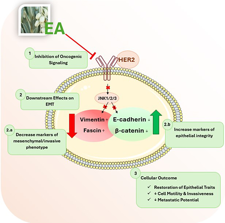 |
Figure 2 Proposed molecular mechanism of EA (Elaeagnus angustifolia) in suppressing cancer cell motility and invasiveness via modulation of HER2/JNK signaling and EMT-related proteins. EA inhibits the activity of HER2 and the JNK1/2/3 signaling cascade, both of which are associated with tumor growth and progression. This inhibition leads to a shift in the expression of key epithelial-mesenchymal transition (EMT) markers, characterized by upregulation of epithelial proteins E-cadherin and β-catenin, and downregulation of mesenchymal proteins vimentin and fascin. These molecular changes contribute to the restoration of epithelial traits and suppression of metastatic potential in cancer cells. |
Fouzat et al (2021) conducted in vitro investigations on triple-negative breast cancer cells utilizing EA extract.3 Exposure to high concentrations of EA (100 and 200 μL/mL) resulted in significant suppression of cell proliferation and induction of apoptosis. The underlying mechanisms involved alterations in cell cycle regulation and modulation of apoptotic markers, specifically upregulation of Bax and cleaved caspase-8, and downregulation of Bcl-2. Consistent with findings in HER2-positive breast cancer cells,21 EA also inhibited colony formation in triple-negative breast cancer cells.3
Importantly, both studies employed normal mammary epithelial cell lines as controls, revealing negligible toxicity of EA extract on non-malignant cells (MCF 10A).3,21 This selective cytotoxicity enhances the therapeutic potential of EA. Despite these encouraging findings, important limitations exist. The majority of data derive from cell culture models, with no in vivo breast cancer studies to date. Moreover, the concentrations of EA extracts required to achieve anti-cancer effects (commonly 100–200 μg/mL) are considerably higher than physiologically achievable and may not be safe for systemic administration in humans. These limitations highlight the gap between preclinical findings and translational application.
Taken together, EA demonstrates promising mechanistic activity in breast cancer, particularly in HER2-positive and TNBC models, but further research in animal models and eventual pharmacokinetic studies are necessary to clarify its therapeutic potential and clinical relevance.
Elaeagnus Angustifolia and Cervical Cancer
Cervical cancer, primarily attributed to persistent human papillomavirus (HPV) infection, remains a significant global health concern.30 Direct evidence for Elaeagnus angustifolia (EA) in cervical cancer is very limited. Research involving EA has primarily focused on other cancer types. The only reported anticancer work in this context was a study that has combined silver nanoparticles with EA fruit extract, demonstrating cytotoxicity against HELA human cervical cancer and PC3 human prostate cell lines.31 Their results showed a dose-dependent cytotoxic effect on both cancer types. Morphological examinations also showed that silver nanoparticles exhibit apoptotic markers such as cell size reduction and bubbling in the plasma membrane.31 However, this combined approach complicates the isolation of EA’s specific effects on cervical cancer. To date, there is a paucity of research exclusively investigating EA’s influence on cervical cancer cells.
Consequently, no in vivo or clinical data exist for EA in cervical cancer, and detailed pathway analysis has not been reported for cervical models. Thus, current findings are preliminary and confounded by the use of nanoparticle formulations; targeted studies that test EA alone across multiple human papillomavirus (HPV)-positive cervical cancer models, report IC50/concentration ranges, include proper nanoparticle controls, and follow with in vivo and pharmacokinetic/toxicity assessment are urgently needed.
Elaeagnus Angustifolia and Hepatocellular Carcinoma
Hepatocellular carcinoma (HCC) is a highly aggressive liver cancer characterized by chronic inflammation and oxidative stress.32,33 It poses a significant global health burden, resulting in over half a million deaths annually.34,35 The treatment of HCC depends on the disease progression stage, with radical interventions reserved for early-stage cases.36 However, advanced-stage hepatocellular carcinoma (HCC) presents limited therapeutic options.37
Given the recognized antioxidant, anti-inflammatory, and anti-mutagenic properties of EA, a study investigated its potential chemopreventive effects against diethylnitrosamine (DEN)-induced HCC in male-Sprague rats in vivo. EA fruit extract was administered orally to rats prior to the induction of HCC.35 The results demonstrated a significant reduction in liver damage biomarkers such as Alfa-fetoprotein (AFP), gamma-glutamyl transpeptidase (GGT), alanine transaminase (ALT), and aspartate transaminase (AST), in EA-treated rats compared to the HCC control group.35 These findings suggest a potential hepatoprotective role for EA in the context of HCC. Furthermore, EA treatment attenuated hepatic lipid peroxidation, thereby mitigating oxidative stress. Concurrently, it elevated glutathione (GSH) levels, a critical antioxidant implicated in carcinogenesis regulation and free radical detoxification. Notably, EA administration reduced liver mass compared to control groups. These findings collectively suggest that EA exhibits potential chemopreventive effects against HCC through mechanisms involving antioxidant and hepatoprotective activities.35
Complementary in vitro data support EA’s cytotoxic effects on hepatocellular carcinoma cell lines. Hydro-alcoholic EA leaf extract reduced viability of HepG2 cells in a dose-dependent manner, with morphological changes consistent with apoptosis.20
Mechanistically, EA treatment increased nitric oxide (NO) release, catalase activity, and intracellular GSH levels, reflecting enhanced redox modulation. It also upregulated pro-apoptotic Bax and downregulated anti-apoptotic Bcl-2 gene expression, indicating activation of intrinsic apoptotic pathways.20 These effects were accompanied by mitochondrial dysfunction and apoptotic morphology further supporting EA’s role in programmed cell death.20
In addition to its antioxidant and apoptotic effects, ellagic acid - a key bioactive constituent of Elaeagnus angustifolia- may influence oncogenic and inflammatory pathways relevant to HCC progression.38 Although direct evidence using whole-plant extracts in HCC models is limited, EA itself has been studied in a carbon tetrachloride (CCl4)-induced HCC rat model, where it demonstrated significant modulation of the tumor microenvironment and hepatic cancer stem cell (HCSC) populations. In this study, Wistar rats were administered EA orally at a dose of 50 mg/kg body weight for five weeks following CCl4-induced hepatic injury.38 EA treatment effectively reduced liver injury biomarkers (ALT, AST, ALP) and tumor markers (AFP, GGT), and restored liver architecture disrupted by carcinogenesis. At the molecular level, EA downregulated pro-tumorigenic and angiogenic genes including TGF-α, TGF-β, and VEGF, while restoring p53 expression- a key tumor suppressor. Immunohistochemical analysis revealed increased caspase-3-positive apoptotic cells and decreased CD44-positive HCSCs, indicating that EA not only promoted apoptosis but also suppressed stemness features associated with therapy resistance and tumor relapse.38 These findings suggest that EA may target HCSC-driven tumor progression by remodeling the inflammatory and genetic landscape of the HCC microenvironment.38 However, further validation using diethylnitrosamine (DEN)-induced models and whole-plant EA extracts is warranted to confirm these effects in broader hepatocarcinogenesis contexts.
Although these findings could be promising, translation to clinical application remains limited. Only one animal study has evaluated EA in HCC models, and most mechanistic insights derive from cell culture studies using high extract concentrations, which may not be physiologically achievable in vivo. Moreover, no pharmacokinetic, bioavailability, or clinical safety data exist for EA in liver cancer patients. Pathway-specific investigations are still lacking and represent critical gaps in the current literature.
Elaeagnus Angustifolia and Oral Cancer
EA has demonstrated promising anti-cancer effects in preclinical oral cancer models using FaDu (pharyngeal squamous cell carcinoma) and SCC25 (tongue squamous cell carcinoma) cell lines.39 EA flower extract significantly inhibited cell proliferation, suppressed colony formation, and induced apoptosis in both cell lines. In this study, a concentration of 100 μg/mL was used across all assays, showing substantial cytotoxicity against FaDu and SCC25 oral cancer cell lines.39
Cell cycle analysis revealed deregulation at multiple checkpoints, with arrest observed in the G1/G2 and S phases, indicating disruption of DNA replication and mitotic progression.39 EA extract also inhibited cell invasion and motility, which was mechanistically linked to reversal of epithelial-to-mesenchymal transition (EMT) and promotion of mesenchymal-to-epithelial transition (MET). This was evidenced by upregulation of E-cadherin- a key epithelial marker- and inhibition of phosphorylated Erk1/Erk2 signaling, which plays a central role in cell proliferation and invasion.39 These molecular changes were accompanied by morphological alterations in both FaDu and SCC25 cells, consistent with reduced metastatic potential.39
Furthermore, EA extract demonstrated anti-angiogenic activity in ovo using the chorioallantoic membrane (CAM) assay, where it significantly reduced neovascularization. This suggests that EA may interfere with tumor vascularization, a critical process in oral cancer progressionin ovo.39
There is limited research on the effects of EA on oral cancer specifically, but some studies suggest that the plant may have potential therapeutic applications in treating oral cancer.21,39 It is important to emphasize that the study was limited to in vitro assays and CAM-based angiogenesis models. No in vivo animal models or clinical trials were conducted, and the extract composition was not fully characterized, leaving a huge gap in the knowledge in which the plant has contributed to the observed effects. Additionally, the study did not assess pharmacokinetics, bioavailability, or systemic toxicity, which are essential for translational relevance.
Elaeagnus Angustifolia and Colorectal Cancer
Colorectal cancer (CRC), the second deadliest cancer globally, arises from the uncontrolled proliferation of glandular epithelial cells in the colon or rectum.40 EA has demonstrated potential anti-cancer effects in CRC.41 In vitro studies using two human CRC KRAS cell lines known as HCT-166 and LoVo, revealed that EA flower extract significantly inhibited cell proliferation, colony formation, and invasion. Treatment with EA water extract at concentrations of 100 μL/mL and 200 μL/mL for 48 hours resulted in dose-dependent reductions in cell viability, as measured by AlamarBlue™ assay. Morphological analysis confirmed cytotoxic effects, and soft agar assays showed suppressed anchorage-independent growth, indicating anti-tumorigenic potential.42
EA extract downregulated key oncogenic and EMT-related proteins, including phosphorylated EGFR, phosphorylated AKT, vimentin, and β-catenin, while upregulating E-cadherin and pan-cadherin. These changes suggest that EA reverses epithelial-to-mesenchymal transition (EMT), thereby reducing cell motility and invasiveness. Western blot analysis confirmed these alterations in both HCT-116 and LoVo cells, supporting EA’s role in modulating EGFR/AKT signaling and EMT pathways.42 KRAS quantification has been shown to correlate with key clinicopathological features in colorectal cancer, including tumor location, infiltration depth, and vascular invasion, underscoring its relevance as a prognostic biomarker.43
In vivo, the study employed a transgenic Drosophila melanogaster model harboring KRAS mutations to evaluate EA’s systemic effects. Unexpectedly, EA treatment increased the survival rate of both wild-type and KRAS-mutant flies, suggesting a potential pro-survival effect in this genetic context.42 While this may reflect EA’s general antioxidant or stress-buffering properties, it raises concerns about its impact on KRAS-driven tumorigenesis and highlights the complexity of its biological actions.
These findings underscore the need for further investigation to reconcile the contrasting in vitro and in vivo outcomes. The absence of mammalian animal models and pharmacokinetic data limits translational interpretation, leaving open questions around the effect of EA and it constitutes on CRC.
Table 1 and Figure 3 summarizes the potential anticancer effects of Elaeagnus angustifolia (EA) extract in various cancer cell lines. While EA has shown promise in inhibiting cell growth, inducing apoptosis, and suppressing invasion in breast, cervical, hepatocellular, oral, and colorectal cancer models, the evidence is primarily derived from in vitro and preclinical studies. Extensive research, including clinical trials, is necessary to fully understand EA’s therapeutic potential and to establish its safety and efficacy in human cancer treatment.
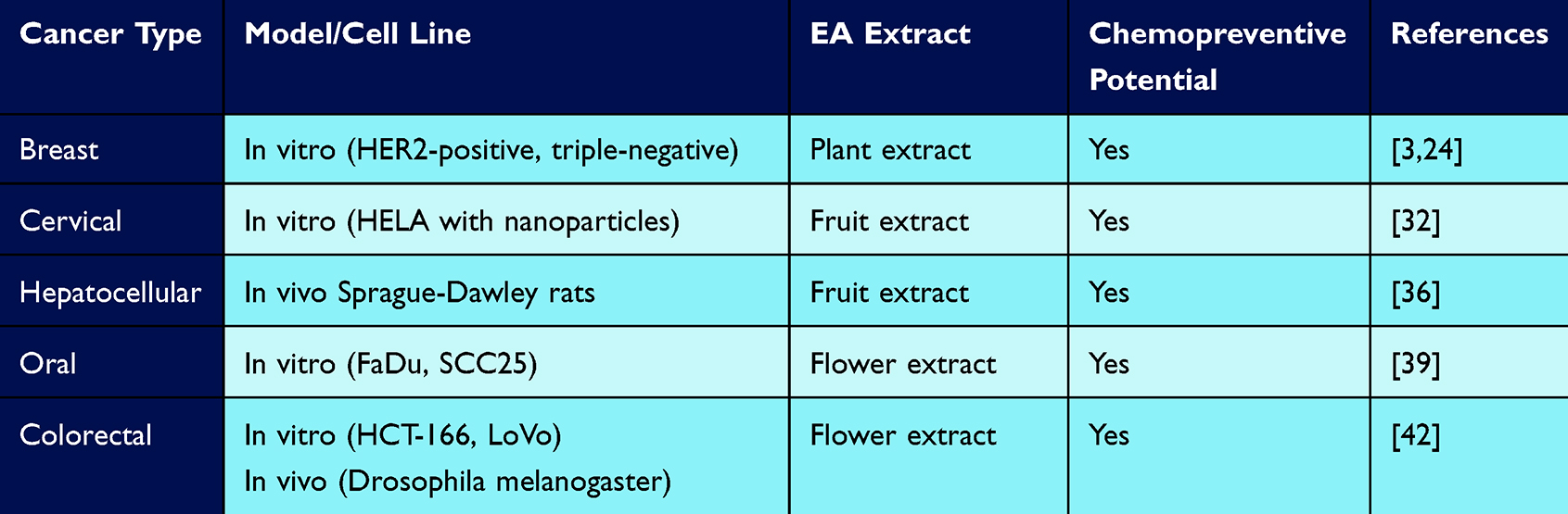 |
Table 1 Summary of Elaeagnus Angustifolia (EA) Studies in Cancer |
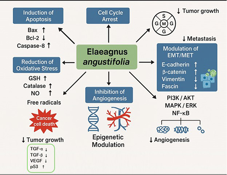 |
Figure 3 Molecular Pathways Targeted by Elaeagnus angustifolia in Cancer Cells. |
Molecular Mechanisms Underlying Elaeagnus Angustifolia’s Anti-Cancer Effects
While the exact mechanisms by which EA exerts its anti-cancer effects are still under investigation, several key pathways have been identified. These include induction of apoptosis, cell cycle arrest, and reduction of oxidative stress.3,24,44 Apoptosis, or programmed cell death, is a critical process through which damaged or unwanted cells are eliminated. EA has been shown to trigger apoptosis in various cancer cell lines by activating a cascade of molecular events leading to cell death. By inducing apoptosis, EA effectively eliminates cancer cells and inhibits tumor growth.21,45 Furthermore, EA can interfere with the cell cycle, the series of events that lead to cell division. By arresting the cell cycle at specific checkpoints, EA prevents cancer cells from proliferating uncontrollably. This cell cycle arrest can contribute to cancer cell death or senescence.21,45 Oxidative stress, caused by an imbalance between free radicals and antioxidants, plays a significant role in cancer development. EA’s antioxidant properties help neutralize harmful free radicals, protecting cells from oxidative damage and reducing the risk of cancer initiation and progression.5,35
As previously mentioned, studies that have investigated the effect of EA on breast cancer show that EA inhibits HER2 and JNK signaling, leading to modulation of EMT-related markers, including increased E-cadherin and β-catenin and decreased vimentin and fascin, which may contribute to reduced metastatic potential. In cervical cancer models, direct evidence for EA’s mechanistic effects is limited. Preliminary findings from a nanoparticle-based formulation suggest cytotoxicity and apoptotic features, but the specific contribution of EA remains unclear. In hepatocellular carcinoma, EA reduce liver damage biomarkers, increase GSH levels, and trigger apoptosis and inhibits proliferation by modulating Bax/Bcl-2 ratios and activating caspase-3. In oral cancer, EA inhibit cell invasion and motility, reverse EMT, and promotr MET by suppressing Erk1/Erk2 signaling. In colorectal cancer, EA reduces proliferation and invasion via modulation of EGFR1, Akt, and β-catenin signaling. It also alters EMT markers, upregulating E-cadherin and downregulating vimentin and β-catenin, indicating a role in metastasis suppression.
In addition to these mechanisms, EA has demonstrated the ability to inhibit angiogenesis, the formation of new blood vessels that supply tumors with nutrients and oxygen. By blocking angiogenesis, EA can starve tumors and prevent their growth and metastasis.6,39 EA has been shown to restricts the formation of new blood vessels by suppressing the production of pro-angiogenic factors such as VEGF,39 thereby limiting tumor growth and potential spread.
EA appears to exert its anticancer effects by modulating key signaling pathways frequently dysregulated in cancer. Notably, EA has been shown to influence the PI3K/AKT, MAPK/ERK,46 and NF-κB47 pathways, which play pivotal roles in cancer cell proliferation, survival, and metastasis.48–50
Furthermore, EA’s potential to influence epigenetic modifications, such as DNA methylation and histone modifications, suggests a broader impact on cancer cell behavior.20 These epigenetic changes can alter gene expression patterns, contributing to the development and progression of cancer.20 Table 2 provides an overview of targeted pathways and their potential therapeutic implications of EA. Despite promising in vitro findings, there is currently no published data on the pharmacokinetics of EA’s bioactive compounds, including their absorption, distribution, metabolism, and excretion.
 |
Table 2 Targeted Cellular Pathways and Potential Therapeutic Implications of Elaeagnus Angustifolia |
The interactions between EA and cancer cells are complex and influenced by various factors. The specific cancer type, disease stage, and even the particular components within the plant extract can significantly impact the observed effects. While the available data suggest promising anticancer properties, a comprehensive understanding of the underlying mechanisms requires further investigation. Consequently, caution should be exercised when interpreting these findings, and additional research, including well-designed clinical trials, is essential to validate the potential therapeutic benefits of EA.
From Bench to Bedside: Exploring the Clinical Promise of Elaeagnus Angustifolia
While few studies explored EA’s therapeutic potential, the evidence base remains relatively small. One clinical trial investigated EA extract as an adjuvant therapy for breast cancer,51 the trial was registered in Iranian Registry of Clinical Trials under code: IRCT2013092414760N1, the Clinical Trial Registry (Registration date: 2013–10-25, 1392/08/03). Another clinical trial was designed to assess the effects of E. angustifolia on postmenopausal women.52 A simple random sampling method was used to assign 60 participants, aged 45–70 and residing in the Alborz province, into intervention and control groups (30 women per group). Over 10 weeks, the intervention group received 15 grams of full-grain E. angustifolia powder daily, while the control group received a placebo consisting of 7.5 grams of corn starch and 7.5 grams of isomalt. Participants with cardiovascular, renal, or metabolic diseases, such as diabetes, as well as those who used psychoactive drugs, smoked, or had lipid levels between 200–300 mg/mL, were excluded. Blood pressure, heart rate, and lipid profiles were measured at baseline, and the levels of estrogen, progesterone, testosterone, FSH, and LH were assessed at the start and end of the trial.52 The trial was registered in the Iranian Registry of Clinical Trials under registration number IRCT2017030932795N2 (Registration date: 2017–09-11, 1396/06/20).52 According to the between-group analyses, there were no significant differences in the studied parameters between the herbal medicine and placebo groups, except for a notable improvement in joint pain in the E. angustifolia group. Within-group analysis revealed a significant increase in FSH levels and the FSH to testosterone ratio, while progesterone levels significantly decreased after 10 weeks of E. angustifolia consumption.52 While another reported positive effects when combining EA and ginger extracts for pain management.6 Additionally, anecdotal reports suggest its use as a complementary or alternative treatment in some cancer patients.6 However, it is essential to interpret these findings with caution due to potential limitations such as small sample sizes, lack of control groups, and variations in treatment protocols. Larger, well-designed randomized controlled trials are needed to establish EA’s safety, efficacy, and optimal dosage in cancer treatment.
Synergistic Enhancement of Cancer Therapy Using Elaeagnus Angustifolia
The potential synergistic interaction between EA and conventional cancer therapies represents a promising avenue for enhanced treatment outcomes. Synergy, defined as a combined effect greater than the sum of individual effects,53 offers the possibility of improved therapeutic efficacy and reduced adverse events.
EA holds promise as a complementary therapy to enhance the efficacy of conventional cancer treatments. Its potent antioxidant properties offer a potential solution to a major challenge associated with chemotherapy and radiation therapy-oxidative stress.6 These treatments, while effective in targeting cancer cells, can also damage healthy tissues by generating harmful free radicals known as reactive oxygen species (ROS) that can injure healthy tissues.54 EA’s well-documented antioxidant properties could potentially neutralize these free radicals, thereby reducing oxidative stress and protecting healthy cells from damage.6 This, in turn, could lead to improved treatment tolerance for patients undergoing chemotherapy or radiation.
Secondly, some studies suggest an additional benefit: EA extracts might modulate the immune system, enhancing the body’s ability to recognize and target cancer cells.25 This immune-modulating effect could work synergistically with immunotherapy, a standard cancer treatment that stimulates the immune system to fight cancer cells.29 By potentially amplifying the immune response, EA could contribute to an overall improvement in the effectiveness of cancer treatment.
Beyond antioxidant and immune system benefits, EA possesses another potential weapon against cancer: its anti-angiogenic properties.5,39 Tumors rely on a network of blood vessels for growth and metastasis.55 EA’s ability to inhibit the formation of these new blood vessels could significantly restrict tumor growth and hinder its ability to spread.55 This characteristic makes EA a promising candidate for combination therapy with chemotherapy and radiation, potentially leading to a more comprehensive attack on cancer.
Furthermore, preclinical studies have suggested that certain natural compounds, including some found in EA, can sensitize cancer cells to the effects of chemotherapy.56–58 Similarly, EA extracts may potentially sensitize cancer cells to the effects of radiation therapy.51 Combined with radiation therapy, this could improve cancer cell killing and better treatment outcomes. Furthermore, EA’s reported hepatoprotective and anti-inflammatory properties might contribute to better treatment tolerability.18,47 These properties could potentially reduce the significant side effects often associated with chemotherapy and radiation, improving a patient’s quality of life during treatment.
EA presents a fascinating prospect as a complementary therapy to enhance conventional cancer treatments. Combining EA with standard therapies offers a multitude of potential benefits. Firstly, EA might augment the therapeutic effects of existing treatments, leading to improved tumor control and overall better outcomes for cancer patients. Secondly, EA’s potential to work alongside conventional therapies might help overcome resistance that cancer cells can develop against single-agent treatments. Ideally, a synergistic effect could be achieved, where the combined action of EA and standard therapies is greater than the sum of their individual effects, potentially leading to better treatment outcomes with lower drug dosages.
However, it’s important to exercise caution. Individual patient responses to EA can vary, and potential interactions with other medications need careful monitoring to ensure safety and efficacy. Further research, particularly well-designed clinical trials, is essential to translate the promising preclinical data on EA into tangible clinical benefits for cancer patients. This research should explore optimal treatment regimens, potential side effects, and interactions with standard therapies to ensure safe and effective use of EA in cancer care.
A Roadmap for Future Research: Optimizing Elaeagnus Angustifolia for Cancer Care
The experimental evidence from animal studies suggests that EA has a decent safety profile, with no significant adverse effects reported.5,23,24 However, it is essential to interpret these findings cautiously, as preclinical studies may not always accurately predict human safety and efficacy. The safety profile of EA in cancer patients is not fully established due to limited clinical data.
Unveiling the secrets of EA holds the key to unlocking its full potential in personalized cancer care. Further research is crucial in two main areas. Firstly, a deeper understanding of the cellular and molecular mechanisms underlying EA’s anti-cancer effects is necessary. This knowledge can illuminate potential therapeutic targets, paving the way for the development of more targeted cancer therapies. Secondly, investigating the potential synergistic effects of EA with established cancer treatments like chemotherapy, radiation, and immunotherapy is another exciting avenue. Synergistic effects occur when combining therapies leads to a greater effect than the sum of their individual effects. This could offer new opportunities for improved treatment outcomes with potentially lower drug dosages and reduced side effects. By identifying biomarkers that predict a patient’s response to EA, alongside exploring synergistic effects with conventional treatments, researchers can pave the way for personalized and more effective cancer management strategies.
Conclusion
EA emerges as a promising player in the fight against cancer, supported by encouraging preclinical evidence. Extracts from this plant exhibit a diverse arsenal of anti-cancer properties, including pro-apoptotic, anti-proliferative, anti-angiogenic, and antioxidant effects, potentially offering significant benefits in cancer therapy. However, the exciting preclinical data requires further validation.
Well-designed clinical trials are essential to establish EA’s safety, efficacy, and pharmacokinetic profile in humans. Such research should aim to unravel its mechanisms of action, determine optimal dosing strategies, and assess potential side effects. Importantly, EA’s integration into personalized medicine frameworks, such as biomarker screening for pathway-specific responsiveness, could enhance its clinical utility and help identify patient subgroups most likely to benefit.
Exploring how EA can be safely integrated with conventional cancer treatments is crucial, particularly in the context of minimizing toxicity and maximizing synergistic effects. Until more robust clinical evidence is available, caution is warranted. Patients considering EA should consult with their healthcare providers to navigate this promising but complex area of cancer care. Collaboration between patients, healthcare professionals, and researchers is key to unlocking the true potential of EA and potentially improving future cancer treatment outcomes.
Abbreviations
CRC, Colorectal cancer; DEN, diethylnitrosamine; EA, Elaeagnus angustifolia; EMT, Epithelial-mesenchymal; HCC, Hepatocellular Carcinoma; HPV, human papillomavirus; MET, mesenchymal-to-epithelial.
Author Contributions
All authors made a significant contribution to the work reported, whether that is in the conception, study design, execution, acquisition of data, analysis and interpretation, or in all these areas; took part in drafting, revising or critically reviewing the article; gave final approval of the version to be published; have agreed on the journal to which the article has been submitted; and agree to be accountable for all aspects of the work.
Funding
This work was funded by grants from Qatar University (QUCP-CHS-2022-483).
Disclosure
The authors declare that the research was conducted in the absence of any commercial or financial relationships that could be construed as a potential conflict of interest.
References
1. Michaud A, Stawicki SP, Izurieta R, Iyer-Raniga U. Global Health Security: Contemporary Considerations and Developments. BoD–Books on Demand; 2024.
2. Dehelean CA, Marcovici I, Soica C, et al. Plant-derived anticancer compounds as new perspectives in drug discovery and alternative therapy. Molecules. 2021;26:1109. doi:10.3390/molecules26041109
3. Fouzat A, Hussein OJ, Gupta I, et al. Elaeagnus angustifolia plant extract induces apoptosis via p53 and signal transducer and activator of transcription 3 signaling pathways in triple-negative breast cancer cells. Front Nutr. 2022;9:871667. doi:10.3389/fnut.2022.871667
4. Nirmala C, Shahar B, Dolma N, Santosh O. Promising underutilized wild plants of cold desert Ladakh, India for nutritional security and health benefits. Applied Food Research. 2022;2:100145. doi:10.1016/j.afres.2022.100145
5. Tehranizadeh ZA, Baratian A, Hosseinzadeh H. Russian olive (Elaeagnus angustifolia) as a herbal healer. Bioimpacts. 2016;6:155. doi:10.15171/bi.2016.22
6. Hamidpour R, Hamidpour S, Hamidpour M, et al. Russian olive (Elaeagnus angustifolia L.): from a variety of traditional medicinal applications to its novel roles as active antioxidant, anti-inflammatory, anti-mutagenic and analgesic agent. J Traditional Complementary Med. 2017;7:24–13. doi:10.1016/j.jtcme.2015.09.004
7. Nazir N, Zahoor M, Nisar M. A review on traditional uses and pharmacological importance of genus Elaeagnus species. Bot Rev. 2020;86:247–280. doi:10.1007/s12229-020-09226-y
8. Saboonchian F, Jamei R, Sarghein SH. Phenolic and flavonoid content of Elaeagnus angustifolia L.(leaf and flower). Avicenna J Phytomed. 2014;4:231.
9. Cansev A, Sahan Y, Celik G, Taskesen S, Ozbey H. Chemical properties and antioxidant capacity of Elaeagnus angustifolia L. fruits. Asian J. Chem. 2011;23:2661–2665.
10. Obón C, Rivera D, Alcaraz F, Attieh L. Beverage and culture Zhourat, a multivariate analysis of the globalization of a herbal tea from the Middle East. Appetite. 2014;79:1–10. doi:10.1016/j.appet.2014.03.024
11. Çakmakçı S, Topdaş EF, Kalın P, et al. Antioxidant capacity and functionality of oleaster (Elaeagnus angustifolia L.) flour and crust in a new kind of fruity ice cream. Int. J. Food Sci. Technol. 2015;50:472–481. doi:10.1111/ijfs.12637
12. Erdemoglu N, Küpeli E, Yeşilada E. Anti-inflammatory and antinociceptive activity assessment of plants used as remedy in Turkish folk medicine. J Ethnopharmacol. 2003;89:123–129. doi:10.1016/S0378-8741(03)00282-4
13. Wallace TC, Bailey RL, Blumberg JB, et al. Fruits, vegetables, and health: a comprehensive narrative, umbrella review of the science and recommendations for enhanced public policy to improve intake. Crit Rev Food Sci Nutr. 2020;60:2174–2211. doi:10.1080/10408398.2019.1632258
14. Kohlmeier L, Kark JD, Gomez-Gracia E, et al. Lycopene and myocardial infarction risk in the EURAMIC Study. Am. J. Epidemiol. 1997;146:618–626. doi:10.1093/oxfordjournals.aje.a009327
15. Gürbüz I, Üstün O, Yesilada E, Sezik E, Kutsal O. Anti-ulcerogenic activity of some plants used as folk remedy in Turkey. J Ethnopharmacol. 2003;88:93–97. doi:10.1016/S0378-8741(03)00174-0
16. Hosseinzadeh H, Ramezani M, Namjo N. Muscle relaxant activity of Elaeagnus angustifolia L. fruit seeds in mice. J Ethnopharmacol. 2003;84:275–278. doi:10.1016/S0378-8741(02)00331-8
17. Natanzi MM, Pasalar P, Kamalinejad M, et al. Effect of aqueous extract of Elaeagnus angustifolia fruit on experimental cutaneous wound healing in rats. Acta Med Iran. 2012;50:589–596.
18. Farzaei MH, Bahramsoltani R, Abbasabadi Z, Rahimi R. A comprehensive review on phytochemical and pharmacological aspects of Elaeagnus angustifolia L. J Pharm Pharmacol. 2015;67:1467–1480. doi:10.1111/jphp.12442
19. Khan SU, Anjum SI, Ansari MJ, et al. Antimicrobial potentials of medicinal plant’s extract and their derived silver nanoparticles: a focus on honey bee pathogen. Saudi J Biol Sci. 2019;26:1815–1834. doi:10.1016/j.sjbs.2018.02.010
20. Ghanghareh M, Zare M. Cytotoxic effects of hydro-alcoholic extract of the leaf of elaeagnus angustifolia in hepatocellular carcinoma cell line (HepG2). Jentashapir J Cell Mol Biol. 2020;11. doi:10.5812/jjcmb.108505
21. Jabeen A, Sharma A, Gupta I, et al. Elaeagnus angustifolia plant extract inhibits epithelial-mesenchymal transition and induces apoptosis via HER2 inactivation and JNK pathway in HER2-positive breast cancer cells. Molecules. 2020;25:4240. doi:10.3390/molecules25184240
22. Alharbi KS, Almalki WH, Makeen HA, et al. Role of medicinal plant‐derived nutraceuticals as a potential target for the treatment of breast cancer. J Food Biochem. 2022;46:e14387. doi:10.1111/jfbc.14387
23. Panahi Y, Alishiri GH, Bayat N, Hosseini SM, Sahebkar A. Efficacy of Elaeagnus Angustifolia extract in the treatment of knee osteoarthritis: a randomized controlled trial. Excli J. 2016;15:203–210. doi:10.17179/excli2015-639
24. Mahboubi M. Elaeagnus angustifolia and its therapeutic applications in osteoarthritis. Ind Crop Prod. 2018;121:36–45.
25. Gupta M, Singh N, Gulati M, et al. Herbal bioactives in treatment of inflammation: an overview. S Afr J Bot. 2021;143:205–225. doi:10.1016/j.sajb.2021.07.027
26. Kaur B, Singh P. Inflammation: biochemistry, cellular targets, anti-inflammatory agents and challenges with special emphasis on cyclooxygenase-2. Bioorg. Chem. 2022;121:105663. doi:10.1016/j.bioorg.2022.105663
27. Amirghofran Z. Medicinal plants as immunosuppressive agents in traditional Iranian medicine. Iran J Immunol. 2010;7:65–73.
28. Zhao Y, Yan B, Wang Z, Li M, Zhao W. Natural polysaccharides with immunomodulatory activities. Mini Revin Med Chem. 2020;20:96–106. doi:10.2174/1389557519666190913151632
29. Sun N-X, Liu H-P, Liu X-H, et al. Immunological activities of polysaccharide extracted from Elaeagnus angustifolia L. Cyta-J Food. 2018;16:995–1002. doi:10.1080/19476337.2018.1516240
30. Castle PE, Einstein MH, Sahasrabuddhe VV. Cervical cancer prevention and control in women living with human immunodeficiency virus. CA Cancer J Clin. 2021;71:505–526. doi:10.3322/caac.21696
31. Erdoğan Ö, Salih P, Cevik O. Green synthesis and characterization of anticancer effected silver nanoparticles with silverberry (elaeagnus angustifolia) fruit aqueous extract. Int. J. Appl. Sci. or Int. J. Pure Appl. 2021;7:391–400. doi:10.29132/ijpas.915005
32. Buendia MA, Neuveut C. Hepatocellular carcinoma. Cold Spring Harb Perspect Med. 2015;5:a021444. doi:10.1101/cshperspect.a021444
33. Llovet JM, et al. Hepatocellular carcinoma. Nat Rev Dis Primers. 2021;7(6).
34. El Dika I, Makki I, Abou-Alfa GK. Hepatocellular carcinoma, novel therapies on the horizon. Chin clin oncol. 2021;10:12. doi:10.21037/cco-20-113
35. Amereh Z, Hatami N, Shirazi FH, et al. Cancer chemoprevention by oleaster (Elaeagnus angustifoli L.) fruit extract in a model of hepatocellular carcinoma induced by diethylnitrosamine in rats. Excli J. 2017;16:1046–1056. doi:10.17179/excli2017-389
36. Luo P, Yin P, Hua R, et al. A Large-scale, multicenter serum metabolite biomarker identification study for the early detection of hepatocellular carcinoma. Hepatology. 2018;67:662–675. doi:10.1002/hep.29561
37. Sim H-W, Knox J. Hepatocellular carcinoma in the era of immunotherapy. Curr Problems in Cancer. 2018;42:40–48. doi:10.1016/j.currproblcancer.2017.10.007
38. Ramadan WS, Alkarim S, Moulay M, et al. Modulation of the tumor microenvironment by ellagic acid in rat model for hepatocellular carcinoma: a potential target against hepatic cancer stem cells. Cancers. 2023;15:4891. doi:10.3390/cancers15194891
39. Saleh AI, Mohamed I, Mohamed AA, et al. Elaeagnus angustifolia plant extract inhibits angiogenesis and downgrades cell invasion of human oral cancer cells via erk1/erk2 inactivation. Nutr Cancer. 2018;70:297–305. doi:10.1080/01635581.2018.1412472
40. Hossain MS, Karuniawati H, Jairoun AA, et al. Colorectal cancer: a review of carcinogenesis, global epidemiology, current challenges, risk factors, preventive and treatment strategies. Cancers. 2022;14:1732. doi:10.3390/cancers14071732
41. Hassan AF, Khalil A, Al Moustafa AE. Elaeagnus angustifolia plant extract inhibits epithelial-mesenchymal transition in human colorectal cancer via β-catenin/jnk signaling pathways. Jordan J. Pharm. Sci. 2023;16:471. doi:10.35516/jjps.v16i2.1528
42. Fouzat A. Elaeagnus Angustifolia extract inhabits cell invasion of human colorectal cancer cells and increases the survival rate of the Drosophila colon cancer model. 2021.
43. Bratei AA, Stefan-van Staden R-I. The importance of KRAS quantification for a clinicopathological characterization in colorectal cancer patients. Medinformatics. 2024;1:20–26. doi:10.47852/bonviewMEDIN32021546
44. Patel S. Plant genus Elaeagnus: underutilized lycopene and linoleic acid reserve with permaculture potential. Fruits. 2015;70:191–199. doi:10.1051/fruits/2015014
45. Arab S, Bahraminasab M, Yazdani A, Abdolshahi A. Effects of whole fruit extract of elaeagnus angustifolia l. on glioblastoma cell lines. J Microbiol Biotechnol Food Sci. 2022;11:e4314–e4314.
46. Zhang H-W, Hu -J-J, Fu R-Q, et al. Flavonoids inhibit cell proliferation and induce apoptosis and autophagy through downregulation of PI3Kγ mediated PI3K/AKT/mTOR/p70S6K/ULK signaling pathway in human breast cancer cells. Sci Rep. 2018;8:11255. doi:10.1038/s41598-018-29308-7
47. Mamashli M, Nasseri S, Mohammadi Y, Ayati S, Zarban A. Anti-inflammatory effects of N-Acetylcysteine and Elaeagnus angustifolia extract on acute lung injury induced by λ-carrageenan in rat. Inflammopharmacology. 2022;30:1759–1768. doi:10.1007/s10787-022-01003-0
48. Bai D, Ueno L, Vogt PK. Akt-mediated regulation of NFkappaB and the essentialness of NFkappaB for the oncogenicity of PI3K and Akt. Int, J, Cancer. 2009;125:2863–2870. doi:10.1002/ijc.24748
49. He Y, Sun MM, Zhang GG, et al. Targeting PI3K/Akt signal transduction for cancer therapy. Signal Transduct. Target. Ther. 2021;6:425. doi:10.1038/s41392-021-00828-5
50. Han -S-S, Yun H, Son D-J, et al. NF-κB/STAT3/PI3K signaling crosstalk in iMycEμ B lymphoma. Mol Cancer. 2010;9:97. doi:10.1186/1476-4598-9-97
51. Salamzadeh J, Kamalinejad M, Babaeian M, et al. The effect of elaeagnus angustifolia L. Cream on radiation-induced skin reactions in women with breast cancer; A preliminary clinical trial. Iran J Pharm Sci. 2017;25–36.
52. Emaminia F, Rezaei A, Badehnoosh B, Ramezani R, Shabani M. The effects of Elaeagnus angustifolia L. whole fruit on the sex hormone profile in menopausal women: a double-blind, randomized, placebo-controlled study. J Ethnopharmacol. 2020;246:112229. doi:10.1016/j.jep.2019.112229
53. Pezzani R, Salehi B, Vitalini S, et al. Synergistic effects of plant derivatives and conventional chemotherapeutic agents: an update on the cancer perspective. Medicina. 2019;55. doi:10.3390/medicina55040110
54. Yang H, Villani RM, Wang H, et al. The role of cellular reactive oxygen species in cancer chemotherapy. J Exp Clin Cancer Res. 2018;37:266. doi:10.1186/s13046-018-0909-x
55. Lugano R, Ramachandran M, Dimberg A. Tumor angiogenesis: causes, consequences, challenges and opportunities. Cell Mol Life Sci. 2020;77:1745–1770. doi:10.1007/s00018-019-03351-7
56. de Oliveira Júnior RG, Christiane Adrielly AF, da Silva Almeida JRG, et al. Sensitization of tumor cells to chemotherapy by natural products: a systematic review of preclinical data and molecular mechanisms. Fitoterapia. 2018;129:383–400. doi:10.1016/j.fitote.2018.02.025
57. Ji A, Xu J. Neuropathic pain: biomolecular intervention and imaging via targeting microglia activation. Biomolecules. 2021;11:1343. doi:10.3390/biom11091343
58. Carradori S, Cairone F, Garzoli S, et al. Phytocomplex characterization and biological evaluation of powdered fruits and leaves from Elaeagnus angustifolia. Molecules. 2020;25:2021. doi:10.3390/molecules25092021
 © 2026 The Author(s). This work is published and licensed by Dove Medical Press Limited. The
full terms of this license are available at https://www.dovepress.com/terms
and incorporate the Creative Commons Attribution
- Non Commercial (unported, 4.0) License.
By accessing the work you hereby accept the Terms. Non-commercial uses of the work are permitted
without any further permission from Dove Medical Press Limited, provided the work is properly
attributed. For permission for commercial use of this work, please see paragraphs 4.2 and 5 of our Terms.
© 2026 The Author(s). This work is published and licensed by Dove Medical Press Limited. The
full terms of this license are available at https://www.dovepress.com/terms
and incorporate the Creative Commons Attribution
- Non Commercial (unported, 4.0) License.
By accessing the work you hereby accept the Terms. Non-commercial uses of the work are permitted
without any further permission from Dove Medical Press Limited, provided the work is properly
attributed. For permission for commercial use of this work, please see paragraphs 4.2 and 5 of our Terms.