
Back to Journals » Clinical Ophthalmology » Volume 20

Effect of Cyclosporine Ophthalmic Solution 0.09% on Signs and Symptoms of Dry Eye Disease in Patients Inadequately Controlled on Cyclosporine Ophthalmic Emulsion 0.05%: Results from a Single-Arm, Open-Label, Phase 4 Study
Authors Johnston J, Adler R, Hessen M  , Nichols KK , White D, Saenz B , Bowden F III, Periman LM , Truett K, Urbieta M, Mitchell B
Received 20 November 2025
Accepted for publication 10 February 2026
Published 4 March 2026 Volume 2026:20 583071
DOI https://doi.org/10.2147/OPTH.S583071
Checked for plagiarism Yes
Review by Single anonymous peer review
Peer reviewer comments 2
Editor who approved publication: Dr Scott Fraser
Josh Johnston,1 Richard Adler,2 Michelle Hessen,3 Kelly K Nichols,4 Darrell White,5 Bobby Saenz,6,7 Frank Bowden III,8 Laura M Periman,9 Kim Truett,10 Maitee Urbieta,11 Brittany Mitchell11
1Georgia Eye Partners, Atlanta, GA, USA; 2Belcara Health, Baltimore, MD, USA; 3Wilmer Eye Institute, Johns Hopkins University, Baltimore, MD, USA; 4School of Optometry, University of Alabama at Birmingham, Birmingham, AL, USA; 5Skyvision Centers, Westlake, OH, USA; 6LASIK San Antonio, San Antonio, TX, USA; 7Rosenberg School of Optometry, San Antonio, TX, USA; 8Bowden Eye & Associates, Jacksonville, FL, USA; 9Periman Eye Institute, Seattle, WA, USA; 10KCT Data, Inc, Alpharetta, GA, USA; 11Sun Pharmaceutical Industries, Inc., Princeton, NJ, USA
Correspondence: Josh Johnston, Georgia Eye Partners, 1100 Johnson Ferry Road NE #108, Atlanta, GA, 30342, USA, Tel +1 404-531-9988, Email [email protected]
Purpose: Cyclosporine ophthalmic emulsion 0.05% (CsA 0.05%) and cyclosporine ophthalmic solution 0.09% (CsA 0.09%) are approved to increase tear production in dry eye disease (DED). We investigated the effect of CsA 0.09% on DED signs and symptoms in patients whose disease was inadequately controlled on CsA 0.05%. In this Phase 4, single-arm, open-label study, adults with DED administered 1 drop of CsA 0.09% in both eyes twice daily for 12 weeks.
Patients and Methods: Patients were ≥ 18 years of age with a history and clinical diagnosis of DED for ≥ 3 months, which was not adequately controlled by treatment with CsA 0.05%. The primary efficacy endpoints were mean changes from baseline in total corneal fluorescein staining (CFS) and modified Symptom Assessment iN Dry Eye (mSANDE) scores at Week 12. Secondary efficacy endpoints included mean changes from baseline in total conjunctival staining score, central CFS score, tear osmolarity, Schirmer test score, and frequency of artificial tear use at Week 12, and patient treatment preference at Week 12. Adverse events (AEs) were monitored.
Results: The intent-to-treat population comprised 124 patients (mean age, 65.6 years). The mean changes from baseline in total CFS and mSANDE scores were statistically significant at Weeks 4, 8, and 12 (P < 0.0001 for all). At Week 12, statistically significant improvements from baseline were also noted for total conjunctival staining score, central CFS score, Schirmer test score, and artificial tear use frequency (P ≤ 0.0029), and 69.4% of patients preferred CsA 0.09%. Most AEs were mild.
Conclusion: Treatment with CsA 0.09% elicited statistically significant improvement from baseline in DED signs and symptoms from Weeks 4 to 12 in patients inadequately controlled on CsA 0.05%. Overall, CsA 0.09% was well tolerated. The findings from this study suggest that switching from CsA 0.05% to CsA 0.09% may improve signs and symptoms in patients with DED.
Plain Language Summary: In this study, patients with dry eye who were still experiencing symptoms while receiving treatment with cyclosporine ophthalmic emulsion 0.05% were switched to cyclosporine ophthalmic solution 0.09% (CsA 0.09%) for 12 weeks. Both formulations use the anti-inflammatory agent cyclosporine, but CsA 0.09% uses a slightly higher concentration that is dissolved into a solution rather than emulsified in an oil like some other dry eye drops. Patients were assessed using eye stains and questionnaires before switching to CsA 0.09%, which served as the baseline for each patient. Patients were then re-examined and completed another questionnaire regarding their dry eye symptoms every 4 weeks during the 12 weeks they received CsA 0.09%. As early as Week 4, patients on average had improvements in dry eye staining and symptom management (based on their questionnaire scores), which were associated with better control of their dry eye symptoms. At the end of the study, 69% of patients preferred CsA 0.09% over their previous eye drops, and CsA 0.09% was well tolerated by patients and was associated with mostly minor ocular side effects; eye irritation after applying the eye drops was reported most frequently. Taken together, these results suggest that treating dry eye early with CsA 0.09% may be beneficial, and some patients who struggle to adequately control their dry eye symptoms with other dry eye drops may benefit from switching their treatment to CsA 0.09%.
Keywords: CEQUA, corneal staining, cyclosporine A, dry eye disease, patient-reported outcome, restasis
Introduction
Dry eye disease (DED) is a multifactorial ocular surface disorder characterized by a loss of tear film homeostasis, with an estimated prevalence in the US of 8.1% (range, 5.3%–14.5%).1,2 DED has many risk factors related to the patient and lifestyle. For example, advancing age increases the odds of developing DED and is associated with meibomian gland dysfunction and tear deficiency.3 Additional risk factors include environmental factors (eg, pollution and low humidity), medications (eg, antihistamines, antidepressants, and isotretinoin), ocular comorbidities (eg, meibomian gland dysfunction and Sjögren syndrome),4,5 and ocular surgeries.6 These factors reduce tear production and/or quality and lead to tear film instability, tear hyperosmolarity, and increased friction and mechanical irritation at the ocular surface.7 These trigger a self-perpetuating cycle of ocular surface inflammation and damage, which produce the symptoms of visual abnormalities and ocular surface discomfort.7 Therefore, treatments for DED must address both etiology and underlying pathophysiology to optimize treatment efficacy and ensure patient satisfaction.8
The US Food and Drug Administration (FDA)–approved treatment options for DED include cyclosporine A formulations (Restasis®, Cequa®, Vevye®)9–11 and lifitegrast ophthalmic solution 5.0% (Xiidra®),12 which inhibits inflammation associated with DED.13 Cyclosporine A is a calcineurin inhibitor immunosuppressant that reduces ocular surface inflammation, resulting in reduced conjunctival epithelial cell apoptosis and increased conjunctival goblet cell density in patients with DED.8,14–18 Reduction in symptoms promotes treatment adherence in patients, improving their quality of life.19,20 Cyclosporine A is highly lipophilic and earlier topical formulations for the treatment of DED relied on castor oil-in-water emulsions, such as cyclosporine ophthalmic emulsion 0.05% (CsA 0.05%; Restasis®; Allergan, Irvine, CA, USA).21,22 CsA 0.05% was approved by the FDA in 2003 to increase tear production in patients with DED.8,9 Adverse events (AEs) including conjunctival hyperemia, eye pain, burning, stinging, and epiphora are common with CsA 0.05% treatment.9,21,22 A topical ophthalmic solution of cyclosporine ophthalmic solution 0.09% (CsA 0.09%; CEQUA®; Sun Pharmaceutical Industries, Inc., Cranbury, NJ, USA) was subsequently developed using a nanomicellar formulation to improve the aqueous solubility of cyclosporine A, reduce local AEs, and improve drug delivery to the ocular surface.10,21 In 2018, CsA 0.09% was approved by the FDA to increase tear production in patients with DED.10,23 Studies directly comparing CsA 0.09% and CsA 0.05% in the treatment of patients with DED are limited. This study investigated changes in DED signs and symptoms with CsA 0.09% in patients whose disease was inadequately controlled on CsA 0.05%.
Materials and Methods
Study Design and Treatment
This was a Phase 4, multicenter, open-label, single-arm study (ClinicalTrials.gov ID: NCT04357795).24 All enrolled patients administered 1 drop of CsA 0.09% in both eyes twice daily for a total of 12 weeks. The study design is depicted in Figure 1.
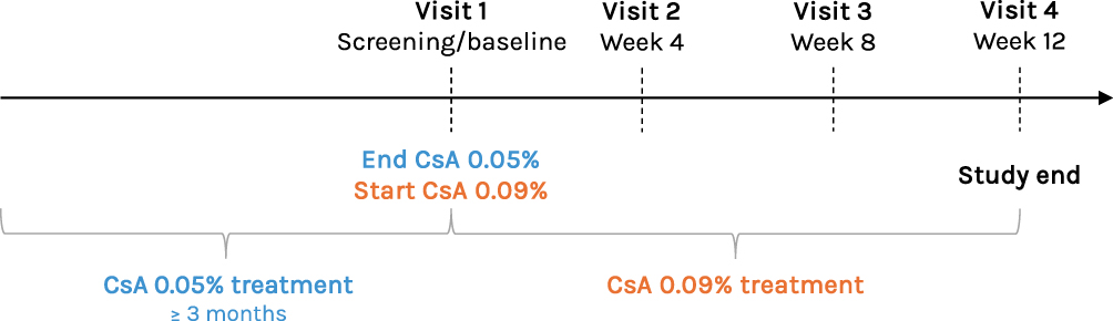 |
Figure 1 Study design. CsA 0.05%, cyclosporine ophthalmic emulsion 0.05%; CsA 0.09%, cyclosporine ophthalmic solution 0.09%. |
This study was conducted in accordance with the Code of Ethics of the World Medical Association (Declaration of Helsinki), and all patients provided written informed consent for study participation. All procedures were performed in compliance with relevant laws and institutional guidelines, and the study protocol was approved by the Sterling Institutional Review Board (Atlanta, GA, USA) on March 10, 2020 (Protocol ID: 7872-*MASTER).
Study Population
Enrolled patients were ≥ 18 years of age with a history and clinical diagnosis of bilateral DED for ≥ 3 months, which was not adequately controlled (ie, still symptomatic and/or exhibiting disease signs) by treatment with CsA 0.05%. Patients must have been on current CsA 0.05% treatment for a period ≥ 3 months prior to the screening/baseline visit. Other key inclusion criteria evaluated at screening/baseline included a best-corrected visual acuity (BCVA) of 20/200 or better in both eyes, a total corneal fluorescein staining (CFS) score ≥ 6 (range, 0–20) and/or an individual zone CFS score ≥ 2 (range, 0–4) in ≥ 1 eye, and a modified Symptom Assessment iN Dry Eye (mSANDE) score ≥ 40 (range, 0–100). Patients were required to discontinue all other DED therapy at screening/baseline, except artificial tears or lid scrubs if they were used routinely and for ≥ 1 month prior to screening/baseline. The use of artificial tears or lid scrubs was to remain stable on study. Use of artificial tears was prohibited within 2 hours before any study visit. Key exclusion criteria included a previous history of treatment failure (no clinically relevant improvement in any DED assessment despite continuous treatment for ≥ 3 months) with discontinuation of or switching from CsA 0.05%, use or initiation of any systemic or topical ocular medication not listed in the exclusion criteria that is known to cause or exacerbate DED within 7 days prior to screening/baseline or during the study, use of topical ocular agents other than artificial tears during the study, active ocular disease other than DED in either eye, periocular or ocular surgery within 6 months of screening/baseline in either eye, punctal plugs or permanent punctal occlusion in either eye, and pregnancy or breastfeeding. Additionally, complete treatment compliance was required during the study. Significant study medication noncompliance resulted in study discontinuation, which was identified by the amount of unused medication returned at each study visit and review of daily patient diaries.
Outcomes and Assessments
Efficacy
The 2 primary efficacy endpoints in this study were the mean changes from baseline in total CFS OU (both eyes) and mSANDE scores at Week 12. Secondary efficacy endpoints included mean changes from baseline in total conjunctival staining score, central CFS score, tear osmolarity, unanesthetized Schirmer test score, and frequency of artificial tear use at Week 12, as well as patient treatment preference at Week 12. All efficacy endpoints were assessed at baseline and Weeks 4, 8, and 12, and patient treatment preference was determined at the end of the study at Week 12.
Corneal fluorescein staining was assessed by measuring corneal epithelial staining 2 to 2.5 minutes after instilling 1 drop of 0.5% sodium fluorescein solution into each eye, followed by adequate blinking. Five corneal areas (central, superior, temporal, nasal, and inferior) were evaluated, and each was scored on a 0 (no staining) to 4 (coalescent lesions) modified National Eye Institute (NEI) grading scale in 0.5-point increments.25 The total CFS score was the sum of all corneal area scores.
The mSANDE used in this study was a 2-item questionnaire that assessed the frequency and severity of ocular dryness and irritation associated with DED. The items were slightly modified from SANDE to “Please indicate how often, over the past week, your eyes felt dry and/or irritated,” and “Please indicate how severe, on average, you felt your symptoms of dryness and/or irritation were over the past week.” The mSANDE was scored on a 0- to 100-mm linear visual analog scale, where 0 represented very low frequency/severity and 100 represented very high frequency/severity.26 For mSANDE, global symptom scores were calculated using the equation √(frequency score × severity score).
Conjunctival staining was assessed approximately 5 minutes after CFS by measuring staining 1 to 4 minutes after instilling 1 drop of 1% lissamine green solution in each eye. Staining was evaluated in 6 conjunctival zones (temporal, superior-temporal, inferior-temporal, superior-nasal, inferior-nasal, and nasal). Conjunctival staining was scored from 0 to 3 in each zone using the Expanded NEI/Industry Workshop scale, and the total score was the sum of all individual zone scores.25
Tear osmolarity was evaluated before dye instillation via the TearLab® Osmolarity System Test, in which nanoliter volumes of tear fluid were collected from each eye using the Osmolarity Test Pen and Test Card. Results were interpreted according to the provided instructions, and values were recorded in mOsm/L.
An unanesthetized Schirmer test was performed in each eye ≥ 5 minutes after conjunctival staining. Strips were placed in both eyes simultaneously, and the amount of wetting on each strip was marked with a line and recorded in mm after 5 minutes.
The number of days and daily frequency of artificial tear use was evaluated at each study visit using patient diaries. The baseline values were determined by a self-assessment that asked patients to report the number of days and average uses per day of artificial tears over the 7 days prior to the screening/baseline visit. Patients were instructed to continue using artificial tears as needed for the duration of the study.
Patient treatment preference was assessed by asking, “Overall, which treatment do you prefer for the management of your dry eyes: your prior treatment or your study treatment?” Responses were recorded.
Safety
Safety assessments included AE monitoring, BCVA measurement, and slit lamp examination, which were completed at baseline and Weeks 4, 8, and 12. The reporting period for AEs was from the time the informed consent form was signed through the final visit (Week 12 or early termination).
At each study visit, AE monitoring was performed by asking the patient how they had been feeling. All AEs observed by the investigator or reported by the patient were documented and graded by the investigator as mild, moderate, or severe. Mild AEs were defined as noticeable discomfort that did not interfere with normal daily activities. Moderate AEs were defined as discomfort that interfered with normal daily activities, and severe AEs were defined as incapacitating and prevented performance of normal daily activities. Any ongoing AEs at the time of study exit were followed until stabilization or resolution. Adverse events were coded using the Medical Dictionary for Regulatory Activities (MedDRA) version 25.1.
Best-corrected visual acuity was assessed at all study visits using the Snellen eye chart and the patient’s current corrective lens prescription. Patients were required to wear the same eyeglasses, if applicable, at each visit, and must have read ≥ 50% of the letters on a single line correctly to accept that visual acuity (VA) line. Refraction must have been performed within 6 months of screening/baseline, and this refraction was used for all VA assessments for the study duration. Snellen VA measurements were converted to the logarithm of the minimum angle of resolution (logMAR) for results reporting.
Routine slit lamp examination was performed at each study visit to evaluate the anterior segment of the eye, including the lids, cornea, conjunctiva, anterior chamber, and lens. Abnormalities were documented and graded.
Statistical Analysis
All statistical analyses and reporting were performed using SAS® version 9.4. Efficacy assessments used the intent-to-treat (ITT) population, which included all patients who received ≥ 1 dose of study treatment and had ≥ 1 postbaseline assessment. Safety assessments used the safety population, which included all patients who received ≥ 1 dose of study treatment.
The sample size was calculated by considering the primary endpoint of the study per baseline eligibility group. A sample size of ≥ 40 patients was required to achieve similar power to the individual endpoint estimates to detect similar differences with a 1.67% level of significance using a 2-sided paired t-test. Continuous variables were summarized using descriptive statistics (n, mean, standard deviation [SD], median, minimum, and maximum) and categorical variables, including binary variables, were summarized with counts and percentages.
Baseline values served as the study control since these assessments were completed, while patients were still affected by prior CsA 0.05% treatment. Changes from baseline were evaluated using the 2-tailed Student’s t-test. For the mean change from baseline in total CFS and Schirmer test scores, scores from each eye were averaged for each time point prior to statistical analysis. The statistical significance level was set at P ≤ 0.05 for all study endpoints. As the trial was a single-arm study, no adjustments for multiple comparisons were made.
Missing data were not imputed for safety or efficacy analyses. Missing or partial dates were not imputed, except for the identification of treatment-emergent AEs.
Results
Patient Demographics and Baseline Characteristics
Of the 155 screened patients, 134 were enrolled and included in the safety population, with 124 in the ITT population (Figure 2). The mean (SD) patient age was 65.6 (11.54) years, with the majority being female (109; 87.9%) and White (109; 87.9%; Table 1). At baseline, patients had been on treatment with CsA 0.05% for a mean (SD; range) duration of 39.4 (40.58; 3–211) months.
 |
Table 1 Patient Demographics and Baseline Characteristics |
 |
Figure 2 Patient disposition. aEarly termination due to the sponsor not providing the study medication. |
Primary Efficacy Endpoints
Both primary efficacy endpoints were met in the study. At baseline, the mean (SD) total CFS score in both eyes combined (OU, means of right eye [OD] and left eye [OS] combined) was 5.7 (3.37). As early as Week 4, the mean (SD) total CFS score OU was significantly lower than baseline by 2.8 (2.70) points (P < 0.0001) and continued to decrease to reach a maximum reduction by 12 weeks of treatment (mean [SD] difference to baseline, −3.1 [2.9] points; P < 0.0001; Table 2 and Figure 3). The total CFS scores for each eye individually were also statistically significant (P < 0.0001) at each time point (Figure 4A and B for right eye and left eye, respectively). Similarly, the mean (SD) mSANDE score was significantly lower than the baseline value of 67.1 (21.05) points at each study visit (all P < 0.0001), with a maximal change of 29.5 (26.38) points by Week 12 (Table 2 and Figure 5).
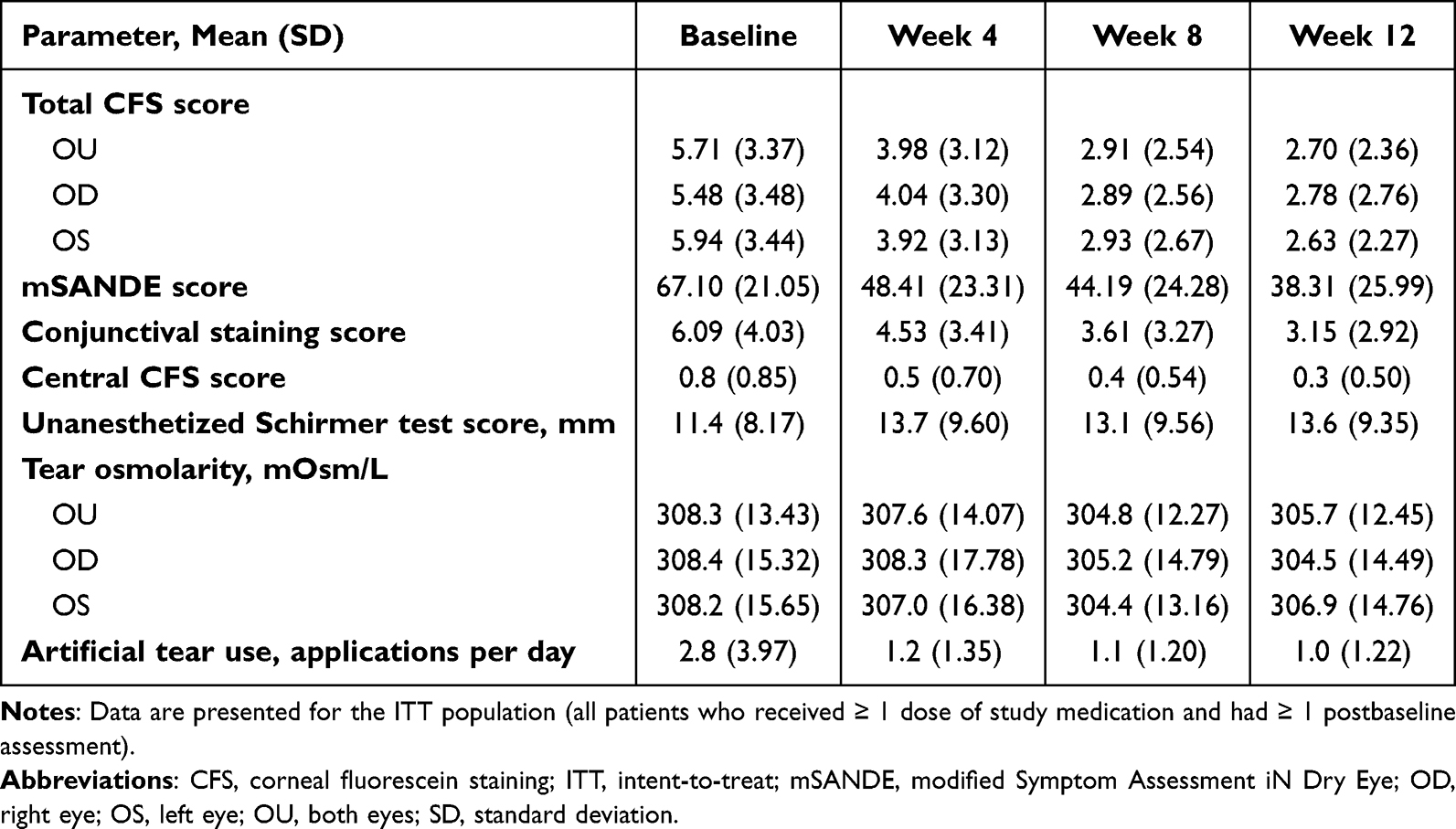 |
Table 2 Summary of Mean Clinical Changes Through Week 12 |
 |
Figure 3 Mean changes from baseline in total corneal fluorescein staining score (primary efficacy endpoint). Dotted lines represent the baseline. CsA 0.09%, cyclosporine ophthalmic solution 0.09%; SD, standard deviation. |
 |
Figure 4 Mean changes from baseline in total corneal fluorescein staining score by eye. (A) Total corneal fluorescein staining score (right eye). (B) Total corneal fluorescein staining score (left eye). Dotted lines represent the baseline. CsA 0.09%, cyclosporine ophthalmic solution 0.09%; SD, standard deviation. |
 |
Figure 5 Mean changes from baseline in modified Symptom Assessment iN Dry Eye score at Week 12 (primary efficacy endpoint). Dotted lines represent the baseline. CsA 0.09%, cyclosporine ophthalmic solution 0.09%; SD, standard deviation. |
Secondary Efficacy Endpoints
The mean (SD) total conjunctival staining score OU (means of OD and OS combined) was 6.1 (4.03) points at baseline and then significantly decreased during treatment (all P < 0.0001), reaching a value of 3.0 (3.30) points by Week 12 (Table 2 and Figure 6A). Patients also experienced significant improvements in central CFS scores. At baseline, the mean (SD) central CFS score OU was 0.8 (0.85) and improved by 0.5 (0.87) points at Week 12 (P < 0.0001 for each; Table 2 and Figure 6B). Statistically significant improvements in the unanesthetized Schirmer test were also observed during treatment (Table 2 and Figure 6C).
 |
Figure 6 Mean changes from baseline in secondary efficacy endpoints. (A) Total conjunctival staining score. (B) Central corneal fluorescein staining score. (C) Unanesthetized Schirmer test score. Dotted lines represent the baseline. CsA 0.09%, cyclosporine ophthalmic solution 0.09%; SD, standard deviation. |
Mean (SD) baseline tear osmolarity was 308.4 (15.32) mOsm/L and 308.2 (15.65) mOsm/L for OD and OS, respectively; mean tear osmolarity OU at baseline was 308.3 (13.43) mOsm/L (Table 2). At Week 12, mean (SD) tear osmolarity significantly decreased by 4.0 (19.44) mOsm/L OD (P = 0.0323) and numerically decreased by 1.7 (18.54) mOsm/L OS (P = 0.3447) and 2.8 (15.62) mOsm/L OU (P = 0.0583; Table 2).
Patients reported decreased artificial tear use during the study. At baseline, the mean (SD) frequency was 2.8 (3.97) applications per day (Table 2). Significant reductions in artificial tear use were reported at each study visit (all P < 0.0001), and the mean (SD) change in frequency was −1.5 (2.4) applications per day by Week 12. By the end of the study, 86 (69.4%) patients preferred CsA 0.09% vs 27 (21.8%) who preferred the prior treatment; the remaining 11 (8.9%) did not indicate a preference (Figure 7).
 |
Figure 7 Patient treatment preference at Week 12. CsA 0.05%, cyclosporine ophthalmic emulsion 0.05%; CsA 0.09%, cyclosporine ophthalmic solution 0.09%. |
Safety Endpoints
During the Phase 4 study, 58 (43.3%) patients reported a total of 84 treatment-emergent AEs, most of which were mild (73.8%) or moderate (21.4%) in severity. Adverse events leading to study discontinuation occurred in 9 (6.7%) patients (Table 3). The most frequently reported AEs were instillation-site irritation (12.7%), instillation-site pain (2.2%), eye irritation (2.2%), sinusitis (2.2%), and presence of vital dye corneal staining (2.2%). All other AEs occurred in < 2% of patients. Two serious AEs (coronavirus disease 2019 and irregular heart rate) were reported by 1 patient each; both were deemed unrelated to study treatment.
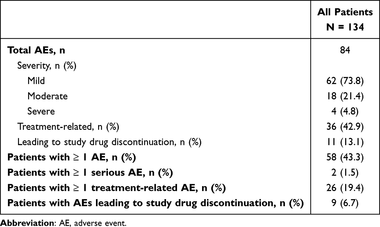 |
Table 3 Overview of Adverse Events |
No patients experienced changes from baseline in BCVA > 0.40 logMAR OU, and no patients had values worse than 20/100 (0.7 logMAR) OU at any study visit. Slit lamp findings were unremarkable and consistent with the expected profile of CsA 0.09%.27,28
Discussion
Cyclosporine A 0.09% treatment was associated with significant improvements in total CFS and mSANDE scores, the coprimary efficacy endpoints, at Week 12 in patients with DED inadequately controlled by CsA 0.05%. Notably, the changes from baseline in both endpoints were statistically significant beginning at Week 4, the earliest time point assessed after screening. Significant improvements were also noted in nearly all secondary efficacy endpoints with CsA 0.09%. Of particular importance is the observed reduction in daily frequency of artificial tear use with CsA 0.09% vs prior CsA 0.05% treatment in patients who continued using artificial tears as needed. Multiple daily administrations of artificial tears over several months are recommended for patients with DED.29 The frequency of artificial tear use in a clinical trial is not commonly evaluated, but a reduction in their use may be clinically meaningful and have a significant impact on patients’ daily lives. In this study, treatment with CsA 0.09% significantly reduced the number of daily artificial tears administrations in patients during the study period.
Changes in tear osmolarity over time with CsA 0.09% were inconsistent, with statistically significant improvement at Week 12 OD, but not OS, and a trend for improvement OU. However, the mean baseline tear osmolarity OU (308.3 mOsm/L) for this study was at the upper limit of the normal range for a healthy population (302 ± 6.3 mOsm/L).30 Therefore, it may not be reasonable to expect a significant decrease within the normal range during CsA 0.09% treatment in patients with DED.
Twice-daily CsA 0.09% was well tolerated in this patient population, consistent with its safety profile in Phase 2b/3 and Phase 3 clinical trials.27,28 Less than half (42.9%) of the observed AEs were deemed related to treatment. Of note, instillation-site pain was reported at a much lower rate in this study (2.2%) than in the Prescribing Information for CsA 0.09% (22%).10 A potential contributor to this discrepancy includes differences in AE terminology coding between MedDRA version 25.1 (which was used in this study) and versions 17.0 and 19.0 used in the pivotal trials.27,28 Two versions of MedDRA are published annually; thus, there are 16 MedDRA versions between 17.0 and 25.1, and 12 versions between 19.0 and 25.1. It is also possible that patients became acclimated to topical cyclosporine treatment during their previous course of CsA 0.05% and did not report instillation-site pain during the current study or that they experienced a partial treatment response to CsA 0.05% leading to corneal healing and decreased pain sensitivity. Additionally, the open-label study design may have resulted in patient bias that influenced AE reporting.
This Phase 4 study adds to the limited data available to help inform treatment decisions for patients with DED receiving CsA 0.09% or CsA 0.05%. A real-world, retrospective analysis of treatment patterns in patients with DED receiving CsA 0.09%, CsA 0.05%, or lifitegrast ophthalmic solution 5% (Xiidra®) demonstrated that patients remained on CsA 0.09% significantly longer and were significantly less likely to discontinue treatment than those on CsA 0.05%.31 In the current analysis, most patients preferred treatment with CsA 0.09% over CsA 0.05%, which may influence treatment adherence and compliance. It should also be noted that the mean duration of previous CsA 0.05% treatment at baseline was 39.4 months, indicating that many patients with DED remained on CsA 0.05% for long time periods despite their inadequate treatment response. However, upon switching to CsA 0.09%, most efficacy endpoints showed significant improvement by Week 4 and continued through Week 12, supporting the conclusion that patient’s DED symptoms were better controlled while on CsA 0.09% compared with their previous treatment. These improvements may be due to the higher concentration of cyclosporine A in CsA 0.09% and/or the increased bioavailability of cyclosporine A due to the nanomicellar formulation.10,21 Therefore, switching to CsA 0.09% early in treatment may benefit patients with an inadequate response to CsA 0.05%.
There are important limitations to note for this study. As a single-arm, open-label study, both patients and investigators were unblinded to treatment, which could bias results, particularly for subjective assessments like mSANDE scores. Indeed, the recency/recall effect (when patients score their symptom management more positively after a period of time) may have biased scores during the 12-week treatment period.32 Similarly, the study population consisted of patients whose DED symptoms were inadequately controlled on their previous DED treatment, which could cause selection bias and enrich for patients who were more likely to score a new treatment favorably. However, while subjective scores improved during the study, several assessments measuring signs of DED also improved, suggesting that symptom improvements were linked to reduced DED burden overall. Additionally, while the patients could have more easily tolerated a higher concentration of CsA, having switched from a lower concentration, the safety outcomes for CsA 0.09% in this study were consistent with previous studies.27,28 Lastly, the majority of patients in the study were White females (88%), potentially limiting the generalizability of the results.
Despite this study’s inherent limitations as a single-arm, open-label trial, Phase 4 studies performing treatment comparisons among DED products are rare. This analysis adds to the literature comparing topical ophthalmic cyclosporine products and provides valuable insight regarding treatment response in patients switching to a different formulation of CsA.
Conclusion
Twice-daily treatment with CsA 0.09% elicited significant improvement in multiple DED signs and symptoms beginning at Week 4 and continuing to the end of the study (Week 12) in patients previously inadequately controlled on CsA 0.05%. Overall, CsA 0.09% was well tolerated in this patient population. The results of this Phase 4 study support the efficacy and safety of CsA 0.09% in the treatment of DED and suggest switching patients to a different formulation of cyclosporine A may improve DED signs and symptoms. Clinicians may consider switching to CsA 0.09% early in the treatment of patients whose DED is not adequately controlled by CsA 0.05%.
Data Sharing Statement
All data that support the findings of this manuscript are available from the corresponding author (Josh Johnston) or Sun Pharmaceutical Industries, Inc. upon reasonable request.
Ethical Considerations
This study was conducted in accordance with the Code of Ethics of the World Medical Association (Declaration of Helsinki). All procedures were performed in compliance with relevant laws and institutional guidelines, and the study protocol was approved by the Sterling Institutional Review Board (Atlanta, GA, USA) on March 10, 2020 (Protocol ID: 7872-*MASTER).
Consent to Participate
All patients provided written informed consent for study participation.
Acknowledgments
We thank the patients who participated and the trial site coordinators for their contributions to the study. Medical writing and editorial support for this manuscript were provided by Jennifer Masucci, VMD, and Ryan P. Reynolds, PhD, of Red Nucleus, under the direction of the authors.
Funding
The study was funded by Sun Pharmaceutical Industries, Inc.
Disclosure
JJ reports consultant fees from Alcon, Aldeyra Therapeutics, Allergan, Avellino, Azura Ophthalmics, Bausch + Lomb, BioTissue, Bruder Healthcare, Dompe, Gaukos, Harrow, Horizon Therapeutics, Johnson & Johnson, Kala Pharmaceuticals, Novaliq, OcuTerra, Orasis Pharmaceuticals, Oyster Point Pharma, Quidel, Seagen, Sight Sciences, Sun Pharma, Tarsus Pharmaceuticals, Thea Pharma, Trukera Medical, Versea Biologics, Visus Therapeutics, and Zeiss; speaker fees from Allergan, BioTissue, Gaukos, Kala Pharmaceuticals, Oyster Point Pharma, Quidel, Sight Sciences, and Sun Pharma; and research fees from Tarsus Pharmaceuticals. He is also a shareholder for LacriSciences. RA reports consultant fees from Allergan, Lumenis, Sun Pharma, and Tarsus Pharmaceuticals; and speaker fees from Allergan, Lumenis, Oyster Point Pharma, and Sun Pharma. MH reports nothing to disclose. KKN reports consultant fees from AbbVie, Alcon, Aldeyra Therapeutics, Azura Ophthalmics, Bausch + Lomb, Bruder Healthcare, Cavalry Biosciences, Cloudbreak, Dompe, HanAll Biopharma, Harrow Health, Nicox, Novaliq, Novartis, Oyster Point Pharma, Palatin, Santec, Shire, Sight Sciences, Sydnexis, Tarsus Pharmaceuticals, TearSolutions, Thea Pharma, Topcon Healthcare, Trukera Medical, Versea, and Xequel Bio; and research fees from Aramis Biosciences, Kowa, Visus/Tenpoint, ScienceBased Health, Sylentis, and TearScience. DW reports consultant fees from Allergan, Eyevance, Kala Pharmaceuticals, Shire/Takeda Pharmaceuticals, Sun Pharma, and TearLab. BS reports consultant and speaker fees from Allergan and Sight Sciences. FB reports speaker fees from Bausch + Lomb and Sun Pharma, and advisory board fees from Sight Sciences. LMP is a shareholder for Eyedetec, MYZE, and Visant Medical; reports research support from Dompe, Kala Pharmaceuticals, Lumenis, Olympic Ophthalmics, and Taurus; and reports consulting fees from Alcon, Allergan/AbbVie, Amgen, Azura Ophthalmics, Bausch, Harrow, Jett, Kala Pharmaceuticals, MD Elite, Mallinkrodt, Nordic Pharma, Nusight Medical, NuVissa, ScienceBased Health, Scope, Sight Sciences, Sun Pharma, Tarsus Pharmaceuticals, Viatris, and Visant. KT reports consultant fees from Sun Pharma. MU and BM are employees of Sun Pharmaceutical Industries, Inc. The authors report no conflicts of interest in this work.
The key results from this paper were presented at the 2024 International Society for Pharmacoeconomics and Outcomes Research congress as a poster presentation with interim findings. The poster is available as follows: [johnston-et-alispor-2024]
References
1. Craig JP, Nichols KK, Akpek EK, et al. TFOS DEWS II definition and classification report. Ocul Surf. 2017;15(3):276–13. doi:10.1016/j.jtos.2017.05.008
2. McCann P, Abraham AG, Mukhopadhyay A, et al. Prevalence and incidence of dry eye and meibomian gland dysfunction in the United States: a systematic review and meta-analysis. JAMA Ophthalmol. 2022;140(12):1181–1192. doi:10.1001/jamaophthalmol.2022.4394
3. Di Zazzo A, Coassin M, Surico PL, Bonini S. Age-related ocular surface failure: a narrative review. Exp Eye Res. 2022;219:109035. doi:10.1016/j.exer.2022.109035
4. Stapleton F, Abad JC, Barabino S, et al. TFOS lifestyle: impact of societal challenges on the ocular surface. Ocul Surf. 2023;28:165–199. doi:10.1016/j.jtos.2023.04.006
5. Stapleton F, Alves M, Bunya VY, et al. TFOS DEWS II epidemiology report. Ocul Surf. 2017;15(3):334–365. doi:10.1016/j.jtos.2017.05.003
6. De Gregorio C, Nunziata S, Spelta S, et al. Unhappy 20/20: a new challenge for cataract surgery. J Clin Med. 2025;14(5):1408. doi:10.3390/jcm14051408
7. Bron AJ, de Paiva CS, Chauhan SK, et al. TFOS DEWS II pathophysiology report. Ocul Surf. 2017;15(3):438–510. doi:10.1016/j.jtos.2017.05.011
8. Jones L, Downie LE, Korb D, et al. TFOS DEWS II management and therapy report. Ocul Surf. 2017;15(3):575–628. doi:10.1016/j.jtos.2017.05.006
9. Restasis® (Cyclosporine Ophthalmic Emulsion) 0.05%. Full Prescribing Information. Allergan; 2024.
10. CEQUA® (Cyclosporine Ophthalmic Solution) 0.09%. Full Prescribing Information. Sun Pharmaceutical Industries. Inc.; 2022.
11. Vevye® (Cyclosporine Ophthalmic Emulsion) 0.1%. Full Prescribing Information. Harrow Eye. 2025.
12. Xiidra® (Lifitegrast Ophthalmic Solution) 5%. Full Prescribing Information. Shire US, Inc.; 2016.
13. Perez VL, Pflugfelder SC, Zhang S, Shojaei A, Haque R. Lifitegrast, a novel integrin antagonist for treatment of dry eye disease. Ocul Surf. 2016;14(2):207–215. doi:10.1016/j.jtos.2016.01.001
14. Kunert KS, Tisdale AS, Gipson IK. Goblet cell numbers and epithelial proliferation in the conjunctiva of patients with dry eye syndrome treated with cyclosporine. Arch Ophthalmol. 2002;120:330–337. doi:10.1001/archopht.120.3.330
15. Kunert KS, Tisdale AS, Stern ME, Smith JA, Gipson IK. Analysis of topical cyclosporine treatment of patients with dry eye syndrome: effect on conjunctival lymphocytes. Arch Ophthalmol. 2000;118:1489–1496. doi:10.1001/archopht.118.11.1489
16. Brignole F, Pisella PJ, De Saint Jean M, Goldschild M, Goguel A, Baudouin C. Flow cytometric analysis of inflammatory markers in KCS: 6-month treatment with topical cyclosporin A. Invest Ophthalmol Vis Sci. 2001;42:90–95.
17. Gao J, Sana R, Calder V, et al. Mitochondrial permeability transition pore in inflammatory apoptosis of human conjunctival epithelial cells and T cells: effect of cyclosporin A. Invest Ophthalmol Vis Sci. 2013;54(7):4717–4733. doi:10.1167/iovs.13-11681
18. Pflugfelder SC, De Paiva CS, Villarreal AL, Stern ME. Effects of sequential artificial tear and cyclosporine emulsion therapy on conjunctival goblet cell density and transforming growth factor-b2 production. Cornea. 2008;27:64–69. doi:10.1097/ICO.0b013e318158f6dc
19. Brown MM, Brown GC, Brown HC, Peet J, Roth Z. Value-based medicine, comparative effectiveness, and cost-effectiveness analysis of topical cyclosporine for the treatment of dry eye syndrome. Arch Ophthalmol. 2009;127(2):146–152. doi:10.1001/archophthalmol.2008.608
20. Trattler W, Katsev D, Kerney D. Self-reported compliance with topical cyclosporine emulsion 0.05% and onset of the effects of increased tear production as assessed through patient surveys. Clin Ther. 2006;28(11):1848–1856. doi:10.1016/j.clinthera.2006.11.016
21. Cholkar K, Gilger BC, Mitra AK. Topical, aqueous, clear cyclosporine formulation design for anterior and posterior ocular delivery. Transl Vis Sci Technol. 2015;4(3):1. doi:10.1167/tvst.4.3.1
22. Mandal A, Gote V, Pal D, Ogundele A, Mitra AK. Ocular pharmacokinetics of a topical ophthalmic nanomicellar solution of cyclosporine (Cequa®) for dry eye disease. Pharm Res. 2019;36(2):36. doi:10.1007/s11095-018-2556-5
23. Han DH. FDA approves Cequa for the treatment of dry eye disease. EMPR. 2018. Available from: https://www.empr.com/home/news/fda-approves-cequa-for-the-treatment-of-dry-eye-disease/. [
24. Effect of Cequa™ in subjects with dry eye disease. ClinicalTrials.gov identifier: NCT04357795. Updated February 6, 2024.
25. Lemp MA. Report of the National Eye Institute/Industry workshop on clinical trials in dry eyes. CLAO J. 1995;21(4):221–232.
26. Schaumberg DA, Gulati A, Mathers WD, et al. Development and validation of a short global dry eye symptom index. Ocul Surf. 2007;5(1):50–57. doi:10.1016/s1542-0124(12)70053-8
27. Goldberg DF, Malhotra RP, Schechter BA, Justice A, Weiss SL, Sheppard JD. A phase 3, randomized, double-masked study of OTX-101 ophthalmic solution 0.09% in the treatment of dry eye disease. Ophthalmology. 2019;126(9):1230–1237. doi:10.1016/j.ophtha.2019.03.050
28. Tauber J, Schechter BA, Bacharach J, et al. A phase II/III, randomized, double-masked, vehicle-controlled, dose-ranging study of the safety and efficacy of OTX-101 in the treatment of dry eye disease. Clin Ophthalmol. 2018;12:1921–1929. doi:10.2147/OPTH.S175065
29. Semp DA, Beeson D, Sheppard AL, Dutta D, Wolffsohn JS. Artificial tears: a systematic review. Clin Optom. 2023;15:9–27. doi:10.2147/OPTO.S350185
30. Baenninger PB, Voegeli S, Bachmann LM, et al. Variability of tear osmolarity measurements with a point-of-care system in healthy subjects systematic review. Cornea. 2018;37:938–945. doi:10.1097/ICO.0000000000001562
31. Karpecki P, Barghout V, Schenkel B, et al. Real-world treatment patterns of OTX-101 ophthalmic solution, cyclosporine ophthalmic emulsion, and lifitegrast ophthalmic solution in patients with dry eye disease: a retrospective analysis. BMC Ophthalmol. 2023;23(1):443. doi:10.1186/s12886-023-03174-y
32. Blome C, Augustin M. Measuring change in quality of life: bias in prospective and retrospective evaluation. Value Health. 2015;18(1):110–115. doi:10.1016/j.jval.2014.10.007
 © 2026 The Author(s). This work is published and licensed by Dove Medical Press Limited. The
full terms of this license are available at https://www.dovepress.com/terms
and incorporate the Creative Commons Attribution
- Non Commercial (unported, 4.0) License.
By accessing the work you hereby accept the Terms. Non-commercial uses of the work are permitted
without any further permission from Dove Medical Press Limited, provided the work is properly
attributed. For permission for commercial use of this work, please see paragraphs 4.2 and 5 of our Terms.
© 2026 The Author(s). This work is published and licensed by Dove Medical Press Limited. The
full terms of this license are available at https://www.dovepress.com/terms
and incorporate the Creative Commons Attribution
- Non Commercial (unported, 4.0) License.
By accessing the work you hereby accept the Terms. Non-commercial uses of the work are permitted
without any further permission from Dove Medical Press Limited, provided the work is properly
attributed. For permission for commercial use of this work, please see paragraphs 4.2 and 5 of our Terms.
Recommended articles

A Randomized, Controlled Trial Comparing Tearcare® and Cyclosporine Ophthalmic Emulsion for the Treatment of Dry Eye Disease (SAHARA)
Ayres BD, Bloomenstein MR, Loh J, Chester T, Saenz B, Echegoyen J, Kannarr SR, Perez VL, Rodriguez TC, Dickerson Jr JE
Clinical Ophthalmology 2023, 17:3925-3940
Published Date: 18 December 2023