Back to Journals » International Journal of Nanomedicine » Volume 20
Dynamic Nanopeptide Assemblies for Trans-Tympanic Drug Delivery
Authors Patel EA ![]() , Shah SV, Poulson TA, Fry HC, Jagasia AA
, Shah SV, Poulson TA, Fry HC, Jagasia AA
Received 20 November 2024
Accepted for publication 3 May 2025
Published 22 July 2025 Volume 2025:20 Pages 9301—9310
DOI https://doi.org/10.2147/IJN.S507576
Checked for plagiarism Yes
Review by Single anonymous peer review
Peer reviewer comments 3
Editor who approved publication: Prof. Dr. RDK Misra
Evan A Patel,1 Swapnil V Shah,1 Trevor A Poulson,1 H Christopher Fry,2 Ashok A Jagasia1
1Department of Otolaryngology, Head and Neck Surgery, Rush University Medical Center, Chicago, IL, 60612, USA; 2Center for Nanoscale Material, Argonne National Laboratory, Lemont, IL, 60439, USA
Correspondence: Evan A Patel, Department of Otolaryngology, Head and Neck Surgery, 610 S. Maple Avenue, Suite 3700, Oak Park, IL, 60304, USA, Tel +1 630-877-7200, Email [email protected]
Aim: Otitis media is a common otolaryngologic diagnosis worldwide. Invasive methods to curtail and treat frequent occurrences are undesirable, thus necessitating the identification and production of a non-invasive approach to treating the disease. Due to tympanic membrane thickness, ototopical drug delivery is challenging. In this preliminary study, formulations integrating nanopeptides and thermoresponsive polymeric hydrogels are utilized to improve the efficiency of trans-tympanic membrane drug delivery.
Methods: Peptides were synthesized using standard Fmoc (fluorenylmethoxycarbonyl protecting group) based solid state peptide synthesis on an automated peptide synthesizer. Ciprofloxacin release was simulated using multiwell microplates with porous inserts. Rate of Ciprofloxacin release was measured over a 48-hour period using a 200 uL solution of peptide fibers and Ciprofloxacin at 1 wt% each, and the labeled peptide at 0.1 wt% in PBS at pH of 7.4. The cytotoxicity of the PA (peptide amphiphile, specifically c16-AHL3K3-CO2H) micelle and fiber with and without ciprofloxacin was investigated by examining epidermal keratinocyte viability in the presence of the material at various concentrations. Laser scanning confocal microscopy was performed with excitation of the calcein dye at 485 nm and the PA-TAMRA (rhodamine labeled peptide) at 515 nm.
Results: We have demonstrated the potential viability of a self-assembled peptide amphiphile hydrogel capable of transitioning from a network of 1D nanoscale fibers to 0D micelles. This dissociative mechanism of action yields a peptide that is an effective cell penetrating peptide (CPP) while temporally controlling the release of the antibiotic ciprofloxacin.
Conclusion: This work highlights the potential utility of the dynamic process of an engineered peptide hydrogel capable of dissociating into CPPs capable of facilitating drug delivery across the tympanic membrane.
Keywords: peptide, self-assembly, tympanic membrane, drug release, cell culture
Introduction
Otitis media (OM) is a condition associated with inflammation of the middle ear, and one of the most common diseases of early childhood, with approximately 20 million annual physician visits.1 Children who are diagnosed with acute otitis media (AOM) during the first year of life have a greater likelihood to develop recurrent OM and chronic OM, in comparison with those children when the first episode of OM occurs after one year.2 Untreated or persistent otitis media can progress to intracranial complications including meningitis, intracranial abscess, subdural or epidural abscess, otitic hydrocephalus, and thrombosis of the dural venous sinuses.3,4 The treatment objective in regard to management of otitis media includes decreasing the severity and duration of symptoms, as well as “improving hearing outcome and to prevent complications”. Currently, oral Amoxicillin and Augmentin are utilized as first-line antibiotic treatment in the management of otitis media5,6 However, for patients that remain refractory to conservative measures such as antibiotic therapy, surgical intervention in the form of myringotomy with respective pressure equalizing (PE) tube placement is sought.7–9 The financial impact of otitis media on the healthcare system remains significant. Specifically, otitis media–related Medicaid expenditures in the United States were $555 million for the 12.5 million covered children younger than 14 years in 1992.10 Also, national expenditures for treatment and disability associated with otitis media exceeded $4 billion.10 Therefore, a non-invasive and simple method of treating OM remains an ambitious goal that would not only alleviate the immediate symptoms but also prevent future complications minimizing long term medical expenditures.
Delivering an antibiotic across the tympanic membrane is a challenging solution. The tympanic membrane is divided into two sections, the superior aspect of the tympanic membrane referred to as the pars flaccida (30–230 microns thick) and the inferior aspect of the tympanic membrane is referred to as the pars tensa (30–90 microns thick).11 Both layers include an outer layer of cells comprised epithelial cells composed of keratin. The greatest challenge lies in finding an antibiotic delivery matrix that can permeate the tympanic membrane to enhance the efficiency of trans-tympanic drug delivery.12 The material should act as not only a good matrix for temporally controlled drug-release but also as a chemical permeation enhancer to “break through” the tympanic membrane. Here, we propose the deployment of a dynamic nanoscale peptide assembly that potentially serves these two roles in delivering the antibiotic ciprofloxacin across the tympanic membrane through a dynamic transition mechanism converting between fibers and micelles, Figure 1.
 |
Figure 1 Proposed function of the temporal controlled release of the cell penetrating peptide assembly and antibiotic for enhanced delivery across the tympanic membrane (TM). Step 1: Assemble the peptide amphiphile, the fluorescently tagged peptide amphiphile, and the antibiotic ciprofloxacin into a hydrogel by elevating the pH. Step 2: Dialyze or decrease the hydrogel conditions to physiological pH. The nanoscale fibers are kinetically trapped and do not immediately disassemble. Step 3: Deliver the nanoscale fibers/hydrogel to the TM where over time the peptide dissociates into micelles, releasing the drug and simultaneously acting as a chemical permeation enhancer. |
Peptide amphiphiles assembled into dense networks of nanoscale fibers from low weight percent solutions demonstrated the ability to carry drugs either specifically (epitope functionalized) or non-specifically (cationic peptides interact with negatively charged cell surface).13 The cationic peptide amphiphile, c16-AHL3K3-CO2H, has previously been demonstrated to convert between micelles and fibers via a pH trigger.14 As a result, a free-flowing peptide solution at pH 7 transforms into a self-supporting hydrogel comprising a dense network of nanoscale fibers at pH 11. The hydrogel remains stable after buffer exchange occurs, returning the pH to 7.4. We propose here to utilize the hydrogel to encapsulate/embed the antibiotic ciprofloxacin and to adhere to the tympanic membrane. Over time, the hydrogel “disassembles” to form micelles releasing the drug at the interface with the tympanic membrane. The cationic nature of the peptide renders the assembly as a potential chemical permeation enhancer (CPE)15 as cationic peptides are well known to exhibit strong interactions with cell membranes.16–19 These peptides can also be classified as cell penetrating peptides (CPPs). Unlike many other CPP investigations, the role of the peptide fiber assembly is critical to the material’s function. Here, we demonstrate that the peptide possesses the two desired characteristics: (1) as a matrix for temporally controlled drug delivery and (2) as a potential adjuvant and chemical permeation enhancer to facilitate trans-tympanic membrane drug delivery.
Materials and Methods
Peptide Synthesis
The peptide c16-AHL3K3-CO2H (PA) and the similar peptide c16-AHL3K2K(TAMRA)-CO2H are synthesized using standard Fmoc-based solid state peptide synthesis on an automated peptide synthesizer, CS Bio CS136X. For PA, the preloaded lysine resin Fmoc-Lys(Boc)-Wang Resin (0.25 mmol, loading capacity = 0.7 mmol/g) was added to the reaction vessel of the automated synthesizer. The synthesis was programmed to undergo sequential deprotection and coupling cycles to build the peptide. Briefly, the base labile Fmoc group was removed from the terminal amino acid via mixing with 10 mL of a 20% to 25% solution (v/v) of piperidine in N,N-dimethylformamide. Coupling was achieved by first preactivating Fmoc protected amino acids (eg Fmoc-Lys(Boc)-OH in solution with HBTU to generate an activated ester Fmoc-Lys(Boc)-O(HBTU) in situ. The mixture was then added to the reaction vessel where it was coupled to the deprotected amino acid on the resin and reacted for 90 minutes. The product, Fmoc-Lys(Boc)-Lys(Boc)-Wang Resin was washed 3× with DMF before proceeding to the next deprotection and coupling cycle. By utilizing the amino acids, Fmoc-Leu-OH, Fmoc-His(trt), Fmoc-Ala-OH, and palmitic acid (c16) we achieved the protected peptide c16-AH(trt)LeuLeuLeuLys(Boc)Lys(Boc)Lys(Boc)-Wang resin. The peptide side chain protecting groups were removed and the peptide itself was cleaved from the resin by suspending the dried resin in a 10 mL solution of TFA (95%), triisopropylsilane (2.5%), and water (2.5%). After 4 hrs, the suspension was filtered. The TFA filtrate was added drop wise to 90 mL of cold (4°C) diethyl ether to precipitate the crude material. The solution was transferred to two conical centrifuge tubes (50 mL) end centrifuged at 3000 g for 3 min yielding white pellets. The ether supernatant was decanted. The remaining pellets were resuspended in fresh, cold diethyl ether (10 mL) and centrifuged. This process was repeated three times to remove as much of the TFA solution as possible. The remaining pellet was air dried overnight to achieve the crude peptide, c16-AHL3K3-CO2H.
To generate the fluorescently labeled c16-AHL3K2K(TAMRA)-CO2H (PA-TAMRA), we began the synthesis with Fmoc-Lys(MTT)-Wang Resin (Chem Impex) which facilitates selective removal of the MTT group in the presence of a mild acid. Using 2% TFA in dichloromethane, the MTT protecting group was removed. A solution of 5(6)-Carboxytetramethylrhodamine (1 mmol, TAMRA, Chem Impex)) was activated with HBTU (1 mmol) in DMF (4 mL) and coupled to the Fmoc-Lys-Wang Resin to afford Fmoc-Lys(TAMRA)-Wang Resin. The synthesis then proceeded using the identical reaction conditions described above to ultimately afford a crude, deep red pellet containing the peptide c16-AHL3K2K(TAMRA)-CO2H.
The peptides were dissolved in water to achieve a concentration of 10 mg/mL for further purification by reversed phase HPLC. Using a linear gradient of acetonitrile (0.1% TFA) into water (0.1% TFA) over a 30 minute period, we used an automatic fractionator to collect the largest peak. We monitored the chromatogram using λ=220 nm for c16-AHL3K3-CO2H and 550 nm for c16-AHL3K2(TAMRA)-CO2H. The fractions were isolated and freeze dried to afford >99% purity of PA-TAMRA (Supplemental Figure S1). To ensure that the correct product was obtained electrospray mass spectroscopy (Advion Expression CMS) indicated a mass of 1601.1 (M+H+) consistent with the calculated value for the chemical formula C86H133N15O14, 1600.02 (Monoisotopic mass). Similar peptides have been synthesized in our group.20
Peptide and Assembly Characterization
UV/visible spectroscopy and fluorescence spectroscopy of PA-TAMRA were measured using a Tecan M200 Plate Reader using a black/clear bottom 96 well plate (Thermo Scientific). 300 μL of a 0.1 wt% (820 μM) solution was loaded into one of the wells for the absorption measurement from 300 to 800 nm with a 2 nm pitch. Three hundred microliter of 0.01 wt% (82 μM) was loaded into a second well for the fluorescence measurement from 500 to 800 nm with data pitch set to 2 nm and excitation wavelength set to 470 nm (Supplemental Figure S2).
Circular dichroism spectroscopy of PA:PA-TAMRA (10:1, 500 μM:50 μM) in PBS buffer at pH 7.4 and 11 was measured using a JASCO 815 CD Spectrophotometer scanning from 260 to 190 nm with a 0.1 nm step at 100 nm/min. An average of 3 scans is reported (Supplemental Figure S3).
Peptide Disassembly
Peptide Solutions of PA:PA-TAMRA (200 μL, 10:1, 1 wt%: 0.1 wt%) were prepared in PBS buffer at pH 7.4 and 11 in 1.5 mL centrifuge tubes. The solutions were heated to 80°C for 15 minutes and cooled. Using Transwell permeable supports (24 well with 0.4 μm porous inserts; Corning), 200 mL of each solution was transferred to the insert while the well was filled with 1 mL of fresh DPBS. The DPBS buffer was removed and stored in a centrifuge tube at various times to observe any PA-TAMRA that passed through the pores, 15, 30, 45, and 60 min, 2, 3, 4, and 5 hrs, 1, 2, 3, 4 days. The absorption of the dye was measured using UV/vis spectroscopy (Cary 50). The values at 520 nm were recorded and plotted vs time. Lifetime measurements were analyzed using a first order exponential function (Origin 9.0).
Drug Release
Ciprofloxacin release was simulated using multiwell microplates with porous (0.4 mm) inserts. Rate of Ciprofloxacin release was measured over a 48-hour period using a 200 uL solution of peptide fibers and Ciprofloxacin at 1 wt% each, and the labeled peptide at 0.1 wt% in PBS. The pH was increased to greater than 10.5 to achieve a drug-loaded hydrogel. The hydrogel suspension was then centrifuged, and the supernatant was replaced with a fresh buffer at a pH 7.4. This step was performed three times to ensure a neutral pH. Our control was a 200 uL suspension of Ciprofloxacin at 1 wt% in PBS.
PA Interaction with Epidermal Keratinocytes
The cytotoxicity of the PA micelle and fiber with and without ciprofloxacin was investigated by examining epidermal keratinocyte viability in the presence of the material at various concentrations. Epidermal keratinocytes (ATCC) were cultured under aseptic conditions according to the manufacturers protocol. The cells were grown to a 70% confluence. At this point, the cells were detached from the culture flask and resuspended in fresh growth media where they were co-incubated in an 8-well microplate with either 0, 10, 100 μM PA solutions and 0 or 1 wt% ciprofloxacin. After 24 hrs, the cells were washed with sterile PBS and labeled with calcein. Laser scanning confocal microscopy was performed with a Zeiss microscope (LSCM-510) with excitation of the calcein dye at 485 nm and the PA-TAMRA (rhodamine labeled peptide) at 515 nm.
Ethics Statement
Institutional Review Board approval was not sought for this research given that the study was basic science research conducted with no human subjects or medical patient data utilized. No animal studies were conducted. There are no conflicts of interest from any authors.
Results
Peptide Design and Assembly
The peptide, c16-AHL3K3-CO2H (PA) has been previously employed in the binding of metalloporphyrin cofactors.14,20 It was discovered during the characterization that unlike many peptide amphiphiles, this peptide demonstrated an ability to transition between two states, a micelle and a fiber, under different pH and buffer conditions. When the peptide dissolves under neutral pH, it yields micelles. Increasing the pH above the pKa of lysine decreases the electrostatic repulsion between neighboring molecules yielding β-sheet rich, kinetically trapped nanoscale fibers. The kinetically trapped fibers make it possible to centrifuge and resuspend the fibers in various buffers at neutral pH. Slowly, the peptides dissociate from the fibers forming either peptide monomers or micelles. This mechanism is proposed here to serve as the primary means of time delayed ototopical drug release from a hydrogel. In an effort to track the molecules in various experiments, the peptide was labeled with a rhodamine dye on the ε-nitrogen of the C-terminal lysine yielding c16-AHL3K2K(TAMRA)-CO2H (PA-TAMRA). Characterization of this new peptide highlighting the purity and identity of the compound, the absorption and fluorescence features, and the pH sensitivity of the secondary structure is featured in the supporting information, Supplemental Figures S1–S3. A 10:1 unlabeled/labeled peptide mixture was employed throughout the investigation, Figure 2A.
 |
Figure 2 (A) Stick renderings of the peptide c16-AHL3K3-CO2H (PA) and c16-AHL3K2K(TAMRA)-CO2H (PA-TAMRA). Color code: Lysine, blue; leucine, green; histidine, red; alanine, yellow; palmitoyl (c16) terminus, gray; rhodamine, purple. (B) Prepared solutions of micelles and fibers. The tubes are inverted to highlight the free-flowing solution containing micelles and the self-supporting hydrogel comprised of nanoscale peptide fibers. (C) Atomic force micrographs (10 μm × 10 μm) height and peak force error renderings of the 10:1 PA:PA-TAMRA mixture. |
Peptide Assembly Dissociation
To test the kinetics of micelle and fiber dissociation two solutions were prepared: 1wt% c16-AHL3K3-CO2H (PA) mixed with 0.1 wt% c16-AHL3K2K(TAMRA)-CO2H in PBS (200 μL) at 1) pH 7.4 and 2) pH 11. The sample prepared at pH 7.4 is free flowing consistent with our previous characterization of micelles while the sample prepared at pH 11 yields a self-supporting hydrogel, Figure 2B. The hydrogel is comprised of a dense network of nanoscale fibers, Figure 2C. We employed a synthetic porous membrane (0.4 μm pores) to demonstrate the ability of the material in the micelle or fiber to dissociate across the membrane into fresh PBS over four days. Here, we find that the micelle dissociates across the membrane at a greater rate than the fiber. Nonetheless, the peptide fibers are confirmed to dissociate across the membrane slowly over time, confirming the ability to engineer a peptide fiber matrix whose dissociation properties can be controlled simply based on nanoscale assembly, fibers vs micelles. By fitting the kinetic data to simple first order kinetics, the micelles yield a dissociative lifetime of τ = 40 hrs while the fibers dissociate with a slower lifetime of τ = 190 hrs, Figure 3A. Thus, a peptide drug delivery system is present where a peptide hydrogel matrix can dissociate approximately five times more slowly than micelles into active cationic peptide/chemical permeation enhancers in solution.
 |
Figure 3 (A) Peptide disassembly of PA micelles (red) and PA fibers (black) over the course of 4 days. (B) Ciprofloxacin transfer across a membrane (red) and release from PA fibers (black) over two days. |
Temporally Controlled Drug Release
By plotting the total amount of ciprofloxacin released from the multiwell micropore plates vs time, the lifetime of drug release is clearly demonstrated to be regulated by the presence of the peptide fibers, Figure 3B. A suspension of ciprofloxacin crosses the membrane with a lifetime of 18 hrs while the drug embedded in the peptide hydrogel regulates the release yielding a lifetime of 70 hrs. Due to the non-covalent association of the drug embedded in the fibers, the rate of drug release is faster than the rate of peptide assembly dissociation.
Tuning Rates of Dissociation and Drug Release
While fiber dissociation into micelles or monomeric peptides occurs slowly over time, it is possible to control the rate of dissociation and drug release by adding a secondary CPE that can interact with the peptide assembly. Sodium dodecyl sulfate (SDS) has been employed as a CPE in formulations for ciprofloxacin delivery across the tympanic membrane.21 It is a negatively charged surfactant and is therefore electrostatically complementary to the positively charged, lysine rich PA. Upon mixing the PA gel and aliquots of SDS, the gelation properties visibly degrade (ie the self-supporting hydrogel transitions to a free flowing solution). The β-sheet rich content is observed to decline with increasing aliquots of SDS as indicated by circular dichroism spectroscopy, Supplemental Figure S4. After >6 equivalents of SDS per peptide (3 mM SDS to 500 μM PA) have been added, the β-sheet content is absent and the peptide yields a random-coil morphology. We propose that the peptide, at this point, is integrated into SDS micelles, which possess a critical micelle concentration of 1.1 mM in PBS.21 We repeated the drug release study in the presence of SDS (200 μL of 1wt% PA, 1wt% SDS, 1wt% Ciprofloxacin) and found the lifetime of drug release to fall in between the fiber and micelle release with τ = 23 hrs, Supplemental Figure S5, suggesting that the addition of a secondary CPE like SDS can promote drug release and may be useful in enhancing the rate of drug delivery if needed.
Chemical Permeation Enhancement
Once the peptide dissociates from the assembly and releases the drug, the peptide can perform its second role as a chemical permeation enhancer. To test the ability of the peptide as a cell permeation enhancer, the peptide assemblies (both micelles and fibers) were incubated with epidermal keratinocytes to determine a possible mechanism, but more importantly the fate of the peptide. Once again, the peptide mixture 1wt% c16-AHL3K3-CO2H (PA) with 0.1 wt% c16-AHL3K2K(TAMRA)-CO2H in PBS was employed with peptides prepared either as fibers or micelles. The peptide assemblies were investigated at different concentrations to gain an estimate at what concentration the peptides begin to penetrate the cell wall. The peptide assemblies and keratinocytes were co-incubated at 37°C for 24 hrs prior to imaging. Laser scanning confocal microscopy was employed to image the fate of the peptide as well as to ensure our cell culture practices were viable.
Cell penetration was of the peptide was compared to a control group of healthy, untreated cells (Figure 4A). Cell penetration was observed at approximately 10 μM concentrations of all peptide morphologies, Figure 4B and C, micelles and fibers, respectively. In panel B, peptide micelles are observed to disrupt and compromise the keratinocytes. In panel C, however, peptide fibers are observed to adhere to the outer membrane of the keratinocytes. Upon increasing the concentration to 100 μM, cell viability dramatically decreased in both micelles and fibers (Figure 4F and G). Introducing ciprofloxacin yields healthy, intact cells, Figure 4E. Peptide fibers in the presence of ciprofloxacin at 10 μM and 100 xM (Figure 4D and H) yield similar results when in the absence of the antibiotic (Figure 4C and H) suggesting that the peptide plays the dominant mechanical role as the chemical permeation enhancer.
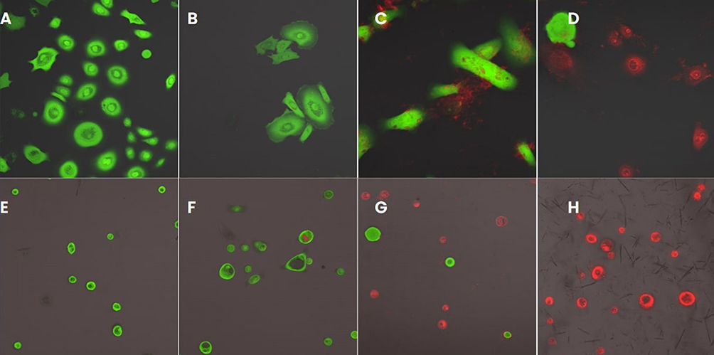 |
Figure 4 Peptide micelles and fibers interfaced with epidermal keratinocytes. Green – calcein dye – indicates a healthy intact membrane. Red – PA-TAMRA – indicates the fate of the peptide (A) Healthy untreated cells, (B) cells + peptide micelles (10 μM), (C) + peptide fibers (10 μM), (D) + peptide fibers (10 μM) + ciprofloxacin (0.1 wt%), (E) + ciprofloxacin (1 wt%), (F) +micelles (100 μM), (G) + peptide fibers (100 μM), (H) + peptide fibers (100 μM) + ciprofloxacin (1 wt%). |
Discussion
Despite the fact that the peptide assemblies at high concentrations decrease cell viability, the tympanic membrane has regenerative properties and is very thick.22 Therefore, the CPE capability of the peptide will only penetrate so far to assist with trans-tympanic drug delivery, while the tympanic membrane itself can regenerate. Furthermore, there is a host of examples present that suggest peptide amphiphiles can be engineered to optimize regenerative capabilities by either stiffening the delivered hydrogel through peptide sequence modification and/or adding a biorecognition epitope to promote cell growth.23 The work presented here highlights the many roles that the peptide can play including CPE and drug delivery.
Cell Penetrating Peptides
Cell penetrating peptides (CPPs) are designed to deliver cargo using various cellular mechanisms that are either energy independent like direct penetration or energy dependent like endocytosis.16 Typically, peptides that are cationic follow the direct penetration mechanism through various means like membrane thinning, pore formation, or micelle formation. Most likely, our peptide follows an energy independent mechanism due to the highly cationic surface. In fact, our LSCM data, Figure 4A and B, highlight that the peptide interacts with the cell membrane without fully disrupting it at lower concentrations, consistent with a direct penetration mechanism. At higher concentrations, we observe the peptide integrating with the membrane but the calcein dye no longer interacts suggesting a dramatically weakened outer membrane. Furthermore, in the case of fiber interaction with keratinocytes at high concentration, we observed peptide integration beyond the cell membrane towards the center of the cell, potentially locating to the nuclear membrane or another intracellular lipid membrane. This suggests that the peptides ultimate fate is the integration into the outer lipid bilayer membrane compromising membrane integrity at high concentrations, allowing the peptide to interact freely with other organelles.
While the mechanism is somewhat typical of other CPPs, we find that the introduction of a temporal component to CPP delivery offers a new approach to regulating the application of the material. Specifically, delivering a CPP to the target region as a self-supporting hydrogel relies on the dissociation of individual CPP molecules before taking effect. Other CPPs, even those modified with alkyl tails like PA in this study, form micelles and dissociate at greater rates than fibers as highlighted in Figure 2A. Therefore, we suggest that engineering CPPs to form weak hydrogels capable of slowly dissociating present an excellent means of controlling not only CPP delivery but also controlling the payload delivery, ciprofloxacin, as highlighted in Figure 2B.
Other Trans Tympanic Membrane Drug Delivery Formulations
The employment of a hydrogel matrix for tympanic membrane drug delivery has been explored prior to this study.24–26 The use of SDS, bupivacaine, and limonene (three chemical permeation enhances) demonstrated the ability to increase ciprofloxacin drug delivery nearly threefold when compared to eardrops of ciprofloxacin alone in chinchillas. When embedding the 3 CPEs in a thermosresponsive Poloxamer hydrogel matrix, temporal control of ciprofloxacin release was demonstrated over longer periods of time facilitating the application of a single dose in the form of ear drops to completely eradicate an infection as opposed to daily use. The thermoresponsive nature of the poloxamer (transitions from a fluid solution at room temperature to a hydrogel at 37°C) makes it a useful drug delivery matrix. However, the additional components complicate the formulation and determining the exact mechanistic nature of the enhanced drug delivery becomes difficult to ascertain. Nonetheless, the employment of a hydrogel matrix showed promise. Our peptide amphiphile plays the role of not only the drug loaded polymer matrix but also eliminates the need for additional CPEs as the peptide plays that role. In fact, in the PA system presented here, the addition of a secondary CPE, SDS, plays a clear role in enhancing the rate of delivery.
Another approach to identify peptides that could potentially lead to active transport across the tympanic membrane utilized phage display.27 The presence of basic amino acid residues (lysine/arginine) was a critical phage property needed for the binding and crossing of the TM similarly to the peptide presented here. Although which peptide sequences have the highest affinity for the plasma membrane have not yet been determined, phage display helped identify that electrostatic interactions played an essential role in crossing the tympanic membrane. The N-terminal and C-terminal motifs were identified as significant for binding and internalization to the TM. The 2 conserved motifs appear to be the ST(K/R)T core motif and the PxxPxxP motif. BPT-4 was an interesting peptide that was characterized due to its proline-repeat motif and in that it is the primary residue in collagen secondary structure reveal that it is possible to translocate peptide-linked small particles across the TM. This was the first comprehensive biopanning for the isolation of TM transiting peptidic ligands. The identified mechanisms offered a unique method of drug delivery into the middle ear. Future iterations of the material could utilize sequences identified through similar phage display/biopanning methods in conjunction with the mechanical properties investigated here that temporally control the release of the peptide and the encapsulated drug.
Conclusion
The peptide-amphiphile, c16-AHL3K3-CO2H (in a 9:1 formulation with the labeled peptide c16-AHL3K2K(TAMRA)-CO2H), was demonstrated here to act as an all-in-one material in which the peptide serves as a drug delivery matrix when assembled into a hydrogel and facilitates drug-delivery while serving as a chemical permeation enhancer (or cell-penetrating peptide) as the peptide dissociates freely from the hydrogel/fiber assembly. Transfer of the peptide across a synthetic membrane was demonstrably slower in fibers than in micelles. Drug delivery across the same membrane was desirably slower in fibers than micelles showcasing the potential application in temporally controlled drug-delivery. The CPP ability was highlighted when the PA was interfaced with epidermal keratinocytes, highlighting the initial interaction and subsequent integration of the peptide into the lipid bilayer membrane. Future investigations will investigate treating animal models with the PA hydrogel/ciprofloxacin mixture to determine how deep the peptide penetrates into the tympanic membrane as well as the efficacy of trans-tympanic drug-delivery. In addition, tuning the rates of peptide disassembly through sequence variation marks an avenue to customize rates of drug delivery from a self-assembled peptide matrix.
Acknowledgment
The funding for this research was in part based on a grant received through the Department of Otolaryngology, Head and Neck Surgery, Rush University, Chicago. Use of the Center for Nanoscale Materials, an Office of Science user facility, was supported by the US Department of Energy, Office of Science, Office of Basic Energy Sciences, under Contract No. DE-AC02-06CH11357.
Author Contributions
All authors contributed to data analysis, drafting or revising the article, have agreed on the journal to which the article will be submitted, given final approval of the version to be published, and agree to be accountable for all aspects of the work.
Disclosure
The authors report no conflicts of interest in this work.
References
1. Auinger P, Lanphear BP, Kalkwarf HJ, Mansour ME. Trends in otitis media among children in the United States. Pediatrics. 2003;112(3):514–520. doi:10.1542/peds.112.3.514
2. Waseem M. Otitis media. Medscape. 2022. Available from: https://emedicine.medscape.com/article/994656-overview.
3. Osma U, Cureoglu S, Hosoglu S. The complications of chronic otitis media: report of 93 cases. J Laryngol Otol. 2000;114(2):97–100. doi:10.1258/0022215001905012
4. Sun J, Sun J. Intracranial complications of chronic otitis media. Eur Arch Otorhinolaryngol. 2013;271(11):2923–2926. doi:10.1007/s00405-013-2778-4
5. Harmes KM, Blackwood RA, Burrows HL, Cooke JM, Van Harrison R, Passamani PP. Otitis media: diagnosis and treatment. Am Fam Physician. 2013;88(7):435–440.
6. Toll EC, Nunez DA. Diagnosis and treatment of acute otitis media: review. J Laryngol Otol. 2012;126(10):976–983. doi:10.1017/S0022215112001326
7. Lambert E, Roy S. Otitis media and ear tubes. Pediatr Clin North Am. 2013;60(4):809–826. doi:10.1016/j.pcl.2013.04.014
8. Steele DW, Adam GP, Di M, Halladay CH, Balk EM, Trikalinos TA. Effectiveness of tympanostomy tubes for otitis media: a meta-analysis. Pediatrics. 2017;139(6):e20170125. doi:10.1542/peds.2017-0125
9. Hoberman A, Preciado D, Paradise JL, et al. Tympanostomy tubes or medical management for recurrent acute otitis media. N Engl J Med. 2021;384(19):1789–1799. doi:10.1056/NEJMoa2027278
10. Bondy J, Berman S, Glazner J, Lezotte D. Direct expenditures related to otitis media diagnoses: extrapolations from a pediatric Medicaid cohort. Pediatrics. 2000;105(6):e72–e72. doi:10.1542/peds.105.6.e72
11. Lim DJ. Human tympanic membrane - An ultrastructural observation. Acta Otolaryngol. 1970;70(3):176–186. doi:10.3109/00016487009181875
12. Kono M, Umar NK, Takeda S, et al. Novel antimicrobial treatment strategy based on drug delivery systems for acute otitis media. Front Pharmacol. 2021;12. doi:10.3389/fphar.2021.640514
13. Sis MJ, Webber MJ. Drug delivery with designed peptide assemblies. Trends Pharmacol Sci. 2019;40(10):747–762. doi:10.1016/j.tips.2019.08.003
14. Solomon LA, Kronenberg JB, Fry HC. Control of heme coordination and catalytic activity by conformational changes in peptide–amphiphile assemblies. J Am Chem Soc. 2017;139(25):8497–8507. doi:10.1021/jacs.7b01588
15. Lane ME. Skin penetration enhancers. Int J Pharm. 2013;447(1–2):12–21. doi:10.1016/j.ijpharm.2013.02.040
16. Desale K, Kuche K, Jain S. Cell-penetrating peptides (CPPs): an overview of applications for improving the potential of nanotherapeutics. Biomater Sci. 2020;9(4):1153–1188. doi:10.1039/D0BM01755H
17. Copolovici DM, Langel K, Eriste E, Langel U. Cell-penetrating peptides: design, synthesis, and applications. ACS Nano. 2014;8(3):1972–1994. doi:10.1021/nn4057269
18. Feger G, Angelov B, Angelova A. Prediction of amphiphilic cell-penetrating peptide building blocks from protein-derived amino acid sequences for engineering of drug delivery nanoassemblies. J Phys Chem B. 2020;124(20):4069–4078. doi:10.1021/acs.jpcb.0c01618
19. Reissmann S, Filatova MP. New generation of cell-penetrating peptides: functionality and potential clinical application. J Pept Sci. 2021;27(5). doi:10.1002/psc.3300
20. Fry HC, Garcia JM, Medina MJ, et al. Self-assembly of highly ordered peptide amphiphile metalloporphyrin arrays. J Am Chem Soc. 2012;134(36):14646–14649. doi:10.1021/ja304674d
21. Yang R, Okonkwo OS, Zurakowski D, Kohane DS. Synergy between chemical permeation enhancers and drug permeation across the tympanic membrane. J Control Release. 2018;289:94–101. doi:10.1016/j.jconrel.2018.06.019
22. Topf MC, Hsu DW, Adams DR, et al. Rate of tympanic membrane perforation after intratympanic steroid injection. Am J Otolaryngol. 2017;38(1):21–25. doi:10.1016/j.amjoto.2016.09.004
23. Matson JB, Stupp SI. Self-assembling peptide scaffolds for regenerative medicine. Chem Commun. 2012;48(1):26–33. PMID: 22080255; PMCID: PMC3355058. doi:10.1039/c1cc15551b
24. Sikorska E, Wyrzykowski D, Szutkowski K, Greber K, Lubecka EA, Zhukov I. Thermodynamics, size, and dynamics of zwitterionic dodecylphosphocholine and anionic sodium dodecyl sulfate mixed micelles. J Therm Anal Calorim. 2015;119(1):507–513. doi:10.1007/s10973-015-4918-0
25. Khoo X, Simons EJ, Chiang HH, et al. Formulations for trans-tympanic antibiotic delivery. Biomaterials. 2013;34(4):1281–1288. doi:10.1016/j.biomaterials.2012.10.025
26. Yang R, Sabharwal V, Okonkwo OS, et al. Treatment of otitis media by transtympanic delivery of antibiotics. Sci Transl Med. 2016;8(356). doi:10.1126/scitranslmed.aaf4363
27. Kurabi A, Pak KK, Bernhardt M, Baird A, Ryan AF. Discovery of a biological mechanism of active transport through the tympanic membrane to the middle ear. Sci Rep. 2016;6(1). doi:10.1038/srep22663
 © 2025 The Author(s). This work is published and licensed by Dove Medical Press Limited. The
full terms of this license are available at https://www.dovepress.com/terms
and incorporate the Creative Commons Attribution
- Non Commercial (unported, 4.0) License.
By accessing the work you hereby accept the Terms. Non-commercial uses of the work are permitted
without any further permission from Dove Medical Press Limited, provided the work is properly
attributed. For permission for commercial use of this work, please see paragraphs 4.2 and 5 of our Terms.
© 2025 The Author(s). This work is published and licensed by Dove Medical Press Limited. The
full terms of this license are available at https://www.dovepress.com/terms
and incorporate the Creative Commons Attribution
- Non Commercial (unported, 4.0) License.
By accessing the work you hereby accept the Terms. Non-commercial uses of the work are permitted
without any further permission from Dove Medical Press Limited, provided the work is properly
attributed. For permission for commercial use of this work, please see paragraphs 4.2 and 5 of our Terms.