
Back to Journals » Journal of Inflammation Research » Volume 19
Dynamic Changes in Serum Choline Acetyltransferase and Acetylcholinesterase in Septic versus Non-Septic ICU Patients: A Prospective Exploratory Study
Authors Zhu Z  , Wang H , Huo Y, Lu B, Zhang Y, Liu L
Received 13 April 2026
Accepted for publication 29 June 2026
Published 23 July 2026 Volume 2026:19 612518
DOI https://doi.org/10.2147/JIR.S612518
Checked for plagiarism Yes
Review by Single anonymous peer review
Peer reviewer comments 3
Editor who approved publication: Dr Xin Du
Ziyi Zhu,1,* Haiping Wang,2,* Yaxin Huo,1 Bingqi Lu,1 Yujun Zhang,3 Lixia Liu1
1Department of Critical Care Medicine, The Fourth Hospital of Hebei Medical University, Shijiazhuang, Hebei, People’s Republic of China; 2Department of Cardiology, The Fourth Hospital of Hebei Medical University, Shijiazhuang, Hebei, People’s Republic of China; 3Department of Critical Care Medicine, The First Hospital of Xinji, Xinji, Hebei, People’s Republic of China
*These authors contributed equally to this work
Correspondence: Lixia Liu, Department of Critical Care Medicine, The Fourth Hospital of Hebei Medical University, No. 12 Jiankang Road, Shijiazhuang, Hebei, 050011, People’s Republic of China, Tel +86 13833186570, Email [email protected]
Objective: To compare serum choline acetyltransferase (ChAT) and acetylcholinesterase (AChE) levels between septic and non-septic critically ill patients, and to evaluate their dynamic changes, associations with inflammatory markers, disease severity, and prognosis in sepsis.
Methods: In this prospective observational study, 87 patients with sepsis and 93 non-septic critically ill controls were enrolled. Blood samples were collected from septic patients within 12 hours (T1), 24 hours (T2), 48 hours (T3), and 7 days (T4) after intensive care unit (ICU) admission, while the non-septic group underwent sampling only at T1. Serum levels of ChAT, AChE, and inflammatory cytokines (IL-6, IL-10, TNF-α) were measured. A linear mixed model was used to analyze dynamic changes, repeated measures correlation (rmcorr) to assess associations between variables, receiver operating characteristic curves to evaluate discriminative ability, and multivariable logistic regression to identify independent influencing factors.
Results: Septic patients had significantly lower ChAT at T1 than non-septic controls (P< 0.001), whereas AChE levels did not differ. Both ChAT and AChE were lower in septic shock than in non-shock septic patients (both P< 0.05). Linear mixed‑model analysis revealed a significant overall group effect for AChE, with lower levels in non‑survivors (group effect, P = 0.013); however, no significant intergroup difference was observed at T1 (P = 0.098). ChAT showed moderate discriminative ability for sepsis, with an area under the ROC curve of 0.70, and was identified as an independent factor associated with sepsis in this exploratory cohort (OR = 0.85, 95% CI: 0.76– 0.94, P = 0.001). The inverse association between ChAT and sepsis was stronger in patients aged ≥ 65 years (interaction P=0.023).
Conclusion: This exploratory study demonstrates that serum ChAT levels are significantly decreased in septic patients, correlate with disease severity, and show moderate discriminative ability for sepsis. Linear mixed‑model analysis revealed that AChE levels were overall lower in non‑survivors than in survivors, although the prognostic value at individual time points was limited. Age modifies the ChAT–sepsis association, which should be considered in clinical evaluation.
Keywords: sepsis, cholinergic anti-inflammatory pathway, choline acetyltransferase, acetylcholinesterase, prognosis
Introduction
Sepsis is defined as a life-threatening organ dysfunction caused by a dysregulated host response to infection. It represents a common and critical clinical syndrome in the field of critical care medicine worldwide and is a major cause of death among patients in intensive care units (ICUs).1,2 In recent years, despite significant advances in early identification,3 fluid resuscitation,4,5 anti-infective therapy,6 and organ support,1,7 the mortality rate of sepsis, particularly septic shock, remains persistently high,8 imposing a substantial medical burden on families and society.9 The pathogenesis of sepsis is complex and involves multiple interconnected mechanisms, including dysregulated inflammatory responses, immunosuppression, mitochondrial dysfunction, and coagulation disorders,1,7,10 among which uncontrolled inflammation is considered the core pathophysiological basis leading to tissue damage and multiple organ dysfunction.
The cholinergic anti-inflammatory pathway (CAP) is a key component of the neuro-immune regulatory network and plays an important role in modulating the systemic inflammatory response in sepsis.11,12 This pathway involves the release of acetylcholine (ACh) by the vagus nerve, which binds to the α7 nicotinic acetylcholine receptor (α7nAChR) on the surface of splenic macrophages, thereby inhibiting the synthesis and release of pro-inflammatory cytokines such as tumor necrosis factor-α (TNF‑α), interleukin-6 (IL‑6), and interleukin-1β (IL‑1β), thus exerting a negative regulatory role in the inflammatory response.13 In the context of sepsis, CAP often exhibits functional impairment or suppression, leading to amplification of the inflammatory response, immune dysfunction, and multiple organ injury.14
Choline acetyltransferase (ChAT) is a key enzyme in the synthesis of acetylcholine within the cholinergic anti-inflammatory pathway, and its activity directly influences the efficiency of acetylcholine production.15 Acetylcholinesterase (AChE) is the key enzyme responsible for hydrolyzing acetylcholine, reflecting the metabolic status of cholinergic neurotransmitters.16 The α7nAChR expressed by macrophages and the ACh generated thereby are components of the non‑neuronal cholinergic system, and have become a research focus due to their potential to regulate inflammatory and immune responses;17 they also represent the final effector target of CAP‑mediated neuroimmune regulation. The present study focuses on changes in ChAT and AChE within the CAP and their clinical significance.
Previous studies have shown that peripheral blood cholinesterase activity is often significantly decreased in patients with sepsis, and its decline is associated with disease severity, organ failure, and short‑term mortality risk.18–20 However, only one small‑sample study (n = 11) has reported changes in serum ChAT activity in septic patients, with results suggesting elevated ChAT levels;15 due to the limited sample size, this conclusion requires validation in larger cohorts. Therefore, prospective observational studies are needed to further clarify the pattern of ChAT changes in sepsis and to examine the dynamic trends of both enzymes and their prognostic implications.
We hypothesized that ChAT and AChE may exhibit distinct dynamic patterns during the course of sepsis. Accordingly, this prospective observational study was designed to compare serum ChAT and AChE levels between septic and non‑septic critically ill patients, to dynamically track the trajectories of these two enzymes over a 7‑day period in septic patients, and to analyze their associations with inflammatory cytokines, disease severity, and prognosis. The aim was to preliminarily explore the potential value of these two enzymes in the clinical evaluation of sepsis.
Materials and Methods
Study Design and Patient Characteristics
This study was a single‑center, prospective, observational cohort study. Patients consecutively admitted to the Intensive Care Unit (ICU) of the Fourth Hospital of Hebei Medical University from December 2024 to May 2025 were enrolled and divided into a sepsis group and a non‑sepsis group according to the Sepsis‑3 criteria. Patients in the sepsis group were diagnosed with sepsis upon ICU admission. Inclusion criteria were: (1) age ≥ 18 years; (2) for the sepsis group, fulfillment of the Sepsis‑3 diagnostic criteria, defined as infection plus an increase in the Sequential Organ Failure Assessment (SOFA) score of ≥ 2 points; (3) newly diagnosed sepsis, with the time interval from diagnosis to enrollment ≤ 2 hours. Exclusion criteria were: (1) a history of neuropsychiatric disorders such as schizophrenia or Alzheimer’s disease, or long‑term use of medications affecting mental status; (2) a history of splenectomy; (3) for patients in the sepsis group, fewer than two time points with valid serum ChAT or AChE measurements; (4) ICU length of stay < 24 hours; and (5) pregnant or breastfeeding patients.
Blood samples were collected from patients in the sepsis group within 12 hours (T1), at 24 hours (T2), 48 hours (T3), and 7 days (T4) after ICU admission. In the non‑sepsis group, a single blood sample was collected within 12 hours of admission. Patients with sepsis were further divided into survivor and non‑survivor groups based on their 28‑day survival status. This study was conducted in accordance with the Declaration of Helsinki and was approved by the Ethics Committee of the Fourth Hospital of Hebei Medical University (Approval No. 2024KS159). Written informed consent was obtained from all patients or their legal proxies. This study was registered in the Chinese Clinical Trial Registry (registration number: ChiCTR2500106909).
Sample Measurement
Venous blood samples were centrifuged at 3000 rpm for 10 min at 4°C. Serum was separated and stored at −80°C until analysis. Serum choline acetyltransferase (ChAT) activity was measured using a commercial colorimetric assay kit (Nanjing Jiancheng Bioengineering Institute, China; Cat. No. A079-2-1) following the manufacturer’s instructions. Absorbance was read with a microplate reader. Serum acetylcholinesterase (AChE) activity was determined using a commercial activity assay kit (Shanghai Zhuocai Biotechnology Co., Ltd., China; Cat. No. ZC-S0384) according to the manufacturer’s protocol. Concentrations of inflammatory cytokines (IL‑6, IL‑10, TNF‑α) were quantified using enzyme‑linked immunosorbent assay (ELISA) kits (Wuhan Boster Biological Technology Co., Ltd., China).
Data Collection
Demographic data (age, sex, body mass index [BMI]), clinical characteristics (type of infection, underlying diseases, surgical history), laboratory parameters (routine blood tests, biochemical profiles, coagulation function, blood gas analysis, etc), disease severity scores (Acute Physiology and Chronic Health Evaluation [APACHE] II score, SOFA score), and prognostic information (28‑day survival status, ICU length of stay, sepsis‑associated acute kidney injury, delirium, etc.) were collected for all patients.
Statistical Analysis
This study was an exploratory analysis, and the sample size was determined based on clinical feasibility and preliminary experimental data; no formal power calculation was performed. All statistical analyses were conducted using R version 4.3.0, Zstats version 1.0, and SPSS version 27.0. Data visualization was performed using GraphPad Prism 10 and R software.
For continuous variables, those following a normal distribution were expressed as mean ± standard deviation (SD), and comparisons between groups were performed using the independent samples t‑test. Non‑normally distributed continuous variables were described as median (interquartile range), and comparisons between two groups were performed using the Mann‑Whitney U-test. Categorical variables were described as frequencies and percentages, and comparisons between groups were performed using the chi‑square (χ2) test. A two‑tailed significance level of α = 0.05 was used for all tests. A linear mixed model was used to analyze the dynamic changes in serum ChAT and AChE levels in patients with sepsis, with patient ID as a random intercept and time point (T1, T2, T3, T4), group (non‑survivor vs survivor), and their interaction as fixed effects. An unstructured covariance matrix was assumed. Post‑hoc pairwise comparisons were adjusted using Bonferroni correction. Repeated measures correlation (rmcorr) was used to assess the within-individual correlations of ChAT and AChE with IL-6, IL-10, and TNF-α, accounting for the non-independence of repeated measurements from the same patient. Results are reported as r_rm, 95% CI, degrees of freedom, and p-values. Receiver operating characteristic (ROC) curves were constructed to assess the discriminative ability of serum ChAT and AChE for sepsis, and the area under the curve (AUC), sensitivity, specificity, and Youden index were calculated. Multivariate logistic regression with forward stepwise selection was used to identify independent factors associated with sepsis, and odds ratios (ORs) with 95% confidence intervals (CIs) were calculated. Model calibration was assessed using the Hosmer‑Lemeshow goodness‑of‑fit test. Bootstrap internal validation (500 resamples) was performed to assess the stability of the final model, including the variables selected in the final model, and the corrected AUC was calculated. Subgroup analyses were performed using multivariate logistic regression, and interaction effects were tested. A P value < 0.05 was considered statistically significant. Missing data were handled using complete‑case analysis, with a low proportion of missing values (<5%) across key variables.
Results
Baseline Characteristics and Clinical Features
Patient Enrollment Process
A total of 226 critically ill patients admitted to the ICU of the Fourth Hospital of Hebei Medical University from December 2024 to May 2025 were screened. According to the inclusion and exclusion criteria, 180 patients were finally enrolled, including 87 patients in the sepsis group and 93 patients in the non‑sepsis group. Among the sepsis group, 27 patients (31.03%) died within 28 days, and 60 patients (68.97%) survived. The patient enrollment process is illustrated in Figure 1.
 |
Figure 1 Patient enrollment flowchart. |
Comparison of Baseline Characteristics Between the Sepsis and Non‑sepsis Groups
Comparison of baseline characteristics between the two groups is shown in Table 1. There were no statistically significant differences between the sepsis group and the non‑sepsis group in terms of age, sex, or BMI (P>0.05). Statistically significant differences were observed between the two groups in albumin, lymphocyte count, fibrinogen, hemoglobin, body temperature, heart rate, platelet count, creatinine, oxygenation index, lactate, total bilirubin, ChAT level, proportion of patients with coronary heart disease, and proportion of patients receiving immunosuppressive therapy (P<0.05). Compared with the control group, the sepsis group had higher body temperature, heart rate, creatinine, fibrinogen, lactate, total bilirubin levels, and a higher proportion of patients receiving immunosuppressive therapy, whereas albumin, lymphocyte count, hemoglobin, platelet count, oxygenation index, ChAT levels, and the proportion of patients with coronary heart disease were lower.
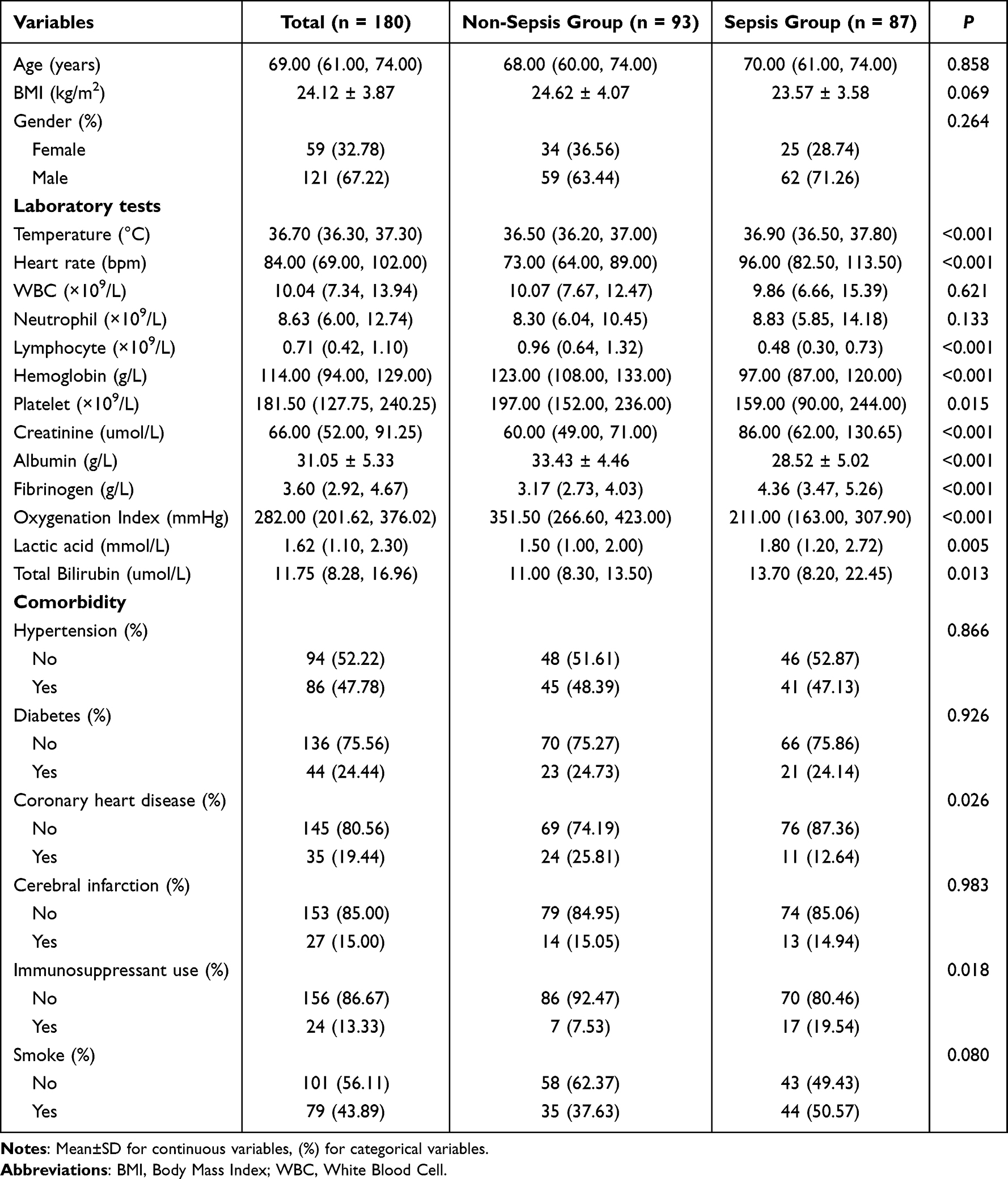 |
Table 1 Comparison of Baseline Characteristics Between Sepsis and Non‑Sepsis Groups |
Comparison of Baseline Characteristics Between Different Prognostic Subgroups in Patients with Sepsis
Among the 87 patients with sepsis, 27 were classified into the non‑survivor group and 60 into the survivor group based on 28‑day survival status. Comparison of baseline characteristics between the two subgroups is shown in Table 2. Statistically significant differences were observed between the survivor and non‑survivor groups in body temperature, lactate level, SOFA score, postoperative admission status, and proportion of patients with septic shock (all P<0.05). Compared with the survivor group, the non‑survivor group had higher lactate levels and SOFA scores, a higher proportion of non‑surgical admissions, a higher proportion of patients with septic shock, and lower body temperature at ICU admission.
 |
Table 2 Comparison of Baseline Characteristics Between Survivors and Non‑Survivors In Septic Patients |
Comparison of Serum ChAT and AChE Levels
Comparison Between the Sepsis and Non‑sepsis Groups
Serum ChAT levels at T1 were significantly lower in the sepsis group than in the non‑sepsis group [15.50 (12.25, 20.70) U/mL vs 21.21 (17.00, 23.57) U/mL, P<0.001], whereas no statistically significant difference was observed in serum AChE levels between the two groups (P>0.05) (Table 3 and Figure 2).
 |
Table 3 Comparison of Serum ChAT and AChE Levels Between Sepsis and Non‑Sepsis Groups at T1 |
 |
Figure 2 Comparison of serum ChAT levels between non-sepsis and sepsis groups. Data are presented as median with interquartile range (Q1-Q3). ChAT levels were significantly lower in the sepsis group (15.5 U/mL, IQR: 12.25–20.70) compared to the non-sepsis group (21.21 U/mL, IQR: 17.00–23.57).****P < 0.001 (Mann–Whitney U-test). |
Comparison Between Survivor and Non-Survivor Subgroups in Sepsis
Among the 87 patients with sepsis, 27 (31.03%) died within 28 days and 60 (68.97%) survived. No statistically significant differences were observed in serum ChAT or AChE levels at T1 between the non‑survivor and survivor groups (P>0.05) (Table 4).
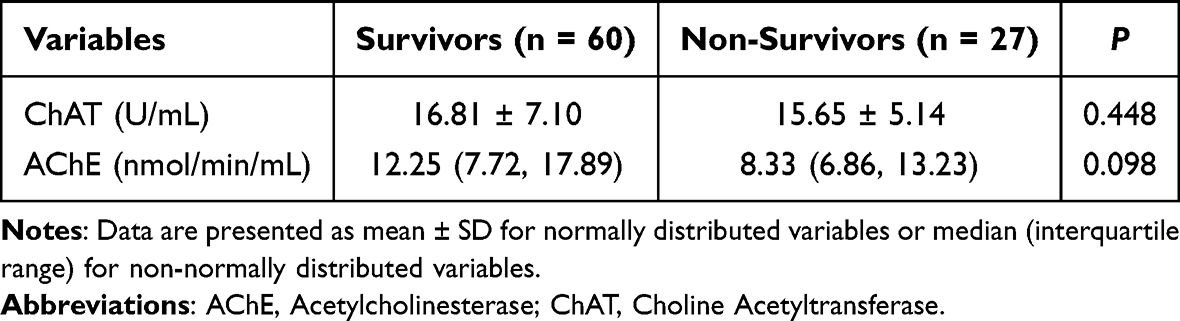 |
Table 4 Comparison of Serum ChAT and AChE Levels Between Survivors and Non‑Survivors at T1 |
Subgroup Analysis
According to the Sepsis-3 diagnostic criteria, the 87 patients with sepsis were further divided into a septic shock group (n = 46) and a non‑shock septic group (n = 41). Serum ChAT levels were significantly lower in the septic shock group than in the non‑shock septic group (15.10 ± 5.78 U/mL vs 17.96 ± 7.07 U/mL, P=0.041). Serum AChE levels were also significantly lower in the septic shock group than in the non‑shock septic group [9.07 (6.00, 13.23) nmol/min/mL vs 12.74 (8.33, 20.09) nmol/min/mL, P=0.005] (Table 5, Figure 3A and B).
 |
Table 5 Comparison of Serum ChAT and AChE Levels Between Septic Shock and Sepsis without shock groups |
 |
Figure 3 Comparison of serum ChAT and AChE levels between septic shock group and sepsis without shock group. (A) Serum ChAT levels. Data are presented as mean ± SD. Patients in the septic shock group (n=46) had significantly lower ChAT levels than those in the sepsis without shock group (n=41) (P = 0.041, independent samples t-test). *P < 0.05. (B) Serum AChE levels. Data are presented as median with interquartile range. AChE levels were significantly lower in the septic shock group (P = 0.005, Mann–Whitney U-test). **P < 0.01. |
Dynamic Trends of Serum ChAT and AChE in Patients with Sepsis
A linear mixed model was used to analyze the dynamic changes in serum ChAT and AChE levels at four time points (T1, T2, T3, and T4) in 87 patients with sepsis. The results are shown in Figure 4.
 |
Figure 4 Dynamic changes of serum AChE (A) and ChAT (B) in sepsis patients with different outcomes. (A) Serum AChE levels in survivors (n=60) and non-survivors (n=27). Data are presented as mean (95% CI). Non-survivors had significantly lower AChE levels overall (group effect, P=0.013), with no significant time effect (P=0.637) or interaction (P=0.992). Both groups showed initial decline followed by partial recovery. (B) Serum ChAT levels. No significant group (P=0.539), time (P=0.359), or interaction effects (P=0.925) were observed. |
Serum AChE: The group effect was statistically significant (F=6.497, P=0.013), indicating that overall AChE levels were significantly lower in the non‑survivor group than in the survivor group. Neither the time effect (F = 0.570, P = 0.637) nor the time‑by‑group interaction effect (F = 0.032, P = 0.992) reached statistical significance. Detailed results of intergroup comparisons at each time point are presented in Table 6. Among the four time points, only at T3 did non‑survivors have significantly lower AChE levels than survivors [8.33 (nmol/min/mL) vs 11.26 (nmol/min/mL), P = 0.048]; no significant differences were observed at T1, T2, or T4. Both groups showed a descriptive trend of an initial decline followed by a partial recovery (Figure 4A).
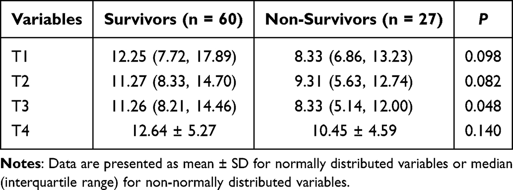 |
Table 6 Comparison of Serum AChE Levels Between Survivors and Non‑Survivors at Each Time Point |
Serum ChAT: The group effect (F=0.380, P=0.539), time effect (F=1.090, P=0.359), and interaction effect (F=0.157, P=0.925) were not statistically significant, indicating that ChAT levels did not differ significantly between the two groups and showed no significant change over time (Figure 4B).
Discriminative Ability of Serum ChAT and AChE for Sepsis
Receiver operating characteristic (ROC) curves were used to evaluate the discriminative ability of serum ChAT and AChE measured within 12 hours of ICU admission (T1) for sepsis, with 87 patients with sepsis as the case group and 93 non‑septic critically ill patients as the control group. The results are shown in Figure 5.
 |
Figure 5 ROC curve of serum ChAT for discriminating sepsis. The area under the curve (AUC) for ChAT was 0.70 (95% CI: 0.62–0.78, P < 0.001). |
The area under the curve (AUC) of ChAT for discriminating sepsis was 0.70 (95% CI: 0.62–0.78, P<0.001). Whereas the AUC of AChE was 0.54 (95% CI: 0.46–0.62, P=0.404), indicating no significant discriminative ability. The AUC for the combination of ChAT and AChE was 0.70 (95% CI: 0.62–0.78), which was identical to that of ChAT alone, suggesting that the addition of AChE did not improve discriminative performance.
These findings indicate that serum ChAT has moderate discriminative ability for sepsis, whereas AChE is not suitable as an independent discriminative marker.
Correlation Analysis of Serum ChAT and AChE with Inflammatory Cytokines in Patients with Sepsis
To account for the within‑patient correlation inherent in repeated measurements, we used repeated measures correlation (rmcorr) to assess the associations of serum AChE and ChAT with inflammatory cytokines.
The rmcorr analysis showed no significant correlations between AChE and IL‑6 (r_rm = −0.04, 95% CI: −0.168 to 0.089, df = 231, p = 0.543), IL‑10 (r_rm = −0.005, 95% CI: −0.133 to 0.124, df = 231, p = 0.94), or TNF‑α (r_rm = −0.067, 95% CI: −0.194 to 0.062, df = 231, p = 0.308). ChAT also showed no statistically significant correlations with any of the inflammatory cytokines (all p > 0.05).
In addition, the rmcorr analysis revealed a significant positive correlation between ChAT and AChE (r_rm = 0.14, 95% CI: 0.011 to 0.264, df = 231, p = 0.033).
Multivariable Logistic Regression Analysis of Factors Associated with Sepsis and Incremental Value Assessment
Using sepsis status as the dependent variable, forward stepwise selection (LR) was used to identify independent influencing factors. Five independent factors were ultimately identified (Table 7 and Figure 6). Model evaluation showed good calibration, as assessed by the Hosmer‑Lemeshow test (χ2 = 14.906, P = 0.061), with a Nagelkerke R2 of 0.726. The full model (including ChAT) yielded an area under the ROC curve of 0.94 (95% CI: 0.91–0.98) for predicting sepsis (Figure 7).
 |
Table 7 Multivariable Logistic Regression Analysis of Factors Associated with Sepsis |
 |
Figure 6 Forest plot of multivariable logistic regression analysis for independent factors associated with sepsis. ChAT, total bilirubin, oxygenation index, heart rate, and hemoglobin were identified as independent factors for sepsis. Decreased levels of ChAT, hemoglobin, and oxygenation index (OR < 1) were associated with increased sepsis risk, while increased levels of total bilirubin and heart rate (OR > 1) were associated with higher sepsis risk. |
 |
Figure 7 ROC curve of the multivariable logistic regression model for predicting sepsis. The ROC curve was based on the multivariable model including ChAT, total bilirubin, oxygenation index, heart rate, and hemoglobin. The area under the curve (AUC) was 0.94 (95% CI: 0.91–0.98). |
To assess the incremental value of ChAT, we compared the AUC of a baseline clinical model (total bilirubin, oxygenation index, heart rate, hemoglobin) with that of a model additionally including ChAT. In the whole cohort, the AUC increased from 0.929 to 0.943, with overlapping confidence intervals indicating no significant improvement. Bootstrap internal validation (500 resamples) was performed to assess the stability of the model, yielding a corrected AUC of 0.933, with a decrease of only 0.01 from the apparent AUC (0.943), suggesting a low risk of overfitting.
Subgroup Analysis
Multivariate logistic regression models (adjusted for total bilirubin, heart rate, hemoglobin, and oxygenation index) were used to evaluate the association between ChAT and sepsis risk across different subgroups. The results are shown in Figure 8. In the overall population, ChAT was significantly negatively associated with sepsis (OR = 0.85, 95% CI: 0.76–0.94, P=0.001). Subgroup analyses revealed that the direction of the association between ChAT and sepsis was consistent across subgroups stratified by sex, hypertension, diabetes, and smoking status, with no significant interactions observed (all P for interaction > 0.05). Notably, a significant interaction was observed for age group (P for interaction = 0.023): among patients aged ≥65 years, the negative association between ChAT and sepsis was stronger (OR = 0.73, 95% CI: 0.62–0.87, P<0.001), whereas the association did not reach statistical significance in patients aged <65 years (OR = 0.82, 95% CI: 0.65–1.03, P=0.089), suggesting that age may modify the association between ChAT and sepsis.
 |
Figure 8 Forest plot of subgroup analysis for the association between ChAT and sepsis risk. ChAT was significantly inversely associated with sepsis risk in the overall population (OR=0.85, 95% CI: 0.76–0.94, P=0.001). A significant interaction was observed for age (P=0.023): the association was stronger in patients aged ≥65 years (OR=0.73, 95% CI: 0.62–0.87, P<0.001) but not significant in those <65 years (OR=0.82, 95% CI: 0.65–1.03, P=0.089). No significant interactions were found for sex, hypertension, diabetes, or smoking (all P>0.05). |
Further stratified analysis by age was performed to evaluate the incremental discriminative ability of ChAT (Table 8). In patients aged ≥65 years, adding ChAT increased the AUC from 0.930 to 0.960; in patients aged <65 years, no notable change in AUC was observed (0.968 vs 0.973). Although the confidence intervals overlapped, the improvement in point estimates was consistent with the significant age interaction (P = 0.023), suggesting an age ‑modifying effect of ChAT.
 |
Table 8 Incremental Discriminative Ability of ChAT by Age Subgroup |
Discussion
In this prospective observational study, we compared serum ChAT and AChE levels between septic and non‑septic critically ill patients, dynamically tracked the changes in these two enzymes over a 7‑day period in patients with sepsis, and analyzed their associations with inflammatory cytokines, disease severity, and prognosis. The main findings are as follows: (1) Serum ChAT levels at T1 were significantly lower in the sepsis group than in the non‑sepsis group, and both ChAT and AChE levels were significantly lower in the septic shock group than in the non‑shock septic group; (2) Serum AChE levels showed a descriptive trend of an initial decrease followed by a partial recovery within 7 days, with overall lower levels in non‑survivors than in survivors, while ChAT levels showed no significant differences between the two groups; (3) ChAT was positively correlated with AChE, whereas neither ChAT nor AChE showed significant correlations with IL‑6, IL‑10, or TNF‑α; (4) ChAT showed moderate discriminative ability for sepsis (AUC = 0.70); (5) Multivariable logistic regression analysis identified ChAT, total bilirubin, heart rate, hemoglobin, and oxygenation index as independent factors associated with sepsis; (6) Subgroup analysis revealed that the inverse association between ChAT and sepsis was more pronounced in elderly patients (≥65 years), with a significant interaction (P for interaction = 0.023).
CAP and Sepsis
CAP is a neuroimmune regulatory mechanism that has attracted extensive attention in recent years.21,22 This pathway inhibits the synthesis and release of inflammatory cytokines through modulation of the vagus nerve. The role of the cholinergic anti‑inflammatory pathway has been validated in the treatment of various diseases.23–25 Choline acetyltransferase (ChAT) and acetylcholinesterase (AChE) are the key enzymes within this pathway.
In the present study, serum ChAT levels were significantly lower in patients with sepsis than in non‑septic critically ill patients, which may be related to CAP dysfunction under septic conditions. In sepsis, excessive release of inflammatory cytokines to the central nervous system may impair central regulation of the peripheral immune system, leading to autonomic nervous system dysfunction and markedly attenuated anti‑inflammatory effects of the cholinergic pathway.14 Further analysis revealed that both ChAT and AChE levels were lower in patients with septic shock than in those with sepsis without shock, suggesting that the degree of cholinergic system impairment is closely associated with disease severity. This finding is also consistent with the potential role of the cholinergic anti‑inflammatory pathway in sepsis.12,26,27
Dynamic Changes of AChE and Their Clinical Significance
In this study, serum AChE levels in patients with sepsis showed a descriptive downward trend within the first 48 hours after ICU admission, followed by a slight increase by day 7. This pattern may reflect initial consumption of the cholinergic system in response to excessive inflammation, with subsequent recovery of cholinergic function after effective anti‑infective treatment and organ support.
Overall AChE levels were lower in non‑survivors than in survivors, with a significant difference at T3 (48 hours). This finding is consistent with previous studies,18–20 suggesting that lower overall AChE levels in non‑survivors may reflect insufficient functional reserve of the cholinergic system. Of note, the above association between AChE and prognosis was derived from a univariable linear mixed model without adjustment for other confounding factors; therefore, its independent prognostic value remains uncertain.
Correlation Between AChE and Inflammatory Cytokines
In this study, ChAT and AChE were found to be significantly positively correlated, which may reflect coordinated changes in the synthetic and degradative components of the cholinergic anti‑inflammatory pathway (CAP) during sepsis, potentially serving as a neuroimmune compensatory mechanism in response to dysregulated inflammation. However, no significant correlations were observed between ChAT or AChE and the inflammatory cytokines (IL‑6, IL‑10, TNF‑α). Possible explanations include: (1) AChE is predominantly synthesized and released by the liver, and its serum levels are influenced by multiple factors, including hepatic function and nutritional status;28,29 (2) the pathogenesis of sepsis is complex, and inflammatory cytokine levels are regulated by various other physiological mechanisms; thus, the serum level of a single enzyme may be insufficient to fully reflect the overall inflammatory response.
Discriminative Ability, Incremental Value, and Age‑Modifying Effect of ChAT
In this study, ChAT showed moderate discriminative ability for sepsis and was identified as an independent factor associated with sepsis. In contrast to the elevated ChAT levels reported by Gabalski et al15 the present study found decreased ChAT levels, which may be attributed to differences in sample size, assay methods, and sampling timing. Incremental value analysis showed that adding ChAT did not significantly improve the discriminative ability of the baseline clinical model in the whole cohort. After age stratification, the AUC increased from 0.930 to 0.960 in patients aged ≥65 years, whereas no notable change was observed in those aged <65 years. Although the confidence intervals still overlapped, the improvement in point estimates was consistent with the significant age interaction, suggesting that the clinical significance of ChAT may be modified by age.
With advancing age, the cholinergic system undergoes a series of changes:30 degeneration of basal forebrain nerve fibers leads to cholinergic neuron dysfunction and reduced ChAT expression.31 These changes diminish the capacity of elderly individuals to buffer inflammatory insults, resulting in reduced cholinergic functional reserve. Consequently, when ChAT levels decrease, the cholinergic system in elderly individuals may be more vulnerable to impairment, leading to an increased risk of sepsis. In younger individuals, however, the cholinergic system maintains relatively adequate functional reserve, and a certain degree of ChAT decline may still be compensated through regulatory mechanisms, thereby accounting for the weaker association with sepsis risk. This finding suggests that age should be taken into consideration when evaluating the association between ChAT and sepsis.
It should be acknowledged that this study represents a post‑hoc analysis in an already diagnosed cohort; therefore, the AUC of ChAT reflects only its discriminative ability in known groups, and its independent diagnostic value warrants further validation. In addition, the subgroup analysis was limited by a relatively small sample size, and the interaction effect should be interpreted as exploratory, requiring confirmation in larger cohorts.
Innovations and Clinical Significance
This prospective exploratory study on serum ChAT and AChE levels in patients with sepsis has the following novel aspects: (1) it is the first to simultaneously measure serum ChAT and AChE levels in septic patients, preliminarily observing changes in the cholinergic system during the pathophysiological process of sepsis; (2) it is the first to dynamically track the trajectories of these two enzymes over a 7‑day period; and (3) it is the first to report a possible age‑modifying effect on the association between ChAT and sepsis.
From a clinical perspective, the present study suggests that decreased ChAT levels may assist in identifying sepsis and assessing disease severity, with particular warning significance in elderly patients, while decreased AChE levels are associated with poor prognosis. In addition, reduced ChAT levels may indirectly suggest impaired function of the cholinergic anti‑inflammatory pathway, and therapeutic strategies such as exogenous ChAT supplementation,15 vagus nerve stimulation,32,33 and α7nAChR agonists34–36 warrant further investigation.
Limitations
This study has several limitations. First, the sample size was limited. As an exploratory study, the sample size was determined primarily based on clinical feasibility and preliminary experimental data, without formal power calculation. The sepsis group comprised only 87 patients, with 27 deaths (31.0%) within 28 days. According to the sample size requirement for multivariate regression analysis (events per variable [EPV] ≥ 10), the number of death events in this study would allow stable inclusion of only 2–3 variables; therefore, multivariate analysis with mortality as the outcome was not performed. The independent associations of ChAT and AChE with prognosis warrant validation in larger sample studies. Second, this was a single‑center prospective study, which may introduce selection bias. Third, the exclusion of patients with ICU stay < 24 hours and those with fewer than two valid ChAT/AChE measurements may have systematically excluded the most severely ill patients who died early and were unable to complete follow‑up sampling, potentially biasing the enrolled cohort toward a relatively stable intermediate group. Fourth, although we enrolled only patients with newly diagnosed sepsis (within 2 hours of diagnosis) and performed the first blood sampling within 12 hours of ICU admission, using ICU admission as the baseline (T1) cannot completely eliminate inter‑individual differences in pathophysiological onset, which may introduce bias when aligning dynamic trajectories. Fifth, serum ChAT and AChE levels may not fully reflect cholinergic functional status in tissues or synaptic clefts, where changes may be more pronounced. Sixth, the lack of longitudinal sampling in the non‑septic group precludes comparison of dynamic trajectories between the two groups. Seventh, despite adjustment for multiple confounders in the multivariable analysis, residual confounding (eg, medication use, nutritional status) may still exist. Eighth, the discriminative ability of AChE for sepsis was limited; it is not recommended as an independent discriminative marker. However, its association with disease severity (shock vs non‑shock) and dynamic trends may still provide certain clinical insights. Ninth, Bootstrap internal validation of the multivariable logistic regression model showed a decrease of only 0.01 in the corrected AUC, suggesting a low risk of overfitting. Nevertheless, the model remains exploratory, and its predictive performance requires external validation in an independent cohort.
Conclusion
This exploratory study suggests that serum ChAT levels are significantly decreased in patients with sepsis and correlate with disease severity, while AChE levels are overall lower in non‑survivors than in survivors. These findings indicate that ChAT and AChE may be associated with sepsis status and certain clinical features; however, their clinical value remains to be confirmed through larger, multicenter studies with more rigorous methodological designs.
Abbreviations
ACh, acetylcholine; AChE, acetylcholinesterase; APACHE II, Acute Physiology and Chronic Health Evaluation II; AUC, area under the curve; BMI, body mass index; CAP, cholinergic anti-inflammatory pathway; ChAT, choline acetyltransferase; ChiCTR, Chinese Clinical Trial Registry; CI, confidence interval; ELISA, enzyme-linked immunosorbent assay; EPV, events per variable; ICU, intensive care unit; IL-6, interleukin‑6; IL-10, interleukin‑10; LR, likelihood ratio (stepwise forward selection method); MV, mechanical ventilation; OR, odds ratio; ROC, receiver operating characteristic; SAKI, sepsis‑associated acute kidney injury; SD, standard deviation; SOFA, Sequential Organ Failure Assessment; TNF‑α, tumor necrosis factor‑α; WBC, white blood cell.
Data Sharing Statement
The datasets generated and/or analyzed during the current study are not publicly available due to patient privacy and ethical restrictions, but are available from the corresponding author on reasonable request.
Ethics Approval and Informed Consent
This study was conducted in accordance with the Declaration of Helsinki and approved by the Ethics Committee of the Fourth Hospital of Hebei Medical University (Approval No. 2024KS159). Written informed consent was obtained from all patients or their legal proxies.
Consent for Publication
No individual person’s data, images, videos, or recordings are presented in this paper. Therefore, consent for publication is not applicable. All patients or their legal proxies provided written informed consent for participation in the study, and were informed that the anonymized results would be submitted for publication.
Author Contributions
Ziyi Zhu: conceptualization, data curation, formal analysis, investigation, methodology, writing – original draft, writing – review & editing. Haiping Wang: conceptualization, data curation, formal analysis, investigation, methodology, writing – review & editing. Yaxin Huo: investigation, data curation – review & editing. Bingqi Lu: investigation, data curation – review & editing. Yujun Zhang: investigation, data curation – review & editing. Lixia Liu: conceptualization, funding acquisition, project administration, supervision – review & editing. All authors met the ICMJE authorship criteria, agreed on the journal to which the article would be submitted, reviewed and agreed on all versions of the paper before submission, during revision, the final version accepted for publication, and any significant changes introduced at the proofing stage, and agree to take responsibility and be accountable for the contents of the article.
Funding
This work was supported by the Science and Technology Project of Hebei Medical University–Hengrui Hebei Innovation and Development Medical Cooperation Program (Grant No. 79).
Disclosure
The authors declare that they have no competing interests.
References
1. Singer M, Deutschman CS, Seymour CW, et al. The Third International Consensus Definitions for Sepsis and Septic Shock (Sepsis-3). JAMA. 2016;315(8):801–17. doi:10.1001/jama.2016.0287
2. Kaukonen KM, Bailey M, Suzuki S, Pilcher D, Bellomo R. Mortality related to severe sepsis and septic shock among critically ill patients in Australia and New Zealand, 2000–2012. JAMA. 2014;311(13):1308–1316. doi:10.1001/jama.2014.2637
3. Yan MY, Gustad LT, Nytrø Ø. Sepsis prediction, early detection, and identification using clinical text for machine learning: a systematic review. J Am Med Inform Assoc. 2022;29(3):559–575. doi:10.1093/jamia/ocab236
4. Arabi YM, Belley-Cote E, Carsetti A, et al. European Society of Intensive Care Medicine clinical practice guideline on fluid therapy in adult critically ill patients. Part 1: the choice of resuscitation fluids. Intensive Care Med. 2024;50(6):813–831. doi:10.1007/s00134-024-07369-9
5. Mekontso Dessap A, AlShamsi F, Belletti A, et al. European Society of Intensive Care Medicine (ESICM) 2025 clinical practice guideline on fluid therapy in adult critically ill patients: part 2-the volume of resuscitation fluids. Intensive Care Med. 2025;51(3):461–477. doi:10.1007/s00134-025-07840-1
6. Liu VX, Fielding-Singh V, Greene JD, et al. The Timing of Early Antibiotics and Hospital Mortality in Sepsis. Am J Respir Crit Care Med. 2017;196(7):856–863. doi:10.1164/rccm.201609-1848OC
7. Gotts JE, Matthay MA. Sepsis: pathophysiology and clinical management. BMJ. 2016;353:i1585. doi:10.1136/bmj.i1585
8. Bauer M, Gerlach H, Vogelmann T, et al. Mortality in sepsis and septic shock in Europe, North America and Australia between 2009 and 2019- results from a systematic review and meta-analysis. Crit Care. 2020;24(1):239. doi:10.1186/s13054-020-02950-2
9. Paoli CJ, Reynolds MA, Sinha M, et al. Epidemiology and Costs of Sepsis in the United States-An Analysis Based on Timing of Diagnosis and Severity Level. Crit Care Med. 2018;46(12):1889–1897. doi:10.1097/CCM.0000000000003342
10. Mira JC, Gentile LF, Mathias BJ, et al. Sepsis Pathophysiology, Chronic Critical Illness, and Persistent Inflammation-Immunosuppression and Catabolism Syndrome. Crit Care Med. 2017;45(2):253–262. doi:10.1097/CCM.0000000000002074
11. Len NV. The cholinergic anti-inflammatory pathway in humans: state-of-the-art review and future directions. Neurosci Biobehav Rev. 2022;136:104622. doi:10.1016/j.neubiorev.2022.104622
12. Zhu Z, Liu L. Exploring the Potential Role of the Cholinergic Anti-Inflammatory Pathway from the Perspective of Sepsis Pathophysiology. J Intensive Care Med. 2025;40(5):571–580. doi:10.1177/08850666251334342
13. Tracey KJ. The inflammatory reflex. Nature. 2002;420(6917):853–859. doi:10.1038/nature01321
14. Ren C, Yao RQ, Zhang H, et al. Sepsis-associated encephalopathy: a vicious cycle of immunosuppression. J Neuroinflam. 2020;17(1):14. doi:10.1186/s12974-020-1701-3
15. Gabalski AH, Tynan A, Tsaava T, et al. Circulating extracellular choline acetyltransferase regulates inflammation. J Intern Med. 2024;295(3):346–356. doi:10.1111/joim.13750
16. Cavalcante SFA, Simas ABC, Barcellos MC, et al. Acetylcholinesterase: the “Hub” for Neurodegenerative Diseases and Chemical Weapons Convention. Biomolecules. 2020;10(3):414. doi:10.3390/biom10030414
17. Kawashima K, Mashimo M, Nomura A, et al. Contributions of Non-Neuronal Cholinergic Systems to the Regulation of Immune Cell Function, Highlighting the Role of α7 Nicotinic Acetylcholine Receptors. Int J Mol Sci. 2024;25(8):4564. doi:10.3390/ijms25084564
18. Neu C, Esper Treml R, Baumbach P, et al. Cholinesterase activities and sepsis-associated encephalopathy in viral versus nonviral sepsis. Can J Anaesth. 2024;71(3):378–389. doi:10.1007/s12630-024-02692-7
19. Zivkovic AR, Decker SO, Zirnstein AC, et al. A Sustained Reduction in Serum Cholinesterase Enzyme Activity Predicts Patient Outcome following Sepsis. Mediators Inflamm. 2018;2018:1942193. doi:10.1155/2018/1942193
20. Zivkovic AR, Bender J, Brenner T, et al. Reduced butyrylcholinesterase activity is an early indicator of trauma-induced acute systemic inflammatory response. J Inflamm Res. 2016;9:221–230. doi:10.2147/JIR.S117590
21. Liu L, Lou S, Fu D, et al. Neuro-immune interactions: exploring the anti-inflammatory role of the vagus nerve. Int Immunopharmacol. 2025;159:114941. doi:10.1016/j.intimp.2025.114941
22. Kelly MJ, Breathnach C, Tracey KJ, et al. Manipulation of the inflammatory reflex as a therapeutic strategy. Cell Rep Med. 2022;3(7):100696. doi:10.1016/j.xcrm.2022.100696
23. Wang Y, Zhan G, Cai Z, et al. Vagus nerve stimulation in brain diseases: therapeutic applications and biological mechanisms. Neurosci Biobehav Rev. 2021;127:37–53. doi:10.1016/j.neubiorev.2021.04.018
24. Zhang H, Cao XY, Wang LN, et al. Transcutaneous auricular vagus nerve stimulation improves gait and cortical activity in Parkinson’s disease: a pilot randomized study. CNS Neurosci Ther. 2023;29(12):3889–3900. doi:10.1111/cns.14309
25. D’Haens G, Eberhardson M, Cabrijan Z, et al. Neuroimmune Modulation Through Vagus Nerve Stimulation Reduces Inflammatory Activity in Crohn’s Disease Patients: a Prospective Open-label Study. J Crohns Colitis. 2023;17(12):1897–1909. doi:10.1093/ecco-jcc/jjad151
26. Kanashiro A, Sônego F, Ferreira RG, et al. Therapeutic potential and limitations of cholinergic anti-inflammatory pathway in sepsis. Pharmacol Res. 2017;117:1–8. doi:10.1016/j.phrs.2016.12.014
27. Wang W, Xu H, Lin H, et al. The role of the cholinergic anti-inflammatory pathway in septic cardiomyopathy. Int Immunopharmacol. 2021;90:107160. doi:10.1016/j.intimp.2020.107160
28. Pohanka M. Butyrylcholinesterase as a biochemical marker. Bratisl Lek Listy. 2013;114(12):726–734. doi:10.4149/bll_2013_153
29. Santarpia L, Grandone I, Contaldo F, et al. Butyrylcholinesterase as a prognostic marker: a review of the literature. J Cachexia, Sarcopenia Muscle. 2013;4(1):31–39. doi:10.1007/s13539-012-0083-5
30. Hampel H, Mesulam MM, Cuello AC, et al. The cholinergic system in the pathophysiology and treatment of Alzheimer’s disease. Brain. 2018;141(7):1917–1933. doi:10.1093/brain/awy132
31. Jamal M, Ito A, Tanaka N, et al. The Role of Apolipoprotein E and Ethanol Exposure in Age-Related Changes in Choline Acetyltransferase and Brain-Derived Neurotrophic Factor Expression in the Mouse Hippocampus. J Mol Neurosci. 2018;65(1):84–92. doi:10.1007/s12031-018-1074-6
32. Zou N, Zhou Q, Zhang Y, et al. Transcutaneous auricular vagus nerve stimulation as a novel therapy connecting the central and peripheral systems: a review. Int J Surg. 2024;110(8):4993–5006. doi:10.1097/JS9.0000000000001592
33. Wu Z, Zhang X, Cai T, et al. Transcutaneous auricular vagus nerve stimulation reduces cytokine production in sepsis: an open double-blind, sham-controlled, pilot study. Brain Stimul. 2023;16(2):507–514. doi:10.1016/j.brs.2023.02.008
34. Sitapara RA, Gauthier AG, Valdés-Ferrer SI, et al. The α7 nicotinic acetylcholine receptor agonist, GTS-21, attenuates hyperoxia-induced acute inflammatory lung injury by alleviating the accumulation of HMGB1 in the airways and the circulation. Mol Med. 2020;26(1):63. doi:10.1186/s10020-020-00177-z
35. Keever KR, Yakubenko VP, Hoover DB. Neuroimmune nexus in the pathophysiology and therapy of inflammatory disorders: role of α7 nicotinic acetylcholine receptors. Pharmacol Res. 2023;191:106758. doi:10.1016/j.phrs.2023.106758
36. Yang A, Wu CH, Matsuo S, et al. Activation of the α7nAChR by GTS-21 mitigates septic tubular cell injury and modulates macrophage infiltration. Int Immunopharmacol. 2024;138:112555. doi:10.1016/j.intimp.2024.112555
 © 2026 The Author(s). This work is published and licensed by Dove Medical Press Limited. The
full terms of this license are available at https://www.dovepress.com/terms
and incorporate the Creative Commons Attribution
- Non Commercial (unported, 4.0) License.
By accessing the work you hereby accept the Terms. Non-commercial uses of the work are permitted
without any further permission from Dove Medical Press Limited, provided the work is properly
attributed. For permission for commercial use of this work, please see paragraphs 4.2 and 5 of our Terms.
© 2026 The Author(s). This work is published and licensed by Dove Medical Press Limited. The
full terms of this license are available at https://www.dovepress.com/terms
and incorporate the Creative Commons Attribution
- Non Commercial (unported, 4.0) License.
By accessing the work you hereby accept the Terms. Non-commercial uses of the work are permitted
without any further permission from Dove Medical Press Limited, provided the work is properly
attributed. For permission for commercial use of this work, please see paragraphs 4.2 and 5 of our Terms.