Back to Journals » Drug Design, Development and Therapy » Volume 20
Drug Polymer Nanoparticles: An Advancement in Biomedical Solutions and Targeted Drug Delivery
Authors Rananaware P, Narayan M, Brahmkhatri V ![]()
Received 23 August 2025
Accepted for publication 4 February 2026
Published 23 February 2026 Volume 2026:20 562785
DOI https://doi.org/10.2147/DDDT.S562785
Checked for plagiarism Yes
Review by Single anonymous peer review
Peer reviewer comments 2
Editor who approved publication: Professor Manfred Ogris
Pranita Rananaware,1 Mahesh Narayan,2 Varsha Brahmkhatri1,3
1Centre for Nano and Material Sciences, Jain University, Jain Global Campus, Bengaluru, Karnataka, India; 2Department of Chemistry and Biochemistry, University of Texas at El Paso, El Paso, TX, 79968, USA; 3Department of Chemistry, Centre of Excellence in Materials & Sensors, CMR Institute of Technology, Bengaluru, Karnataka, 560037, India
Correspondence: Mahesh Narayan, Department of Chemistry and Biochemistry, University of Texas at El Paso, El Paso, TX, 79968, USA, Email [email protected] Varsha Brahmkhatri, Department of Chemistry, Centre of Excellence in Materials & Sensors, CMR Institute of Technology, Bengaluru, Karnataka, 560037, India, Email [email protected]
Abstract: Polymer nanoparticles (PNPs) are compact particulate systems typically ranging from 10 to 1000 nm in size and have emerged as versatile platforms in modern biomedical research. Their growing importance stems from a unique combination of physicochemical properties, including tunable size, surface functionality, high drug loading capacity, and favourable biocompatibility. These features enable PNPs to act as efficient matrix carriers capable of encapsulating, protecting, and co-delivering a wide variety of therapeutic agents, including small molecules, proteins, and nucleic acids, within a single targeted delivery system. One of the key advantages of PNPs lies in their ability to improve both pharmacokinetic and pharmacodynamic profiles of drugs. By controlling drug release, enhancing solubility of poorly water soluble compounds, and reducing premature degradation or clearance, PNP-based systems can increase therapeutic efficacy while minimizing systemic toxicity. Targeting ligands can be incorporated on the nanoparticle surface to promote site-specific drug delivery, further improving treatment outcomes. A range of preparation techniques has been developed for the fabrication of advanced PNPs. These methods are generally classified according to the underlying particle formation mechanism, including polymerization-based approaches that generate nanoparticles directly and techniques that utilize preformed polymers. Advances in nanotechnology and polymer chemistry have enabled precise control over nanoparticle composition, morphology, and surface characteristics, leading to the development of sophisticated colloidal drug delivery systems. The integration of diverse nanomaterials into PNP formulations has further expanded their functional scope, significantly influencing the pharmacological and biopharmaceutical behavior of encapsulated drugs. Owing to their biocompatibility and design flexibility, PNPs have found broad applications in the treatment of cancer, neurodegenerative diseases, central nervous system disorders, and other complex medical conditions. This review elaborates on these aspects, highlighting the potential of PNPs as adaptable and powerful tools in next-generation therapeutic strategies.
Keywords: polymer nanoparticles, liposomes, drug delivery, targeted therapy, disease cure
Introduction
The class of natural or artificial materials known as polymers is made up of large molecules known as macromolecules. The size of polymer nanoparticles (PNPs) ranges from 1 to 1000 nm. Active chemicals trapped at the surface are adsorbed onto the polymeric core. Polymers possess advantageous characteristics when used as carriers in targeted medication delivery systems, making them a suitable choice of material in such systems. Polymer nanoparticles have different shapes, are easy to produce and engineer, and show interesting biomedical properties.1 These macromolecular materials dissolve, entrap, encapsulate, adsorb, or chemically attach therapeutically active ingredients.2
Polymers are light materials with several very interesting and versatile properties and can be used in a vast number of various applications. PNPS are formed variously by molding to construct diverse structures like monolayers, bilayers, thin films, and a nano-coated film that is synthesized by different methods such as film casting, spin-coating, dip-coating, and printing.3 Conducting polymers are a key element in various sectors, eg, electronics, sensors, photonics, pollution control, environment, and biotechnology.4,5 Due to their nanosize, these polymer-based nanoparticles possess superlative traits. Without changing the nature of the materials, the bulk polymer is converted into a nano-sized polymer that provides tantalizing new features. By transforming polymers into polymer nanoparticles, physicochemical property modifications will ultimately introduce the functions of nanoscience and nanotechnology.6,7
PNPs are efficient tools in terms of transportation and targeting of drugs, proteins, and genes to a specific cell that is needed. Their minute size not only allows them to remain stable during blood circulation but also facilitates their penetration through cell membranes. Polymers are essentially perfect materials to fabricate various molecular designs, which can be further combined with special properties for more efficient medicinal applications.8 Nanomedicines were created based on PNPS across various types of nanoparticles,9 hydrogel nanoparticles,10 metal-organic frameworks (MOFs),11 liposomes,12 drug nanoparticles,13–15 etc as well as different sizes and shapes.
As depicted in Figure 1, the PNPs can be developed and synthesized in many different forms such as spherical polymer micelles, dendrimers, nanodiscs, nanospheres, nanoring polymersomes, and nanorods. These figures are what mainly cause the remarkable difference of their functions.
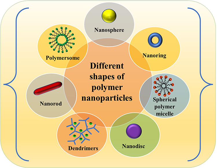 |
Figure 1 Different shapes of polymeric nanoparticles. |
Improved drug pharmacokinetics and bioavailability, less toxicity, and the potential to increase therapeutic dosage are all benefits of PNP-based drug delivery systems converted into nanomedicines.16
PNPs’ enhanced stability and simplicity of manufacture position them as a substantial advance over conventional oral and intravenous administration techniques. With reduced toxicity and adverse drug reactions, they can be utilised in drug delivery processes, including for tissue engineering and organ distribution.
Functionalization of PNPs can occur using drugs, biomolecules, targeting agents, and other nanoparticles as shown in Figure 2. Their large surface area and small size aggregation, however, make physical handling difficult in both liquid and dry powder form. The main disadvantage is the preparation process’s usage of organic solvents, which can damage physiological systems and the environment, and ruin some pharmaceutical drug molecules.2,17,18
 |
Figure 2 Possible conjugations of polymer nanoparticles with nanoparticles, drug molecules, and targeting agents. |
Polymer-based nanoparticles are a colloidal system made from natural or synthetic polymers. They retain significant advantages over other nanocarriers like micelles, liposomes, and inorganic nanosystems. The polymer has two types of nanoparticles, viz., natural and synthetic PNPs, which differ based on the preparation of nanoparticles. The natural polymer which has been produced naturally from plant origin or animal origin that dissolves in water is named natural hydrophilic polymer. For examples- starch,19 algin,20 pectin,21 xanthan gum,22 insulin, agarose etc. are natural hydrophilic polymers. The choice of polymer in polymeric nanoparticle (PNP) synthesis is a critical factor that directly influences the physicochemical properties, stability, and biological performance of the nanoparticles. Biodegradable polymers, such as poly(lactic-co-glycolic acid) (PLGA), polycaprolactone (PCL), and natural polymers like chitosan and alginate, are commonly selected due to their biocompatibility and safe degradation products. The polymer’s molecular weight, hydrophilicity or hydrophobicity, and functional groups determine drug loading efficiency, release kinetics, and surface characteristics, which in turn affect cellular uptake and biodistribution. Additionally, polymers can be tailored or functionalized to provide stimuli-responsiveness, targeting capabilities, or prolonged circulation times. Therefore, polymer selection is guided by the intended therapeutic application, the nature of the encapsulated drug, and the desired pharmacokinetic and pharmacodynamic profile, ensuring optimal efficacy and safety of the PNP formulation.
Synthetic polymers, including Polyacrylic acid (PAA), Polyethene glycol (PEG), Polyvinyl pyrrolidone (PVP), and Polylactic acid (PLA), are chemically synthesized and utilized in biotechnology, pharmacology, and chemistry. These polymers, extracted from regenerative sources, have been approved by the FDA as drug conjugated carriers for clinical us.23
- Polyacrylic acid (PAA) is a homopolymer of acrylic acid which is cross-linked with allyl ether propylene. It has the potential of absorbing and retaining water and also swells many times to its original volume so it is used in disposable diapers.24
- Polyvinyl pyrrolidone (PVP) is a binder used in tablet formation, synthesized by polymerizing vinylpyrrolidone in water or isopropanol. Its molecular weight ranges from 40,000 to 360,000, and it is widely available in different grades.25
- Polyethene glycol (PEG) is a promising material for drug delivery due to its high drug loading ability, biocompatibility, biodegradability, extended circulation half-life, and simple functionalization.26
- Polylactic acid (PLA) is a significant polymer suitable for synthesizing polymeric nanoparticles due to its biodegradability and biocompatibility properties. Due to its low molecular weight, PLA is chosen as a drug carrier due to its shorter degradation time, accelerating the formation of a required drug delivery system in multiple formulations.24
- Polycaprolactone (PCL) - PCL is biodegradable polyester. PCL is formed by ring-opening polymerization of ɛ-caprolactone by using a catalyst like stannous octanoate. Variousdrugs can be encapsulated within PCL used for targeted drug delivery. PCL is conjugated with starch to obtain a good biodegradable material.27
- Poly(lactic-co-glycolic acid) (PLGA)- PLGA, a biocompatible and biodegradable polymer, is under study as a delivery vehicle for proteins, drugs, and macromolecules like peptides, RNA, DNA, and RNA due to its tunable mechanical properties and its long-term stability.28,29 Drugs were released at specific doses without surgery, allowing active PLGA degradation. The physical properties of the polymer-drug matrix were adjusted to achieve the desired dosage and release intervals, based on factors such as polymer molecular weight, lactide-glycolide ratio, and drug concentration, depending on the type of drug.30,31
Synthetic hydrophobic polymers are those which are not soluble in water or other polar solvents such as epoxides, polystyrene, polyvinylchloride (PVC), polystyrene, polyisobutylcyanoacrylates, polymethyl (methcyanoacrylates), poly (butylcyanoacrylates), and poly (alkyl cyanoacrylates).32,33
Drug therapies have long been a cornerstone of medicine, yet more than half of the currently approved medications suffer from low solubility and weak permeability, limiting their clinical utility.34 Polymer-drug conjugates, particularly those utilizing drug-loaded polymeric nanoparticles, present a highly promising therapeutic strategy. This method involves the attachment of sensitive bioactive molecules—including proteins, peptides, hormones, enzymes, and growth factors—to a hydrophilic polymer backbone through reversible, physiologically degradable linkers. These linkages protect encapsulated and potentially fragile therapeutic agents from premature enzymatic degradation and significantly prolong their circulation half-life, thereby enhancing the potential for intestinal absorption and systemic bioavailability. It is to be noted that a sustained presence in the bloodstream increases the likelihood that the conjugate will interact with its intended molecular target, establishing a controlled delivery system that enhances therapeutic efficacy while also narrowing the therapeutic window. By meticulously adjusting essential physicochemical properties such as surface charge, hydrodynamic diameter, and biocompatibility, these polymer-drug conjugates can achieve targeted and effective delivery to the specific site of action.35,36
Given their unique pharmacokinetic behavior, these entities are regarded as novel chemical entities distinct from the parent compound, and they are incorporated into standard pharmaceutical formulations by being physically entrapped within the carrier matrix.13,37 Ongoing investigation is directed at enhancing the absorption of the drug, achieving disease-targeted delivery, improving solubility, minimizing toxicity, and amplifying the therapeutic efficacy of bioactive agents by surmounting biological barriers.38–40 The employment of nanoparticles in biomedicine is underpinned by their size-dependent physical and chemical characteristics, which modify their biological behavior in ways not observed with bulk materials.41
Polymer is a macromolecule that is composed of repeating units that are organized in a chain-like structure exhibiting multiple compositions and properties. Polymeric nanoparticles (PNPs) represent a significant advancement in the field of drug delivery and are also utilized in biosensing and bioimaging applications.42 Their growing importance in targeted therapies is attributed to their ability to accumulate in specific tissues, exhibit low toxicity, and retain drugs effectively. The method of production typically corresponds to the chemical characteristics of the therapeutic compound, allowing for the use of either natural or fully synthetic polymers to achieve targeted delivery. Techniques such as surface modification and drug encapsulation enhance the therapeutic capacity of these nanoparticles while optimizing their distribution and retention in diseased tissues.43
This review highlights the potential of PNPs for the delivery of poorly soluble, hydrophobic compounds. Although such drugs often suffer from limited bioavailability, their integration with nanoparticles can markedly improve solubilization, stability, and therapeutic indices. We focus on synthetic and loading techniques that facilitate a smooth transition of these compounds from the formulation stage to clinical impact, ensuring efficient delivery to the intended site of action while minimizing off-target effects.
Synthesis Strategies of Polymer Nanoparticles
Because of their high molecular weight and inability to pass across lipid membranes, water-soluble phytochemicals with poor absorption qualities are frequently found in drugs. Over 40% of newly developed molecules suffer from slow uptake and low water solubility, resulting in suboptimal exposure and drug delivery. To enhance the clinical usefulness of these compounds, the formulation of drug-polymer nanoparticles has become indispensable.44–47 These carriers stand out due to their elevated surface-to-volume ratio, their capacity to entrap active agents, and the modulation of quantum confinement effects. By shielding the drug from harsh conditions—such as extreme pH, enzymatic cleavage, and systemic degradation—they facilitate transport to the target site after administration. Achieving nanoparticles with the desired stability, release profile, and tissue targeting mandates careful selection of fabrication techniques. Mastery of these parameters is key to transforming pharmacologically promising molecules into effective therapeutics.48–50
Dispersion of Preformed Polymers
This synthesis method enhances various factors such as ligand density, biodistribution, pharmacokinetics, structural integrity, chemical uniformity, and regulated biodegradability, all of which offer significant benefits for pharmaceutical applications.51 In this technique, five various methods are prevalent.
Solvent Evaporation Method
To create emulsions, the polymer solution is prepared in volatile solvents. To create an oil-in-water or water-in-oil emulsion, emulsions are specifically prepared in aqueous solutions using emulsifying agents or surfactants such as poly (vinyl alcohol), gelatin, or polysorbate-80.52 Following the solvent’s evaporation from the polymer, these emulsions are transformed into nanoparticle suspensions, which are subsequently permitted to diffuse through the continuous emulsion phase.53,54 The water-oil-water method was used to produce water-soluble drug-loaded nanoparticles.55
Spontaneous Emulsification
This technique is an advanced solvent evaporation process referred to as solvent diffusion. It involves the creation of polymer nanoparticles within the oil phase by combining water with a water-miscible solvent, such as methanol or acetone, alongside inorganic solvents like dichloromethane or chloroform.56 When the saturated polymer is emulsified in aqueous solutions containing stabilizers, the solvent migrates to the external phase, leading to the formation of nanocapsules or nanospheres, which is influenced by the ratio of oil to polymer. In the end, the removal of the solvent occurs via either evaporation or filtration, which is contingent upon its boiling point.
Salting Out
In this methodology, both emulsification and solvent diffusion techniques are utilized. An aqueous gel is created by dissolving the polymer alongside the active pharmaceutical ingredient in a solvent such as acetone. This process is then augmented by the addition of salting-out agents, which may be classified as either electrolytes or non-electrolytes. The inclusion of a colloidal stabilizer promotes the diffusion of acetone. To further enhance acetone diffusion and facilitate the formation of nanospheres, the oil-water emulsion is subsequently diluted with water. The extraction of the solvent and salting-out agents is performed via cross-flow filtration, highlighting the importance of selecting an appropriate salting-out agent to ensure the drug’s effectiveness.57
Nano-Precipitation
This method, commonly known as solvent displacement, is utilized in the production of polymeric nanoparticles (PNPs).58 The underlying principle resides in the interfacial deposition of a semi-polar solvent that is compatible with water, originating from a lipophilic solution. PNP synthesis requires solvent and non-solvent phases, with polymers like polylactide being commonly used,59,60 biodegradable polyesters,61,62 poly(lactide-co-glycolide),63,64 polyalkyl-cyanoacrylate65,66 and eudragit.67 In certain cases, polymers are copolymerized to reduce the recognition of nanoparticles by a reticular endothelial system to avoid drug clearance out of the body.68 This method is a straightforward, reproducible, and efficient method for creating nanospheres and nanocapsules.50
Dialysis
By dissolving the polymer in an organic solvent, the dialysis technique distributes small, thin polymeric nanoparticles. Solvents such as dimethyl sulfoxide, dimethylformamide, and dimethylacetamide are utilised to create poly (lactide)-b-poly (ethylene oxide) and poly (γ-glutamic acid) nanoparticles. Nevertheless, this method presents considerable drawbacks, including limited capacity for drug loading and the instability of nanoparticles.69–71
Desolvation Method
A widely utilized approach for producing polymeric nanoparticles is the desolvation method. This technique involves the addition of a desolvating agent, such as ethanol or a concentrated solution of organic salts, to alter the charge and pH of the system. In practice, a solution containing the protein and the drug is gradually combined with ethanol until turbidity occurs, signaling a shift in the concentration of hydrogen ions. To ensure the stability of the resulting coacervates, glutaraldehyde is employed, with a required minimum concentration of 40%. The nanoparticles must then undergo a reaction time of 24 hours to achieve adequate stability. Subsequently, any surplus cross-linking agent and unbound drug are eliminated from the nanoparticles via centrifugation.57,72 The nanoparticle powder is obtained by freeze-drying the nanosuspension, with the inclusion of 5% mannitol serving as a cryoprotectant during this process.
Supercritical Fluid Technology
Since polymer dispersion necessitates the use of hazardous organic solvents, environmentally safe, supercritical fluid technology was developed for PNP synthesis. This process creates extremely pure PNPs without the need for organic solvents by using environmentally benign solvents.73 Rapid expansion of supercritical solution (RESS) and rapid expansion of supercritical solution into a liquid solvent (RESOLV) are the two principles that underpin the production of PNPs using supercritical fluid technology. A solution that extends into the surrounding air is created when the solute dissolves in a supercritical fluid. Rapid pressure decrease, homogenous nucleation, and high supersaturation all aid in the creation of well-dispersed nanoparticles.74
Poly (perfluropolyether diamide) droplets were created using RESS.75 The RESOLV technique, which expands the supercritical solution into a liquid solvent rather than ambient air, has been developed as a modification of the RESS method, which generates micro-scaled products, to address its primary shortcomings.56 Despite being widely used in tissue engineering and drug administration, high molecular weight polymers are difficult to use because of their poor solubility in supercritical fluids, as explained by the RESOLV technique. Among the medications developed in PNPS using supercritical technology were indomethacin,76 growth factors,77,78 griseofulvin,79 ibuprofen,80 dexamethasone,81 and amoxicillin.82,83 These medications showed encouraging outcomes in terms of drug delivery.
Ionotropic Gelatin Technique
Polymers play a primary role in designing an oral delivery system. In this context, the use of natural polymers like chitosan and alginates instead of toxic chemical polymers in the oral delivery system enhances the permeation effect, enzyme inhibitory ability, and mucoadhesive property of the drug. Briefly, based on the solubility of the drug and polymer, they are dissolved in a weakly acidic medium or water, and the resultant solution is added to the solution containing counter ions and stabilizer dropwise under constant stirring. Spherical-shaped particles are formed due to the complexation of oppositely charged species, which results in gelation and precipitation. The particle size is reduced to the nanometric range by sonicating the resultant solution. The nanosuspension produced is freeze-dried using 5% mannitol as a cryoprotectant to obtain a fine powder of Nanoparticles.84
Production of Various PNPs by Natural Polymers
Albumin nanoparticles are formed in an external-oil suspension using one of two procedures viz., chemical treatment in vegetable oil or thermal treatment at high temperatures (958°C–1708°C), stabilization, aqueous medium, or iso-octane emulsions. The nanospheres are produced by homogenizing the oil phase containing albumin droplets and thermally stabilizing them by heating at 1758°C to 1808°C for 10 minutes.85 The mixture is cooled, diluted with ethyl ether, and separated by centrifugation. The drug molecules, which are not sensitive to heat, are treated with heat, resulting in the preparation of nanoparticles by emulsifying the serum albumin aqueous solution in cottonseed oil at 258°C.86 The cross-linking agent’s formaldehyde or 2,3-butadiene was used to denature albumin, resulting in particles that were stirred, isolated, and dried by lyophilization. These particles were not as fast as those releasing the drug doxorubicin,86 hence, the purification step is the major difficulty in the removal of the cottonseed oil.87 A technique was identified based on the isolation of natural macromolecules, in which the step of purification was simplified.88 The procedure involves an aqueous solution of albumin, emulsified in chloroform with stabilizers and hydroxypropyl cellulose, and then cross-linked with glutaraldehyde, but lacks advantages over other techniques due to the need for chlorinated solvents.89
Sodium alginate, a water-soluble polymer, can form gels when mixed with multivalent cations.90 To prepare alginate nanoparticles, sodium alginate solution is added to calcium chloride solution, resulting in particles ranging from 1 to 5 nm in size.91 Gelatine is induced in a low calcium concentration, creating invisible calcium alginate gels. The internal gelation method or emulsification is used to prepare alginate particles, which can be achieved at room temperature without specific equipment.92 However, washing the nanoparticles is a major problem, and new approaches have been developed to address this issue.57
Chitosan nanoparticles have been developed to encapsulate proteins like bovine serum albumin, tetanus, diphtheria toxoid,93 insulin,94 anticancer agents,95 vaccines,96 and nucleic acids.97 They are enhanced by the absorption of peptides like insulin and calcitonin across the nasal epithelium.72 Chitosan nanoparticles are prepared by spontaneously forming complexes between chitosan and polyanions.98 The gelation of a chitosan solution is dispersed in an oil emulsion,99 resulting in small, spherical chitosan nanoparticles like tripolyphosphate. However, the method’s drawback is the difficulty in eradicating organic solvents during particle separation, which can cause toxicity.100
High Drug Loading Strategies
Post-Loading
Drug-loading nanocarriers can be created using porous-structured materials like carbon nanoparticles, MOF, hydrogel nanoparticles, and silica nanoparticles as shown in Figure 3.101,102 These materials offer high surface area, straightforward functionalization chemistry, and adjustable pore size and volume.103,104 Other non-porous materials, like proteins and polypeptides have also been studied for post-loading procedures.105,106
Doxorubicin, commonly administered in a PEGylated liposomal formulation, plays a significant role in the treatment of various cancers, including leukemia.107,108 However, the drug’s high hydrophilicity complicates its encapsulation within hydrophobic polymer matrices. Research indicates that micelles formed by the coprecipitation of Doxorubicin with PLGA-PEG can have loading capacities as low as 0.51% and loading efficiencies near 23%.109 Despite these challenges, recent advancements in drug delivery techniques have improved the loading capacity of Doxorubicin, and several nanoparticle systems have shown a rapid release profile for the drug. Additionally, the conjugation of Doxorubicin to polymers is further complicated by the drug’s complex structure, which features three hydroxyl groups, two phenolic hydroxyls, one ketone, and one amine group. Furthermore, its susceptibility to pH variations, heat, metal ions, and light adds complexity to its conjugation chemistry. One conjugation strategy is to couple the terminal carboxylate of PLA with Doxo by creating an amide linkage through the -NH2 of Doxo.110
Co-Loading
Figure 3 illustrates various drug loading strategies developed during nanoparticle formation, including various systems. Formation of nanoparticles with a co-loading strategy includes proteins,79 drug-polymer conjugate,111 solid-lipids,112 proteins,79 pure drugs,113 drug-silsesquioxane conjugate,114 and polymers.115,116 Covalent binding and electrostatic co-loading strategies have successfully achieved 18.5 to 100.0% drug loadings with nanoparticle sizes ranging from 29 to 400 nm.
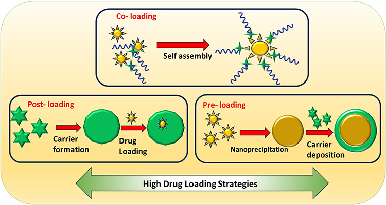 |
Figure 3 High drug loading strategies for polymeric nanoparticles. |
Drug-polymer nanoparticles self-assemble amphiphilic drug-polymer molecules by taking use of the characteristics of hydrophilic polymers and hydrophobic medicines. DOX, 7-ethyl-10-hydroxycamptothecin, and camptothecin are a few examples.111 A low molecular weight oligoethylene glycol chain and SN38 were used to create a drug-polymer conjugate, which resulted in nanoparticles with 36% drug loading.117 Using flash nanoprecipitation, carboxymethyl cellulose-based nanoparticles demonstrated a 29.5% drug loading.111 For self-assembly, carboxymethyl cellulose was coupled to the hydrophobic anticancer medication cabacitaxel. For cisplatin-loaded silica nanoparticles with a drug loading between 35–47%, a drug-silsesquioxane conjugate was created.114
Drugs can be encapsulated in polymers using nanoprecipitation, which uses non-covalent hydrophobic interactions.118 Nonetheless, the majority of systems have low drug loadings—typically less than 10%. Improving medication loading requires careful control of mixing time. The non-ionic surfactant D-αtocopheryl polyethene glycol 1000 succinate (TPGS) is frequently utilised to enhance drug loading in PNPS. Nanoparticles with drug loadings ranging from 20 to 80% have been created using a variety of medications and polymers.115,116 A quick, scalable, and continuous bottom-up method for creating monodispersed nanoparticles with adjustable particle sizes and high drug loading is flash nanoprecipitation.111,119 For flash nanoprecipitation, two mixing devices—a multi-inlet vortex mixer and a restricted impinging jet mixer—have been widely utilised. λ-cyhalothrin loaded poly(ethylene glycol)-poly(d,l-lactide) (PEG-PDLLA) nanoparticles were produced using a multi-inlet vortex mixer, achieving 49.7% drug loading and 99% encapsulation efficiency.120
Pre-Loading
One technique for creating drug nanoparticles and stabilising and protecting them is the core-shell strategy.121 Benefits of this structure include protection against degradation and regulated medication release.122,123 Drug delivery, biosensing, imaging, and diagnostics can all benefit from the use of polymers as the shell material because of their biocompatibility, biodegradability, and ease of production.29
Camptothecin (Cpt), a topoisomerase I inhibitor from Camptotheca acuminate, has shown anticancer activity in animal models.124 However, it has low aqueous solubility in its active lactone form and is rapidly converted to its carboxylate form, producing toxic and inactive molecules.125,126 While Cpt–polymer nanoparticles have been prepared to overcome the solubility limit of the drug, nanoparticles prepared with conventional coupling chemistry are plagued by various heterogeneities. To achieve a better-controlled polymerization, Beta-diiminate Zinc [(BDI-II) ZnN] Transcranial Magnetic Stimulation (TMS) was tested. However, the (BDI-II) ZnN (TMS)2/Cpt-mediated resulted in only 61% Cpt incorporation, indicating the inefficient formation of Cpt–Zn complexes during the initiation step.127
The drug-loaded nanospheres or nanocapsules can be produced by simple, safe, and reproducible techniques. The selection of PNPs depends on the drug’s physicochemical properties, yield and entrapment potential, the use of less toxic reagents, and simplification methods. These techniques are now available for drug-loaded PNPs, showing great promise for drug delivery systems. Nanoparticles have shown promise in improving drug delivery systems, as demonstrated in the results discussed in Table 1.
 |
Table 1 Different Synthesis Methods of PNPs |
Formulation optimization is a critical step in developing effective polymer nanoparticle (PNP) drug delivery systems, as it directly influences stability, drug loading, release behavior, and in vivo performance. Polymer-to-drug ratio is one of the primary parameters optimized during formulation. Lower polymer-to-drug ratios may increase drug loading but often lead to particle aggregation, burst release, or poor encapsulation. Conversely, very high polymer content improves nanoparticle stability and encapsulation efficiency but can dilute the drug payload and require higher dosing volumes. Most reported PLGA- or PEG–PLGA-based systems achieve optimal performance at polymer-to-drug ratios ranging from 5:1 to 20:1, depending on drug hydrophobicity and molecular weight. The goal is to identify a ratio that balances high encapsulation with acceptable particle size and reproducible batch-to-batch characteristics. Drug loading efficiency (DLE) and encapsulation efficiency (EE) are key quantitative metrics. EE reflects how much of the initially added drug is retained within the nanoparticles, while DLE indicates the drug content relative to the total nanoparticle mass. Hydrophobic drugs generally show higher EE in polymeric matrices due to favorable polymer–drug interactions, whereas hydrophilic drugs often require strategies such as polymer modification, double emulsion techniques, or ionic complexation to improve retention. Optimization typically involves adjusting solvent systems, polymer molecular weight, and preparation method (eg, nanoprecipitation vs emulsion evaporation) to minimize drug loss during fabrication.
Selection criteria for final polymeric nanoparticles go beyond loading metrics. Particle size (typically 80–250 nm for systemic delivery), narrow size distribution (low PDI), surface charge, and colloidal stability in physiological media are essential. Release kinetics must align with the therapeutic goal, whether sustained release, stimuli responsiveness, or rapid payload availability. Biocompatibility, biodegradability, and use of polymers with regulatory acceptance (such as PLGA, chitosan, or PEGylated systems) are crucial for translational relevance. Ultimately, the optimized formulation is selected based on a combined assessment of physicochemical stability, drug loading performance, reproducibility, and in vitro and in vivo efficacy, ensuring both scientific robustness and clinical feasibility.
To strengthen reproducibility and translational relevance, formulation optimization of PNPs should systematically include polydispersity index (PDI), stability assessments under different conditions, and complementary physicochemical and biological parameters, alongside polymer/drug ratio and drug loading metrics.
Polydispersity index (PDI) is a critical indicator of size uniformity and formulation quality. For drug delivery applications, a PDI ≤ 0.20 is generally considered acceptable, while values ≤ 0.10 indicate highly monodisperse systems. Low PDI is essential for predictable biodistribution, reproducible cellular uptake, and consistent drug release. During optimization, PDI is strongly influenced by polymer molecular weight, solvent–antisolvent ratio, stirring or sonication energy, and surfactant concentration. Formulations with high drug loading but elevated PDI are often deprioritized in favor of slightly lower loading but better size uniformity.
Stability under different conditions is equally important. Colloidal stability is typically evaluated in aqueous buffers (PBS, pH 7.4), cell culture media containing serum proteins, and across physiologically relevant pH values (pH 5.0–6.8 for endosomal or tumor microenvironments). Stable PNPs show minimal changes in particle size, PDI, and zeta potential over time (24–72 h short-term; weeks to months for storage studies). Temperature stability at 4 °C, 25 °C, and 37 °C is also assessed to simulate storage, handling, and in vivo conditions. PEGylation or surface charge optimization is often used to suppress aggregation and protein corona–induced instability.
Additional parameters enhancing reproducibility include zeta potential (typically ±20–30 mV for electrostatic stability), drug release kinetics under sink conditions, and batch to batch consistency. Encapsulation efficiency (EE), drug loading efficiency (DLE), and polymer/drug ratio must be reported together with preparation method details to allow meaningful comparison across studies. Morphological confirmation by TEM or SEM further validates size measurements obtained from DLS.
For final formulation selection, researchers prioritize nanoparticles that combine low PDI, high colloidal stability, reproducible drug loading, controlled release, and compatibility with biologically relevant environments. Reporting these parameters in a standardized manner not only improves experimental reproducibility but also facilitates regulatory evaluation and clinical translation of polymeric nanoparticle-based drug delivery systems.
Application of Polymeric Nanoparticles
In inflammation, the immune system will be activated for abnormally long periods (months, years), which produce different proinflammatory cytokines that will induce damages to healthy tissue.146 Growing evidence reveals that inflammation is the underlying cause of most chronic diseases.147,148 PNPs are used in the treatment of inflammation, targeted drug delivery, neurodegenerative disease, tissue engineering, infectious disease, and cancer as represented in Figure 4. Most of the therapeutic strategies were managing inflammation work by neutralizing the activity of cytokines, downregulating cellular pathways that produce proinflammatory cytokines, or upregulating cellular pathways for the biosynthesis of anti-inflammatory cytokines.149,150 Due to the lack of specificity, conventional therapies (non-steroidal anti-inflammatory drugs (NSAIDs), glucocorticoids) present numerous adverse effects, many novel anti-inflammatory molecules suffer from low bioavailability. Nanocarriers offer possibilities of overcoming these challenges by optimizing site-specific delivery and improving drug solubility.151,152 The design is an approach of such nanocarriers are often adapted from the microenvironment of the inflamed tissue, which is characterized by acidic pH value, increasing permeability, and high presence of reactive oxygen species (ROS).152 Here, naproxen was primarily modified with phenylboronic acid (a ROS-sensitive ligand) and then conjugated to activated dextran (a pH-sensitive polymer) to form dual-stimuli responsive NPs.153 Similarly, dual-responsive smart particles were encapsulated indomethacin as an adjunct anti-inflammatory therapy to doxorubicin-loaded NPs.154 Pioglitazone-loaded Poly(lactic-co-glycolic acid) Nanoparticle (PLGA NPs) were significantly more effective than oral pioglitazone to decrease the number of atherosclerotic plaques. Pioglitazone represents an antidiabetic drug that has been shown to stabilize atherosclerotic plaque and prevent ruptures.155 Pioglitazone-NPs regulated the inflammation response in vivo by decreasing the number of pro-inflammatory monocytes and slightly increasing the anti-inflammatory monocytes.156
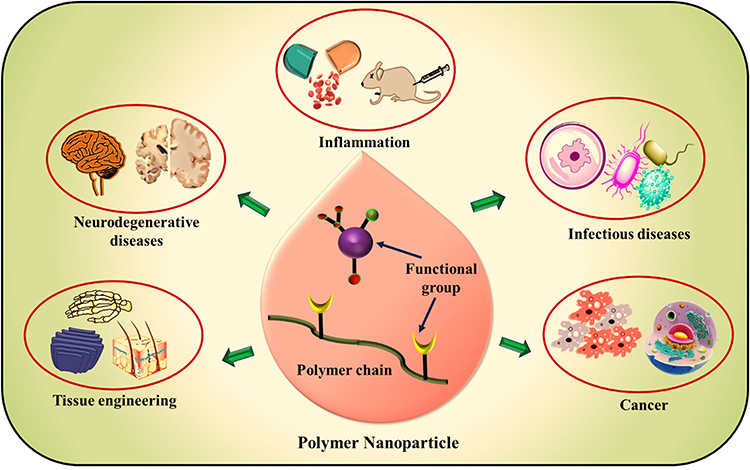 |
Figure 4 Contribution of drug–polymer nanoparticles to managing diseases. |
PNPs have gained attention in the treatment of pain. Pain is often a lifelong disease, which drastically worsens the quality of life of its sufferers.157 Numerous anti-inflammatory drugs (including Non-steroidal anti-inflammatory drugs (NSAIDs), steroids, opioids) have been formulated into biodegradable PNPs [PLGA, PLA, chitosan, Poly(N-isopropyl acrylamide (PNIPAm)] to regulate administered doses through the controlled release mechanism.158
PNPs for Infectious Diseases
Antibiotic multidrug resistance (AMR) is a significant global health threat due to uncontrolled antibiotic use.159 Some bacterial species have evolved into “superbugs” that can resist any available antibiotics.160 Mechanisms for AMR include enzyme production, efflux protein overexpression, gene mutations, and microbial biofilm formation.161 Nanobiotechnology offers potential for designing formulations with physicochemical properties that facilitate drug penetration, protect against enzymes, and prolong antibiotic circulation time.162 Encapsulation of vancomycin into gelatin nanocarriers improved the survival rate of S. aureus-infected zebra fish. These mechanisms are being explored as potential targeting strategies for developing new antimicrobial therapies.163
Moreover, several bacteria and fungi are known to form biofilms around their cell structures to protect themselves from the immune cells of the host. The microbial biofilm consists of polysaccharides, DNA, and proteins, which form the so-called extracellular polymeric substances (EPS) matrix.164 Biofilm-targeting described it was exploited in nanobiotechnology to develop novel antimicrobials with better therapeutic efficacies.164 Nanotechnological principles are applied to circumvent obstacles in the treatment of viral infections. Ritonavir and Efavirenz were incorporated into pH-sensitive polysaccharide-based nanoparticles (NPs) to enhance the solubility of these drugs.103 In this study, various derivatives of cellulose acetate, modified with differing quantities of substituents such as carboxylic, acetate, and/or butyrate groups, were evaluated for particle formation. The findings indicated that an increase in the polymer’s hydrophobic characteristics leads to heightened crystallinity, which subsequently reduces the solubility of the antiviral-loaded NPs.165 Another research team developed acyclovir-loaded PLGA NPs integrated into biocompatible buccal films made from polymer-based nanoparticles aimed at biomedical applications, utilizing cellulose derivatives, PEG 200, or Carbopol 974P to enhance the bioavailability of acyclovir. In vitro, ex vivo, and in vivo studies consistently demonstrated a controlled release of the drug from the buccal films, resulting in a threefold increase in plasma concentration in vivo when compared to free acyclovir.166 Similar strategies are being explored in antiviral treatments for HIV/AIDS.To have prepared stealth NPs loaded with, lamivudine, raltegravir, nevirapine, and zidovudine using Poly(ethylene glycol)/poly(methyl-methacrylate) (PEG-PMMA) and poly(ethylene-glycol)-polycaprolactone (PEG-PCL) polymers, it was shown that the antiviral NPs effectively inhibited HIV-1 infections in vitro.167
PNPs for Neurodegenerative Disease
Neurological disorders, including neuroinflammatory, neurodegenerative, and neoplastic diseases, affect the central nervous system and brain. As the population ages, the incidence of these disorders increases, making them the most expensive medical conditions globally. Neurodegenerative diseases, such as Alzheimer’s168 and Parkinson’s,169 result from progressive neuronal cell loss and have significant social and economic impacts. Current treatments only improve symptoms but do not cure these diseases due to impaired functioning of proteins and enzymes. Drugs encountering the blood-brain barrier (BBB) have limited beneficial substances, making further efforts to develop systems to enable drug passage through the BBB.170
In order to increase bioavailability, decrease adverse effects, and lower medicine dosage, nanoparticles such Poly(glycolic acid) (PGA), PLA, and PLGA nanoparticles are being created to deliver therapeutic agents at precise places. These nanoparticles can deliver medications to treat neurological diseases by passing across the blood-brain barrier (BBB).171 Drug absorption that is size-dependent is made possible by the interactions that functionalised NPs can have with different BBB components. Because PLA-NPs have trans-activating transcriptor peptide on their surface, they improve the transport of neurotrophic peptides across the blood-brain barrier (BBB), which in turn improves BBB permeability and penetration of the central nervous system (CNS).172 In the brain, loperamide and rhodamine-123 are efficiently transported by PEGylated PLA-NPs coupled with a glyco-heptapeptide. Functionalised PLA-NPs with two targeting peptides—one specific to the blood-brain barrier and the other having a high affinity for the amyloid beta peptide 1–42—are used to treat Alzheimer’s disease (AD).173,174 Their potential as a targeted treatment is supported by their efficacy in both in vitro and in vivo studies.175 Because of its focused distribution, biomedical applications, minimal cytotoxicity, controlled release, and biocompatibility, PLGA-NPs are frequently used.176 Memantine (MEM) was employed in PEG-PLGA-NPs to treat AD; it reduced beta-amyloid plaques both in vitro and in vivo and had a slower release profile than free drug solutions.177
A new drug delivery system for treating Parkinson’s disease (PD) has been developed using PLGA-NPs loaded with ropinirole.178 This system can revert PD-like symptoms in an animal model assay. Functionalizing PLGA-NPs can also restore their function through lysosomes.179 These new drugs are being developed to provide early diagnosis and regress Alzheimer’s disease (AD). Curcumin-PLGA-NPs can pass through the BBB, interrupting A-beta aggregates.180,181 Quercetin, functionalized with PLGA-NPs or rosmarinic acid, could be a potential candidate for AD treatment.182,183
Lysosomal storage disorders (LSDs) are a group of around 50 pathologies caused by inherited gene mutations in genes that normally codify lysosomal enzymes. Most LSD outbreaks occur during childhood and 75% of them have serious neurological consequences, leading to progressive neurodegeneration, physical deterioration, potential death, and functional impairment.184 This highlights the need for more research into developing efficient ERT systems. Nanoparticles (NPs) can efficiently deliver drug molecules, diagnostic agents, enzymes, proteins, and nucleic acids to the CNS, making them more stable and safer than other nanocarrier systems like quantum dots.185
PNPs for Cancer Treatment
One of the major drawbacks of conventional radio- and chemotherapy is the difficulty of discriminating cancer cells from healthy cells. There are other characteristics of cancer cells that can be used as targeting strategies, such as the acidic extracellular environment of cancer cells, their higher temperature than normal cells, the enzymes associated with cancer, the surface molecules expressed on cancer cells, hypoxic conditions, and reductive oxygen species (ROS). The effect of enhanced permeability and retention (EPR) alone does not appear to be as effective in humans.186 PNPs are highly modified systems that can be manipulated into a certain size and surface architecture to suit the cancer microenvironment.187 Polymer-based nanoparticles for biomedical applications uptake is to design delivery systems with cancer-recognizing molecules for active targeting. Several polymeric NP delivery systems of anticancer drugs (doxorubicin, paclitaxel, camptothecin) have been investigated for targeting the various type of cancers.188 Furthermore, the well-known anticancer drug curcumin has been formulated into cancer-targeting delivery systems using different biocompatible polymers (PLGA, lecithin, fibroin). Encapsulating curcumin loaded into PLGA NPs and functionalizing their surface with a target ligand to block the P-glycoprotein efflux mechanism increased the cellular uptake and effectiveness of curcumin when compared to the nontarget curcumin-NPs.189 Functionalized curcumin-loaded PEG-lecithin-PLGA NPs with an aptamer, which has a high affinity to adhere to epithelial cells. Compared to free curcumin, the curcumin nanoformulation with the targeting ligand showed a sixfold increase in bioavailability because of better mucoadhesion to cancer cells in the colon.190 NPs for the treatment of breast cancer are found by combining three targeting approaches. NPs targeting liver cancer were prepared by reversibly crosslinked polymer and folic acid NPs loaded with paclitaxel, which would de-crosslink under the reductive condition of the intracellular medium of cancer. PNPs provide a broad platform for the design of drug-delivery vehicles based on the pathophysiology of cancer cells.188,191
PNPs for Tissue Engineering
Chitosan, a natural biodegradable polymer, has been approved by the FDA for use in various pharmaceutical formulations.192 The advancement of bone tissue engineering significantly relies on innovative scaffold designs, including a newly developed scaffold utilizing nanospheres aimed at facilitating the regeneration of bone tissue lost due to injury or disease. This composite scaffold not only promotes the formation of new bone tissue but also enhances the process of bone conduction. Additionally, bioactive glass-ceramic nanoparticles (nBGC) play a crucial role in this field, as they more closely mimic the natural composition of bone compared to metal nanoparticles. A double-layered scaffold composed of bioactive glass combined with polyvinyl alcohol and silk fibroin has shown promise in enhancing the proliferation and differentiation of bone marrow cells.193
Moreover, both bone marrow and umbilical cord tissue have been identified as effective stimulants for bone formation and angiogenesis. Various materials, including alginate, cross-linked dextran, polycaprolactone (PCL), chitosan (PCL-CHI), poly(lactic-co-glycolic acid) (PLGA), collagen, and fibrin, have been integrated with bioactive glass to develop scaffolds for bone-tissue engineering. In vivo studies show that nanocellulose composite systems can inhibit Multiple Drug Resistance (MDR) bacteria-derived wound infections and improve skin regeneration. These nanocomposite materials demonstrate significant capabilities in promoting skin tissue repair. Polymer micelles, generated through self-assembly and emulsion evaporation techniques, are advantageous for the efficient delivery of therapeutics in disease management. The critical micelle concentration (CMC) is a vital factor in drug administration, as lower CMC values correlate with improved pharmacokinetic stability.192,194 A specialized group was established utilizing adjustable drug delivery micelles, which improved tumor endocytosis and penetration, surmounted biological barriers, boosted immune responses, and effectively inhibited tumor growth, metastasis, and recurrence in both in vivo and in vitro settings.195 Additionally, a micelle-based drug delivery system with dual-targeting capabilities—aiming at both mitochondria and cellular structures—has been developed for the treatment of pancreatic ductal adenocarcinoma. The Dendrimer–camptothecin (CPT) conjugate, which combines camptothecin with polyamidoamine dendrimers, has demonstrated enhanced drug penetration characteristics and increased antitumor efficacy. This system has been shown to improve drug distribution at both cellular and subcellular levels, thereby augmenting its therapeutic effectiveness against PDA.196
Elsewhere, modified cisplatin and dendrimers were chemically linked to create stimulating responsive clustered nanoparticles (NPs) for antitumor use, primarily concentrating in tumor sites and releasing drugs to inhibit tumor growth.192
PNPs are crucial to contemporary drug and gene delivery systems because they increase drug accumulation in particular organs and tissues, which reduces adverse effects, improves bioavailability, and minimizes drug breakdown. With a variety of preparation techniques and biomedical uses, they are synthesised utilising both natural and synthetic polymers as shown in Figure 5. While functionalised PNPs provide precise drug release by targeting particular cellular receptors, especially in neurological diseases and cancer disorders, irregular PNPs have characteristics including slow degradation and reactivity to stimuli. Functionalised PNPs have enormous potential, especially when it comes to treating various ailments as shown in Table 2.
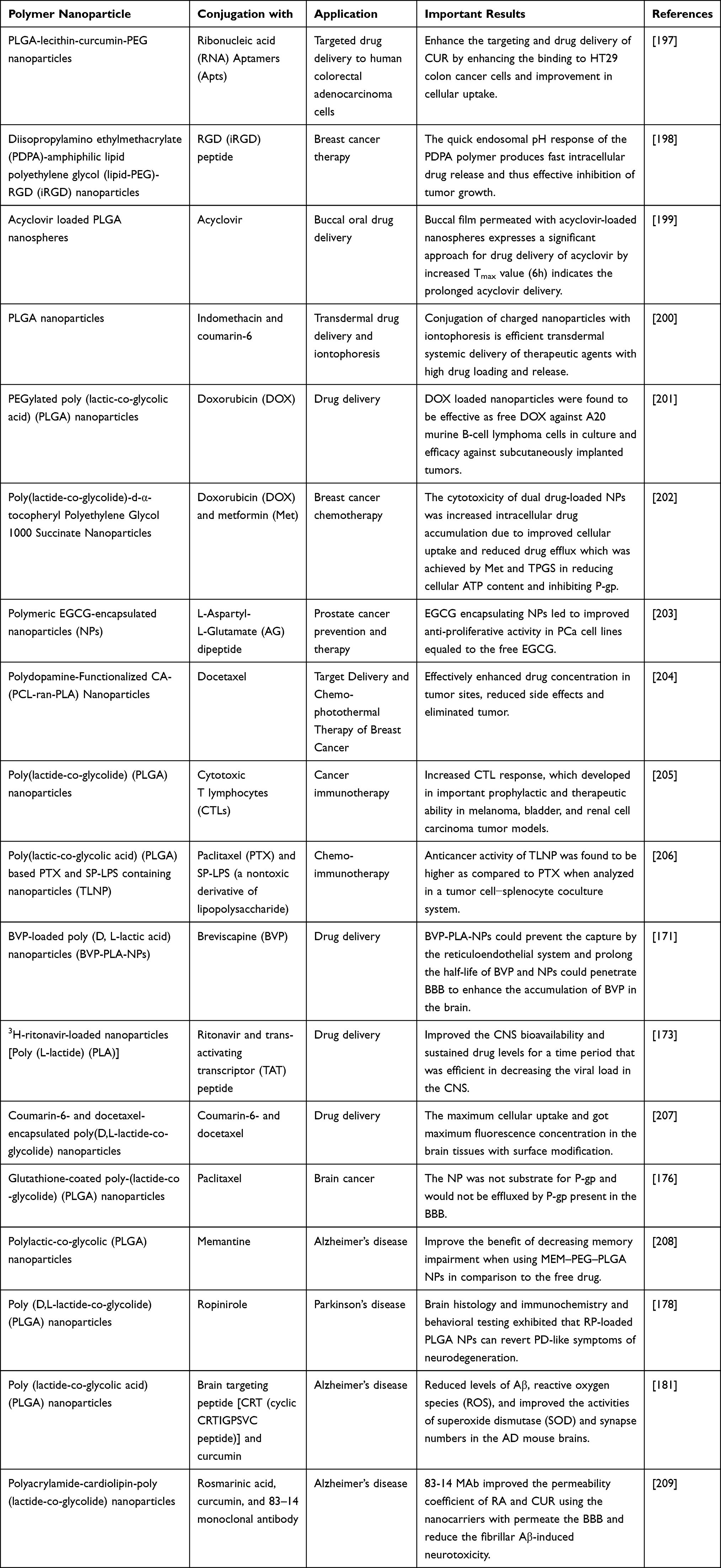 |
Table 2 Conjugation of PNPs for Biomedical Applications |
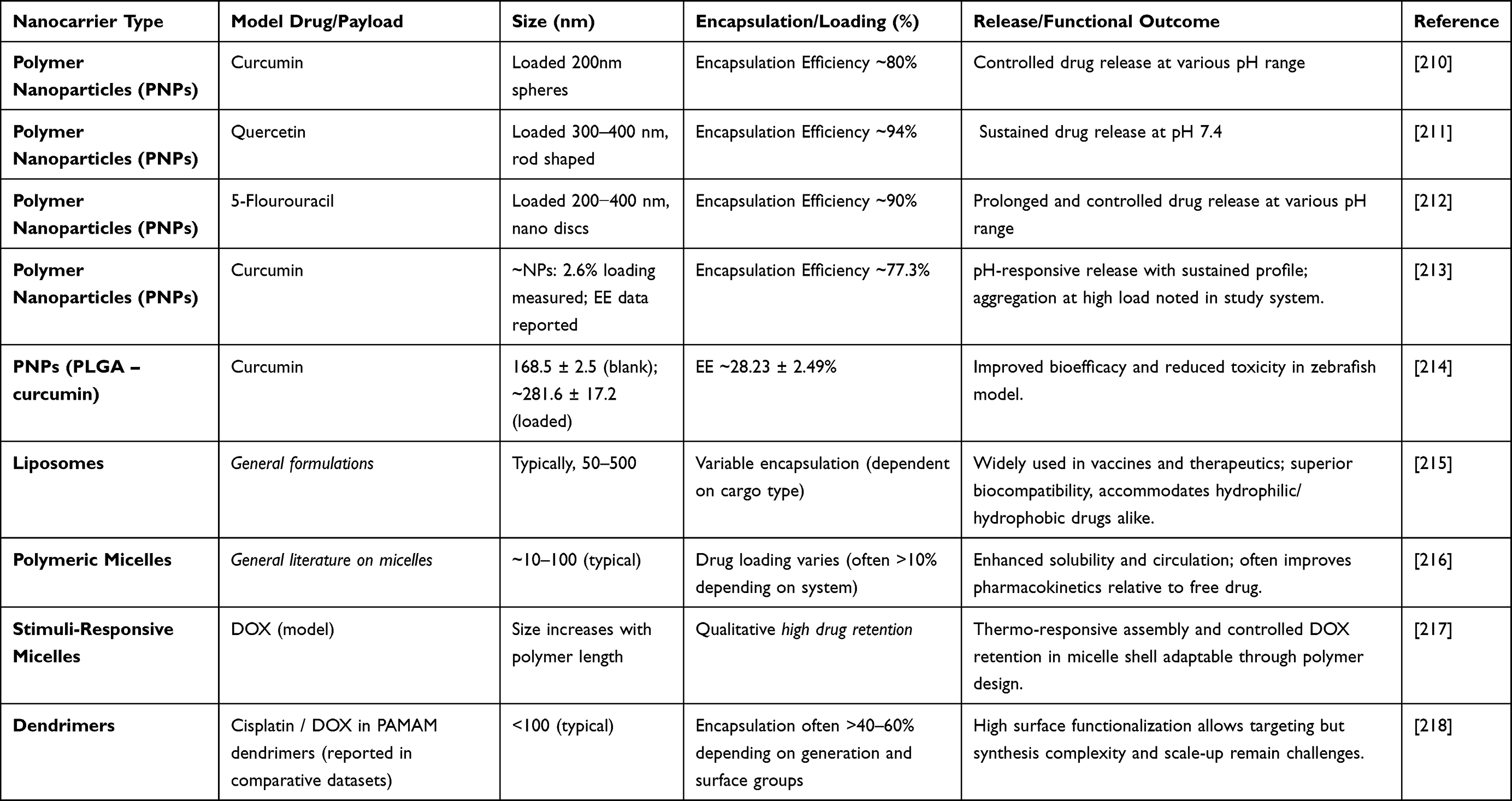 |
Table 3 Comparison of PNPS with Other Nanocarrier Systems Like Liposomes, Micelles, Dendrimers |
 |
Figure 5 Drug–polymer nanoparticle systems employed in diverse biomedical applications. |
Comparison Between PNPs and Other Nanocarrier Systems
PNP based drug delivery systems differ from other established nanocarriers such as liposomes, micelles, and dendrimers in ways that highlight both their novelty and their translational relevance as summarized in Table 3. PNPs offer high structural stability due to their solid polymeric matrix, which allows sustained and controlled drug release over extended periods. In contrast, liposomes, while clinically successful, are relatively fragile systems. They are prone to leakage, fusion, and oxidation, especially during storage and circulation. PNPs therefore provide better control over drug retention and release kinetics, which is critical for chronic therapies and precision dosing. Compared to polymeric micelles, PNPs exhibit superior stability in vivo. Micelles rely on non-covalent selfassembly and can dissociate upon dilution below their critical micelle concentration, leading to premature drug release. PNPs, formed from preassembled polymers or polymerization processes, remain intact under physiological conditions, enabling more predictable pharmacokinetics and reduced off-target toxicity.
Dendrimers offer precise molecular architecture and multivalent surface functionality, but their synthesis is complex, costly, and difficult to scale. Additionally, cationic dendrimers often raise concerns related to cytotoxicity and hemolysis. PNPs, by comparison, can be produced using scalable and industry-compatible methods such as nanoprecipitation or emulsion techniques, using FDA-approved biodegradable polymers like PLGA, chitosan, or PEGylated systems. This makes PNPs more attractive for clinical translation. From a translational perspective, PNPs stand out due to their formulation flexibility. They can encapsulate hydrophobic and hydrophilic drugs, support co-delivery of multiple therapeutics, and allow surface modification for active targeting or stealth behavior. Their compatibility with regulatory expectations, established manufacturing pipelines, and long-term safety data further strengthens their clinical potential. Overall, while liposomes, micelles, and dendrimers each address specific therapeutic needs, PNPs uniquely balance stability, versatility, scalability, and safety. This combination positions polymer nanoparticles as a robust and clinically relevant nanocarrier platform, bridging the gap between advanced nanomedicine design and real-world therapeutic application.
In vivo Investigations on Drug Polymeric Nanoparticles
Biological evaluation should include comparative studies to clearly support cytotoxicity and safety claims. Blank PNPs must be tested alongside drug-loaded PNPs and free drug to confirm that observed effects arise from the therapeutic agent and not the polymer carrier. Blank PNPs generally serve as biocompatibility controls and should show minimal cytotoxicity across relevant concentration ranges. Drug-loaded PNPs should be compared with free drug at equivalent doses to demonstrate improved efficacy, controlled release, or enhanced cellular uptake. Inclusion of uptake, apoptosis, and time-dependent assays further strengthens interpretation. Such comparative biological assessments are essential for reproducibility and translational relevance. The Table 4 summarizes recent in-vivo studies on polymeric nanoparticles, highlighting animal models, dosing routes, and therapeutic outcomes. Across cancer and metabolic disease models, polymeric nanoparticles consistently improve drug efficacy, bioavailability, and safety compared with free drugs, supporting their strong translational potential. While a few polymeric nanoparticle formulations have reached late stage clinical evaluation, broad clinical translation remains limited compared with lipid nanoparticles. Regulatory challenges include ensuring large scale reproducible manufacturing, comprehensive safety profiling, and clear demonstration of benefit over existing therapies. However, advances in both patented designs and ongoing trials indicate a growing trajectory for PNPs in precision medicine and diversified therapeutic areas.[ref}
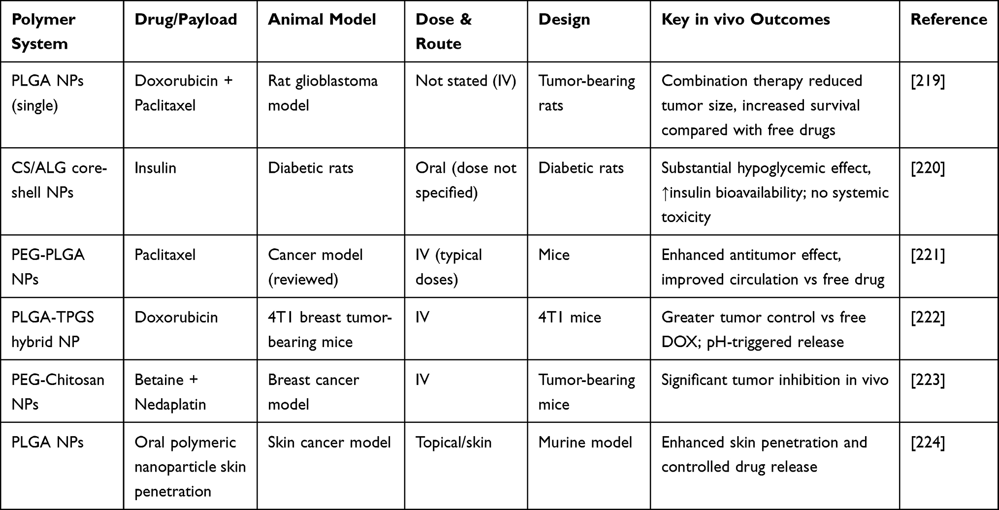 |
Table 4 In-vivo Studies on Polymeric Nanoparticles |
Conclusion
Polymeric nanoparticles (PNPs) have emerged as versatile and effective platforms for targeted drug delivery, offering clear advantages in improving drug stability, bioavailability, and site-specific therapeutic action. Their performance is strongly governed by particle size, surface functionalization, polymer chemistry, and synthesis route, all of which determine biological interactions and therapeutic outcomes. This review highlights the diversity of polymer systems and nanoconjugates currently explored for biomedical applications, emphasizing how rational design enables precise control over drug loading and release at disease-specific sites.
From a clinical translation perspective, the use of biodegradable and regulatory-accepted polymers such as PLGA, PEG, and chitosan has accelerated progress toward human applications. However, large scale manufacturing, batch reproducibility, long term stability, and standardized characterization remain key regulatory challenges. Comprehensive toxicity evaluation, including chronic exposure and immunogenicity studies, is essential to meet safety requirements set by regulatory agencies.
Beyond oncology, PNPs show strong potential in treating neurodegenerative disorders, infectious diseases, inflammatory conditions, metabolic disorders, and gene-based therapies. Their ability to integrate electrical, optical, and magnetic functionalities further expands their applicability in diagnostics, imaging, and theranostics. Future research should focus on clinically relevant disease models, harmonized regulatory frameworks, and environmentally safe manufacturing processes. Despite existing challenges, continued interdisciplinary efforts will position PNP based nanomedicines as integral components of next generation, precision driven healthcare.
Acknowledgment
VB acknowledges CMR Institute of Technology for providing research facilities and infrastructure.
Author Contributions
All authors made a significant contribution to the work reported, whether that is in the conception, acquisition of data, analysis and interpretation, or in all these areas; took part in drafting, revising or critically reviewing the article; gave final approval of the version to be published; have agreed on the journal to which the article has been submitted; and agree to be accountable for all aspects of the work.
Funding
We are thankful to Jain University, Bangalore, India, and the NANOMISSION project (SR/NM/NS-20/2014) for providing facilities. VB acknowledges TARE-SERB. TAR/2018/000547 and MN acknowledges NIH 1R16GM145575-01.
Disclosure
The authors report no financial or personal conflicts that could have impacted the research presented here.
References
1. Bennet D, Kim S. Polymer nanoparticles for smart drug delivery. Appl Nanotechnol Drug Delivery. 2014;8:1.
2. Jawahar N, Meyyanathan S. Polymeric nanoparticles for drug delivery and targeting: a comprehensive review. Int J Health Allied Sci. 2012;1(4):217. doi:10.4103/2278-344X.107832
3. Adhikari C. Polymer nanoparticles-preparations, applications and future insights: a concise review. Polym Plast Technol Eng. 2021;60(18):1996–26.
4. Schmid G. Nanoparticles: From Theory to Application. John Wiley & Sons; 2011.
5. Zhang Q, Chuang KT. Adsorption of organic pollutants from effluents of a Kraft pulp mill on activated carbon and polymer resin. Adv Environ Res. 2001;5(3):251–258. doi:10.1016/S1093-0191(00)00059-9
6. Schadler LS, Kumar SK, Benicewicz BC, et al. Designed interfaces in polymer nanocomposites: a fundamental viewpoint. MRS Bulletin. 2007;32(4):335–340. doi:10.1557/mrs2007.232
7. Hejtmancik JF, Nickerson JM. Progress in Molecular Biology and Translational Science Molecular Biology of Eye Disease Preface. San Diego: Elsevier Academic Press Inc; 2015:XIX–XX.
8. Peer D, Karp JM, Hong S, et al. Nanocarriers as an Emerging Platform for Cancer Therapy. In: Nano-Enabled Medical Applications. Jenny Stanford Publishing; 2020:61–91.
9. Liu Y, Yang G, Jin S, et al. Development of High‐Drug‐Loading Nanoparticles. ChemPlusChem. 2020;85(9):2143–2157. doi:10.1002/cplu.202000496
10. Hamidi M, Azadi A, Rafiei P. Hydrogel nanoparticles in drug delivery. Adv Drug Delivery Rev. 2008;60(15):1638–1649. doi:10.1016/j.addr.2008.08.002
11. Steinmetz NF. Viral nanoparticles as platforms for next-generation therapeutics and imaging devices. Nanomedicine: nanotechnology. Biol Med. 2010;6(5):634–641.
12. Kalyane D, Raval N, Maheshwari R, et al. Employment of enhanced permeability and retention effect (EPR): nanoparticle-based precision tools for targeting of therapeutic and diagnostic agent in cancer. Mater Sci Eng C. 2019;98:1252–1276. doi:10.1016/j.msec.2019.01.066
13. Canal F, Sanchis J, Vicent MJ. Polymer–drug conjugates as nano-sized medicines. Curr Opin Biotechnol. 2011;22(6):894–900. doi:10.1016/j.copbio.2011.06.003
14. Wang H, Li B, Sun Y, et al. NIR-II AIE Luminogen-Based Erythrocyte-Like Nanoparticles with Granuloma-Targeting and Self-Oxygenation Characteristics for Combined Phototherapy of Tuberculosis. Adv Mater. 2024;36(38):2406143. doi:10.1002/adma.202406143
15. Deng Y, Li B, Zheng H, et al. Multifunctional Prussian blue nanoparticles loading with Xuetongsu for efficient rheumatoid arthritis therapy through targeting inflammatory macrophages and osteoclasts. Asian J Pharm Sci. 2025;20(3):101037. doi:10.1016/j.ajps.2025.101037
16. Van Ngo H, Nguyen PK, Van Vo T, et al. Hydrophilic-hydrophobic polymer blend for modulation of crystalline changes and molecular interactions in solid dispersion. Int J Pharm. 2016;513(1–2):148–152. doi:10.1016/j.ijpharm.2016.09.017
17. Mahendra C, Murali M, Manasa G, et al. Antibacterial and antimitotic potential of bio-fabricated zinc oxide nanoparticles of Cochlospermum religiosum (L.). Microb Pathogenesis. 2017;110:620–629. doi:10.1016/j.micpath.2017.07.051
18. Kayser O, Lemke A, Hernandez-Trejo N. The impact of nanobiotechnology on the development of new drug delivery systems. Current Pharm Biotechnol. 2005;6(1):3–5. doi:10.2174/1389201053167158
19. Pareta R, Edirisinghe M. A novel method for the preparation of starch films and coatings. Carbohydr Polym. 2006;63(3):425–431. doi:10.1016/j.carbpol.2005.09.018
20. Rowe RC, Sheskey P, Quinn M. Handbook of Pharmaceutical Excipients. Libros Digitales-Pharmaceutical Press; 2009.
21. Braconnot H. Recherches sur un nouvel acide universellement répandu dans tous les végétaux. In: Annales de Chimie Et de Physique. 1825:173–178
22. Barrere G, Barber C, Daniels M. Molecular cloning of genes involved in the production of the extracellular polysaccharide xanthan by Xanthomonas campestris pv. campestris. Int J Biol Macromol. 1986;8(6):372–374. doi:10.1016/0141-8130(86)90058-9
23. Massadeh S, Alaamery M. Polymer nanoparticles for targeted gene delivery. Nanotechnol. Drug Deliv. 2016;1:1–20.
24. Erothu H, Kumar AC. Hydrophilic polymers. Biomed Applicat Polym Mater Composit. 2016;2016:416.
25. Chowhan Z. Role of binders in moisture‐induced hardness increase in compressed tablets and its effect on in vitro disintegration and dissolution. J Pharmaceut Sci. 1980;69(1):1–4. doi:10.1002/jps.2600690102
26. Ghasemi R, Abdollahi M, Emamgholi Zadeh E, et al. mPEG-PLA and PLA-PEG-PLA nanoparticles as new carriers for delivery of recombinant human Growth Hormone (rhGH). Sci Rep. 2018;8(1):1–13. doi:10.1038/s41598-018-28092-8
27. McKeen LW. Film Properties of Plastics and Elastomers. William Andrew; 2017.
28. Bouissou C, Rouse JJ, Price R, et al. The influence of surfactant on PLGA microsphere glass transition and water sorption: remodeling the surface morphology to attenuate the burst release. PharmA Res. 2006;23(6):1295–1305. doi:10.1007/s11095-006-0180-2
29. Jain RA. The manufacturing techniques of various drug loaded biodegradable poly (lactide-co-glycolide)(PLGA) devices. Biomaterials. 2000;21(23):2475–2490. doi:10.1016/S0142-9612(00)00115-0
30. Allison SD. Effect of structural relaxation on the preparation and drug release behavior of poly (lactic-co-glycolic) acid microparticle drug delivery systems. J Pharmaceut Sci. 2008;97(6):2022–2035. doi:10.1002/jps.21124
31. Mundargi RC, Babu VR, Rangaswamy V, et al. Nano/micro technologies for delivering macromolecular therapeutics using poly (D, L-lactide-co-glycolide) and its derivatives. J Control Release. 2008;125(3):193–209. doi:10.1016/j.jconrel.2007.09.013
32. Fatima S, Quadri SN, Parveen S, et al. Polymeric nanoparticles for potential drug delivery applications in cancer. Nanoformulation Strategies Cancer Treatment. 2021;65–88.
33. Bochicchio D, Panizon E, Monticelli L, et al. Interaction of hydrophobic polymers with model lipid bilayers. Sci Rep. 2017;7(1):1–9. doi:10.1038/s41598-017-06668-0
34. Connors R, Elder E. Delivery of Poorly Soluble or Poorly Permeable Drugs. Fall Church, VA: Technology Catalyst International Incorporation; 2003.
35. Duncan R. Polymer conjugates as anticancer nanomedicines. Nat Rev Cancer. 2006;6(9):688–701. doi:10.1038/nrc1958
36. Owen S, Dalam Rowe RC, Sheskey PJ, Owen SC. Handbook of Pharmaceutical Excipient. London: Pharmaceutical Press; 2005:471–473.
37. Li C, Wallace S. Polymer-drug conjugates: recent development in clinical oncology. Adv Drug Delivery Rev. 2008;60(8):886–898. doi:10.1016/j.addr.2007.11.009
38. Duncan R. The dawning era of polymer therapeutics. Nat Rev Drug Discov. 2003;2(5):347–360. doi:10.1038/nrd1088
39. Abioye O, Tangyie Chi G, T. Kola-Mustapha A, et al. Polymer-drug nanoconjugate–an innovative nanomedicine: challenges and recent advancements in rational formulation design for effective delivery of poorly soluble drugs. Pharmal Nanotechnol. 2016;4(1):38–79. doi:10.2174/2211738504666160213001714
40. Haag R, Kratz F. Polymer therapeutics: concepts and applications. Angew Chem Int Ed. 2006;45(8):1198–1215. doi:10.1002/anie.200502113
41. Yildirimer L, Thanh NTK, Loizidou M, et al. Toxicology and clinical potential of nanoparticles. Nano Today. 2011;6(6):585–607. doi:10.1016/j.nantod.2011.10.001
42. Jain KK, Jain KK. The Handbook of Nanomedicine. Vol. 404. Springer; 2008.
43. Moreno-Vega A-I, Gomez-Quintero T, Nunez-Anita RE, et al. Polymeric and ceramic nanoparticles in biomedical applications. J Nanotechnol. 2012;2012:1.
44. Shakeri A, Sahebkar A. Nanotechnology: a successful approach to improve oral bioavailability of phytochemicals. Recent Pat Drug Deliv Formul. 2016;10(1):4–6. doi:10.2174/1872211309666150611120724
45. Savjani KT, Gajjar AK, Savjani JK. Drug Solubility: Importance and Enhancement Techniques. Vol. 2012. International Scholarly Research Notices; 2012.
46. Saraf S, Saraf S. Applications of novel drug delivery system for herbal formulations. Fitoterapia. 2010;81(7):680–689. doi:10.1016/j.fitote.2010.05.001
47. Kumari A, Yadav SK, Yadav SC. Biodegradable polymeric nanoparticles based drug delivery systems. Colloids Surf B. 2010;75(1):1–18. doi:10.1016/j.colsurfb.2009.09.001
48. De Jong WH, Borm PJ. Drug delivery and nanoparticles: applications and hazards. Int J Nanomed. 2008;3(2):133. doi:10.2147/IJN.S596
49. Devarajan PV, Jain S. Targeted Drug Delivery: Concepts and Design. Springer; 2015.
50. Rao JP, Geckeler KE. Polymer nanoparticles: preparation techniques and size-control parameters. Prog Polym Sci. 2011;36(7):887–913. doi:10.1016/j.progpolymsci.2011.01.001
51. Lee CC, MacKay JA, Fréchet JMJ, et al. Designing dendrimers for biological applications. Nat Biotechnol. 2005;23(12):1517–1526. doi:10.1038/nbt1171
52. Scholes P, Coombes AGA, Illum L, et al. The preparation of sub-200 nm poly (lactide-co-glycolide) microspheres for site-specific drug delivery. J Control Release. 1993;25(1–2):145–153. doi:10.1016/0168-3659(93)90103-C
53. Allémann E, Leroux J-C, Gurny R, et al. In vitro extended-release properties of drug-loaded poly (DL-lactic acid) nanoparticles produced by a salting-out procedure. PharmA Res. 1993;10(12):1732–1737. doi:10.1023/A:1018970030327
54. Anton N, Benoit J-P, Saulnier P. Design and production of nanoparticles formulated from nano-emulsion templates—a review. J Control Release. 2008;128(3):185–199. doi:10.1016/j.jconrel.2008.02.007
55. Zambaux MF, Bonneaux F, Gref R, et al. Influence of experimental parameters on the characteristics of poly(lactic acid) nanoparticles prepared by a double emulsion method. J Control Release. 1998;50(1–3):31–40. doi:10.1016/S0168-3659(97)00106-5
56. Jeevanandam J, Chan YS, Danquah MK. Nano-formulations of drugs: recent developments, impact and challenges. Biochimie. 2016;128:99–112. doi:10.1016/j.biochi.2016.07.008
57. Reis CP, Neufeld RJ, Ribeiro AJ, et al. Nanoencapsulation I. Methods for preparation of drug-loaded polymeric nanoparticles. Nanomedicine. 2006;2(1):8–21.
58. Fessi H, Puisieux F, Devissaguet JP, et al. Nanocapsule formation by interfacial polymer deposition following solvent displacement. Int J Pharm. 1989;55(1):R1–R4. doi:10.1016/0378-5173(89)90281-0
59. Seyler I, Appel M, Devissaguet J-P, et al. Macrophage activation by a lipophilic derivative of muramyldipeptide within nanocapsules: investigation of the mechanism of drug delivery. J Nanopart Res. 1999;1(1):91–97. doi:10.1023/A:1010016128378
60. Legrand P, Lesieur S, Bochot A, et al. Influence of polymer behaviour in organic solution on the production of polylactide nanoparticles by nanoprecipitation. Int J Pharm. 2007;344(1–2):33–43. doi:10.1016/j.ijpharm.2007.05.054
61. Blouza IL, Charcosset C, Sfar S, et al. Preparation and characterization of spironolactone-loaded nanocapsules for paediatric use. Int J Pharm. 2006;325(1–2):124–131. doi:10.1016/j.ijpharm.2006.06.022
62. Zili Z, Sfar S, Fessi H. Preparation and characterization of poly-ɛ-caprolactone nanoparticles containing griseofulvin. Int J Pharm. 2005;294(1–2):261–267. doi:10.1016/j.ijpharm.2005.01.020
63. Nehilla BJ, Bergkvist M, Popat K, et al. Purified and surfactant-free coenzyme Q10-loaded biodegradable nanoparticles. Int J Pharm. 2008;348(1–2):107–114. doi:10.1016/j.ijpharm.2007.07.001
64. Yallapu MM, Gupta BK, Jaggi M, et al. Fabrication of curcumin encapsulated PLGA nanoparticles for improved therapeutic effects in metastatic cancer cells. J Colloid Interface Sci. 2010;351(1):19–29. doi:10.1016/j.jcis.2010.05.022
65. Yordanov GG, Dushkin CD. Preparation of poly(butylcyanoacrylate) drug carriers by nanoprecipitation using a pre-synthesized polymer and different colloidal stabilizers. Colloid Polym Sci. 2010;288(9):1019–1026. doi:10.1007/s00396-010-2226-6
66. Deepak V, Gurunathan S, Gurunathan S, Gurunathan S. Purification, immobilization, and characterization of nattokinase on PHB nanoparticles. Bioresour Technol. 2009;100(24):6644–6646. doi:10.1016/j.biortech.2009.06.057
67. Chang J, Jallouli Y, Kroubi M, et al. Characterization of endocytosis of transferrin-coated PLGA nanoparticles by the blood–brain barrier. Int J Pharm. 2009;379(2):285–292. doi:10.1016/j.ijpharm.2009.04.035
68. de Assis DN, Mosqueira VCF, Vilela JMC, et al. Release profiles and morphological characterization by atomic force microscopy and photon correlation spectroscopy of 99mTechnetium-fluconazole nanocapsules. Int J Pharm. 2008;349(1–2):152–160. doi:10.1016/j.ijpharm.2007.08.002
69. Lee J, Cho EC, Cho K. Incorporation and release behavior of hydrophobic drug in functionalized poly (D, L-lactide)-block–poly (ethylene oxide) micelles. J Control Release. 2004;94(2–3):323–335. doi:10.1016/j.jconrel.2003.10.012
70. Akagi T, Kaneko T, Kida T, et al. Preparation and characterization of biodegradable nanoparticles based on poly (γ-glutamic acid) with L-phenylalanine as a protein carrier. J Control Release. 2005;108(2–3):226–236. doi:10.1016/j.jconrel.2005.08.003
71. Jeong YI, Cho C-S, Kim S-H, et al. Preparation of poly(DL -lactide- co -glycolide) nanoparticles without surfactant. J Appl Polym Sci. 2001;80(12):2228–2236. doi:10.1002/app.1326
72. Weber C, Coester C, Kreuter J, et al. Desolvation process and surface characterisation of protein nanoparticles. Int J Pharm. 2000;194(1):91–102. doi:10.1016/S0378-5173(99)00370-1
73. York P. Strategies for particle design using supercritical fluid technologies. Pharm Sci Technol Today. 1999;2(11):430–440. doi:10.1016/S1461-5347(99)00209-6
74. Sun Y-P. Supercritical Fluid Technology in Materials Science and Engineering: Syntheses: Properties, and Applications. Crc Press; 2002.
75. Chernyak Y, Henon F, Harris RB, et al. Formation of perfluoropolyether coatings by the rapid expansion of supercritical solutions (RESS) process. Part 1: experimental results. Ind Eng Chem Res. 2001;40(26):6118–6126. doi:10.1021/ie010267m
76. Cabezas L, Fernández V, Mazarro R, et al. Production of biodegradable porous scaffolds impregnated with indomethacin in supercritical CO2. J Supercrit Fluids. 2012;63:155–160. doi:10.1016/j.supflu.2011.12.002
77. Hile DD, Amirpour ML, Akgerman A, et al. Active growth factor delivery from poly (D, L-lactide-co-glycolide) foams prepared in supercritical CO2. J Control Release. 2000;66(2–3):177–185. doi:10.1016/S0168-3659(99)00268-0
78. Kanczler J, Barry J, Ginty P, et al. Supercritical carbon dioxide generated vascular endothelial growth factor encapsulated poly (DL-lactic acid) scaffolds induce angiogenesis in vitro. Biochem Biophys Res Commun. 2007;352(1):135–141. doi:10.1016/j.bbrc.2006.10.187
79. Zhao D, Zhao X, Zu Y, et al. Preparation, characterization, and in vitro targeted delivery of folate-decorated paclitaxel-loaded bovine serum albumin nanoparticles. Int J Nanomed. 2010;5:669. doi:10.2147/ijn.s12918
80. Velasco D, Benito L, Fernández-Gutiérrez M, et al. Preparation in supercritical CO2 of porous poly (methyl methacrylate)–poly (l-lactic acid)(PMMA–PLA) scaffolds incorporating ibuprofen. J Supercrit Fluids. 2010;54(3):335–341. doi:10.1016/j.supflu.2010.05.012
81. Silva SS, Duarte ARC, Mano JF, et al. Design and functionalization of chitin-based microsphere scaffolds. Green Chem. 2013;15(11):3252–3258. doi:10.1039/c3gc41060a
82. Cardea S, Sessa M, Reverchon E. Supercritical phase inversion to form drug-loaded poly (vinylidene fluoride-co-hexafluoropropylene) membranes. Ind Eng Chem Res. 2010;49(6):2783–2789. doi:10.1021/ie901616n
83. Reverchon E, Cardea S, Rappo ES. Production of loaded PMMA structures using the supercritical CO2 phase inversion process. J Membr Sci. 2006;273(1–2):97–105. doi:10.1016/j.memsci.2005.09.042
84. Rajan M, Raj V. Encapsulation, characterisation and in-vitro release of anti-tuberculosis drug using chitosan-poly ethylene glycol nanoparticles. Int J Pharm Pharm Sci. 2012;4(4):255–259.
85. Patil GV. Biopolymer albumin for diagnosis and in drug delivery. Drug Dev Res. 2003;58(3):219–247. doi:10.1002/ddr.10157
86. Widder K, Flouret G, Senyei A. Magnetic microspheres: synthesis of a novel parenteral drug carrier. J Pharmaceut Sci. 1979;68(1):79–82. doi:10.1002/jps.2600680124
87. Allemann E, Gurny R, Doelker E. Drug-loaded nanoparticles: preparation methods and drug targeting issues. Eur J Pharm Biopharm. 1993;39(5):173–191.
88. Marty J, Jj M, Rc O. Nanoparticles-a new colloidal drug delivery system. Pharm Acta Helv. 1978;53(1):17–23.
89. Longo WE, Iwata H, Lindheimer TA, et al. Preparation of hydrophilic albumin microspheres using polymeric dispersing agents. J Pharmaceut Sci. 1982;71(12):1323–1328. doi:10.1002/jps.2600711205
90. Aslani P, Kennedy RA. Studies on diffusion in alginate gels. I. Effect of cross-linking with calcium or zinc ions on diffusion of Acetaminophen. J Control Release. 1996;42(1):75–82. doi:10.1016/0168-3659(96)01369-7
91. Kwok KK, Groves MJ, Burgess DJ. Production of 5–15 µm diameter alginate-polylysine microcapsules by an air-atomization technique. PharmA Res. 1991;8(3):341–344. doi:10.1023/A:1015841531372
92. Reis C, Neufeld RJ, Ribeiro AJ, et al. Insulin-alginate nanospheres: influence of calcium on polymer matrix properties. in
93. Soppimath KS, Aminabhavi TM, Kulkarni AR, et al. Biodegradable polymeric nanoparticles as drug delivery devices. J Control Release. 2001;70(1–2):1–20. doi:10.1016/S0168-3659(00)00339-4
94. Vinogradov SV, Zeman A, Batrakova E, et al. Polyplex Nanogel formulations for drug delivery of cytotoxic nucleoside analogs. J Control Release. 2005;107(1):143–157. doi:10.1016/j.jconrel.2005.06.002
95. Janes KA, Fresneau MP, Marazuela A, et al. Chitosan nanoparticles as delivery systems for doxorubicin. J Control Release. 2001;73(2–3):255–267. doi:10.1016/S0168-3659(01)00294-2
96. Vila A, Sánchez A, Janes K, et al. Low molecular weight chitosan nanoparticles as new carriers for nasal vaccine delivery in mice. Eur J Pharm Biopharm. 2004;57(1):123–131. doi:10.1016/j.ejpb.2003.09.006
97. Mansouri S, Lavigne P, Corsi K, et al. Chitosan-DNA nanoparticles as non-viral vectors in gene therapy: strategies to improve transfection efficacy. Eur J Pharm Biopharm. 2004;57(1):1–8. doi:10.1016/S0939-6411(03)00155-3
98. Calvo P, Remuñan-López C, Vila-Jato JL, et al. Chitosan and chitosan/ethylene oxide-propylene oxide block copolymer nanoparticles as novel carriers for proteins and vaccines. PharmA Res. 1997;14(10):1431–1436. doi:10.1023/A:1012128907225
99. Tokumitsu H, Ichikawa H, Fukumori Y, et al. Preparation of gadopentetate-loaded chitosan nanoparticles for gadolinium neutron capture therapy of cancer using a novel emulsion droplet coalescence technique. in
100. El-Saadony MT, et al. Chitosan nanoparticles: green synthesis, biological activities, and sustainable frontiers in targeted drug delivery and cancer nanomedicine – a comprehensive review. Mater Today Bio. 2025;35:102358.
101. Gu J, Su S, Li Y, et al. Hydrophilic mesoporous carbon nanoparticles as carriers for sustained release of hydrophobic anti-cancer drugs. Chem Commun. 2011;47(7):2101–2103. doi:10.1039/C0CC04598E
102. Zhao P, Wang L, Sun C, et al. Uniform mesoporous carbon as a carrier for poorly water soluble drug and its cytotoxicity study. Eur J Pharm Biopharm. 2012;80(3):535–543. doi:10.1016/j.ejpb.2011.12.002
103. Pan Y-J, Chen -Y-Y, Wang D-R, et al. Redox/pH dual stimuli-responsive biodegradable nanohydrogels with varying responses to dithiothreitol and glutathione for controlled drug release. Biomaterials. 2012;33(27):6570–6579. doi:10.1016/j.biomaterials.2012.05.062
104. Palanikumar L, Kim HY, Oh JY, et al. Noncovalent surface locking of mesoporous silica nanoparticles for exceptionally high hydrophobic drug loading and enhanced colloidal stability. Biomacromolecules. 2015;16(9):2701–2714. doi:10.1021/acs.biomac.5b00589
105. Lv S, Li M, Tang Z, et al. Doxorubicin-loaded amphiphilic polypeptide-based nanoparticles as an efficient drug delivery system for cancer therapy. Acta Biomater. 2013;9(12):9330–9342. doi:10.1016/j.actbio.2013.08.015
106. Ren D, Dalmau M, Randall A, et al. Biomimetic design of protein nanomaterials for hydrophobic molecular transport. Adv FunctMater. 2012;22(15):3170–3180. doi:10.1002/adfm.201200052
107. Dalgıç AD. Development of a New Targeted Liposomal Delivery System of Doxorubicin Modified with Lymphoma Cell Specific Antibody. 2014.
108. Lorusso D, Di Stefano A, Carone V, et al. Pegylated liposomal doxorubicin-related palmar-plantar erythrodysesthesia (‘hand-foot’syndrome). Ann Oncol. 2007;18(7):1159–1164. doi:10.1093/annonc/mdl477
109. Yoo HS, Park TG. Biodegradable polymeric micelles composed of doxorubicin conjugated PLGA–PEG block copolymer. J Control Release. 2001;70(1–2):63–70. doi:10.1016/S0168-3659(00)00340-0
110. Yoo HS, Oh JE, Lee KH, et al. Biodegradable nanoparticles containing doxorubicin-PLGA conjugate for sustained release. PharmA Res. 1999;16(7):1114–1118. doi:10.1023/A:1018908421434
111. Bteich J, McManus SA, Ernsting MJ, et al. Using flash nanoprecipitation to produce highly potent and stable cellax nanoparticles from amphiphilic polymers derived from carboxymethyl cellulose, polyethylene glycol, and cabazitaxel. Mol Pharmaceut. 2017;14(11):3998–4007. doi:10.1021/acs.molpharmaceut.7b00670
112. Huxford RC, deKrafft KE, Boyle WS, et al. Lipid-coated nanoscale coordination polymers for targeted delivery of antifolates to cancer cells. Chem Sci. 2012;3(1):198–204. doi:10.1039/C1SC00499A
113. Huang P, Wang D, Su Y, et al. Combination of small molecule prodrug and nanodrug delivery: amphiphilic drug–drug conjugate for cancer therapy. J Am Chem Soc. 2014;136(33):11748–11756. doi:10.1021/ja505212y
114. Della Rocca J, Huxford RC, Comstock‐Duggan E, et al. Polysilsesquioxane nanoparticles for targeted platin‐based cancer chemotherapy by triggered release. Angew Chem Int Ed. 2011;50(44):10330–10334. doi:10.1002/anie.201104510
115. Liu F, Park J-Y, Zhang Y, et al. Targeted cancer therapy with novel high drug-loading nanocrystals. J Pharmaceut Sci. 2010;99(8):3542–3551. doi:10.1002/jps.22112
116. Zhu Z, Li Y, Li X, et al. Paclitaxel-loaded poly (N-vinylpyrrolidone)-b-poly (ε-caprolactone) nanoparticles: preparation and antitumor activity in vivo. J Control Release. 2010;142(3):438–446. doi:10.1016/j.jconrel.2009.11.002
117. Zhang H, Wang J, Mao W, et al. Novel SN38 conjugate-forming nanoparticles as anticancer prodrug: in vitro and in vivo studies. J Control Release. 2013;166(2):147–158. doi:10.1016/j.jconrel.2012.12.019
118. Guo Y, Luo J, Tan S, et al. The applications of Vitamin E TPGS in drug delivery. Eur J Pharm Sci. 2013;49(2):175–186. doi:10.1016/j.ejps.2013.02.006
119. Chow SF, Wan KY, Cheng KK, et al. Development of highly stabilized curcumin nanoparticles by flash nanoprecipitation and lyophilization. Eur J Pharm Biopharm. 2015;94:436–449. doi:10.1016/j.ejpb.2015.06.022
120. Chen G, Yu B, Lu C, et al. Controlled synthesis of Fe3O4 @ZIF-8 nanoparticles for drug delivery. CrystEngComm. 2018;20(46):7486–7491. doi:10.1039/C8CE01302K
121. Wibowo D, Hui Y, Middelberg APJ, et al. Interfacial engineering for silica nanocapsules. Adv Colloid Interface Sci. 2016;236:83–100. doi:10.1016/j.cis.2016.08.001
122. Chen F-P, Ou S-Y, Tang C-H. Core–shell soy protein–soy polysaccharide complex (nano) particles as carriers for improved stability and sustained release of curcumin. J Agri Food Chem. 2016;64(24):5053–5059. doi:10.1021/acs.jafc.6b01176
123. Zhu Y, Shi J, Shen W, et al. Stimuli‐responsive controlled drug release from a hollow mesoporous silica sphere/polyelectrolyte multilayer core–shell structure. Angew Chem. 2005;117(32):5213–5217. doi:10.1002/ange.200501500
124. Hertzberg RP, Caranfa MJ, Hecht SM. On the mechanism of topoisomerase I inhibition by camptothecin: evidence for binding to an enzyme-DNA complex. Biochemistry. 1989;28(11):4629–4638. doi:10.1021/bi00437a018
125. Mi Z, Burke TG. Differential interactions of camptothecin lactone and carboxylate forms with human blood components. Biochemistry. 1994;33(34):10325–10336. doi:10.1021/bi00200a013
126. Mi Z, Burke TG. Marked interspecies variations concerning the interactions of camptothecin with serum albumins: a frequency-domain fluorescence spectroscopic study. Biochemistry. 1994;33(42):12540–12545. doi:10.1021/bi00208a002
127. Tong R, Tang L, Gabrielson NP, et al. Polymer–Drug Nanoconjugates. Pharma Sci Encyclopedia. 2010;2010:1–32.
128. Pandey G, Singh S, Hitkari G. Synthesis and characterization of polyvinyl pyrrolidone (PVP)-coated Fe 3 O 4 nanoparticles by chemical co-precipitation method and removal of Congo red dye by adsorption process. Int Nano Lett. 2018;8(2):111–121. doi:10.1007/s40089-018-0234-6
129. Escalona-Rayo O, Fuentes-Vázquez P, Jardon-Xicotencatl S, et al. Rapamycin-loaded polysorbate 80-coated PLGA nanoparticles: optimization of formulation variables and in vitro anti-glioma assessment. J Drug Delivery Sci Technol. 2019;52:488–499. doi:10.1016/j.jddst.2019.05.026
130. Szczęch M, Szczepanowicz K. Polymeric core-shell nanoparticles prepared by spontaneous emulsification solvent evaporation and functionalized by the layer-by-layer method. Nanomaterials. 2020;10(3):496. doi:10.3390/nano10030496
131. Saqib M, Ali Bhatti AS, Ahmad NM, et al. Amphotericin B loaded polymeric nanoparticles for treatment of leishmania infections. Nanomaterials. 2020;10(6):1152. doi:10.3390/nano10061152
132. Moncalvo F, Espinoza MIM, Cellesi F. Nanosized delivery systems for therapeutic proteins: clinically validated technologies and advanced development strategies. Front Bioeng Biotechnol. 2020;8:89. doi:10.3389/fbioe.2020.00089
133. Akbari A, Wu J. Ovomucin nanoparticles: promising carriers for mucosal delivery of drugs and bioactive compounds. Drug Delivery Transl Res. 2017;7(4):598–607. doi:10.1007/s13346-017-0406-3
134. Mert O, Lai SK, Ensign L, et al. A poly (ethylene glycol)-based surfactant for formulation of drug-loaded mucus penetrating particles. J Control Release. 2012;157(3):455–460. doi:10.1016/j.jconrel.2011.08.032
135. Damelin LH, Fernandes MA, Tiemessen CT. Alginate microbead-encapsulated silver complexes for selective delivery of broad-spectrum silver-based microbicides. Int J Antimicrob Agents. 2015;46(4):394–400. doi:10.1016/j.ijantimicag.2015.05.016
136. Das Neves J, Amiji M, Bahia MF, et al. Assessing the physical–chemical properties and stability of dapivirine-loaded polymeric nanoparticles. Int J Pharm. 2013;456(2):307–314. doi:10.1016/j.ijpharm.2013.08.049
137. Rolland A, Gibassier D, Sado P, et al. Purification et propriétés physico-chimiques des suspensions de nanoparticules de polymere. J Pharm Belg. 1986;41:94–105.
138. Prabha S, Zhou W-Z, Panyam J, et al. Size-dependency of nanoparticle-mediated gene transfection: studies with fractionated nanoparticles. Int J Pharm. 2002;244(1–2):105–115. doi:10.1016/S0378-5173(02)00315-0
139. Konan YN, Berton M, Gurny R, et al. Enhanced photodynamic activity of meso-tetra (4-hydroxyphenyl) porphyrin by incorporation into sub-200 nm nanoparticles. Eur J Pharm Sci. 2003;18(3–4):241–249. doi:10.1016/S0928-0987(03)00017-4
140. Krause H-J, Schwarz A, Rohdewald P. Interfacial polymerization, a useful method for the preparation of polymethylcyanoacrylate nanoparticles. Drug Dev Ind Pharm. 1986;12(4):527–552. doi:10.3109/03639048609048026
141. Sekar V, Rajendran K, Vallinayagam S, et al. Synthesis and characterization of chitosan ascorbate nanoparticles for therapeutic inhibition for cervical cancer and their in silico modeling. J Ind Eng Chem. 2018;62:239–249. doi:10.1016/j.jiec.2018.01.001
142. Molpeceres J, Guzman M, Aberturas MR, et al. Application of central composite designs to the preparation of polycaprolactone nanoparticles by solvent displacement. J Pharmaceut Sci. 1996;85(2):206–213. doi:10.1021/js950164r
143. Ekman B, Sjöholm I. Improved stability of proteins immobilized in microparticles prepared by a modified emulsion polymerization technique. J Pharmaceut Sci. 1978;67(5):693–696. doi:10.1002/jps.2600670533
144. Perez C, Sanchez A, Putnam D, et al. Poly (lactic acid)-poly (ethylene glycol) nanoparticles as new carriers for the delivery of plasmid DNA. J Control Release. 2001;75(1–2):211–224. doi:10.1016/S0168-3659(01)00397-2
145. Traeger A, Voelker S, Shkodra-Pula B, et al. Improved bioactivity of the natural product 5-lipoxygenase inhibitor hyperforin by encapsulation into polymeric nanoparticles. Mol Pharmaceut. 2020;17(3):810–816. doi:10.1021/acs.molpharmaceut.9b01051
146. Talekar M, Tran T-H, Amiji M. Translational nano-medicines: targeted therapeutic delivery for cancer and inflammatory diseases. AAPS J. 2015;17(4):813–827. doi:10.1208/s12248-015-9772-2
147. Murakami M, Hirano T. The molecular mechanisms of chronic inflammation development. Front Immunol. 2012;3:323. doi:10.3389/fimmu.2012.00323
148. Hunter P. The inflammation theory of disease: the growing realization that chronic inflammation is crucial in many diseases opens new avenues for treatment. EMBO Rep. 2012;13(11):968–970. doi:10.1038/embor.2012.142
149. Dinarello CA. Anti-inflammatory agents: present and future. Cell. 2010;140(6):935–950. doi:10.1016/j.cell.2010.02.043
150. Hanke T, Dehm F, Liening S, et al. Aminothiazole-Featured Pirinixic Acid Derivatives As Dual 5-Lipoxygenase and Microsomal Prostaglandin E 2 Synthase-1 Inhibitors with Improved Potency and Efficiency in Vivo. J Med Chem. 2013;56(22):9031–9044. doi:10.1021/jm401557w
151. Rivas CJM, Tarhini M, Badri W, et al. Nanoprecipitation process: from encapsulation to drug delivery. Int J Pharm. 2017;532(1):66–81. doi:10.1016/j.ijpharm.2017.08.064
152. Ulbrich W, Lamprecht A. Targeted drug-delivery approaches by nanoparticulate carriers in the therapy of inflammatory diseases. J Royal Soc Interface. 2010;7(suppl_1):S55–S66. doi:10.1098/rsif.2009.0285.focus
153. Lee S, Stubelius A, Hamelmann N, et al. Inflammation-responsive drug-conjugated dextran nanoparticles enhance anti-inflammatory drug efficacy. ACS Appl Mater Interfaces. 2018;10(47):40378–40387. doi:10.1021/acsami.8b08254
154. Feng L, Wang Y, Luo Z, et al. Dual stimuli‐responsive nanoparticles for controlled release of anticancer and anti‐inflammatory drugs combination. Chemistry. 2017;23(39):9397–9406. doi:10.1002/chem.201701524
155. Tian Y, Chen T, Wu Y, et al. Pioglitazone stabilizes atherosclerotic plaque by regulating the Th17/Treg balance in AMPK-dependent mechanisms. Cardiovascular Diabetol. 2017;16(1):1–9. doi:10.1186/s12933-017-0623-6
156. Nakashiro S, Matoba T, Umezu R, et al. Pioglitazone-Incorporated Nanoparticles Prevent Plaque Destabilization and Rupture by Regulating Monocyte/Macrophage Differentiation in ApoE −/− Mice. Arteriosclerosis Thrombosis Vasc Biol. 2016;36(3):491–500. doi:10.1161/ATVBAHA.115.307057
157. Crofford LJ. Chronic pain: where the body meets the brain. Transact Am Clin Climatol Assoc. 2015;126:167.
158. Andreu V, Arruebo M. Current progress and challenges of nanoparticle-based therapeutics in pain management. J Control Release. 2018;269:189–213. doi:10.1016/j.jconrel.2017.11.018
159. Abe IA, Mathurin K, Paulin DS, et al. Molecular characterization and in silico analysis of mutations associated with extended-spectrum beta-lactamase resistance in uropathogenic Escherichia coli and Klebisiella pneumoniae in two hospitals. Int J Genet Mol Biol. 2021;13(1):9–20. doi:10.5897/IJGMB2020.0207
160. Aslam B, Wang W, Arshad MI, et al. Antibiotic resistance: a rundown of a global crisis. Infect Drug Resist. 2018;11:1645. doi:10.2147/IDR.S173867
161. Koo H, Allan RN, Howlin RP, et al. Targeting microbial biofilms: current and prospective therapeutic strategies. Nat Rev Microbiol. 2017;15(12):740–755. doi:10.1038/nrmicro.2017.99
162. Gupta A, Mumtaz S, Li C-H, et al. Combatting antibiotic-resistant bacteria using nanomaterials. Chem Soc Rev. 2019;48(2):415–427. doi:10.1039/C7CS00748E
163. Zhang X, Song J, Klymov A, et al. Monitoring local delivery of vancomycin from gelatin nanospheres in zebrafish larvae. Int J Nanomed. 2018;13:5377. doi:10.2147/IJN.S168959
164. Liu Y, Shi L, Su L, et al. Nanotechnology-based antimicrobials and delivery systems for biofilm-infection control. Chem Soc Rev. 2019;48(2):428–446. doi:10.1039/C7CS00807D
165. Mazumder S, Dewangan AK, Pavurala N. Enhanced dissolution of poorly soluble antiviral drugs from nanoparticles of cellulose acetate based solid dispersion matrices. Asian J Pharm Sci. 2017;12(6):532–541. doi:10.1016/j.ajps.2017.07.002
166. Al-Dhubiab BE, Nair AB, Kumria R, et al. Formulation and evaluation of nano based drug delivery system for the buccal delivery of Acyclovir. Colloids and Surfaces B. Biointerfaces. 2015;136:878–884. doi:10.1016/j.colsurfb.2015.10.045
167. Ogunwuyi O, Kumari N, Ka S, et al. Antiretroviral drugs-loaded nanoparticles fabricated by dispersion polymerization with potential for HIV/AIDS treatment. Infect Dis. 2016;9:S38108.
168. Carradori D, Balducci C, Re F, et al. Antibody-functionalized polymer nanoparticle leading to memory recovery in Alzheimer’s disease-like transgenic mouse model. Nanomedicine. 2018;14(2):609–618.
169. Newland B, Newland H, Werner C, et al. Prospects for polymer therapeutics in Parkinson’s disease and other neurodegenerative disorders. Prog Polym Sci. 2015;44:79–112.
170. Polchi A, Magini A, Mazuryk J, et al. Rapamycin loaded solid lipid nanoparticles as a new tool to deliver mTOR inhibitors: formulation and in vitro characterization. Nanomaterials. 2016;6(5):87. doi:10.3390/nano6050087
171. Liu M, Li H, Luo G, et al. Pharmacokinetics and biodistribution of surface modification polymeric nanoparticles. Arch Pharmacal Res. 2008;31(4):547–554. doi:10.1007/s12272-001-1191-8
172. Kulkarni SA, Feng -S-S. Effects of surface modification on delivery efficiency of biodegradable nanoparticles across the blood–brain barrier. Nanomedicine. 2011;6(2):377–394. doi:10.2217/nnm.10.131
173. Rao KS, Reddy MK, Horning JL, et al. TAT-conjugated nanoparticles for the CNS delivery of anti-HIV drugs. Biomaterials. 2008;29(33):4429–4438. doi:10.1016/j.biomaterials.2008.08.004
174. Tosi G, Costantino L, Rivasi F, et al. Targeting the central nervous system: in vivo experiments with peptide-derivatized nanoparticles loaded with Loperamide and Rhodamine-123. J Control Release. 2007;122(1):1–9. doi:10.1016/j.jconrel.2007.05.022
175. Zhang C, Wan X, Zheng X, et al. Dual-functional nanoparticles targeting amyloid plaques in the brains of Alzheimer’s disease mice. Biomaterials. 2014;35(1):456–465. doi:10.1016/j.biomaterials.2013.09.063
176. Geldenhuys W, Mbimba T, Bui T, et al. Brain-targeted delivery of paclitaxel using glutathione-coated nanoparticles for brain cancers. J Drug Targeting. 2011;19(9):837–845. doi:10.3109/1061186X.2011.589435
177. Sánchez-López E, Ettcheto M, Egea MA, et al. Memantine loaded PLGA PEGylated nanoparticles for Alzheimer’s disease: in vitro and in vivo characterization. J Nanobiotechnol. 2018;16(1):1–16. doi:10.1186/s12951-018-0356-z
178. Barcia E, Boeva L, García-García L, et al. Nanotechnology-based drug delivery of ropinirole for Parkinson’s disease. Drug Delivery. 2017;24(1):1112–1123. doi:10.1080/10717544.2017.1359862
179. Bourdenx M, Daniel J, Genin E, et al. Nanoparticles restore lysosomal acidification defects: implications for Parkinson and other lysosomal-related diseases. Autophagy. 2016;12(3):472–483. doi:10.1080/15548627.2015.1136769
180. Barbara R, Belletti D, Pederzoli F, et al. Novel Curcumin loaded nanoparticles engineered for Blood-Brain Barrier crossing and able to disrupt Abeta aggregates. Int J Pharm. 2017;526(1–2):413–424. doi:10.1016/j.ijpharm.2017.05.015
181. Huang N, Lu S, Liu X-G, et al. PLGA nanoparticles modified with a BBB-penetrating peptide co-delivering Aβ generation inhibitor and curcumin attenuate memory deficits and neuropathology in Alzheimer’s disease mice. Oncotarget. 2017;8(46):81001. doi:10.18632/oncotarget.20944
182. Sun D, Li N, Zhang W, et al. Design of PLGA-functionalized quercetin nanoparticles for potential use in Alzheimer’s disease. Colloids and Surfaces B. Biointerfaces. 2016;148:116–129. doi:10.1016/j.colsurfb.2016.08.052
183. Kuo Y-C, Rajesh R. Targeted delivery of rosmarinic acid across the blood–brain barrier for neuronal rescue using polyacrylamide-chitosan-poly (lactide-co-glycolide) nanoparticles with surface cross-reacting material 197 and apolipoprotein E. Int J Pharm. 2017;528(1–2):228–241. doi:10.1016/j.ijpharm.2017.05.039
184. Martin-Banderas L, Holgado MA, Durán-Lobato M, et al. Role of nanotechnology for enzyme replacement therapy in lysosomal diseases. A focus on Gaucher’s disease. Curr Med Chem. 2016;23(9):929–952. doi:10.2174/0929867323666160210130608
185. Ulbrich K, Holá K, Šubr V, et al. Targeted drug delivery with polymers and magnetic nanoparticles: covalent and noncovalent approaches, release control, and clinical studies. Chem Rev. 2016;116(9):5338–5431. doi:10.1021/acs.chemrev.5b00589
186. Uthaman S, Huh KM, Park I-K. Tumor microenvironment-responsive nanoparticles for cancer theragnostic applications. Biomater Res. 2018;22(1):1–11. doi:10.1186/s40824-018-0132-z
187. Zhong Y, Meng F, Deng C, et al. Ligand-directed active tumor-targeting polymeric nanoparticles for cancer chemotherapy. Biomacromolecules. 2014;15(6):1955–1969. doi:10.1021/bm5003009
188. Calzoni E, Cesaretti A, Polchi A, et al. Biocompatible polymer nanoparticles for drug delivery applications in cancer and neurodegenerative disorder therapies. J Funct Biomater. 2019;10(1):4. doi:10.3390/jfb10010004
189. Punfa W, Yodkeeree S, Pitchakarn P, et al. Enhancement of cellular uptake and cytotoxicity of curcumin-loaded PLGA nanoparticles by conjugation with anti-P-glycoprotein in drug resistance cancer cells. Acta Pharmacol Sin. 2012;33(6):823–831. doi:10.1038/aps.2012.34
190. Subramanian N, Sreemanthula JB, Balaji B, et al. A strain-promoted alkyne–azide cycloaddition (SPAAC) reaction of a novel EpCAM aptamer–fluorescent conjugate for imaging of cancer cells. Chem Commun. 2014;50(80):11810–11813. doi:10.1039/C4CC02996H
191. Alsuraifi A, Curtis A, Lamprou DA, et al. Stimuli responsive polymeric systems for cancer therapy. Pharmaceutics. 2018;10(3):136. doi:10.3390/pharmaceutics10030136
192. Zheng X, Zhang P, Fu Z, et al. Applications of nanomaterials in tissue engineering. RSC Adv. 2021;11(31):19041–19058. doi:10.1039/D1RA01849C
193. Singh B, Pramanik K. Development of novel silk fibroin/polyvinyl alcohol/sol–gel bioactive glass composite matrix by modified layer by layer electrospinning method for bone tissue construct generation. Biofabrication. 2017;9(1):015028. doi:10.1088/1758-5090/aa644f
194. Lu Y, Park K. Polymeric micelles and alternative nanonized delivery vehicles for poorly soluble drugs. Int J Pharm. 2013;453(1):198–214. doi:10.1016/j.ijpharm.2012.08.042
195. Dai L, Li X, Yao M, et al. Programmable prodrug micelle with size-shrinkage and charge-reversal for chemotherapy-improved IDO immunotherapy. Biomaterials. 2020;241:119901. doi:10.1016/j.biomaterials.2020.119901
196. Li X, Ouyang Z, Li H, et al. Dendrimer-decorated nanogels: efficient nanocarriers for biodistribution in vivo and chemotherapy of ovarian carcinoma. Bioact Mater. 2021;6(10):3244–3253. doi:10.1016/j.bioactmat.2021.02.031
197. Li L, Xiang D, Shigdar S, et al. Epithelial cell adhesion molecule aptamer functionalized PLGA-lecithin-curcumin-PEG nanoparticles for targeted drug delivery to human colorectal adenocarcinoma cells. Int J Nanomed. 2014;9:1083. doi:10.2147/IJN.S59779
198. Yao Y, Saw PE, Nie Y, et al. Multifunctional sharp pH-responsive nanoparticles for targeted drug delivery and effective breast cancer therapy. J Mat Chem B. 2019;7(4):576–585. doi:10.1039/C8TB02600A
199. Al-Dhubiab BE, Nair AB, Kumria R, et al. Formulation and evaluation of nano based drug delivery system for the buccal delivery of Acyclovir. Colloids Surf B Biointerfaces. 2015;136:878–884.
200. Tomoda K, Terashima H, Suzuki K, et al. Enhanced transdermal delivery of indomethacin using combination of PLGA nanoparticles and iontophoresis in vivo. Colloids Surf B Biointerfaces. 2012;92:50–54. doi:10.1016/j.colsurfb.2011.11.016
201. Park J, Fong PM, Lu J, et al. PEGylated PLGA nanoparticles for the improved delivery of doxorubicin. Nanomedicine. 2009;5(4):410–418.
202. Shafiei-Irannejad V, Samadi N, Salehi R, et al. Reversion of Multidrug Resistance by Co-Encapsulation of Doxorubicin and Metformin in Poly(lactide-co-glycolide)-d-α-tocopheryl Polyethylene Glycol 1000 Succinate Nanoparticles. Pharm Res. 2018;35(6):119. doi:10.1007/s11095-018-2404-7
203. Sanna V, Singh CK, Jashari R, et al. Targeted nanoparticles encapsulating (-)-epigallocatechin-3-gallate for prostate cancer prevention and therapy. Sci Rep. 2017;7(1):41573. doi:10.1038/srep41573
204. Kong N, Deng M, Sun X-N, et al. Polydopamine-Functionalized CA-(PCL-ran-PLA) Nanoparticles for Target Delivery of Docetaxel and Chemo-photothermal Therapy of Breast Cancer. Front Pharmacol. 2018;9. doi:10.3389/fphar.2018.00125
205. Kim H, Niu L, Larson P, et al. Polymeric nanoparticles encapsulating novel TLR7/8 agonists as immunostimulatory adjuvants for enhanced cancer immunotherapy. Biomaterials. 2018;164:38–53. doi:10.1016/j.biomaterials.2018.02.034
206. Roy A, Singh MS, Upadhyay P, et al. Combined Chemo-immunotherapy as a Prospective Strategy To Combat Cancer: a Nanoparticle Based Approach. Mol Pharmaceut. 2010;7(5):1778–1788. doi:10.1021/mp100153r
207. Kulkarni SA, Feng SS. Effects of surface modification on delivery efficiency of biodegradable nanoparticles across the blood-brain barrier. Nanomedicine. 2011;6(2):377–394.
208. Sánchez-López E, Ettcheto M, Egea MA, et al. Memantine loaded PLGA PEGylated nanoparticles for Alzheimer’s disease: in vitro and in vivo characterization. J Nanobiotechnol. 2018;16(1):32.
209. Kuo Y-C, Tsai H-C. Rosmarinic acid- and curcumin-loaded polyacrylamide-cardiolipin-poly(lactide-co-glycolide) nanoparticles with conjugated 83-14 monoclonal antibody to protect β-amyloid-insulted neurons. Mater Sci Eng C. 2018;91:445–457. doi:10.1016/j.msec.2018.05.062
210. Rananaware P, Bauri S, Keri R, et al. Polymeric curcumin nanospheres for lysozyme aggregation inhibition, antibacterial, and wound healing applications. Environ Sci Pollut Res. 2024;31(34):46625–46640. doi:10.1007/s11356-023-29160-x
211. Rananaware P, Pandit P, Naik S, et al. Anti-amyloidogenic property of gold nanoparticle decorated quercetin polymer nanorods in pH and temperature induced aggregation of lysozyme. RSC Adv. 2022;12(36):23661–23674. doi:10.1039/D2RA03121C
212. Rananaware P, Naik S, Mishra L, et al. Polymeric Nanodiscs Comprising 5-Fluorouracil for Inhibition of Protein Aggregation and Their Anti-Alzheimer’s Activity in the Drosophila Model. ACS Chem Neurosci. 2025;16(3):342–354. doi:10.1021/acschemneuro.4c00458
213. Kuddushi M, Kanike C, Xu BB, et al. Recent advances in nanoprecipitation: from mechanistic insights to applications in nanomaterial synthesis. Soft Matter. 2025;21(15):2759–2781. doi:10.1039/D5SM00006H
214. Singha A, Harshitha M, Kalladka K, et al. Exploring the potential of curcumin-loaded PLGA nanoparticles for angiogenesis and antioxidant proficiency in zebrafish embryo (Danio rerio). Future J Pharm Sci. 2024;10(1):167. doi:10.1186/s43094-024-00727-w
215. Senjab RM, AlSawaftah N, Abuwatfa WH, et al. Advances in liposomal nanotechnology: from concept to clinics. RSC Pharmaceutics. 2024;1(5):928–948. doi:10.1039/D4PM00176A
216. Cho S, Rasoulianboroujeni M, Kang RH, et al. From Conventional to Next-Generation Strategies: recent Advances in Polymeric Micelle Preparation for Drug Delivery. Pharmaceutics. 2025;17(10):1360. doi:10.3390/pharmaceutics17101360
217. Jamirad G, Seif M, Montazeri A. Fine-tuning hydrophilic–hydrophobic balance in stimuli-responsive PEG-PNIPAM micelles for controlled drug delivery. J Mol Liq. 2025;436:128239. doi:10.1016/j.molliq.2025.128239
218. Omolo CA, Ismail EA, Amuhaya EK, et al. Revolutionizing drug delivery: the promise of surface functionalized dendrimers in overcoming conventional limitations. Eur Polym J. 2025;235:114079. doi:10.1016/j.eurpolymj.2025.114079
219. Malekpour MR, Hosseindoost S, Madani F, et al. Combination nanochemotherapy of brain tumor using polymeric nanoparticles loaded with doxorubicin and paclitaxel: an in vitro and in vivo study. Eur J Pharm Biopharm. 2023;193:175–186. doi:10.1016/j.ejpb.2023.11.002
220. Wang Y, Li H, Rasool A, et al. Polymeric nanoparticles (PNPs) for oral delivery of insulin. J Nanobiotechnol. 2024;22(1):1. doi:10.1186/s12951-023-02253-y
221. Kesharwani P, Kumar V, Goh KW, et al. PEGylated PLGA nanoparticles: unlocking advanced strategies for cancer therapy. Mol Cancer. 2025;24(1):205. doi:10.1186/s12943-025-02410-x
222. Fernandes RS, Arribada RG, Silva JO, et al. In Vitro and In Vivo Effect of pH-Sensitive PLGA-TPGS-Based Hybrid Nanoparticles Loaded with Doxorubicin for Breast Cancer Therapy. Pharmaceutics. 2022;14(11):2394. doi:10.3390/pharmaceutics14112394
223. Fahmy SA, Ramzy A, El Samaloty NM, et al. PEGylated Chitosan Nanoparticles Loaded with Betaine and Nedaplatin Hamper Breast Cancer. Vitro in Vivo Studies ACS Omega. 2023;8(44):41485–41494. doi:10.1021/acsomega.3c05359
224. Todaria M, Awasthi R. PLGA nanoparticles as promising drug delivery carrier: the future of skin cancer treatment. J Umm Al-Qura Univ App Sci. 2025. doi:10.1007/s43994-025-00245-2
 © 2026 The Author(s). This work is published and licensed by Dove Medical Press Limited. The
full terms of this license are available at https://www.dovepress.com/terms
and incorporate the Creative Commons Attribution
- Non Commercial (unported, 4.0) License.
By accessing the work you hereby accept the Terms. Non-commercial uses of the work are permitted
without any further permission from Dove Medical Press Limited, provided the work is properly
attributed. For permission for commercial use of this work, please see paragraphs 4.2 and 5 of our Terms.
© 2026 The Author(s). This work is published and licensed by Dove Medical Press Limited. The
full terms of this license are available at https://www.dovepress.com/terms
and incorporate the Creative Commons Attribution
- Non Commercial (unported, 4.0) License.
By accessing the work you hereby accept the Terms. Non-commercial uses of the work are permitted
without any further permission from Dove Medical Press Limited, provided the work is properly
attributed. For permission for commercial use of this work, please see paragraphs 4.2 and 5 of our Terms.
Recommended articles
Bio-Inspired Nanodelivery Platform: Platelet Membrane-Cloaked Genistein Nanosystem for Targeted Lung Cancer Therapy

Gao R, Lin P, Yang W, Fang Z, Gao C, Cheng B, Fang J, Yu W
International Journal of Nanomedicine 2024, 19:10455-10478
Published Date: 16 October 2024
From Pioneering Discoveries to Innovative Therapies: A Journey Through the History and Advancements of Nanoparticles in Breast Cancer Treatment
Basingab FS, Alshahrani OA, Alansari IH, Almarghalani NA, Alshelali NH, Alsaiary AH, Alharbi N, Zaher KA
Breast Cancer: Targets and Therapy 2025, 17:27-51
Published Date: 21 January 2025
Progress in the Application of Novel Nanomaterials in Targeted Therapy for Liver Cancer

Wei X, Cao W, Wang S, Zhang Y, Gao Z, Wang S, Yao L, Zhang Z, Li X, Deng W, Xie Y, Li M
International Journal of Nanomedicine 2025, 20:2623-2643
Published Date: 3 March 2025
Exosomes as Emerging Therapeutic Strategies in Primary Osteoporosis: A Narrative Review
Chen G, Li H, Chen R, Chen G
Drug Design, Development and Therapy 2025, 19:9099-9115
Published Date: 7 October 2025
Bridging the Gap: The Role of Advanced Formulation Strategies in the Clinical Translation of Nanoparticle-Based Drug Delivery Systems
Herdiana Y
International Journal of Nanomedicine 2025, 20:13039-13053
Published Date: 29 October 2025