
Back to Journals » International Journal of General Medicine » Volume 18
Diagnostic and Prognostic Predictive Value of Serum Selenium and Redox Biomarkers in Retinal Vein Occlusion
Authors Shen D, Li M, Sun W, Tao Z
Received 3 March 2025
Accepted for publication 18 August 2025
Published 19 September 2025 Volume 2025:18 Pages 5731—5742
DOI https://doi.org/10.2147/IJGM.S522918
Checked for plagiarism Yes
Review by Single anonymous peer review
Peer reviewer comments 2
Editor who approved publication: Dr Daniela Opriș-Belinski
Danqing Shen,1,* Meixiu Li,1,* Wencai Sun,2 Zhifu Tao3
1Department of Ophthalmology, Taicang TCM Hospital Affiliated to Nanjing University of Chinese Medicine (Taicang Hospital of Traditional Chinese Medicine), Taicang, Suzhou, Jiangsu, People’s Republic of China; 2Department of Nephrology, Taicang TCM Hospital Affiliated to Nanjing University of Chinese Medicine (Taicang Hospital of Traditional Chinese Medicine), Taicang, Suzhou, Jiangsu, People’s Republic of China; 3Department of Ophthalmology, The First Affiliated Hospital of Soochow University, Suzhou, Jiangsu, People’s Republic of China
*These authors contributed equally to this work
Correspondence: Wencai Sun, Email [email protected] Zhifu Tao, Email [email protected]
Objective: To investigate the diagnostic efficacy and prognostic predictive value of serum selenium (Sse) and redox biomarkers in retinal vein occlusion (RVO).
Methods: A prospective analysis was conducted on 112 RVO patients (RVO Group) admitted to the hospital between January 2022 and February 2024, along with 112 non-RVO ophthalmic patients (non-RVO Group) during the same period. The levels of Sse and redox factors [high mobility group protein B1 (HMGB1), reduced glutathione (GSH), nitric oxide (NO), and superoxide dismutase (SOD)] were compared between the two groups, and the receiver operating characteristic (ROC) curves were plotted to analyze their diagnostic efficacy for RVO. The RVO Group received treatment and was divided into poor prognosis group and good prognosis group based on prognosis. The changes in Sse and redox biomarker levels were compared to analyze factors influencing RVO patient prognosis, and a nomogram was constructed to validate the calibration curve.
Results: Compared with the non-RVO group, the levels of Sse, GSH, and SOD were lower, while the levels of HMGB1 and NO were higher in the RVO group (P < 0.05). The ROC curve showed that Sse and various oxidative stress factors had good diagnostic efficacy for RVO. In the Poor Prognosis Group, Sse, GSH, NO, and SOD levels were lower than those in the Good Prognosis Group, while HMGB1 levels were higher than those in the Good Prognosis Group (P < 0.05). Cox regression analysis showed that all these indicators were factors influencing the prognosis of RVO patients (all P < 0.05). Additionally, the consistency of theroc curves constructed using these factors was good, and the calibration curves were close to the ideal curve.
Conclusion: The combined detection of Sse and redox biomarkers can effectively assist in RVO diagnosis, while their dynamic changes have important clinical value in prognosis evaluation.
Keywords: serum selenium, redox factors, retinal vein occlusion, high mobility group protein B1, reduced glutathione
Introduction
Retinal vein occlusion (RVO) is a common fundus vascular disease in the field of ophthalmology. Depending on the site of occlusion, RVO can be classified into central retinal vein occlusion (CRVO) and branch retinal vein occlusion (BRVO).1 This condition, characterized by retinal vascular blockage, can lead to pathological changes such as retinal edema, hemorrhage, ischemia, and neovascularization. In severe cases, it may result in vision loss, significantly impairing the patient’s quality of life.2 Surveys indicate3 that the RVO primarily affects the elderly population, ranking as the second most prevalent retinal vascular disease globally, with up to 160,000 new cases annually. Therefore, timely and accurate diagnosis and treatment of RVO are of substantial social and economic significance. Although the clinical symptoms of RVO are significant, its pathogenesis is complex and has not yet been fully understood. A large number of studies have found4–7 that oxidative stress and inflammatory responses are central pathological mechanisms in the onset and progression of RVO. Prolonged exposure of retinal vascular endothelial cells to reactive oxygen species (ROS) and other free radicals can lead to endothelial dysfunction, the release of inflammatory mediators, and activation of the coagulation system. These processes are interrelated and collectively contribute to thrombosis and vascular occlusion, thereby exacerbating retinal damage.
With the advancement of medical research, clinical practice has increasingly focused on identifying novel biomarkers to aid in the diagnosis and assessment of RVO. Serum selenium (Sse) and redox factors as potential biomarkers have attracted increasing attention for their potential value as biomarkers. Sse is an essential trace element that participates in a variety of biological processes and plays a pivotal role in the antioxidant defense system. Through its interaction with selenoproteins, Sse mitigates oxidative damage caused by free radicals and regulates immune function.8 As a metabolically active tissue, retinal cells are extremely susceptible to oxidative stress. Previous studies have shown9 that selenium deficiency may lead to aggravated retinal damage and play an important role in the occurrence of various ophthalmic diseases. Therefore, changes in Sse may be closely related to the occurrence and progression of RVO and become a potential indicator for diagnosing diseases and evaluating prognosis. Redox factors include high-mobility group protein B1 (HMGB1), reduced glutathione (GSH), nitric oxide (NO), and superoxide dismutase (SOD). HMGB1 is a key pro-inflammatory damage-associated molecular pattern (DAMP); GSH serves as the major intracellular antioxidant; NO is involved in vascular tone regulation, and its reduced bioavailability is associated with endothelial dysfunction; while SOD is a crucial enzyme for scavenging superoxide anions. These biomarkers play essential roles in maintaining the homeostasis of the retinal microenvironment.10–13 Studies have shown that patients with RVO exhibit significant oxidative stress and inflammatory activation. Theoretically, this condition should be accompanied by the depletion or reduced activity of antioxidant factors such as Sse, GSH, and SOD, as well as elevated levels of pro-inflammatory or damage-associated markers such as HMGB1 and NO.14 However, there is still a lack of systematic research evaluating the diagnostic efficacy of this specific set of biomarkers for RVO and their predictive value for patient prognosis.
This study aimed to fill this gap by investigating the roles of Sse, HMGB1, GSH, NO, and SOD in the diagnosis and prognostic assessment of RVO. By evaluating the patterns of change in these biomarkers in RVO patients and their diagnostic and prognostic performance, this research is expected to provide a more sensitive and specific biomarker panel for early detection, disease monitoring, and individualized prognosis of RVO, while also deepening the understanding of the oxidative stress and inflammatory mechanisms underlying its pathogenesis.
Materials and Methods
Study Subjects
According to the sample size calculation formula: n = [(Zα + Zβ)² × p(1 − p)] / δ² [where n represents the minimum required sample size, Zα is the z-score corresponding to the type I error probability (α), Zβ is the z-score corresponding to the type II error probability (β), and p refers to the expected or literature-reported AUC value], the required sample size for this study was calculated to be 224 cases. This study is a single-center prospective observational analysis. It included 112 patients diagnosed with retinal vein occlusion (RVO) who were treated at the hospital between January 2022 and February 2024, as well as 112 ophthalmology patients examined during the same period who were ruled out for RVO via fluorescein fundus angiography (FFA). All procedures adhered to the ethical standards outlined in the 1964 Declaration of Helsinki and its subsequent amendments. The study complied with the requirements for observational research outlined in the International Ethical Guidelines for Health-related Research Involving Humans. The study protocol was reviewed and approved by the Ethics Committee of Taicang TCM Hospital Affiliated to Nanjing University of Chinese Medicine (TTCMLS (2021) No. 052), which written informed consent was obtained from all participants.
Inclusion Criteria
(1) RVO patients met the diagnostic criteria for RVO in the Retinal Vein Occlusion Guidelines.15 Fundus fluorescein angiography (FFA) clearly showed retinal vein dilation, tortuosity, delayed or interrupted blood flow, accompanied by characteristic fundus changes such as retinal hemorrhage and edema. Patients also presented clinical symptoms including sudden, painless vision loss; (2) Age ≥18 years; (3) First onset of RVO in a single eye; (4) Complete clinical data available at initial visit (including comprehensive ophthalmic examination records and laboratory test results); (5) Clear consciousness and normal intellectual development; (6) No clear contraindications to routine ophthalmic examinations and subsequent treatments involved in this study.
Exclusion Criteria
(1) Presence of retinal artery occlusion; (2) Active keratitis, conjunctivitis, uveitis, or other severe ocular inflammatory diseases; (3) Uncontrolled liver or kidney dysfunction (ALT/AST > 2 times the upper limit of normal, serum creatinine > 133 μmol/L), active hematological disorders, acute or chronic active infectious diseases (such as active hepatitis, tuberculosis, HIV infection), or malignancies; (4) History of ocular surgery or systemic treatment within 3 months prior to screening with anti-inflammatory drugs (eg, glucocorticoids, immunosuppressants), anticoagulants/antiplatelet agents (eg, warfarin, clopidogrel, aspirin); (5) Presence of color blindness, color weakness, amblyopia, or other non-RVO ocular diseases severely affecting visual function; (6) Women who are pregnant or breastfeeding.
Note: All inclusion and exclusion criteria were assessed independently by two researchers based on medical records. In case of disagreement, a third senior researcher made the final decision to ensure consistency in criteria application. Data extraction was performed using standardized forms by trained personnel, with dual data entry and verification to minimize errors and bias. Research data were stored in a password-protected encrypted database accessible only to authorized study personnel.
General Information
- Relevant data of patients who met the inclusion and exclusion criteria were collected from medical records, including gender, age, body mass index (BMI), disease duration, comorbidities (hypertension, diabetes, coronary heart disease), smoking history, drinking history, intraocular pressure, and best corrected visual acuity at the initial diagnosis. For patients in the RVO group, IOP and BCVA of the affected eye at initial diagnosis were recorded. For patients in the non-RVO group, IOP and BCVA of either eye were collected (if data for both eyes were available, the first recorded eye or a randomly selected eye was used).
- Related definitions: ① BMI = body weight (kg) / height squared (m2); ② Hypertension: in accordance with the diagnostic criteria for hypertension in the 2024 ESC Guidelines for the Management of Elevated Blood Pressure and Hypertension,16 or have been receiving regular antihypertensive treatment with well-controlled blood pressure (office blood pressure < 140/90 mmHg). ③ Diabetes: In accordance with the diagnostic criteria for diabetes in the Chinese Expert Consensus on Diabetes Classification in clinical practice,16,17 or have been receiving regular antidiabetic treatment with relatively stable glycemic control [glycated hemoglobin (HbA1c) < 9.0%]. ④ Coronary heart disease: in accordance with the diagnostic criteria in the Chinese expert consensus on the rehabilitation treatment stable coronary artery disease;17,18 ⑤ Smoking history: current smokers (those who smoked within 1 month prior to screening) or individuals with a past smoking history (quit less than 5 years ago) and a cumulative smoking amount of ≥100 cigarettes; ⑥ Drinking history: consumed alcohol at least once per week over the past 6 months, with an average intake of ≥10 g of pure alcohol per occasion.
Laboratory Testing
On the day of the examination, 4 mL of fasting venous blood (after ≥8 hours of fasting) was collected from all patients. Serum and plasma samples were obtained by centrifuging at 3000 r/min and 10 min using a centrifuge (Model: 4–16S, National Registration number: 20152134, Manufacturer: Sigma Laboratory Centrifuge Co., Ltd). The separated serum/plasma samples were immediately aliquoted and stored in an ultra-low temperature freezer at −80 °C (Model: Forma 902, Manufacturer: Thermo Scientific) until analysis. All samples were protected from repeated freeze-thaw cycles (no more than twice). All assays were completed within four weeks of sample collection. ① Enzyme-linked immunosorbent assay (ELISA) was used to measure D-dimer (D-D), C-reactive protein (CRP), serum creatinine (Scr), and HMGB1 levels, with assay kits (Catalog No.: ST51011) provided by Shanghai Yiyan Biotechnology Co., Ltd. ② Fasting blood glucose (FBG) levels were detected by glucose oxidase method, with assay kits (Catalog No.: E1010) provided by Beijing Pulilai Gene Technology Co., Ltd. ③ High-performance liquid chromatography was used to measure glycosylated hemoglobin (HbAlc) and Sse levels. Assay kits (Catalog No.: AC17801) were provided by Suzhou Scifyn Medical Devices Co., Ltd. ④ The levels of GSH, NO and SOD were measured by colorimetric method. Assay kits (Catalog No.: NM-W-0407) were provided by Nanjing Jiancheng Bioengineering Institute.
All procedures were performed strictly following the manufacturer’s instructions. Standard curves were validated prior to testing, and each assay included standard and blank controls. Internal quality control samples showed intra-assay coefficients of variation (CV) < 10% and inter-assay CV < 15%.
Comprehensive Treatment Plan
All 112 RVO patients in the RVO Group were given a comprehensive treatment plan and underwent routine ophthalmic examinations before treatment, including visual acuity examination, intraocular pressure measurement, and optical coherence tomography (OCT) to assess central retinal thickness (CRT). Levofloxacin eye drops [Concentration: 0.488% (5 mL: 24.4 mg); National Registration No.: H20243581; Manufacturer: Shandong Guangming Pharmaceutical Co., Ltd.] were used to clean the conjunctival sac 3 days before injection, 6 times a day. Compound tropicamide eye drops (Concentration: 5 mL: tropicamide 25 mg; National Registration No.: H20055546; Manufacturer: Shenyang Xingqi Ophthalmic Co., Ltd). were used to dilate the pupil 30 minutes before injection. Intravitreal injection was performed in a sterile laminar flow operating room. Using a specialized needle, the injection site was selected 3.5–4.0 mm posterior to the limbus on the scleral surface. Oxybuprocaine hydrochloride eye drops (Concentration: 1 mL: 4 mg; National Registration No.: H21023202; Manufacturer: Suzhou Ocuvision Biotech Co., Ltd). were used for surface anesthesia. After disinfecting the conjunctival sac, 0.05 mL of ranibizumab injection (Concentration: 10 mg/mL; National Registration No.: S20240034; Manufacturer: Qilu Pharmaceutical Co., Ltd). was slowly administered. Following the injection, the puncture site was pressed with a sterile cotton swab for 30 seconds, and the visual acuity and intraocular pressure of the operated eye were checked to ensure that there was no obvious bleeding or other abnormalities. After the operation, the patient was applied with tobramycin dexamethasone eye ointment (Concentration: 3 g: tobramycin 9 mg; National Registration No.: H20020496; Manufacturer: Qilu Pharmaceutical Co., Ltd). to the operated eye and bandaged with gauze; and levofloxacin eye drops were used for 7 consecutive days, 4 times a day.
Prognostic Assessment for RVO Patients
After treatment, patients were instructed to undergo regular outpatient follow-ups on postoperative days 1, 7, and 14. After 6 months of treatment, follow-up was conducted with reference to relevant guidelines on diabetic retinopathy screening and ocular management.19 During each follow-up, the examination results were recorded. Good prognosis: Best-corrected visual acuity (BCVA) recovered to ≥50% of the pre-onset level; fundus fluorescein angiography (FFA) revealed that retinal vein diameter had essentially returned to normal, with no leakage or only mild leakage; no recurrence of retinal/macular edema; and no neovascularization, retinal detachment, vitreous hemorrhage, or neovascular glaucoma. Poor prognosis: BCVA recovered to <50% of the pre-onset level; FFA indicated persistent retinal circulatory abnormalities (eg, venous tortuosity and dilation, extensive non-perfusion areas, persistent leakage, or recurrence of macular edema), or the occurrence of any severe complications such as retinal detachment, vitreous hemorrhage, or neovascular glaucoma.
Statistical Analysis
Statistical analysis was performed using SPSS version 25.0. Categorical data were expressed as [n] or [n (%)], and comparisons between groups were conducted using the χ²-test. The Shapiro–Wilk test was used to assess the normality of continuous variables. Data conforming to a normal distribution were presented as mean±standard deviation (SD). Between-group comparisons were conducted using the least significant difference t-test (LSD-t) or Bonferroni-corrected t-test, while one-way ANOVA was used for comparisons among multiple groups. In cases of unequal variances, Welch’s t-test or Mann–Whitney U-test were applied. Non-normally distributed data were expressed as median and interquartile range [M (P25, P75)], and the Mann–Whitney U-test was used for comparisons between groups. A p-value < 0.05 was considered statistically significant. Receiver operating characteristic (ROC) curves were plotted, and the area under the curve (AUC), along with the 95% confidence interval (95% CI), was calculated to evaluate the diagnostic performance of Sse and oxidative stress-related factors for RVO. Subsequently, changes in Sse and oxidative stress indicators were compared in patients with different prognoses in the RVO group. Collinearity analysis was performed for indicators with significant differences. Variables without collinearity were included in a Cox proportional hazards regression model to calculate the hazard ratio (HR), 95% CI, and concordance index (C-index). A nomogram model for predicting RVO prognosis was constructed using appropriate modules in SPSS, and a calibration curve was plotted to assess the agreement between predicted probabilities and actual outcomes (with the ideal curve being a 45-degree diagonal). Bonferroni correction was applied when comparing multiple indicators or conducting multiple ROC analyses to control for type I error. The dataset used in this study had high completeness, with no missing values for key variables (Sse, oxidative stress markers, and prognosis indicators). For a very small proportion of missing non-key variables (<2%), listwise deletion was applied.
Results
General Information and Laboratory Tests
General Information
There was no significant difference in gender, age, BMI, disease duration, comorbidities, smoking history, drinking history, intraocular pressure, or best corrected visual acuity at initial diagnosis between the RVO Group and the non-RVO Group (χ2/ t = 0.072, 1.040, −0.514, −1.427, 0.187, 1.211, 0.419, 0.314, 0.289, 0.888, −1.784, respectively; all P >0.05), indicating that the two groups were comparable. Detailed results are presented in Table 1.
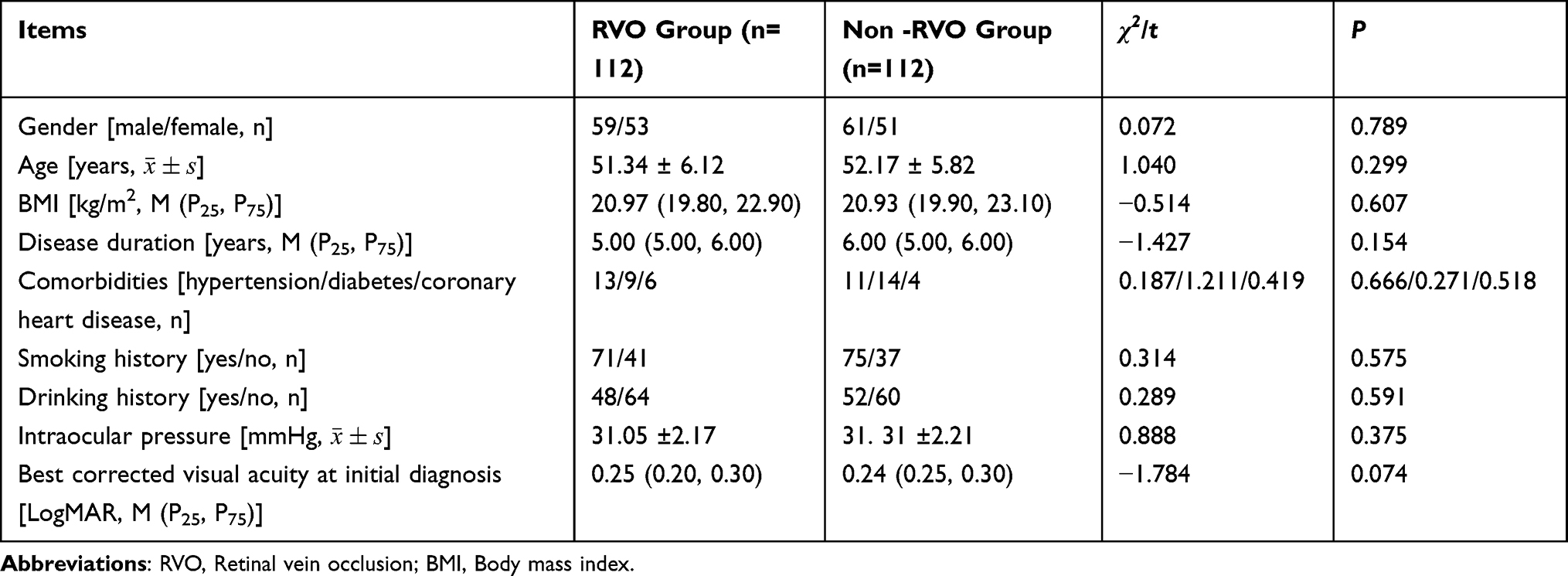 |
Table 1 Comparison of General Information Between the Two Groups |
Laboratory Tests
There were no significant differences in D-D, CRP, Scr, FBG, and HbAlc levels between the RVO Group and the non-RVO Group (t = −0.291, −1.464, 1.219, 1.705, 1.587, respectively, all P >0.05), indicating that the two groups were clinically comparable. Detailed results are shown in Table 2.
 |
Table 2 Comparison of Laboratory Test Results Between the Two Groups |
Diagnostic Value of Sse and Redox Factors in Patients with RVO
Comparison of Sse and Redox Factor Levels Between the Two Groups
To elucidate differences in Sse and redox factor levels between the RVO Group and the non-RVO group, results showed that the Sse [(82.61 ± 8.75) μg/L], GSH [(153.34 ± 24.01) mg/L], and SOD [(17.14 ± 1.55) mU/L] levels in the RVO Group were significantly lower compared with the non-RVO Group [(91.05 ± 11.58) μg/L, (172.71 ± 23.46) mg/L, (18.04 ± 1.73) mU/L], (t = 10.821, 11.769, 9.568, respectively, P<0.05). However, the levels of HMGB1 [(16.39 ± 1.70) mmol/L] and NO [(70.01 ± 7.46) μmol/L] were significantly higher in the RVO Group than the non-RVO Group [(15.79 ± 1.15) mmol/L, (59.48 ± 10.28) μmol/L], and the difference was statistically significant (t = 3.094, 8.774, respectively, P < 0.05). These findings suggest that Sse, HMGB1, GSH, NO, and SOD may be associated with RVO. Detailed results are presented in Table 3.
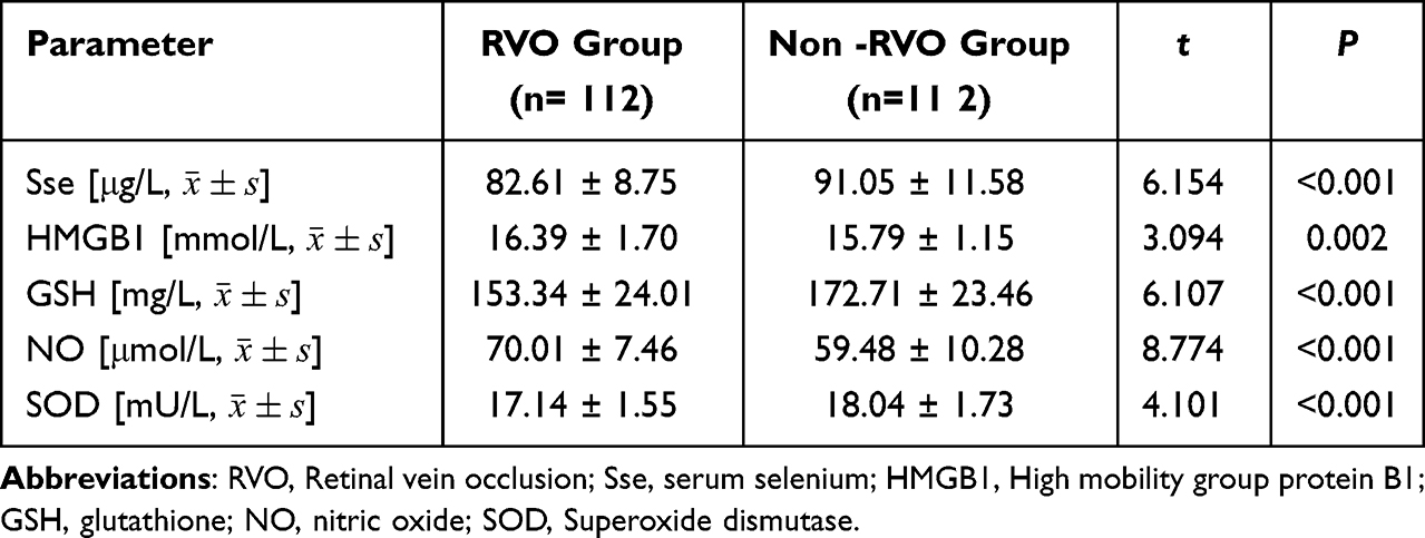 |
Table 3 Comparison of Sse and Redox Factor Levels Between the Two Groups |
Roc Analysis of Sse and Redox Factors in Diagnosing RVO
The ROC curve was drawn, and it was found that the AUC values of Sse, HMGB1, GSH, NO and SOD for diagnosing RVO were 0.710 (95% CI: 0.646–0.770), 0.614 (95% CI: 0.541–0.688), 0.717 (95% CI: 0.633–0.771), 0.798 (95% CI: 0.745–0.839), and 0.654 (95% CI: 0.596–0.712), respectively, all exceeding 0.6. These results indicate that these factors have good diagnostic efficacy for RVO, as shown in Figure 1.
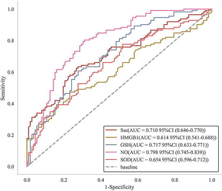 |
Figure 1 ROC Analysis of Sse and Oxidative Stress Markers for the Diagnosis of RVO. |
Prediction of Prognosis with RVO Using Sse and Redox Factors
Analysis of Prognosis with RVO
All 112 RVO patients in the RVO Group received comprehensive treatment. Among them, 23 patients had poor prognosis, accounting for 20.54%, and were classified into the Poor Prognosis Group; 89 patients had good prognosis, accounting for 79.46%, and were classified into the Good Prognosis Group. The changes in Sse, HMGB1, GSH, NO, and SOD indicators were measured in both groups.
Changes in Sse and Redox Factors with Different Prognoses in the RVO Group
The levels of Sse [(92.45 ± 9.07) μg/L], GSH [(156.34 ± 14.83) mg/L], NO [(71.41 ± 7.56) 71.41 ± 7.56] and SOD [(20.14 ± 1.83) mU/L] in the Poor Prognosis Group were lower than those in the Good Prognosis Group [(99.05 ± 10.63) μg /L, (169.71 ± 17.42) mg/L, (89.48 ± 9.02) μmol /L, (23.03 ± 2.04) mU/L, respectively], and the differences were statistically significant (t = 2.730, 3.375, 8.831, 6.178, respectively, all P <0.05). However, the level of HMGB1 [(15.69 ± 1.65) mmol/L] was higher than that in the Good Prognosis Group [(13.29 ± 1.54) mmol/L], and the difference was statistically significant (t = 6.566, P <0.05), indicating that Sse, HMGB1, GSH, NO, SOD may be related to the prognosis of RVO patients. Details are shown in Table 4.
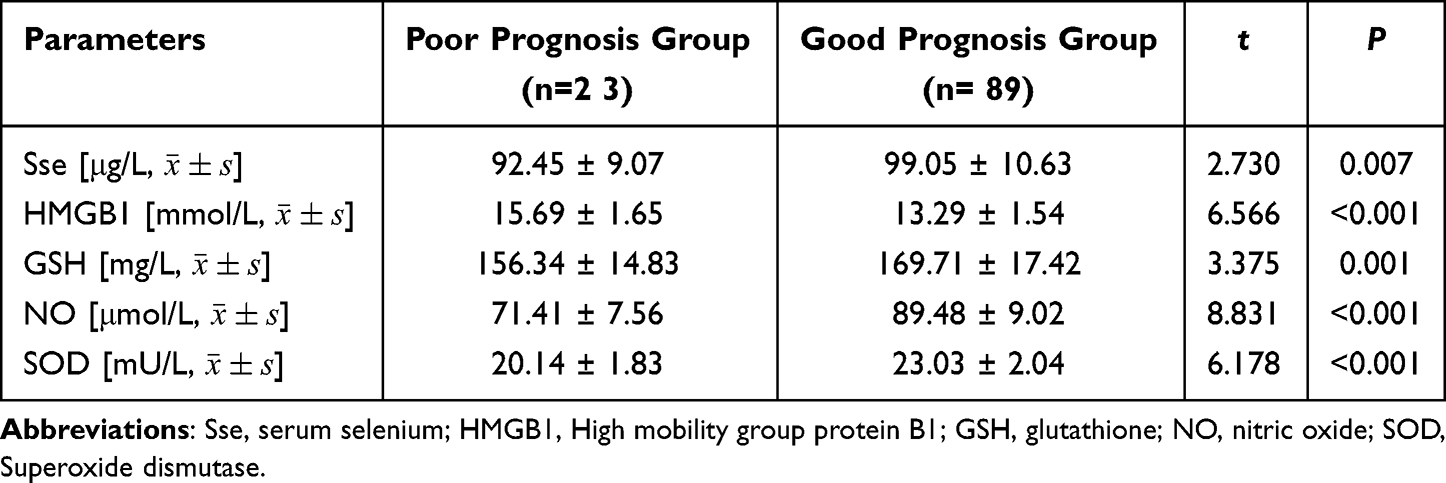 |
Table 4 Comparison of Sse and Oxidative Stress Marker Levels in RVO Patients with Different Prognoses |
Dependent and Independent Variables, and Collinearity Analysis
The prognosis of patients in the RVO group was used as the dependent variable, with good prognosis coded as 0 and poor prognosis as 1. The levels of Sse, HMGB1, GSH, NO, and SOD were assigned as independent variables, and all were treated as continuous measured values. Collinearity analysis revealed that these independent variables did not exhibit collinearity (variance inflation factor [VIF] ≤ 10, tolerance ≥ 0.1), indicating that they could be included in the Cox proportional hazards regression model, as shown in Table 5.
 |
Table 5 Dependent and Independent Variables and Collinearity Analysis |
Cox Proportional Hazards Regression Analysis of Factors Influencing the Prognosis of Patients with RVO
Multivariate Cox regression analysis showed that the levels of Sse, HMGB1, GSH, NO, and SOD were all independent factors influencing the prognosis of patients with RVO (all P < 0.05), suggesting that these variables could be incorporated into the prognostic model for RVO patients, as shown in Table 6.
 |
Table 6 Cox Proportional Hazards Regression Analysis of Prognostic Factors in RVO Patients |
Nomogram and Calibration Curve for Predicting the Prognosis of RVO Patients Based on Sse and Oxidative Stress Markers
Based on the above analysis results, a risk prediction nomogram model for the prognosis of RVO patients was constructed, and a calibration curve was drawn. It was found that the calibration curve of the nomogram model for predicting the prognosis of RVO patients was close to the ideal curve, indicating that the nomogram has good consistency and predictive ability, and the model has high accuracy, as shown in Figure 2A and B).
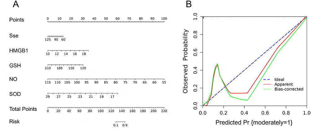 |
Figure 2 Nomogram and Calibration Curve Based on Sse and Oxidative Stress Markers for Predicting the Prognosis of RVO Patients. (A) Nomogram for predicting the prognosis of RVO patients based on Sse and oxidative stress markers; (B) Calibration curve for assessing the agreement between predicted and observed prognosis in RVO patients. |
Discussion
As an ophthalmic disease with a high risk of blindness, the pathogenesis of retinal vein occlusion (RVO) involves complex processes such as vascular endothelial damage, hemodynamic changes, and local inflammatory response. In recent years, oxidative stress has been recognized as an important pathophysiological mechanism. Clinical scholars have pointed out that free radical damage caused by oxidative stress plays a key role in the occurrence and progression of ophthalmic diseases.20
This study pointed out that the levels of Sse, GSH, and SOD in the RVO group were lower than those in the non-RVO group. Previous studies have shown that the level of Sse in RVO patients is lower, which is consistent with the conclusion of this study.21 The reason may be related to the oxidative damage and inflammatory response of retinal blood vessels. Sse is a key component of selenoproteins and is widely involved in the antioxidant system in the body. Sse helps remove free radicals and reduce cell damage caused by oxidative stress by combining it with antioxidant enzymes such as glutathione peroxidase in the body. Low selenium levels lead to weakened antioxidant capacity, thereby aggravating retinal blood vessel damage, promoting thrombosis, and further aggravating vision loss. Similarly, GSH, as an important intracellular antioxidant, is rapidly consumed in response to increased oxidative stress caused by impaired retinal blood flow in RVO patients. Its decreased concentration is negatively correlated with the extent of retinal damage.22 SOD is a key enzyme responsible for eliminating superoxide anions. Its decreased activity directly reflects elevated local oxidative stress in the retina, which may further exacerbate vascular endothelial injury.23
This study found that HMGB1 and NO levels were higher in the RVO group than in the non-RVO group. HMGB1, as a major pro-inflammatory cytokine, reflects the inflammatory state and oxidative stress level in the retina of RVO patients when elevated, and may impair vascular endothelial integrity by activating inflammatory pathways.24 As a critical vasodilator, nitric oxide (NO) also plays a noteworthy role in RVO. A study by Khaloo P et al25 reported that serum NO levels in patients with diabetic retinopathy (DR) were significantly lower than in healthy controls and were associated with increased risk of ischemia. Moreover, other reports26 have noted that patients with RVO complicated by central retinal artery occlusion exhibited significantly lower NO levels than those with isolated RVO, suggesting that NO deficiency may exacerbate ischemic injury. However, in the present study, the overall NO levels in the RVO group were higher than those in the control group. This discrepancy may be related to the disease stage, RVO subtype, or the detection methods used in this study. Notably, a study by Goulopoulos et al27 emphasized that endothelial dysfunction is common in RVO patients and is closely related to NO function, although whether NO is elevated or decreased may be influenced by multiple factors. Nonetheless, changes in NO levels clearly reflect local retinal blood flow status and endothelial dysfunction, making it a valuable marker for evaluating vascular damage in RVO.
ROC analysis in this study revealed that Sse, HMGB1, GSH, NO, and SOD all demonstrated significant diagnostic value for RVO, with AUCs ranging from 0.614 to 0.798. In contrast, traditional inflammatory or hypercoagulable markers such as D-D and CRP did not show significant differences between groups, which differs from some previous literature.28 This may indicate that, in specific patient populations or disease states, such as those focusing on oxidative stress pathways. Sse in combination with HMGB1, GSH, NO, and SOD may offer unique diagnostic value, and may also reflect the specificity of the study population or detection techniques. These findings highlight the need for future research to conduct more detailed stratified analyses (eg, based on RVO type, severity, or complications) and to standardize detection methods. Despite these limitations, the data from this study further support the high combined diagnostic performance of Sse and the aforementioned redox-related markers in RVO.
Additionally, multivariate Cox proportional hazards regression confirmed that Sse, HMGB1, GSH, NO, and SOD were independent prognostic factors for patients with RVO. These findings are consistent with previous studies. For instance, a randomized trial29 reported that patients with poor RVO prognosis showed further decreases in Sse levels, suggesting persistent impairment of antioxidant capacity. Likewise, poor prognosis was often associated with significantly reduced GSH levels, indicating severe compromise of the antioxidant defense system.30 In vascular-related diseases, reduced NO levels are commonly linked to poor outcomes, reflecting worsening local circulation and hypoxia.31 Furthermore, high HMGB1 expression has been associated with poor prognosis in various diseases, and in RVO, it may contribute to retinal injury by promoting sustained inflammation.32 In this study, lower SOD levels were also predictive of worse outcomes, likely due to persistent oxidative stress and inadequate scavenging capacity. The nomogram constructed based on these factors demonstrated good consistency, with the calibration curve closely approximating the ideal 45-degree line, preliminarily demonstrating the potential of multi-parameter integration. Its promising predictive performance suggested it may serve as a valuable auxiliary tool in clinical practice. Recently, LUNEGOVA et al33 also highlighted the core interaction between oxidative stress and inflammation in retinal vascular diseases, further supporting the multi-marker analysis strategy employed in this study.
However, the interpretation of this study’s findings should be approached with caution due to several limitations. First, the study population was relatively homogeneous. As a single-center prospective study, all samples were obtained from patients treated at our hospital, which limits the generalizability (external validity) of the results to broader populations, such as those from different regions, ethnic backgrounds, or healthcare settings. Second, there was insufficient control of confounding factors. Although the two groups showed comparability in baseline characteristics and certain laboratory parameters, unmeasured or uncontrolled confounders—such as dietary selenium intake, detailed medication history, environmental influences, and the status of other micronutrients—may have affected biomarker levels. Future prospective studies should aim to better match or adjust for these potential confounders. Third, the follow-up period was relatively short. Prognostic evaluation was limited to 6 months, which is appropriate for assessing short-term outcomes following the acute phase of RVO but may be inadequate for evaluating long-term prognosis. The natural course of RVO and its long-term outcomes require extended observation. Finally, this study focused primarily on serum biomarker levels, without an in-depth investigation of their roles or interactions within the retinal microenvironment.
Future research should validate these findings in larger, multicenter, prospective cohorts. Extended follow-up durations and mechanistic investigations using animal models or in vitro experiments are needed to elucidate how these biomarkers function and interact locally in the retina. Moreover, future studies should explore how these biomarkers can be incorporated into clinically practical diagnostic models or prognostic risk stratification tools to more accurately assess their real-world value in the clinical management of RVO.
Conclusion
In summary, this study demonstrated significant differences in the levels of Sse and oxidative stress-related factors (HMGB1, GSH, NO, and SOD) between the RVO and non-RVO groups, as well as between patients with poor and good prognoses. These findings suggest that the above biomarkers may have potential value in diagnosing RVO and predicting the prognosis of patients undergoing comprehensive treatment. The results further highlight the critical role of oxidative stress and inflammatory responses in the pathophysiological process of RVO and may provide preliminary evidence for the development of auxiliary diagnostic and prognostic tools based on multi-biomarker panel analysis.
Funding
This study was supported by Suzhou 2022 Annual 28th Batch of Science and Technology Development Plan Guidance Project (SKYD2022060), 2023 Taicang Science and Technology Bureau Directive Project (TC2023JCYL06) and Suzhou Wumen Medical School Research Special Fund (SKYXD2022039). The sponsor had no role in study design, data analysis, or manuscript preparation.
Disclosure
The authors declare that they have no known competing financial interests or personal relationships that could have appeared to influence the work reported in this paper.
References
1. Li TJ, Sun YC, Ma QY, et al. Clinical observation of Dan-Hong Hua-Yu oral solution in treating retinal vein occlusion. Medicine. 2020;99(21):e20173. doi:10.1097/MD.0000000000020173
2. Do DV, Gordon C, Suner IJ, et al. Proliferative diabetic retinopathy events in patients with diabetic macular edema: post hoc analysis of vista and VIVID trials. J Vitreoretin Dis. 2022;6(4):295–301. doi:10.1177/24741264221093914
3. Ai J, Cao Y, Zhang C, et al. Deciphering the interplay of gut microbiota and metabolomics in retinal vein occlusion. Microbiol Spectr. 2024;12(8):e0005224. doi:10.1128/spectrum.00052-24
4. Xiong X, Chen X, Ma H, et al. Metabolite changes in the aqueous humor of patients with retinal vein occlusion macular edema: a metabolomics analysis. Front Cell Dev Biol. 2021;9:762500. doi:10.3389/fcell.2021.762500
5. Shrestha S, Poddar E, Bashyal B, et al. Bilateral central retinal vein occlusion as an initial presentation of Waldenstrom macroglobulinemia: a case report. J Med Case Rep. 2023;17(1):59. doi:10.1186/s13256-023-03778-4
6. Spiewak D, Witek K, Drzyzga L, Mrukwa-Kominek E. An analysis of optical coherence tomography angiography (OCT-A) perfusion density maps in patients treated for retinal vein occlusion with intravitreal aflibercept. Diagnostics. 2023;13(19). doi:10.3390/diagnostics13193100
7. Wang XL, Cai FR, Gao YX, Zhang J, Zhang M. Changes and significance of retinal blood oxygen saturation and oxidative stress indexes in patients with diabetic retinopathy. World J Diabetes. 2022;13(5):408–416. doi:10.4239/wjd.v13.i5.408
8. Niu T, Shi X, Liu X, Wang H, Liu K, Xu Y. Porous Se@SiO(2) nanospheres alleviate diabetic retinopathy by inhibiting excess lipid peroxidation and inflammation. Mol Med. 2024;30(1):24. doi:10.1186/s10020-024-00785-z
9. Younesian O, Khodabakhshi B, Abdolahi N, et al. Decreased serum selenium levels of COVID-19 patients in comparison with healthy individuals. Biol Trace Elem Res. 2022;200(4):1562–1567. doi:10.1007/s12011-021-02797-w
10. Mohammad G, Kowluru RA. Involvement of high mobility group box 1 protein in optic nerve damage in diabetes. Eye Brain. 2022;14:59–69. doi:10.2147/EB.S352730
11. Wojciechowski AM, Bell BA, Song Y, et al. Inducible RPE-specific GPX4 knockout causes oxidative stress and retinal degeneration with features of age-related macular degeneration. Exp Eye Res. 2024;247:110028. doi:10.1016/j.exer.2024.110028
12. Sgambellone S, Khanfar MA, Marri S, et al. Histamine H(3) receptor antagonist/nitric oxide donors as novel promising therapeutic hybrid-tools for glaucoma and retinal neuroprotection. Biomed Pharmacother. 2024;180:117454. doi:10.1016/j.biopha.2024.117454
13. Lopez-Contreras AK, Arevalo-Simental DE, Pacheco-Moises FP, et al. Evaluation of ocular and systemic oxidative stress markers in patients with diabetic retinopathy. Life. 2024;14(12). doi:10.3390/life14121588
14. Li S, Sun D, Chen S, et al. UCP2-SIRT3 signaling relieved hyperglycemia-induced oxidative stress and senescence in diabetic retinopathy. Invest Ophthalmol Vis Sci. 2024;65(1):14. doi:10.1167/iovs.65.1.14
15. Studnicka J, Nemcansky J, Vyslouzilova D, Ernest J, Veith M, Nemec P. Retinal Vein Occlusion Guidelines. Cesk Slov Oftalmol. 2024;80(6):298–305. doi:10.31348/2024/42
16. McEvoy JW, McCarthy CP, Bruno RM, et al. 2024 ESC Guidelines for the management of elevated blood pressure and hypertension. Eur Heart J. 2024;45(38):3912–4018. doi:10.1093/eurheartj/ehae178
17. Yang ST, Deng C, He BB, Chen X, Li X, Zhou ZG. [Application of the Chinese Expert consensus on diabetes classification in clinical practice]. Zhonghua Nei Ke Za Zhi. 2023;62(9):1085–1092. doi:10.3760/cma.j.cn112138-20230131-00043
18. Professional Committee of Cardiopulmonary P, Rehabilitation of the Chinese Rehabilitation Medical A, Cardiovascular Rehabilitation Committee of the Chinese Medical A. [Chinese expert consensus on the rehabilitation treatment and nursing practice for stable coronary artery disease]. Zhonghua Nei Ke Za Zhi. 2023;62(12):1406–1417. doi:10.3760/cma.j.cn112138-20230817-00064
19. Alqahtani TF, Alqarehi R, Mulla OM, et al. Knowledge, attitude, and practice regarding diabetic retinopathy screening and eye management among diabetics in Saudi Arabia. Cureus. 2023;15(9):e46190. doi:10.7759/cureus.46190
20. Deng L, Gupta V, Abyadeh M, et al. Oxidative stress induced dysfunction of protein synthesis in 661W mice photoreceptor cells. Proteomes. 2023;11(2):12. doi:10.3390/proteomes11020012
21. Pfister C, Dawczynski H, Schingale FJ. Selenium deficiency in lymphedema and Lipedema–a retrospective cross-sectional study from a specialized clinic. Nutrients. 2020;12(5):1211. doi:10.3390/nu12051211
22. Otero-Albiol D, Carnero A. Cellular senescence or stemness: hypoxia flips the coin. J Exp Clin Cancer Res. 2021;40(1):243. doi:10.1186/s13046-021-02035-0
23. Wang YC, Zhou CQ, Li RR, et al. The role of serum metrics in anti- VEGF treatment for macular edema induced by retinal vein occlusion. BMC Ophthalmol. 2023;23(1):173. doi:10.1186/s12886-023-02921-5
24. Zhou M, Zhang Y, Tang R, et al. HMGB1/TLR4 signaling affects regulatory T cells in acute lung injury. J Inflamm Res. 2021;14:1551–1561. doi:10.2147/JIR.S302967
25. Khaloo P, Qahremani R, Rabizadeh S, et al. Nitric oxide and TNF-alpha are correlates of diabetic retinopathy independent of hs-CRP and HbA1c. Endocrine. 2020;69(3):536–541. doi:10.1007/s12020-020-02353-x
26. Baek SU, Kwon S, Kim YK, Jeoung JW, Park KH. Diurnal change of retinal vessel density related to hemodynamic variation in treatment-naive low-teens normal-tension glaucoma. Sci Rep. 2023;13(1):10613. doi:10.1038/s41598-023-37214-w
27. Gouliopoulos N, Siasos G, Moschos MM, et al. Endothelial dysfunction and impaired arterial wall properties in patients with retinal vein occlusion. Vasc Med. 2020;25(4):302–308. doi:10.1177/1358863X20913609
28. Sacconi R, Simona F, Forte P, Querques G. Retinal vein occlusion following two doses of mRNA-1237 (Moderna) immunization for SARS-Cov-2: a case report. Ophthalmol Ther. 2022;11(1):453–458. doi:10.1007/s40123-021-00441-3
29. Liu Z, Li J, Lin S, Wu Y, He D, Qu P. PI3K regulates the activation of NLRP3 inflammasome in atherosclerosis through part-dependent AKT signaling pathway. Exp Anim. 2021;70(4):488–497. doi:10.1538/expanim.21-0002
30. Srejovic JV, Muric MD, Jakovljevic VL, et al. Molecular and cellular mechanisms involved in the pathophysiology of retinal vascular disease-interplay between inflammation and oxidative stress. Int J Mol Sci. 2024;25(21):11850. doi:10.3390/ijms252111850
31. Gong X, Pu X, Wang J, et al. Enhancing of Nanocatalyst-Driven Chemodynaminc Therapy for Endometrial Cancer Cells Through Inhibition of PINK1/Parkin-Mediated Mitophagy. Int J Nanomed. 2021;16:6661–6679. doi:10.2147/IJN.S329341
32. Li X, Su X, Xia F, et al. Bibliometric and visual analysis of diabetes mellitus and pyroptosis from 2011 to 2022. Eur J Med Res. 2023;28(1):235. doi:10.1186/s40001-023-01175-7
33. Lunegova DA, Gvozdev DA, Senin II, et al. Antioxidant properties of the soluble carotenoprotein AstaP and its feasibility for retinal protection against oxidative stress. FEBS J. 2025;292(2):355–372. doi:10.1111/febs.17335
 © 2025 The Author(s). This work is published and licensed by Dove Medical Press Limited. The
full terms of this license are available at https://www.dovepress.com/terms
and incorporate the Creative Commons Attribution
- Non Commercial (unported, 4.0) License.
By accessing the work you hereby accept the Terms. Non-commercial uses of the work are permitted
without any further permission from Dove Medical Press Limited, provided the work is properly
attributed. For permission for commercial use of this work, please see paragraphs 4.2 and 5 of our Terms.
© 2025 The Author(s). This work is published and licensed by Dove Medical Press Limited. The
full terms of this license are available at https://www.dovepress.com/terms
and incorporate the Creative Commons Attribution
- Non Commercial (unported, 4.0) License.
By accessing the work you hereby accept the Terms. Non-commercial uses of the work are permitted
without any further permission from Dove Medical Press Limited, provided the work is properly
attributed. For permission for commercial use of this work, please see paragraphs 4.2 and 5 of our Terms.