
Back to Journals » Journal of Multidisciplinary Healthcare » Volume 19
Development and Clinical Evaluation of a Double-Helix Drainage Tube Securement Patch for Patients Undergoing Head and Neck Surgery
Authors Wang Q, Ma HL, Li M, Ma YN
Received 20 March 2026
Accepted for publication 27 May 2026
Published 12 June 2026 Volume 2026:19 610923
DOI https://doi.org/10.2147/JMDH.S610923
Checked for plagiarism Yes
Review by Single anonymous peer review
Peer reviewer comments 2
Editor who approved publication: Professor Tilakavati Karupaiah
Qing Wang, Hong-Li Ma, Min Li, Ya-Nan Ma
Department of Otolaryngology - Head and Neck Surgery, Shanxi Bethune Hospital, Shanxi Academy of Medical Sciences, Third Hospital of Shanxi Medical University, Tongji Shanxi Hospital, Taiyuan, 030032, People’s Republic of China
Correspondence: Ya-Nan Ma, Department of Otolaryngology - Head and Neck Surgery, Shanxi Bethune Hospital, Shanxi Academy of Medical Sciences, Third Hospital of Shanxi Medical University, Tongji Shanxi Hospital, Taiyuan, Shanxi, 030032, People’s Republic of China, Tel +86 18434371082, Fax +86 18834895914, Email [email protected]
Objective: To design a double‑helix drainage tube securement patch and assess its clinical efficacy in patients after head‑neck surgery.
Methods: Based on biomechanical theories, we developed a composite securement patch with bilateral 1‑cm‑wide, 3.5‑cm‑long spiral strips, an elastic connecting strap, and lateral pressure‑distributing wing patches (area ≥ 12 cm2). This non‑randomized controlled trial recruited 125 patients with postoperative drainage tubes after head‑neck surgery in a tertiary Grade‑A hospital in Shanxi Province from June 2024 to June 2025 via convenience sampling. Patients were divided by admission sequence into a control group (n=64) with conventional 3M‑tape bridge‑type fixation and a study group (n=61) with the double‑helix patch. Primary outcomes included securement time, adverse event (AE) incidence, traction‑related pain frequency, skin injury rate, General Comfort Questionnaire (GCQ) scores, patient satisfaction, and cost‑effectiveness.
Results: The study group showed markedly lower AE incidence (p< 0.001), shorter single securement time (1.75± 2.81 min vs 2.88± 2.93 min, p=0.043), higher GCQ scores (93.26± 9.23 vs 84.39± 14.82, p< 0.001), better satisfaction (p< 0.001), and 43.2% lower consumable costs (p< 0.001). Traction‑related pain frequency and skin injury rate showed no inter‑group differences (p> 0.05). No significant between-group difference was observed in the duration of drainage tube indwelling (p = 0.055).
Conclusion: The double‑helix patch enables quick, stable drainage tube fixation. Its dynamic flexibility preserves head‑neck mobility, lowers drainage‑related AEs and secondary incision injury risks, cuts costs, and improves patient comfort and satisfaction, warranting wider clinical use.
Keywords: adverse events, drainage tube, head and neck cancer, satisfaction, securement device
Introduction
Head and neck cancer (ICD-10: C00–C14) is the seventh most common malignancy worldwide, and its associated disease burden continues to increase.1,2 Global cancer statistics indicate that approximately 930,000 new cases of head and neck squamous cell carcinoma (HNSCC) were reported in 2020, accounting for 4.9% of all malignancies, with radical surgery serving as the primary treatment modality in approximately 35.7% of early-stage disease.3 However, the complex anatomical structures and limited surgical exposure within the otolaryngologic and oral and maxillofacial regions make these procedures technically demanding.4,5 Postoperative hematoma occurs in approximately 7.3–12.5% of cases and may impair normal wound healing.
To facilitate the evacuation of accumulated blood and fluid, negative-pressure drainage tubes are routinely placed intraoperatively. The characteristics of postoperative drainage also serve as important indicators of surgical outcomes and prognosis.6 Consequently, effective securement of drainage tubes and maintenance of unobstructed drainage are essential for accurate drainage monitoring and constitute a critical component of postoperative nursing care.7,8
In current clinical practice, securement of postoperative head and neck drainage tubes typically involves two sequential steps. Primary fixation is first achieved by the surgeon through suture anchoring, followed by secondary securement performed by nursing staff. Because cervical drainage tubes are relatively long and the drainage bulb is comparatively heavy, secondary fixation is commonly achieved using 3M adhesive tape applied via the bridge-type taping method or the butterfly cross-taping method, or through the use of commercially available products such as “Xinpigu” dressing patches. However, these conventional approaches have recognized limitations, including a tendency toward loosening, suboptimal aesthetic appearance, and edge curling.9 In addition, cervical drainage tubes inevitably move during routine head rotation, often causing traction on the tube. Conventional securement methods have a limited capacity to accommodate the degree of rotational mobility required in the cervical region.
To address these limitations, a double-helix drainage tube securement patch was developed and applied in patients undergoing head and neck surgery with indwelling drainage tubes, demonstrating favorable clinical outcomes. The device has been granted a utility model patent (ZL 2022 2 1971982.5) and issued Medical Device Registration Certificate No. 20202140330. Manufacturing is commissioned to Henan Tuoren Best Medical Device Co., Ltd. The clinical application of the novel double-helix securement patch and the conventional 3M tape bridge-type fixation method are shown in Figure 1 for visual comparison.
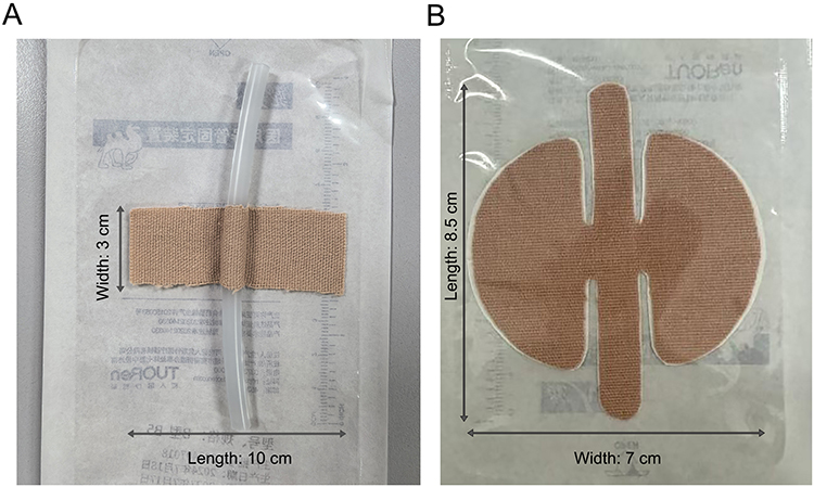 |
Figure 1 Comparative images of two drainage tube fixation methods. (A) Conventional bridge-type fixation with 3M adhesive tape; (B) Physical photograph of the double-helix drainage tube securement patch. |
Materials and Methods
Design of the Double-Helix Drainage Tube Securement Patch
The double-helix drainage tube securement patch is composed of three core components: (Figure 2). The spiral winding strip (Figure 2A, label 1): a bilateral strip (1 cm in width, 3.5 cm in length) designed to wrap the drainage tube in a spiral manner, providing stable fixation without compressing the tube. The pressure-distributing lateral wing patch (Figure 2A, label 2): a semi-circular patch (area ≥ 12 cm2) with evenly distributed ventilation holes (Figure 2A, label 4) to reduce skin irritation and improve comfort. The elastic connecting strap (Figure 2A, label 3): a central elastic band that connects the two lateral wings, allowing dynamic compliance with head and neck movement while maintaining tube stability.
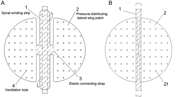 |
Figure 2 Structural design of the double-helix drainage tube securement patch. (A) 1, spiral winding strip; 2, pressure-distributing lateral wing patch; 3, elastic connecting strap; 4, ventilation hole. (B) 1, 3M adhesive tape; 2, skin-adhering wing patch; 21, ≥ 21 ventilation holes per lateral wing patch. |
The device has an overall circular configuration measuring 8.5 cm in length and 7.0 cm in width. It is primarily composed of a medical pressure-sensitive adhesive, elastic fabric, and a release liner. The lateral wing patches are fabricated from a hypoallergenic medical adhesive dressing material, whereas the spiral winding strip is constructed from an elastic adhesive material.
The spiral winding strip is positioned centrally within the device, with one strip located on both the upper and lower aspects. Each strip measures 3.5 cm × 1.0 cm. During application, the strip is helically wrapped around the drainage tube to form an “S”-shaped configuration, thereby allowing greater freedom of head and neck movement. The lateral wing patches are located on both sides of the device, each with a surface area of at least 12 cm2. This design allows the patch to conform closely to the skin contour while increasing the contact area with the skin. Each lateral wing patch contains more than 21 ventilation holes, which help reduce the risk of skin injury caused by excessive local skin temperature or humidity during use. The connecting strap is positioned centrally and links the spiral winding strip to the lateral wing patches. It measures 1.5 cm × 1.5 cm and enables bridge-type elevation fixation, thereby enhancing the overall stability of tube securement. During clinical application, iterative refinements have been implemented, including the replacement of the original lateral wing patch material with a highly breathable medical elastic adhesive material to further improve skin tolerance and comfort.
Study Population
A non-randomized controlled study design was adopted, and participants were recruited using convenience sampling. A total of 125 postoperative patients with indwelling drainage tubes from the Department of Otolaryngology–Head and Neck Surgery at a tertiary Grade A hospital in Shanxi Province were enrolled between June 2024 and June 2025. Participants were allocated according to admission order: 64 patients admitted between June and December 2024 who underwent head and neck surgery with postoperative drainage tube placement were assigned to the control group, whereas 61 patients admitted between January and June 2025 were assigned to the study group. This study was approved by the institutional ethics committee (approval number: YXLL-2023-033).
Inclusion criteria included age ≥ 18 years, intact consciousness and the ability to communicate, first-time head and neck surgery, no history of rubber allergy, and voluntary participation with consent provided by both patients and their family members.
Exclusion criteria included a history of cervical spondylosis or limb movement dysfunction, the presence of concurrent malignancies, or skin infection at the operative site. Written informed consent was obtained from all participants prior to enrollment.
Application Methods
A drainage tube management team was established, consisting of 3 personnel with more than 10 years of experience in otolaryngology–head and neck surgery and holding at least a bachelor’s degree: the head nurse, the deputy head nurse, and the departmental teaching supervisor. Team members reviewed relevant literature to develop implementation guidelines for drainage tube management. The departmental teaching supervisor subsequently trained the primary nurses through didactic instruction and one-on-one guidance, covering the structure, application procedures, indications, precautions, and management of unexpected situations associated with both the conventional securement method and the double-helix securement patch. Competency was assessed using simulation mannequins, and only nurses who met the required standards were permitted to participate in data collection. Standardized implementation was maintained through scheduled evaluations and random spot checks throughout the study period.
Study Group
The study group received drainage tube securement using the double-helix securement patch. After returning to the ward, patients were positioned supine, and the primary nurse inspected the condition of the dressing. A tube label was affixed to the drainage tube at the edge of the dressing, and an additional marker was placed 10 cm from the wound to facilitate subsequent assessment of tube displacement. A skin protectant was applied to the intended securement site 15 cm from the dressing edge and allowed to dry for 10–15 seconds. The appropriate securement length was determined based on the maximum range of head and neck movement (Figure 3). The release liner was removed, and one lateral wing patch was applied to secure one side. The upper spiral winding strip was then wrapped around the drainage tube from bottom to top, and the lower strip from top to bottom, with each successive wrap overlapping the preceding layer by one half of its width. The drainage tube was elevated, and the opposite lateral wing patch was applied, ensuring bilateral alignment. The connecting strap was used to achieve bridge-type elevation fixation, after which the patch was molded to conform to the anatomical contour.
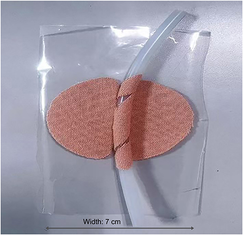 |
Figure 3 Schematic illustration of the application procedure for the double-helix drainage tube securement patch. |
The drainage bulb was placed in the breast pocket of the patient’s hospital gown and secured to the collar using a securement clip. A tube label was affixed 2 cm proximal to the drainage bulb. The primary nurse provided health education to the patient and family members regarding relevant precautions and encouraged and assisted the patient with active or passive movement. The securement patch was replaced weekly or immediately if loosening or edge lifting occurred. During removal, the lateral wing patches were peeled using the “zero-degree” technique, which refers to securing the drainage tube parallel to the skin surface without bending, thereby minimizing skin traction and mechanical irritation and the adhesive beneath was dissolved with an alcohol swab or adhesive remover until the patch was completely separated from the skin.
Control Group
The control group received conventional bridge-type fixation using 3M hypoallergenic medical adhesive tape. The procedure was performed as follows: two horizontal strips of tape were placed parallel to each other across the drainage tube and secured to the surrounding skin, forming a “bridge” over the tube. A third strip was then wrapped vertically around the drainage tube and the two horizontal strips, creating a stable but rigid fixation. The tape was cut into strips measuring 3 cm × 10 cm. After returning to the ward, a tube label was affixed to the drainage tube at the dressing edge. For primary securement, the fixation site was determined according to the maximum range of head and neck movement. The tubing at the designated site was positioned along the center of the tape surface, and the tape was wrapped 360° around the tube, elevating it 0.5 cm above the skin surface, with 0.5 cm of the tape adhered to itself. The remaining ends of the tape were pressed firmly onto the skin on both sides.
For secondary securement, the integrated securement clip of the negative-pressure drainage tube was attached to the collar of the patient’s hospital gown, and the drainage bulb was placed in the breast pocket to reduce gravitational traction. The bridge-type securement patch was replaced daily or immediately if loosening occurred. The removal procedure and health education content were the same as those implemented in the study group.
Outcome Measures and Data Collection
Adverse Event Incidence
Adverse events (AEs) included drainage tube displacement, kinking, and unplanned removal. Tube displacement was determined by the attending physician, and the displacement rate was calculated as the number of patients with tube displacement divided by the total number of patients with indwelling drainage tubes. Kinking was defined as bending or folding of the tube that impeded drainage during the indwelling period, and the kinking rate was calculated in the same manner. Complete exteriorization of the drainage tube was classified as unplanned removal. From the time of the patient’s return to the ward until the attending physician confirmed eligibility for tube removal, the primary nurse inspected the drainage tube at each shift. Any abnormal findings were jointly assessed by the primary nurse and the ward physician, and the results were documented accordingly.
Frequency of Postoperative Traction Pain
Traction pain was defined as pain resulting from pulling, stretching, or displacement of body tissues. A Drainage Tube Traction Pain Observation Form was developed for this study. The primary nurse instructed patients on how to identify traction pain and subsequently recorded the number of episodes during each shift until removal of the drainage tube. Two investigators independently calculated the frequency of postoperative traction pain for each patient.
Single-Episode Tube Securement Time
After the patient returned to the ward and the primary nurse determined the securement site, timing for the control group commenced with the initiation of tape cutting, whereas timing for the study group commenced with removal of the release liner. Timing concluded once the drainage tube had been completely secured. When edge lifting or detachment of the securement dressing occurred, the dressing was replaced promptly, and timing for the replacement began when the previous dressing was removed. For patients who underwent multiple dressing replacements or had more than one drainage tube, the single-episode securement time was calculated as the mean of all recorded durations.
Skin Injury
A Drainage Tube Securement Site Skin Observation Form was designed for this study. The primary nurse assessed the periwound skin at the securement site during each shift. Abnormal findings, including erythema, pruritus, rash, or skin breakdown, were promptly managed and documented.
Postoperative Comfort
On the second postoperative day, the primary nurse administered the General Comfort Questionnaire (GCQ), developed by Kolcaba, to assess patient comfort. The GCQ includes psychological, physiological, and environmental domains of comfort and consists of 28 items rated on a 4-point Likert scale, with total scores ranging from 28 to 112. Higher scores indicate greater levels of comfort. The Cronbach’s α coefficient for the scale is 0.96.10
Postoperative Satisfaction
Before discharge, the primary nurse administered a self-developed Head and Neck Surgery Postoperative Tube Management Satisfaction Questionnaire (total score range: 0–10; 8–10 = highly satisfied, 6–8 = moderately satisfied, <6 = dissatisfied). The overall satisfaction rate was calculated as (number of highly satisfied patients + number of moderately satisfied patients) ÷ total number of patients × 100%.
Economic Outcomes
Consumable costs associated with drainage tube securement during the indwelling period were compared between the two groups. The bridge-type taping method utilized 3M elastic medical adhesive tape (10 cm × 500 cm, Cat. No. 27333-100; 3M International Trading (Shanghai) Co., Ltd.), with an approximate per-episode cost of ¥1.46. The double-helix securement patch had an approximate per-episode cost of ¥5.00.
Statistical Methods
All data were independently entered by two investigators and verified prior to analysis using SPSS version 26.0 (IBM Corp., Armonk, NY, USA). Continuous variables with a normal distribution were expressed as mean ± standard deviation (SD) and compared using the Student’s t-test. Categorical variables were expressed as frequencies and percentages and compared using the chi-square test. A two-sided p value < 0.05 was considered statistically significant.
Results
Comparison of General Characteristics (Table 1)
No significant differences were observed between the two groups in age, sex, occupational status, or educational level (p > 0.05), indicating comparable baseline characteristics between the groups (Table 1).
 |
Table 1 Baseline Characteristics of Patients in Both Groups |
Comparison of Outcome Indicators
The mean single-episode securement time was 1.75 ± 2.81 minutes in the study group and 2.88 ± 2.93 minutes in the control group (p = 0.043). The incidence of AEs was 6.56% in the study group compared with 42.19% in the control group (p < 0.001). Patient satisfaction and GCQ scores were significantly higher in the study group than in the control group. The mean cost of securement consumables was significantly lower in the study group (p < 0.001), representing a reduction of 43.2%. No statistically significant differences were observed between the two groups in surgical site, number of drainage tubes, frequency of traction pain, degree of skin injury, and duration of tube indwelling ((p = 0.055) (Table 2).
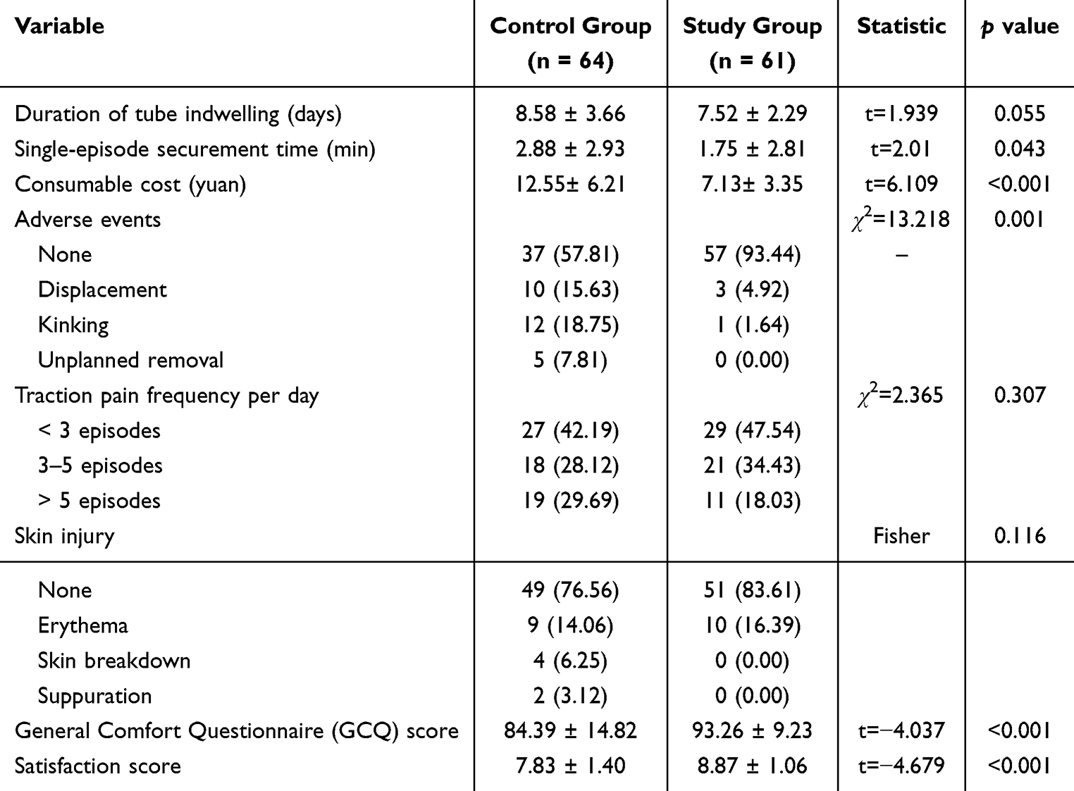 |
Table 2 Comparison of Clinical Outcome Indicators Between Both Groups |
Discussion
The Double-Helix Securement Patch Reduces Postoperative Drainage Tube-Related Adverse Events
The head and neck region is characterized by loose tissue structures and multiple potential spaces; therefore, adequate postoperative drainage of serosanguineous fluid from interstitial spaces represents a key nursing priority following head and neck surgery.11 Appropriate tube securement, ensuring the absence of displacement, kinking, and unplanned removal while maintaining unobstructed drainage, is essential for effective drainage management. The present findings indicate that the double-helix securement patch significantly reduced the incidence of drainage tube–related AEs compared with the bridge-type taping method (p < 0.001).
In the traditional approach, a rectangular 3M adhesive patch is applied using the bridge-type taping technique; the relatively limited contact area between the patch and the skin allows gravitational traction generated by the drainage bulb to cause edge lifting and detachment, which may lead to tube displacement or unplanned removal. The spiral winding structure of the double-helix securement patch increases the contact area between the adhesive material and the drainage tube. In addition, the semicircular configuration of the bilateral lateral wing patches reduces edge curling and adhesive lifting commonly observed with conventional rectangular patches. Even when nursing staff temporarily release the securement clip to empty the drainage bulb, the lateral wing patches and double-helix structure continue to maintain stable tube fixation, thereby substantially improving tube stability. Securing the drainage bulb to the chest pocket with the securement clip further decreases gravitational traction on the tubing and prevents compression or kinking during patient repositioning. Collectively, these design features reduce drainage tube–related AEs and enhance the reliability of tube securement.
The Double-Helix Securement Patch Improves Postoperative Comfort and Satisfaction
Patients undergoing head and neck surgery commonly experience persistent postoperative pain with multiple triggers; routine activities such as coughing, swallowing, or turning the head may provoke discomfort. In addition, the gravitational pull of the drainage bulb exerts traction on the wound and surrounding skin, generating additional nociceptive stimuli.12 Anticipation of pain may contribute to anxiety and other negative emotional responses, which can diminish confidence in early recovery and potentially delay rehabilitation, thereby conflicting with the principles of enhanced recovery after surgery (ERAS).13,14
In the present study, the frequency of traction-related pain was lower in the study group than in the control group. The “S”-shaped configuration created by the double-helix securement patch provides dynamic compliance while preserving maximal head and neck mobility, thereby expanding the allowable range of movement and reducing the psychological burden. This design also reduces the common postoperative reluctance to mobilize, facilitating early ambulation and contributing to improved overall comfort and patient satisfaction (p < 0.001).
The Double-Helix Securement Patch Reduces Consumable Expenditure and Improves Nursing Efficiency
Medical consumables include disposable hygienic materials, implantable devices, and reusable but wear-prone medical items used in clinical care.15 Adhesive patches and securement devices of various sizes and materials are essential for postoperative drainage tube fixation and require replacement at regular intervals or whenever complications occur. As shown in Figure 4, consumable costs increased with the duration of drainage tube indwelling. Among patients with indwelling durations exceeding three days, consumable costs in the study group were lower than those in the control group, representing an overall reduction of 43.2%. Although the single-unit price of the double-helix securement patch was higher than that of conventional 3M tape, the traditional bridge-type fixation required daily replacement, whereas the double-helix patch could be retained for up to one week under normal conditions and only replaced when loosening or edge lifting occurred. Although the difference in tube indwelling duration was only approximately 12% between the two groups, the greatly reduced replacement frequency of the new patch substantially decreased cumulative material consumption. Accordingly, the 43.2% reduction in overall consumable expenditure was mainly attributed to the prolonged service cycle and fewer replacement times of the double-helix patch, rather than merely the slight difference in indwelling days.
 |
Figure 4 Comparison of drainage tube securement consumable costs between both groups. Notes: Control group (blue dots, n = 64); Study group (orange dots, n = 61). Error bars represent the standard deviation (SD) of consumable costs. The fitted dotted lines illustrate the trend of increasing cost with longer indwelling duration in both groups. |
As The simplified structural design of the securement patch also facilitates straightforward application and eliminates the need for repeated tube securement procedures by nursing staff. The mean time required for a single securement episode was significantly shorter in the study group than in the control group (p = 0.043). Accordingly, use of the double-helix securement patch reduces departmental expenditure and operational costs, decreases nursing time devoted to tube management, and improves overall nursing efficiency.
In addition to the observed reductions in adverse events and consumable costs, the superior performance of the double-helix securement patch in reducing loosening, edge curling, and poor aesthetic appearance is attributed to both its structural configuration and material properties. The spiral winding strip and large-area lateral wings distribute force evenly to prevent tension and lifting, while the elastic connecting strap accommodates head and neck movement. The hypoallergenic, breathable adhesive material further enhances long-term adhesion and comfort.
Conclusion
The double-helix securement patch yielded concrete quantitative clinical benefits in practical application. Compared with conventional 3M tape bridge-type fixation, the patch significantly lowered the incidence of drainage tube-related adverse events (6.56% vs 42.19%, p < 0.001), shortened single securement time (1.75 ± 2.81 min vs 2.88 ± 2.93 min, p = 0.043), improved General Comfort Questionnaire scores and patient satisfaction, and reduced consumable costs by 43.2%. No significant intergroup differences were observed in traction-related pain frequency, skin injury rate, and duration of postoperative drainage tube indwelling. The patch enables stable tube fixation, preserves functional head and neck mobility, and lowers the risk of secondary incision injury while improving patient comfort and nursing efficiency. Several limitations of this study should be acknowledged. First, this was a single-center non-randomized controlled trial with convenience sampling and group allocation based on admission sequence, which may carry a potential risk of selection bias. Second, we failed to further collect and adjust for potential confounding factors such as seasonal variation and temporal changes in surgical techniques, and relevant supplementary confounding variable data could not be provided due to clinical data accessibility constraints. In addition, the study population was limited to patients undergoing head and neck surgery, which may restrict the generalizability of the results. Therefore, well-designed multicenter randomized controlled studies with systematic control of confounding factors are warranted in future research to confirm the clinical value of this securement patch. Moreover, this single-center study was only performed in patients undergoing head and neck surgery at a tertiary hospital, so the generalizability of the findings to other anatomical drainage sites and different levels of healthcare institutions is limited. Further multicenter investigations are required to validate the applicability of this patch in broader clinical scenarios.
Abbreviations
AE, adverse event; ERAS, enhanced recovery after surgery; GCQ, General Comfort Questionnaire; HNSCC, head and neck squamous cell carcinoma; SD, standard deviation.
Data Sharing Statement
All data generated or analysed during this study are included in this article. Further enquiries can be directed to the corresponding author.
Ethics Approval and Consent to Participate
The study was conducted in accordance with the Declaration of Helsinki (as was revised in 2013). The study was approved by Ethics Committee of the Shanxi Bethune Hospital (Approval Number: YXLL-2023-033). Written informed consent was obtained from all participants.
Acknowledgments
We are particularly grateful to all the people who have given us help on our article. Henan Tuoren Best Medical Device Co., Ltd. did not provide any research funding. The company only offered qualified manufacturing materials and structural optimization suggestions for product improvement, and did not participate in study design, data analysis, manuscript drafting or result interpretation.
Funding
Shanxi Health Commission Scientific Research (No. 2023070).
Disclosure
The authors declare that they have no competing interests for this work.
References
1. Zhang Y, Liu J, Liu S, et al. Clinical practice guidelines for the treatment of squamous cell carcinoma of the head and neck: an assessment based on the AGREE II, AGREE-REX tools and the RIGHT checklist. Front Oncol. 2024;14:1442657. doi:10.3389/fonc.2024.1442657
2. Gormley M, Creaney G, Schache A, Ingarfield K, Conway DI. Reviewing the epidemiology of head and neck cancer: definitions, trends and risk factors. Br Dent J. 2022;233(9):780–11. doi:10.1038/s41415-022-5166-x
3. Dong Y, Song L, Jing X, Yang H. Interpretation of updates in the 2023 NCCN clinical practice guidelines for head and neck cancers (Version 1). Med J West China. 2023;35(10):1412–1417.
4. Kwon J, Farquhar D, Tasoulas J, Hackman TG. Post-surgical readmission risk factors in otolaryngology/head and neck surgery. Head Neck. 2024;46(10):2407–2414. doi:10.1002/hed.27706
5. Dunn LA, Ho AL, Pfister DG. Head and neck cancer: a review. JAMA. 2026;335(6):531–541. doi:10.1001/jama.2025.21733
6. Sethia R, Freeman T, Mead K, et al. Patient-directed home drain removal in head and neck surgery. Laryngoscope. 2021;131(11):2471–2477. doi:10.1002/lary.29556
7. Greedy J. The role of the clinical nurse specialist in head and neck cancer care. Br Dent J. 2022;233(9):806–811. doi:10.1038/s41415-022-5143-4
8. Tu Y, Jiang T, Zhao G, et al. Negative pressure drainage vs. nasal packing after septoplasty: a randomized clinical trial. Rhinology. 2025;63(5):543–550. doi:10.4193/Rhin25.112
9. Zhuang H, Niu C, Jiang X, Jing X, Long L. Design and application of head dressing and drainage tube fixation sticker. Chin J Nurs. 2020;55(7):1111–1113.
10. Kolcaba KY. Holistic comfort: operationalizing the construct as a nurse-sensitive outcome. ANS Adv Nurs Sci. 1992;15(1):1–10. doi:10.1097/00012272-199209000-00003
11. Huang J. Nursing research progress on fixation methods of abdominal drainage tube. Modern Nurse. 2022;29(10):24–27. doi:10.19791/j.cnki.1006-6411.2022.28.006
12. Zhang H, Liu Y, Zhang H, et al. Application of harness-style neck drainage tube fixation sticker in patients undergoing extended radical thyroidectomy. Chin Nurs Res. 2022;36(12):2247–2250.
13. Bossi P, Giusti R, Tarsitano A, et al. The point of pain in head and neck cancer. Crit Rev Oncol Hematol. 2019;138:51–59. doi:10.1016/j.critrevonc.2019.04.001
14. Yeo CJ, Cameron JL, Lillemoe KD, et al. Does prophylactic octreotide decrease the rates of pancreatic fistula and other complications after pancreaticoduodenectomy? Results of a prospective randomized placebo-controlled trial. Ann Surg. 2000;232(3):419–429. doi:10.1097/00000658-200009000-00014
15. Tu Q, Zhou Y, Qian Z, Liao J, Ji W. Application and effect evaluation of “SPD integrated supply and distribution” mode of medical consumables in clinical nursing units. Chin Nurs Manag. 2016;16(3):415–418.
 © 2026 The Author(s). This work is published and licensed by Dove Medical Press Limited. The
full terms of this license are available at https://www.dovepress.com/terms
and incorporate the Creative Commons Attribution
- Non Commercial (unported, 4.0) License.
By accessing the work you hereby accept the Terms. Non-commercial uses of the work are permitted
without any further permission from Dove Medical Press Limited, provided the work is properly
attributed. For permission for commercial use of this work, please see paragraphs 4.2 and 5 of our Terms.
© 2026 The Author(s). This work is published and licensed by Dove Medical Press Limited. The
full terms of this license are available at https://www.dovepress.com/terms
and incorporate the Creative Commons Attribution
- Non Commercial (unported, 4.0) License.
By accessing the work you hereby accept the Terms. Non-commercial uses of the work are permitted
without any further permission from Dove Medical Press Limited, provided the work is properly
attributed. For permission for commercial use of this work, please see paragraphs 4.2 and 5 of our Terms.