Back to Journals » International Journal of Nanomedicine » Volume 20
Designing Multifunctional Microneedles in Biomedical Engineering: Materials, Methods, and Applications
Authors Liu L ![]() , Wang F, Chen X, Liu L, Wang Y, Bei J, Lei L
, Wang F, Chen X, Liu L, Wang Y, Bei J, Lei L ![]() , Zhao Z, Tang C
, Zhao Z, Tang C
Received 22 April 2025
Accepted for publication 25 June 2025
Published 4 July 2025 Volume 2025:20 Pages 8693—8728
DOI https://doi.org/10.2147/IJN.S531898
Checked for plagiarism Yes
Review by Single anonymous peer review
Peer reviewer comments 2
Editor who approved publication: Dr Kamakhya Prakash Misra
Ling Liu,1 Fangyan Wang,1 Xiang Chen,1 Liangle Liu,2 Yibing Wang,1 Jingyi Bei,1 Lanjie Lei,1 Zhangwei Zhao,2 Chengxuan Tang2
1Key Laboratory of Artificial Organs and Computational Medicine in Zhejiang Province, Shulan International Medical College, Institute of Translational Medicine, Zhejiang Shuren University, Hangzhou, Zhejiang, 310015, People’s Republic of China; 2Department of Spine Surgery, The Third Affiliated Hospital of Wenzhou Medical University, Wenzhou, 325200, People’s Republic of China
Correspondence: Lanjie Lei, Email [email protected] Chengxuan Tang, Email [email protected]
Abstract: This review focuses on the emerging technology of multifunctional microneedles (MNs) within the biomedical engineering (BME) field, highlighting their potential in drug delivery, diagnostics, and therapeutics. Previous studies have explored MNs in various applications; however, their diverse functionalities across different material types and advanced application domains have been rarely comprehensively explored. This review bridges this gap by providing insights into the application of MNs in materials science, drug delivery, diagnostic monitoring, and tissue engineering. The unique properties and skin effects of various inorganic (eg, silicon, metals) and organic materials (eg, polysaccharides, polymers, proteins) used in MNs are examined. The analysis emphasizes the advantages of different MN materials, ie, their biocompatibility, degradation rates, and application specificity. In addition, the preparation processes and application scenarios of each MN type, such as minimally invasive drug delivery in transdermal applications and their benefits in tissue engineering for promoting repair, regeneration, and precise delivery of cells and growth factors in tissues like skin, cartilage, muscle, bone, and nerves, are discussed. Furthermore, this review explores the innovative use of MNs in brain–computer interfaces—an area not yet thoroughly examined. This novel application offers significant opportunities in neuroscience and clinical practice. Overall, this review provides valuable insights into the current research landscape and unexplored areas of MNs, contributing to future advancements in BME.
Keywords: biomedical engineering, multifunctional microneedles, minimally invasive technique, brain–computer interface
Graphical Abstract:

Introduction
Biomedical engineering (BME) is an emerging interdisciplinary subject that combines the principles of physics, chemistry, mathematics, and computers with those of engineering disciplines in biological, medical, behavioral, and health research to solve problems in the medical field. Consequently, the field of BME is growing rapidly. For example, tissue-engineered meniscus scaffolds have been prepared by compositing a 3D-printed polycaprolactone (PCL) matrix with bone marrow mesenchymal stem cells (MSCs). Here, The MSC-PCL scaffold complex was used synergistically with an independently developed controllable dynamic stress-addition device to enhance the connective tissue growth factor and transforming growth factor-beta 3 cytokines, effectively preventing articular cartilage degeneration after meniscectomy.1 Bionic optoelectronic 3D scaffolds with Si nanostructures have also been developed to repair cranial defects,2 and micro- and nanoporous membranes have been used to separate cells and biomolecules, as well as in biomimicry, bioassay, and biosensing applications.3 Gene-editing techniques—such as CRISPR-Cas9 gene-editing—can correct genetic defects, enhance cellular functions, and provide cell-specific properties to improve the therapeutic efficacy of tissue engineering.4 Stem cell-derived organoids with the structural and functional characteristics of natural organs can now be generated using 3D cell culture technology, making them beneficial in studying extracellular mechanisms and genetic regulation.5 In multidisciplinary efforts, artificial intelligence-based tools have been developed to analyze single-cell-resolution spatial transcriptomics (ST) data, identify spatial cell types and tissue modules, characterize the gene expression profiles of each analyte cell and its neighbors, and gain a better understanding of the spatial structure of tissues and intercellular interactions.6
Nonetheless, BME still has certain limitations. Key problems in gene editing include its low delivery efficiency, poor ability to target specific tissues, need to optimize instrumental enzymes, and immunogenicity.7 Nerve regeneration and repair can be extremely complex processes, and the failure to achieve innervation in current BME constructs has limited their use in some neurologically functional tissues and organs—such as the heart and liver. In the case of transdermal patches, only low-molecular-weight and lipophilic drug molecules can penetrate the stratum corneum—the main skin barrier—whereas macromolecular drugs face challenges of their own.8
Microneedles (MNs) are minute needle-shaped medical devices that can be made from a wide range of materials. They are characterized by their minimal invasiveness, efficient and precise drug delivery, high safety, wearability, and convenience. Compared with intramuscular, subcutaneous, and intradermal injections, MNs have great application prospects in transdermal drug delivery.9 They are also currently employed in some diagnostic systems. For example, researchers have inserted MN arrays (MNAs) under the skin to monitor blood glucose concentrations.10 In a broader sense, MNs allow direct and painless measurement of many biomarkers in the tissue interstitial fluid, opening new possibilities for disease diagnosis.11 In transdermal collagen induction therapy—particularly in the field of aesthetics—MNs can be used to inflict mechanical damage to the skin, thereby stimulating a wound-healing cascade response. This process promotes collagen formation,12 which improves skin texture, minimizes stretch marks, and reduces acne scarring.13 MNs can also be combined with other technologies. For example, when gold MNs are combined with radiofrequency irradiation, they open skin channels to promote drug absorption, while the radiofrequency energy stimulates collagen regeneration, the two effects working together to improve skin laxity, reduce wrinkles, and correct other skin problems.14 However, despite the enormous potential of MN technology in enhancing drug transdermal absorption, it faces several challenges in BME applications (Figure 1). Specifically, the mechanical strength and stability of MNs must be improved further to ensure that they do not break or deform during insertion into the skin. Precise control of drug-loading and release rates in MNs are also a major problem to be resolved. The challenges of 3D printing MNs include optimization of the design parameters, biocompatibility, safety, regulatory measures, and production feasibility.15
 |
Figure 1 Systematic overview of multifunctional microneedle biomedical application Created in BioRender. Liu, l. (2025) https://BioRender.com/k04j947. |
In the next section, we provide an overview of the materials and methods used to prepare MNs and summarize their key characteristics—such as their mechanical properties, geometrical parameters, drug-loading and release properties, biocompatibility, and degradability. After describing drug delivery applications, we take a closer look at the use of MNs in BME for different tissue types and brain–computer interfaces (BCIs). Finally, we briefly discuss the remaining challenges and future developments.
Materials and Methods
Materials
Inorganic materials
Silicon
Silicon (Si) was used to make the original MNs and is still the most commonly used material today. Owing to the high dimensional accuracy of Si, the geometric parameters of Si-based MNs can be controlled at the micrometer level during micromachining. Such Si-based MNs have been used to collect tiny liquid samples—such as blood, tissue fluids, and interstitial fluids—as they can accurately pierce specific skin layers to collect the required sample volume.16 Moreover, the biocompatibility of Si-based MNs can be improved using techniques such as surface modification and coating. For example, polyethylene glycol (PEG) coatings can reduce the direct contact of Si-based MNs with the skin and decrease the probability of irritation and immune reactions.17 Additionally, Si is chemically see and does not react easily with drugs, cosmetics, or other substances, ensuring the effectiveness of these materials. This stability allows the use of Si-based MNs in different chemical environments. For example, beauty products containing hyaluronic acid (HA) or collagen, can be introduced into the skin with better absorption after Si-based MNs pierce the stratum corneum.
However, Si-based MNs have several major shortcomings. First, Si-based MNAs are conventionally fabricated using microelectromechanical systems (MEMS) that can be expensive and difficult to scale-up. Second, the smooth surfaces of Si-based MNs can make high-dose drug delivery difficult.18 Third, Si-based MNs are fragile and can break when injected into the skin, which can cause foreign body reactions such as abscesses or granulomas. Because the human body is unable to break down large silica fragments, this can sometimes cause more scarring and fibrosis.19 Fourth, Si-based MNs are not long enough to reach capillaries to draw blood for testing. Finally, manufacturing methods are not compatible with rapid prototyping and are not as flexible as other methods such as 3D printing.20
Metals
Metallic MNs are typically micron-sized and arranged in arrays. Stainless steel and titanium alloys have high mechanical and tensile strengths; consequently, stainless steel and titanium MNs can maintain their shape and structure. These hard materials can pierce the skin easily but are also malleable, unlike Si.21 For example, when performing radiofrequency microneedling, a bendable metal MNs can closely conform to the skin to ensure uniform energy delivery, improve facial wrinkles, reduce pores, tighten laxity, etc. Metallic materials also exhibit excellent fatigue resistance and can withstand repeated use with little damage. Gold and silver have good electrical conductivity and are therefore suitable for MN applications with electrical stimulation functionality as well as for the detection and acquisition of bioelectrical signals. For example, a multifunctional MN carried by tannin@ZnO particles (TZ@mMN) coupled with a self-powered friction nanogenerator—that is, a triboelectric nanogenerator (TENG)—loaded with an electrical stimulation device on an MN has resulted in a novel integrated self-powered MN device that can accelerate the healing of infected diabetic wounds (TZ @mMN-TENG).22 Metallic MNs can also be combined with other materials or technologies for multifunctional integration. One research group developed a multifunctional HEMC-based MNs patch—specifically, HA@EPL&Mg-MOF@CUR, comprising HA, ε-poly-l-lysine (EPL), porous magnesium metal–organic framework (Mg–MOF), and curcumin (CUR)—that integrated sustained drug release, multiple therapeutic approaches, and natural biomaterials for treating recurrent oral ulcers.22 This patch could be integrated with sensors and microfluidic chips to build multifunctional MN systems for disease diagnosis and treatment.
Unfortunately, metal MNs are not cost-effective from a long-term healthcare cost and risk management point of view. Additionally, the dynamic recovery state of each individual varies, and the dosage cannot be accurately controlled. Limited by their material properties, they are usually prepared into solid MNs rather than hollow MNs, restricting their drug-loading capabilities. Moreover, certain metals can cause adverse immune reactions when piercing the skin.
Novel preparation techniques—such as electroplating and electrochemical etching—have also been developed to simplify the manufacturing process and reduce costs. For precise drug delivery, a plastic film or oil-based ointment can be used to seal the site of administration to retain moisture and maintain the stratum corneum in a softened state, thus prolonging drug penetration by keeping the microporous tracts open for longer periods. A suitable liquid reservoir can also be designed—especially for liquid drugs that are unstable—to hold the drugs in a safe and stable condition. Additionally, hollow designs or coating techniques have also been used to increase drug-loading and release capabilities.
Organic Materials
Polysaccharides
Polysaccharides-based materials are soft and cause less physical damage to the skin during piercing, thereby reducing user discomfort. These materials are available from a variety of sources and are cheaper than inorganic materials. For example, cellulose—the basic component of plant tissue—can be extracted from wood, cotton, bacteria, and algae. Chitosan—which is derived from chitin and also found in fungal cell walls23—is a linear biopolymer formed from β-glucosamine and N-acetyl-β-glucosamine linked by a β-(1,4) bond, with molecular weights between 300 and 1000 kDa. Chitosan-based MNs exhibit several desirable properties including nontoxicity, biocompatibility, biodegradability, and natural abundance. For instance, a study demonstrated that chitosan-based MNs could efficiently deliver therapeutic agents to promote wound healing and skin regeneration. Another study showed that chitosan MNs are suitable for various biomedical applications owing to their excellent biocompatibility and biodegradability. Chitosan’s positive charge allows it to interact with the negatively charged cell membrane, enhancing drug penetration. However, chitosan’s insolubility in neutral pH water necessitates a multi-step production process. Despite this limitation, studies on the development of new fabrication techniques and surface modifications to improve the performance and applicability of polysaccharides-based MNs are continuing.24
Chitosan
To address this problem, Alasekaran et al successfully fabricated spherical MNs plastic structures using a one-time molding process from an aqueous solution of chitosan under neutral pH (6.0) conditions.25 Owing to physical crosslinking of the chitosan chains, the MNs exhibited extended release kinetics beyond 72 h, suggesting their suitability for sustained drug release, which is particularly important for treatments that require prolonged administration.
Building on this work, Peddapapannagari et al investigated the drug release kinetics from in-situ modulated agar/chitosan–bacterial cellulose patches. They found that incorporating agar and chitosan into the bacterial cellulose network changed its microstructure and crystallinity, which in turn affected the overall swelling, drug loading, and release properties. Chitosan-modulated bacterial cellulose exhibited non-Fickian first-order kinetics, with slower release for water-soluble drugs and extended release for water-insoluble drugs over 14 days. This reported study highlights the potential to tailor the release kinetics for both water-soluble and less soluble or insoluble drugs.23
Hyaluronic Acid
HA is commonly used to prepare hydrogel-based MNs because they can then swell subcutaneously without dissolving, thus increasing their drug-loading performance. HA itself consists of disaccharide units composed of d-glucuronic acid and N-acetylglucosamine and these two monosaccharides are linked by β-1,3-glycosidic linkages to form a linear polysaccharide chain. Owing to its intrinsic viscoelasticity, biodegradability, and non-allergenicity (non-immunogenicity), HA has a wide range of biomedical applications—including skin treatment, drug manufacturing, food additive production, cosmetics, and skin care. Its permeability is due primarily to its low-molecular-weight—that is, low-molecular-weight HA is suitable for transdermal delivery systems and high-molecular-weight HA is suitable for topical delivery systems.26 Consequently, MNs made from HA with suitable molecular weights can satisfy different mechanical properties and release requirements.
However, polysaccharides-based materials can be less stable and more susceptible to degradation caused by environmental factors—such as temperature, humidity, and micro-organisms. Under higher temperature and humidity conditions, polysaccharides-based MNs can absorb water and become soft or even dissolve. Many micro-organisms can also break down polysaccharides-based materials, affecting the drug-release performance of MNs. Moreover, owing to the poor stability of glycan, the corresponding MNs must be stored under more stringent conditions—specifically, low-temperature, dry, and sterile conditions—which increases the costs as well as the storage and transportation complexities. The physical and chemical properties of polysaccharides-based materials can also pose processing problems. For example, saccharides exhibit high viscosity and can suffer from adhesion problems, which in turn can affect the molding quality of the MNs. Moreover, because saccharide materials have low melting points and glass transition temperatures, the processing temperature and time must be accurately controlled to avoid unwanted structural deformation or property changes in the MNs. Compared with metal and Si materials, the processing accuracy of polysaccharides-based materials can be more difficult to control.
However, polysaccharides-based materials can be less stable and more susceptible to degradation caused by environmental factors. Recent studies have explored ways to enhance the stability and performance of polysaccharides-based MNs. One study developed a novel methacryloyl chitosan hydrogel MNs (CSMA HMNs) patch for sustained drug release. The patch exhibited excellent morphological characteristics and strong mechanical properties, with a concentration of only 3% (w/v) CSMA. In vitro experiments showed that the patch could produce a sustained drug release of 80% within 24 hours.24
Polymers
Polymeric materials are considered to be more promising than inorganic materials for MN fabrication as they can be used to produce solid, coated, soluble, and hollow MNs. Polymeric materials used for MN materials include polymeric-co-glycolic acid (PLGA), PCL, polyglycolic acid (PGA), polyvinylpyrrolidone (PVP), and polyvinyl alcohol (PVA). These polymers exhibit excellent biocompatibility, which can limit the use of invasive devices for transdermal drug delivery.27 Owing to their viscoelastic properties, polymer-based MNs are less vulnerable to skin shear-induced failure than Si-based or metallic MNs.23 In clinical applications, MN patches for transdermal drug delivery maintain a good fit and ensure sustained drug release. Moreover, compared to inorganic materials and polysaccharides, the polymer degradation rate can be precisely controlled through their chemical structure, to match the therapeutic needs and drug- release requirements of MN systems. For cases where long-term drug release is required, polymers with slower degradation rates can be selected—such as PLGAs. PLGAs themselves are copolymers of poly lactic acid (PLA) and PGA, which exhibit properties and degradation characteristics that differ from those of simple mechanical mixtures of PLA and PGA. Consequently, PLGA-based MNs can be used to treat diseases such as oral cancer, diabetes, and hypertension.28 Conversely, short-term treatment implies rapid drug release, and polymers with faster degradation rates—such as PVP—should be chosen. For example, researchers have reported the controlled-release of nifedipine via MNs to rapidly lower blood pressure.29 The processing precision of polymer materials depends on their material properties and processing techniques, and it can be difficult to achieve the same precision as that of Si materials. Compared with metal materials and polysaccharides, polymers exhibit weaker corrosion resistance and poorer stability, and their service life and therapeutic effects can be influenced by the in-vivo biological environment, degradation, aging, and other phenomena. The polymer preparation process is also more complex, requiring multiple steps—including polymerization, molding, and purification—and more stringent processing conditions. Additionally, the low hardness and stiffness of most polymer materials can cause the MNs to flex and fail to penetrate the skin when improperly designed or inserted. Consequently, researchers designing polymer-based MNs must consider their biocompatibility while simultaneously improving their stiffness—for example, HA exhibits high biocompatibility,30 and chitosan can reduce immune and inflammatory reactions. Moreover, the fabrication process must be strictly controlled to prevent the introduction of impurities and other factors affecting biocompatibility.
For instance, Kim et al demonstrate that PLGA-HA composite MNs exhibit enhanced mechanical stability and sustained drug-release properties, and are thus suitable for application in transdermal vaccine delivery. Li et al report that PVP-chitosan MNs can efficiently deliver proteins with minimal immune response, showing potential for application in transdermal protein delivery.25
Proteins
Some proteins with favorable amino acid sequences can self-assemble under suitable conditions to spontaneously form fine MN structures. Protein-based MNs can efficiently encapsulate proteins and store them in a biologically active state without cold storage, thus minimizing transportation costs. The excellent biocompatibility and biodegradability of these MNs31 result in fewer side effects. Many protein materials can also perform active functions—for example, the filipin protein contains several biologically active sequences that can promote cell adhesion, proliferation, and migration—which help wound healing. Collagen is the primary protein component of skin and responsible for maintaining its elasticity and for locking in moisture. Thus, collagen-based MNs can replenish collagen loss in the skin owing to aging and other factors. Moreover, the unique triple-helical structure of collagen enables better elasticity and tensile strength of collagen-based MNs. Compared to other protein materials, collagen can withstand certain external forces without deformation. Consequently, collagen-based MNs can maintain their shape even after being introduced into the skin and penetrating the stratum corneum. Unfortunately, they are highly sensitive to proteases—such as collagenase—which can rapidly break down collagen-based MNs in physiological environments. However, enzyme inhibitors or protectants can be added during MN preparation to slow the enzymatic process. You et al used MN patches to deliver extracellular vesicles loaded with col1a1-type collagen mRNAs.32 The MNs performed the dual roles of protective agent and delivery vehicle, helping the mRNA reach the dermis and perform its function—that is, protein replacement therapy for photodamaged skin. Gelatin is a protein mixture obtained by the acidic (type A) or basic (type B) hydrolysis of collagen, its molecular weight ranging from 15,000 to 400,000 Da. Low-molecular-weight gelatins form only soft gels, whereas high-molecular-weight gelatins provide sufficient strength for certain applications.33 Consequently, gelatin is commonly used to manufacture MNs because of its low antigenicity and non-carcinogenicity. Conversely, gelatin-based MNs are insufficiently stable and can be prone to water absorption, swelling, and deformation in humid environments, have poor structural stability, and are vulnerable to degradation during long-term storage. To address these problems, Wang et al modified gelatin by using glutamine aminotransferase to chemically crosslink the glutamine residues in gelatin with the primary ammonia of polylysine.34 This method enhanced the degree of crosslinking between the gelatin molecules, improved the stability and water resistance of the MNs, and reduced their swelling and deformation in humid environments. Another problem is that protein-based materials can have complex surface properties. Consequently, the drug-loading and release behavior of different proteins varies enormously. Moreover, a variety of factors can affect protein-drug interactions and therefore their drug-loading and release capabilities.
In recent years, new advances have been made in protein-based MN research. Kim et al developed silk protein-based MNs. Through modification and processing, they enhanced the mechanical strength and stability while maintaining biological activity of the silk protein. The silk protein MNs have good biocompatibility and biodegradability and can precisely control drug release rate. Researchers have found that proteins can be modified to improve their performance as MN materials. By introducing functional groups or modifying amino acid sequences, their mechanical strength, stability, and drug-loading capacity can be enhanced.26
Types of MNs
Solid MNs
Solid MNs can be produced by laser etching Si, metal, and other materials. The first reported MNs were solid MNs for transdermal drug delivery, whereby the MNs penetrated the skin and were then removed, leaving a temporary channel of micropores in the skin. The medicine was then applied to the skin, penetrated the dermis through the channels, and entered the bloodstream. Solid MNs are mechanically strong and pierce the skin easily. However, because of their poor biocompatibility and non-degradability, broken pieces of MNs that remain in the skin can cause infections and can be difficult to remove. The drug delivery efficiency also decreases when the skin micropores contract elastically. Currently, solid MNs are widely used in the field of aesthetics but less so in drug delivery applications.27
Dissolving MNs
Dissolving MNs have traditionally been produced by encapsulating drugs in biodegradable polymers.28 After penetrating the stratum corneum, the polymer, forming the needle structure, dissolves to release the encapsulated drug. Owing to their action mechanism, dissolving MNs overcome several limitations of solid MNs as they do not require additional manipulation after insertion. Moreover, their dissolution within the skin reduces the risk of injury due to post-administration needling.29
Hydrogel-Forming MNs
Hydrogel-forming MNs (HF-MNs) are characterized by their crosslinked hydrogel structure that collects interstitial fluids during dermal application. The hydrogel is soluble,30 and the swollen MNs can be completely removed from the skin. Consequently, HF-MNs can be used for diagnostics and transdermal drug delivery without the need to deposit polymeric materials.31 Al-Kasasbeh et al clinically evaluated the repeated application and prolonged wearing of HF-MNs.32 Even after repeated application on human skin, there were no dermal side effects or long-term disruptions of the skin barrier. Unlike dissolving MNs, drugs in HF-MNs are loaded in the substrate rather than on the MNs tip. The drug reservoirs—which can be fabricated as membranes, tablets, or lyophilized sheets—dissolve upon contact with the interstitial fluid in skin, and the drug is released across the hydrogel network, ultimately achieving intradermal delivery.33
Coated MNs
Drug-coated MNs can be used to deliver small molecules,34 proteins,35 and vaccines.36 Because drugs coated on the MNs remain active for long periods, a lower drug dose may be feasible.37 However, the amount of drug that can be carried by a coated MN is limited (up to 1 mg, whereas 33 mg for a dissolving MN).38 Coated MNs use the “coat and apply” technology, whereby the drug formulation is uniformly applied to the MN surface before its application to the skin. When the coated MNs penetrate the skin, the coating gradually dissolves, releasing the drug and depositing it in the skin tissue.39
Hollow MNs
Hollow MNs have an elongated needle-like structure with a hollow channel in the center.40 Their ultrafine size and low invasiveness can reduce patient discomfort. Additionally, hollow MNs have demonstrated an excellent ability to modulate drug dosage and release cycles, but face production challenges and potential risks—including needle breakages and tube blockages.41 Practically, the drug is injected into the hollow portion of the MNs, and upon piercing the skin, the drug is released to act directly on the epidermis or dermis. This process can be succinctly summarized as a “poke and flow” mechanism, as described by Pamornpathomkul et al in 2017.39
Various materials used for MNs preparation are summarized in Table 1.
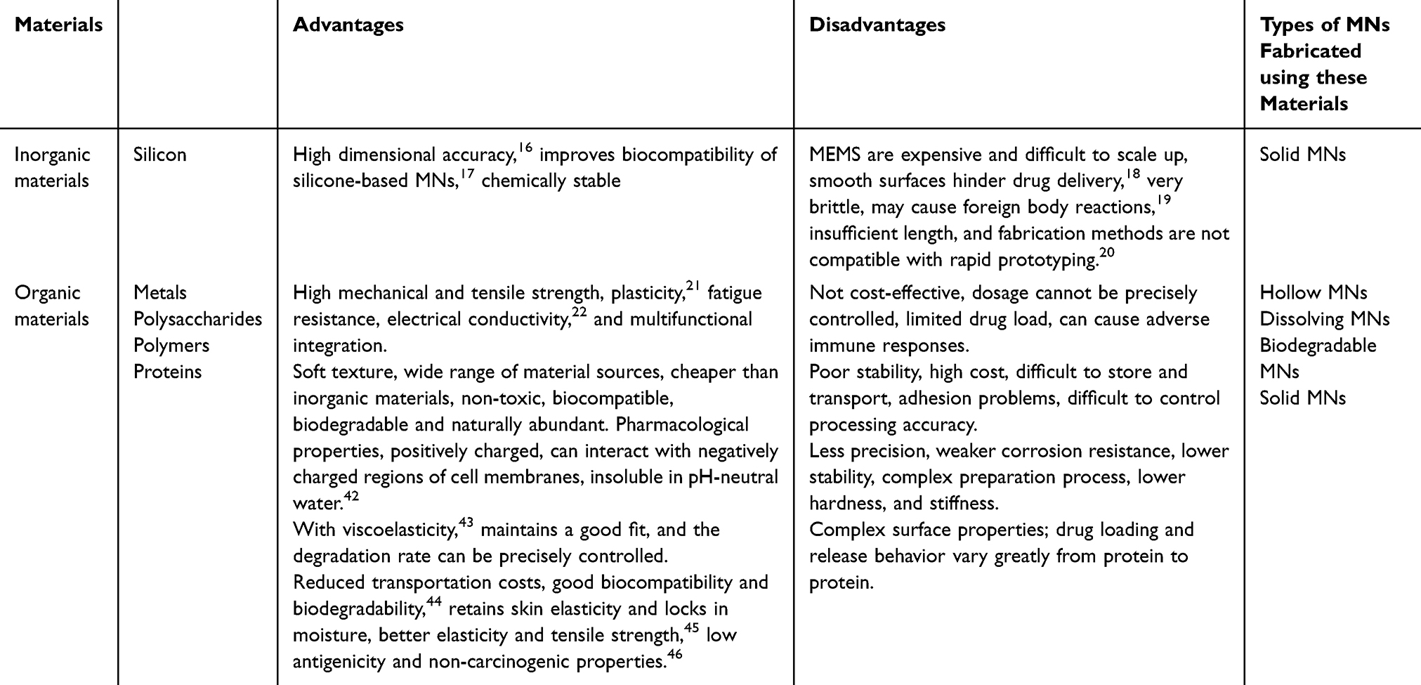 |
Table 1 Advantages and Disadvantages of MNs with Different Materials |
Methodology: Preparation and Characteristics of MNs
Preparation of MNs
When designing MN systems for skin penetration, several key factors need to be considered. First, material selection is crucial, as this impacts the system’s mechanical properties, biocompatibility, and stability. Second, performance characteristics—including the mechanical properties and geometric parameters—determine the MN’s penetration ability and efficiency. Third, manufacturing feasibility is essential, as it can limit the final design. The array layout, matrix density, and total MN count also play major roles in the system’s performance. MN systems are commonly classified by their array density, needle length/height, and morphological features. Other parameters such as their hydrodynamics, penetration, and production costs are also important. Their final design is constrained by the manufacturing process and material stability.
MNs can be divided into planar and flat categories. In-plane MNs can be complex to fabricate as 2D arrays but are easily integrated with other devices. Out-of-plane MNs are simpler to manufacture at high densities but have inferior aspect ratios and heights compared to in-plane MNs made using conventional techniques.20 A critical analysis of the current state of the field reveals several challenges. The complexity of fabricating in-plane MNs limits their large-scale production, despite their excellent integration potential. Conversely, the relatively poor aspect ratios and heights of out-of-plane MNs restrict their application in scenarios where deep penetration is required. Moreover, balancing all the design factors—including the material selection, performance, and costs, remains a considerable challenge in MN system design.
3D Printing
Photopurpose 3D printing—one of the oldest and most widely-used additive manufacturing technologies—employs UV light to cure light-sensitive liquid resin layer-by-layer for MN structure formation. Ren proposed a method to prepare flexible hollow MNs (HMNs) for psoriasis treatment using high-precision 3D printing technology combined with a dual-molding process with a reduced drug dosage. The core of the study was to solve the problems of the traditional HMN manufacturing process, which is complicated, costly, and difficult to mass-produce, through the high flexibility and customization capability of 3D printing technology. As shown in Figure 2A, the application of 3D printing technology can be demonstrated using the HMN preparation process and the efficient drug delivery capability and biosafety of HMNs in psoriasis treatment can be demonstrated using in-vivo fluorescence imaging and in-vitro porcine skin penetration experiments (Figure 2A).47 Although the technology can be used to produce complex MN shapes with high precision and accuracy, it exhibits a relatively slow printing speed. Fused deposition modeling (FDM) 3D printing is the most affordable technology, commonly used for electrochemical sensor manufacturing. A thermoplastic filament is heated, melted, extruded through a nozzle, and deposited layer-by-layer on a platform along a specific path.48 It can use various thermoplastic polymers, is cost-effective, reliable, and fast, and uses relatively inexpensive materials. However, its printing accuracy is inferior to that of light-cured 3D printing.
 |
Figure 2 Microneedling for psoriasis treatment. A (a) Flowchart of HMNs for bimorphic process and in vivo experimental fabrication. (b) Penetration experiments of HMNs on porcine skin in vitro. Adapted from Ren Y, Li J, Chen Y, et al. Customized flexible hollow microneedles for psoriasis treatment with reduced-dose drug. Bioeng Transl Med. 2023;8(4):e10530. © 2023 The Authors. Bioengineering & Translational Medicine published by Wiley Periodicals LLC on behalf of American Institute of Chemical Engineers, CC-BY4.0 license. 47 B (a) Preparation of dynamic MN for bilayer microbiota-assisted gases. (b) SEM image of the microneedle array. C (a) Fabrication process of MNs loaded with B Subtilis containing pAD plasmid. (b) Confocal laser scanning microscopy images of skin sections treated with different MNs. Adapted from Alperovitz CH, David NB, Ramot Y, Gross A, Mizrahi B. Living microneedles for intradermal delivery of beneficial bacteria. ACS Biomater Sci Eng. 2025;11(2):1232–1241. Copyright © 2025 The Authors. Published by American Chemical Society. CC-BY 4.0.49 |
In the current 3D printing field, both photopurpose and FDM 3D printing have their own advantages and limitations. Photopurpose 3D printing offers high-precision fabrication, which is crucial for applications that require intricate MN structures. However, its slow speed can be a bottleneck for large-scale production. Conversely, FDM 3D printing provides an affordable and fast alternative, making it accessible to a wide range of users. However, its lower accuracy restricts its use in high-end applications where fine details are essential. There is a clear trade-off between precision and speed/cost in these two dominant 3D printing technologies, and future research should aim to develop methods that can balance these factors. In psoriasis treatment, Zheng et al developed microbial microengine-based MNs (MM-MNs) that use gas-producing microorganisms to propel drugs deep into the skin, significantly enhancing transdermal penetration and delivery efficiency. These MNs enable precise control of drug release by regulating glucose concentration while maintaining favorable biocompatibility without causing noticeable tissue damage or toxic reactions. In an animal model, MM-MNs effectively delivered drugs for psoriasis treatment, rapidly alleviated symptoms, and significantly improved pathologic features (Figure 2B) As shown in Figure 2B and C, the application of microneedle technology in drug delivery and bioengineering is illustrated. In Figure 2B, the process of creating microneedle arrays involves applying and spin-coating B. Subtilis pellets, followed by drying and demolding to form the microneedle array. This method allows for the incorporation of beneficial bacteria into the microneedles, enhancing their functionality. The scanning electron microscopy images provide a detailed view of the microneedle structure, showcasing their precise and intricate design.49
Figure 2C further demonstrates the potential of microneedles in delivering genetic material, such as plasmids, into the skin. The fluorescence microscopy images compare the performance of empty microneedles and those loaded with BS-GFP (green fluorescent protein), highlighting the efficient delivery and expression of the genetic material over time. These results indicate the microneedles’ effectiveness in facilitating transdermal delivery and their promising applications in various biomedical and clinical scenarios.50 As shown in Figure 3A(a), the 3D printing method utilizes a computer and projector that employ 405nm blue light to form microneedles on a photosensitive substrate.51
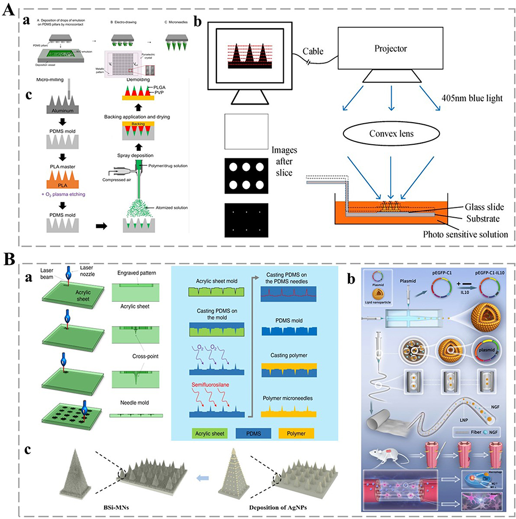 |
Figure 3 Fabrication methods of MNs. A (a) 3D printing method. Adapted from Onesto V, Di Natale C, Profeta M, Netti PA, Vecchione R. Engineered PLGA-PVP/VA based formulations to produce electro-drawn fast biodegradable microneedles for labile biomolecule delivery. Prog Biomater. 2020;9(4):203–217. Copyright © 2020, The Author(s). Creative Commons CC BY license.51 (b) Micro-injection molding method. Adapted from Yao W, Li D, Zhao Y, et al. 3D printed multi-functional hydrogel microneedles based on high-precision digital light processing. Micromachines. 2019;11(1):17. © 2019 by the authors. Licensee MDPI, Basel, Switzerland. This article is an open access article distributed under the terms and conditions of the Creative Commons Attribution (CC BY) license (http://creativecommons.org/licenses/by/4.0/).52 (c) Deposition method. Adapted from Kim MJ, Park SC, Rizal B, et al. Fabrication of circular obelisk-type multilayer microneedles using micro-milling and spray deposition. Front Bioeng Biotechnol. 2018;6:54. © 2018 Kim, Park, Rizal, Guanes, Baek, Park, Betz and Choi. This is an open-access article distributed under the terms of the Creative Commons Attribution License (CC BY).53 B (a) Electroplating and laser cutting methods. Adapted from Nejad HR, Sadeqi A, Kiaee G, Sonkusale S. Low-cost and cleanroom-free fabrication of microneedles. Microsyst Nanoengin. 2018;4(1):17073. Copyright © 2018, The Author(s). Creative Commons CC BY license.54 (b) Electrostatic spinning method. Adapted from Sun Y, Wu J, Zhou L, et al. Genetically engineered electrospinning contributes to spinal cord injury repair by regulating the immune microenvironment. Front Bioeng Biotechnol. 2024;12:1415527. Copyright © 2024 Sun, Wu, Zhou, Wang, Wang, Sun, Xu, Zhang, Jiang, Zhu, Xi, Gu and Chen. This is an open-access article distributed under the terms of the Creative Commons Attribution License (CC BY).55 (c) Lithography and etching methods. Adapted from Cheng W, Wang X, Zou S, et al. Fabrication of black silicon microneedle arrays for high drug loading. J Funct Biomater. 2023;14(5):245. © 2023 by the authors. Licensee MDPI, Basel, Switzerland. This article is an open access article distributed under the terms and conditions of the Creative Commons Attribution (CC BY) license (https://creativecommons.org/licenses/by/4.0/).18 |
Micro-Injection Molding for Polymer Materials
Micro-injection molding technology has the advantages of high precision, high efficiency, and strong shear stress fields, which can improve the mechanical properties and production efficiency of polymer micro-components, making it especially suitable for the manufacture of high-performance micro-components such as MNAs.56 Additionally, the technology can also achieve performance optimization by regulating the microstructure, affording it good prospects for industrial application.57 However, three major bottlenecks exist—specifically, the persistently high cost of high-precision molds (nano-featured molds > $20,000/set), thermal processing limitations for the integration of bioactive ingredients, and the exponential increase in microcavity filling defect rate with decreasing size (>12% defect rate for cavities below 50 μm).58 Although the technology currently holds 83% of the medical MNs market share (2022 data), it faces two paradoxes—that is, the contradiction between the need for clinical personalization and the high fixed costs of the molds, and the trend toward bio-integration and the compatibility of thermal processing processes.59 Recent advances in the industry have shown that composite mold technology (3D printed inserts + CNC base molds) is capable of reducing costs by 40%, but its feature resolution is still limited to the 20–50 μm range, which is insufficient to meet the needs of cutting-edge research.60 Academics have proposed a shift in process focus toward the synergistic innovation of adjustable mold systems and cryogenic machining.
In addition to micro-injection molding, micro-molding is a crucial technique for manufacturing polymer-based MNs. This method uses molds to shape polymer materials into desired micro-needle structures. It offers high precision and can produce complex geometries with good reproducibility. The micro-molding process can be optimized by adjusting temperature, pressure, and molding time to achieve optimal MNs characteristics. Researchers have employed micro-molding techniques to fabricate MNs from various polymer materials, demonstrating effectiveness in transdermal drug delivery applications.61 This method shows promise in addressing challenges of micro-injection molding, particularly in cost and feature resolution. However, further investigations are required to enhance the biocompatibility and mechanical strength of MNs produced through micro-molding for biomedical applications. Figure 3A(b) illustrates the deposition method, where BSI-MNs are created and AgNPs are deposited on them, adding specific nanoparticles to boost microneedle functionality.52
Deposition
This technique employs a deep reactive ion etching process to fabricate sharp, hollow Si MNs that are valuable in minimally invasive diagnostic applications. There are two main methods of etching substrate materials using photoresists as a mask—namely, chemical etching and dry etching. Chemical etching removes unwanted portions through chemical reactions, whereas dry etching relies on physical impact and reaction with gas molecules. After the etching has been completed, the MNs is then obtained by removing the remaining photoresist. To prepare Si-based MNs, planar MNs are first formed using laser patterning and alkali etching. Subsequently, nanowire structures are created on the surface by silver-catalyzed chemical etching to form novel black Si MNs (BSi-MNs).18 Sarabi et al post-processed 3D-printed PLA MNAs by chemical etching to enhance their geometric accuracy. This chemical etching used a potassium hydroxide (KOH) solution to remove the excess created during the printing process and optimized the shape and size of the MNs to improve their performance in transdermal drug delivery and skin puncture applications.61
The fabrication of MNs using deep reactive ion etching has shown potential in the field of minimally invasive diagnostics. However, the process can be complex and costly, which limits its mass production. Fabrication of BSi-MNs through multiple etching steps increases the complexity of fabrication. Additionally, the long-term stability and biocompatibility of these etched MNs need to be further investigated to ensure their safety and efficacy in practical applications. In Figure 3A(c), the electrostatic spinning method involves elements like plasmid, lipid nanoparticles, pEGFP-C1, and IL10 to produce polymer microneedles with unique properties enhancing drug delivery.53
Electroplating and Laser-Cutting for Metallic Materials
The conductive metal seed crystal layer and release layer are first sputtered sequentially on the substrate, patterned by alternating layers of photoresist lithography and metal electrodeposition to build the MN structure, after which the final photoresist is removed to release the MNs.20 However, layer alignment accuracy in multi-step processes remains a bottleneck, as achieving tolerances of less than 5 µm is challenging for industrial-scale production.62 Material compatibility limitations are restricted to precious metals (eg, gold and nickel) owing to the requirement of seed crystal layers. Structural defects in high aspect ratio MNs arise from residual stresses caused by repeated deposition.63 Figure 3B(a) depicts the electroplating and laser cutting method, showing the steps from aluminum to the spray deposition of polymer drug solution.54
Laser-cutting pre-programmed 2D/3D digital models can be used to achieve precise cuts, with post-processing (deburring, cleaning, polishing) to improve surface properties.64 The challenge with laser cutting is that thermal damage induces micro-cracks in parts smaller than 100 µm, reducing the mechanical integrity. Moreover, material waste can exceed 40% in complex 3D structures, and Post-processing can account for 30–35% of the total production time.65 Industry trends show increasing use of hybrid manufacturing (eg, laser-electroplating combinations) to mitigate the weaknesses of individual methods, although interfacial consistency between layers remains an unresolved issue.
Electrostatic Spinning for Polymers, Proteins, and Polysaccharides-Based Materials
In electrostatic spinning, a precursor solution forms a jet under strong electric field conditions. As it travels, the solvent evaporates, and the polymer/protein/ polysaccharides solidifies. The resulting fibers are deposited on a collector to create an MN-like structure. Parameters such as the electric field strength and solution concentration control the diameter, length, and orientation of the MNs.66 This technique has clear advantages in its ability to precisely control the MN characteristics. However, it faces challenges in scalability for large-scale production. Moreover, the potential impact of strong electric fields on the biological activity of proteins and polysaccharides in the precursor solution needs further investigation. In Figure 3B(b), the micro-injection molding method is demonstrated, illustrating the process from PDMS pillars to the creation of polymer microneedles.55
Photolithography and Etching for Si and Other Materials
Photolithography and etching (for Si and other materials) consist of two core stages—that is picture transfer and material etching. The photoresist-coated substrate is selectively exposed (eg, using UV light) through a mask plate, which triggers a chemical change in the exposed area; after development, the unexposed area is removed to form a photoresist template for the MN pattern, which is then used to guide an isotropic/anisotropic etch (eg, plasma etching) to transfer the pattern to the substrate.67 However, the technology still has some challenges. First, the resolution is limited. Traditional ultraviolet lithography faces difficulties in sub-10-nanometer processes owing to light diffraction, prompting a reliance on extreme ultraviolet (EUV) lithography, which has grown exponentially in cost and complexity. Second, masks also have limitations. Multiple patterning techniques (eg, double/quadruple patterning) introduce alignment errors and reduce yield, and EUV mask defects remain a key bottleneck. Third, material compatibility. Emerging materials (2D semiconductors, flexible substrates) require new photoresist chemicals and etchants, but process standardization is lagging. Fourth, there are several environmental considerations. Toxic etchants (eg, hydrofluoric acid) and photoresist solvents require expensive waste disposal processes, which contradict green manufacturing trends. As shown in Figure 3B(c), the lithography and etching method uses a laser beam to create an engraved pattern on an acrylic sheet, forming a needle mold. This method is precise and enables the creation of detailed microneedle structures.18 The various MN preparation methods are summarized in Table 2.
 |
Table 2 Various MNs Preparation Methods and Their Respective Advantages and Disadvantages |
Characteristics of MNs
Mechanical Properties
Strength
The strength of MNs can be influenced by multiple factors. Material properties clearly play a crucial role. Metal MNs, with their high yield strength and modulus of elasticity, deform minimally and resist fracture under external forces. Their strength can be enhanced through alloying, processing, or compositing.68 Polymer-based MNs strength depends on the polymer molecular weight and crystallinity; for example, high-molecular-weight and high-crystallinity PLA MNs exhibit better strength for skin piercing.69 The structural strength of Si and (PEG diacrylate) (PEGDA) MNAs was investigated in detail using ANSYS finite element analysis software. The results showed that the Si MNs, owing to their high mechanical strength and modulus of elasticity, exhibited minimal deformation under external forces and were able to effectively resist flexural failure, similar to the properties of metallic MNs. By contrast, PEGDA MNs experienced considerable deformation and buckling failure at loads exceeding 0.1 N, which indicated their relative lack of strength and toughness. Additionally, the Si MNs exhibited excellent performance in penetrating the simulated skin, generating a stress much greater than the skin resistance (3.18 MPa), and were able to penetrate the skin with ease, whereas the PEGDA MNs exhibited some penetration ability albeit at a lower load (0.1 N).70
Critically, although existing research has pinpointed diverse factors that can impact the strength of MNs, there is a dearth of a holistic understanding regarding the interactions among these factors. For instance, in real-world applications, the way in which different factors jointly affect the MNs strength remains ambiguous. Moreover, the practice of using simulated substrates in puncture tests is problematic. These simulated substrates fail to accurately mimic the intricate mechanical properties of human skin. Consequently, such tests can lead to inaccurate assessments of the strength and effectiveness of MNs.
Hardness
Hardness refers to the ability of MNs to resist localized deformation, typically measured using spectrometry, microscopic manipulation, or mass testing equipment. The mass-testing method involves fixing the MNs to its base, applying pressure via a probe, and recording the pressure-displacement data using high-precision sensors to plot a force-displacement curve. The force for a certain deformation, obtained from the curve, reflects the MNs hardness.71 Morphological changes after external force application can be observed under a microscope; low-hardness MNs bend, deform, or are damaged under low-pressure conditions.72
Metal MNs are generally harder than polymer-based MNs. However, polymers offer good biocompatibility and degradability, which are beneficial in specific applications. Consequently, material selection should be based on the application requirements. For polymer-based MNs, the hardness is influenced by the polymer molecular weight; higher-molecular-weight polymers usually exhibit greater hardness and strength but lower flexibility and processability.73 Crosslinking or adding reinforcing phases (eg, nanoparticles) can enhance polymer hardness.74 The structural design, including the MNs shape, size, and needle-tip angle, can also impact the hardness. Sharper-tipped and thicker-bodied MNs are harder but can increase skin resistance and patient discomfort.
Current research shows a clear trade-off between the hardness of MNs and other properties. For example, although metal MNs are hard, they lack the biocompatibility and degradability of polymer-based MNs. The relationship between the polymer molecular weight and hardness also presents a dilemma, as high-hardness polymers may sacrifice processability. Moreover, the structural design to increase hardness can have a negative impact on the patient experience. Future research should focus on developing materials or designs that can optimize multiple properties simultaneously, such as creating polymers with high hardness without compromising flexibility and processability or designing MNs with high hardness and low skin resistance.
Toughness
The toughness of MNs can be defined as the ability to resist fracture when penetrating the skin.75 It reflects the material’s capacity for plastic deformation and energy absorption before breaking, as distinct from the hardness which measures the surface resistance to localized deformation. High-toughness MNs can better withstand external forces during use, enhancing their safety and reliability, as the skin’s elasticity and complex structure subject MNs to multi-directional forces. Insufficient toughness can lead to MNs breakage in the skin, causing inflammation and infection. Common toughness tests include the single-pin tensile test, Charpy impact test, and simulated skin puncture test. The single-pin tensile test measures the maximum load and elongation before fracture; the Charpy impact test evaluates the energy absorption under impact; and the simulated skin puncture test (using materials like PDMS), focuses on the overall toughness in a real-skin-like environment and reflects fracture resistance under repeated piercing or skin deformation.76
Multiple factors can affect the toughness of MNs. The material nature is crucial, with metal alloys sometimes being brittle and polymers generally being tougher.77 The MNs geometry also matters, as thicker and shorter MNs are more fracture-resistant, whereas longer and thinner MNs are more prone to breakage. The MNs preparation process—such as the molding method for polymer-based MNs—can also influence their toughness.
The current testing methods focus primarily on mimicking the mechanical behavior during MNs penetration. However, they may not fully capture the complex in-vivo environment, such as the interaction with skin cells and biological fluids. Regarding material selection, although polymers are generally considered tougher, more research is needed to optimize their properties for specific applications. The impact of the preparation process on toughness has been acknowledged, but there remains a lack of in-depth understanding of precisely how different steps can precisely affect the MNs toughness.
Geometric Parameters
Length
MNs range from 50–900 μm in length and are generally of the order of 1 μm in diameter78 The MNs length can have an enormous effect on its performance and application. Shorter MNs face less resistance during the puncture process and require less mechanical strength. In transdermal drug delivery, they can transport drugs through the cornea without affecting deep nerves and blood-rich tissues.79 They are also suitable for delivering vaccines that act on epidermal immune cells. In biosensors, short MNs can detect surface biosignals such as sweat composition.80 Conversely, long MNs can penetrate the epidermis and reach deep into the dermis, enabling the delivery of large molecules such as genetic drugs that act at the cellular level.81 They can also deliver vaccine components to the dermis with more immune cells for a stronger immune response. In biosensors, long MNs can access deeper interstitial fluids to detect signals such as blood glucose levels.
Current research has clearly demonstrated the different advantages of short and long MNs in drug delivery and biosensing. However, there has been a lack of in-depth studies on the long-term effects of MNs use, especially for long, deeper-penetrating MNs. The potential damage to tissues, risk of infection, and impact on the body’s normal physiological functions need further investigation. Additionally, the standardization of MNs manufacturing—especially in terms of the ss length and mechanical properties—remains insufficient, which affects the reproducibility and reliability of experimental results and clinical applications.
Density
The density of MNs can be defined as the number of MNs per unit area, and it can impact the drug-delivery efficiency enormously. A higher density means more MNs available for skin puncture, which improves the drug-delivery efficiency. In transdermal drug delivery, an appropriate MN density can reduce skin damage, pain, inflammation,82 and the chances of infection while still effectively puncturing the stratum corneum to promote drug absorption.83 It also allows the skin to heal better.84 Groups with many MNs can reduce the mechanical strength of individual MNs, as the reduced distance between the MNs and changes in their mutual support and stability owing to external forces mean that the MNs can be more prone to damage.85
Shape
The shape of the MNs is a key parameter that determines their performance and therapeutic efficacy.86 For example, pyramid-shaped MNs with hexagonal bases exhibit higher compressive stresses and lower critical buckling loads compared to those with square or triangular bases, resulting in considerably shallow penetration depths.86 Compared to hexagonal MNs, square and triangular MNs with sharp edges show greater ability to penetrate the skin.15 Moreover, compared to pyramid-shaped MNs, conical MNs require less force to penetrate the skin.87 These differences can be attributed to variations in the MNs–skin contact area.
Drug-Loading and Release Properties
Drug-Loading Capacity
Drug recovery poses a major challenge in preparing MNs for drug delivery, influenced by multiple factors. The small size and capacity of MNs patches inherently limit the administration dose, typically to less than 1 mg per patch, though it can reach up to 10 mg.88 This limitation is problematic for high-dose drugs. Many drugs can diffuse through water-soluble MNs matrices, complicating their encapsulation and localization within the MNs.89 Owing to the single-material nature of most MNs, drug-substrate interactions restrict the co-encapsulation of different drugs. Coated MNs capsules can only deliver approximately 1 mg of a drug, whereas hollow MNs capsules offer the advantage of continuous infusion or “on-demand” drug delivery. The penetration of MNs through the skin barrier depends primarily on the passive diffusion of the biological agent into the skin, making it difficult to administer large doses, and a substantial part of the dose can be lost at the skin surface.90 Critically, the current limitations in drug dose, encapsulation, and delivery efficiency suggest that more research is needed to develop MNs with improved drug-loading capacity, better encapsulation methods, and enhanced delivery mechanisms.
Drug Release Performance
The development of controlled-release MNs systems can be influenced enormously by the inherent properties of the matrix material, binding affinity of the drug, and in-vivo environment. In the absence of external stimuli, drug release from MNs is typically adjusted through delayed diffusion, solubilization, and degradation control.91
Nonetheless, MNs can achieve rapid drug release. For example, Wang et al developed bubble MNs (BMNs) with a substrate-integrated bubble structure to concentrate drugs at the needle tip, enhancing delivery efficiency. BMNs achieved > 80% drug release within 20s. Moreover, in-vivo BMNs reached 79%, 83%, and 86% efficiency after 10, 20, and 30s, respectively, considerably outperforming traditional MNs (TMNs), highlighting BMNs’ practical potential.89
As shown in Figure 4A(a and b), the design of the BMNs can achieve a centralized distribution of the drug at the tip of the needle through the bubble structure, which improves the drug-release efficiency. This design enables the BMNs to release the drug rapidly when inserted into the skin, a considerable improvement on the drug-release efficiency of conventional TMNs (Figure 4A(a and b)). Such minimally invasive systems produce tiny pores in the skin to overcome the barrier properties of the stratum corneum, their application greatly reducing the distance for drug diffusion, so that the drug can quickly reach its point of action near the epidermis and dermis.92 Drug release can also be precisely regulated by changing the length, density, and shape of the MNs and the amount of the loaded drug. A well-designed MNA can ensure even distribution of the drug at an accurate dosage into the skin, whereas traditional ointments can be hampered by uneven skin surfaces, hair, or different diffusion rates.93 MNs can also selectively target different skin layers. For example, short MNs can release drugs into the epidermis to treat superficial skin diseases such as mild eczema,94 whereas long MNs can release drugs deep into the dermis to treat deep skin infections95 or deliver vaccines.96 The material used in MNs can also have an impact on the drug release properties. Yang et al developed a zinc-CUR framework (ZnMOF) MNs patch for hair growth promotion that achieved a stable, sustained release of zinc ions (Zn2+) and CUR through the creation of temporary pores in the skin. As shown in Figure 4A(c), the MNs can achieve effective drug release by encapsulating and delivering ZnMOF particles. The penetration ability of the MNs and the encapsulation stability of the drug are the key factors in achieving controlled drug release (Figure 4A(c)).97 MNs made of biodegradable materials break down gradually after entering the skin, releasing the drug slowly, whereas MNs made of non-biodegradable materials may need to be manually removed after the drug release process, although they can sometimes be more useful, such as when rapid drug delivery is required or interference from residual materials is undesirable. Yang et al developed a degradable MN, made of photocrosslinked gelatin methacrylamide (GelMA), which was capable of slow and sustained drug release in response to high reactive oxygen species and overexpression of matrix metalloproteinases (MMPs) in the pathological microenvironment. This MN degraded gradually in the skin, avoiding the inconvenience of manual removal at the end of drug release. As shown in Figure 4B, the MN could achieve effective treatment of hypertrophic scarring through responsive release of drugs (Figure 4B).98
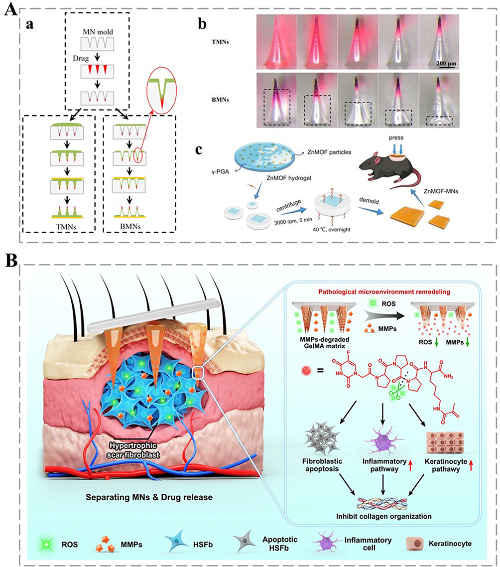 |
Figure 4 Drug loading. A (a) Manufacturing TMS and BMN manufacturing processes. (b) Concentration/viscosity and bubble height analysis of PVA solutions. (a) and (b) adapted from Wang QL, Zhu DD, Liu XB, Chen BZ, Guo XD. Microneedles with controlled bubble sizes and drug distributions for efficient transdermal drug delivery. Scientific Reports. 2016;6(1):38755. Copyright © 2016, The Author(s). Creative Commons CC BY license.89 (c) Synthesis mechanism of ZnMOF-MNs. Adapted from Yang Y, Wang P, Gong Y, et al. Curcumin-zinc framework encapsulated microneedle patch for promoting hair growth. Theranostics. 2023;13(11):3675–3688. ©2025 Ivyspring International Publisher. Creative Commons Attribution License (https://creativecommons.org/licenses/by/4.0/).97 (B) Schematic representation of MNs therapy to enhance tissue repair by slow release of drugs, induction of fibroblast apoptosis, and inhibition of collagen synthesis. Adapted from Yang ZR, Suo H, Fan JW, et al. Endogenous stimuli-responsive separating microneedles to inhibit hypertrophic scar through remodeling the pathological microenvironment. Nat Commun. 2024;15(1):2038. Copyright © 2024, The Author(s). Creative Commons CC BY license.98 |
Biocompatibility
Biocompatibility refers to the ability of materials to make contact with living organisms—primarily skin tissues in the case of MNs—without causing harmful reactions. Biocompatible materials coexist harmoniously with tissues without causing immunogenic reactions, in which the body attempts to reject the MNs as a foreign object.99 HA-based MNs blend well with human tissue, as HA can be degraded in vivo by hyaluronidase in a natural process100 Finer and shorter MNs—generally between tens of microns and several millimeters in length—cause less physical trauma compared to traditional injection needles and allow quicker skin healing afterward.101 Chemically stable materials prepared without using harmful organic solvents are also advantageous in terms of their chemical compatibility and safety.102 Moreover, MNs with good biocompatibility should not induce transient inflammatory and immune reactions. Li et al developed a PFG/M MN patch with good biocompatibility, which was embedded with Fe/PDA@GOx@HA (Fe/Polydopamine@Glucose Oxidase@Hyaluronic Acid) and AP-MSNs (Amine-modified) at the tip and base, respectively. Mesoporous Silica Nanoparticles, exhibited excellent antimicrobial and immunomodulatory properties. No major inflammatory response or tissue damage was evident in both the cell and animal experiments, indicating that the MNs system could coexist harmoniously with living organisms without inducing immune rejection while promoting the healing of infected wounds.103 The biocompatibility of MNs was demonstrated in a study by Haghniaz et al, where MNs composed of GelMA and silicate nanosheets exhibited good biocompatibility. These properties indicate that MNs can integrate well with tissues and will not be recognized by the body as a foreign body that triggers a rejection reaction, further demonstrating their potential as biocompatible materials.104
Cytotoxicity
Cytotoxicity, the harmful effects of a material (or its extracts) on cells, is a crucial safety concern. Cytotoxic MNs can damage skin cells, impede therapeutic effects, and cause serious health issues. Cytotoxicity stems from chemical and physical factors. Chemically, some MNs materials release harmful chemicals like unreacted monomers, residual catalysts, or additives upon contact. In-vivo degradation products of MNs materials can also be toxic. Metallic MNs may release metal ions; at high concentrations, these ions disrupt normal cell functions such as membrane permeability105 and enzymatic activity.106 Physically, MNs with sharp tips or long dimensions cause excessive cell damage. Overly long MNs can penetrate below the dermis, harming deeper-tissue cells and blood vessels, leading to inflammation and cellular necrosis.
While the understanding of MNs cytotoxicity has advanced, there are still gaps. For instance, the long-term effects of low-level, chronic exposure to MNs-released substances need further investigation. Moreover, standardized testing methods for cytotoxicity across different MNs materials are lacking, which hinders accurate risk assessment.
Skin Irritation
MNs can cause adverse skin reactions, such as erythema107 edema,108 pruritus,109 soreness,110 during skin contact and penetration. Insertion often makes nearby skin turn red, which is a typical sign of irritation. The degree of irritation is influenced by the size, shape, (material’s) chemical properties, and biocompatibility of MNs, as well as the penetration method (speed, angle, and depth). Rapid or improper-angled punctures may cause more severe damage and higher irritation risks. Less biocompatible MN materials are more likely to trigger immune responses leading to irritation.111
Critically, while current research has identified these factors, there is a lack of standardized guidelines on optimal MNs design and insertion methods to minimize skin irritation. The interaction between different material properties and the immune system also needs more in-depth study. This knowledge gap hinders the wide-scale application of MNs in the medical field.
Degradability
MNs eliminate the step of manual removal after application as degradable MNs naturally break down,112 reducing patient discomfort and infection risks.113 However, the chemical structure and composition of the material, as well as environmental factors, can affect the rate of degradation. Consequently, in drug delivery applications, MNs degradability can be leveraged to control the drug release rate. For example, in tissue applications, the degradation of HA and chitosan results in the formation of small molecules that promote tissue repair and regeneration. Conversely, irritating degradation products can cause local inflammatory reactions.114
Current research inadequately addresses the trade-off between tunable degradation rates and structural integrity. For instance, overly rapid degradation of chitosan-based MNs may compromise drug delivery precision.115 Despite claims of “biocompatibility”, few studies systematically evaluate long-term inflammatory responses to degradation byproducts, especially in immunocompromised populations.116 Most MNs degradation studies rely on idealized in vitro conditions (eg, controlled pH/temperature), neglecting variability in human tissue microenvironments.117
Results: Multifunctional MNs in BME
Drug Delivery
Transdermal Delivery
MNs offer four drug-delivery methods to the epidermis: knotting-sticking (solid MNs), knotting-release (soluble and frozen MNs), coating delivery (coated MNs), and injection delivery (hollow and gel-based MNs).118 The primary mechanism of MNs drug delivery is skin-barrier disruption and subsequent drug release into the upper dermis for systemic absorption.119 Drug release from MNs post-skin-barrier crossing can be either dissolution-or diffusion-based. Non-biodegradable solid MNs release drugs by diffusion, while coated and biodegradable MNs do so by dissolution.120 Solid non-degradable MNs can create skin microchannels, facilitating passive drug diffusion through them.121 These microchannels significantly enhance drug effectiveness by enabling unhindered passage through the stratum corneum.
Transdermal drug delivery via MNs is highly advantageous; it is non-invasive, convenient, avoids first-pass metabolism and gastrointestinal degradation, and is patient-friendly owing to painlessness, self-administration, and single-dose slow release.122
This advanced technology has far-reaching impacts. It improves patient compliance, especially for those averse to injections or oral medications. The slow-release feature also provides more stable drug levels in the body. Future research could focus on optimizing MNs design for specific drugs, exploring new materials for better biocompatibility and drug-loading capacity, and investigating the long-term effects of microchannel creation on skin health.
Precision and Responsive Release
Spatial Control
MNs can precisely deliver drugs to specific skin depths. Because it can be designed in different lengths, it can pass through the outermost stratum corneum of the skin and release the drug directly into the epidermis or dermis, which can effectively prevent the drug from being degraded or wasted before it reaches the target location. Some protein drugs like insulin can be delivered locally to reach an effective concentration in the skin through MNs delivery.
Stimulus Response Systems
MNs can respond to a variety of endogenous or exogenous stimuli to enable drug delivery. In diabetes treatment, glucose-responsive MNs can detect changes in blood glucose concentration. When blood glucose increases, the MNs changes its structure or properties accordingly to accelerate the release of insulin. Whereas, when blood glucose decreases, drug release slows down. It releases insulin dynamically with the help of pH-sensitive polymers such as phenylboronic acid complexes.123 pH/enzyme-responsive microneedling technology accelerates anti-inflammatory drug release in acidic/inflammatory microenvironments or enzyme-enriched lesions through acid-triggered degradation or protein conformational shifts of inorganic nanomaterials. For example, mesoporous silica nanoparticles (MONs) are used as novel antitumor drug carrier materials with neoantigenic peptides attached to the surface via disulfide bonds, which are rapidly cleaved in a highly reduced tumor microenvironment (TME) to achieve precise release of anticancer drugs.124 This approach significantly improves the drug’s ability to kill tumor cells while reducing adverse effects on normal cells.
Sustained Release
MNs can be designed to release the drug at a stable rate through the selection and design of the material. For example, degradable MNs (eg, GelMA hydrogel) can achieve a long-lasting drug effect through the gradual disintegration of the material, and the controlled release system of MNs can regulate the rate of drug release based on environmental factors, such as temperature and pH. By using materials that are sensitive to these factors, MNs can dynamically adjust the rate of drug release in response to changes in the local environment of the skin (eg, changes in pH at the site of inflammation), allowing for more targeted drug delivery.122
Biological Detection and Sensing
Body Fluid Sampling
In terms of body fluid sampling, the MNs can be used to extract a wide range of body fluids. For example, to collect interstitial tissue fluid (ISF), it can puncture the skin’s stratum corneum and create a series of micropores in the skin with a slight suction to reach near the dermis, allowing ISF to ooze out, and subsequently collect these fluids.125 In terms of sweat collection, sweat is produced by sweat glands located in the dermis and subcutaneous tissue layers that extend into the epidermis. It helps to regulate body temperature, excrete metabolic wastes, and maintain skin acidity.81 MNAs can be pierced into the skin near the sweat glands to collect sweat efficiently. Moreover, compared to traditional methods, MNs extraction of sweat can reduce the interference of external factors on the composition of sweat, and more accurately detect the electrolytes, metabolites, and other components therein.MNs have also been used in blood collection, and some MNs have been designed to pierce superficial blood vessels in the skin to obtain a small sample of blood. This approach causes far less pain and trauma than traditional blood collection needles and is particularly suitable for situations where frequent blood testing is required, such as blood glucose monitoring in diabetic patients.126
Biosensor Integration
The use of MNs for biosensing is based on the fact that its tiny size can painlessly penetrate the surface of the skin to reach tissue areas with biosignals. The principle is to modify the MNs surface with biosensitive materials that can recognize specific biomarkers (eg, glucose, proteins, etc). In recent years, the application of MNs in biosensing has been expanding; for example, MNAs fabricated by 3D printing technology have been used to develop portable, fast-response field assay point-of-care (POC) biosensors capable of real-time monitoring of biomarkers (such as blood glucose and ketone bodies), exhibiting enormous potential in disease diagnosis and health management applications. As shown in Figure 5A(a), the combination of an MNA and POC biosensor fabricated by 3D printing technology was demonstrated. Figure 5A(b) shows the application of a 3D-printed MNA in drug delivery and monitoring applications. Moreover, fluorescence images reveal the wider drug diffusion and deeper penetration (in rat skin) of a 3D-printed micro-heater integrated MNs patch. This technology could not only deliver drugs, but it could also monitor drug release in real time, advancing disease diagnosis and health management (Figure 5A).127 Zheng et al developed an MN that was successfully applied to biosensing, and by combining it with electrochemical test strips, the accurate detection of glucose and alcohol in skin interstitial fluids could be realized, offering new possibilities for minimally invasive and continuous health monitoring applications.
 |
Figure 5 Biosensing. A (a) Schematic of MNAs with immediate (POC) biosensor technology. (b) In vitro device and fluorescent dye images in skin. Adapted Sarabi MR, Nakhjavani SA, Tasoglu S. 3D-printed microneedles for point-of-care biosensing applications. Micromachines. 2022;13(7). © 2022 by the authors. Licensee MDPI, Basel, Switzerland. This article is an open access article distributed under the terms and conditions of the Creative Commons Attribution (CC BY) license (https://creativecommons.org/licenses/by/4.0/).127 (B) Devices for monitoring glucose or alcohol levels in interstitial skin fluids. C (a) Enlarged view of MNs glucose sensor and structure. (b) MN alcohol sensors stably detect alcohol and interfering substances. Adapted from Zheng M, Zhang Y, Hu T, Xu C. A skin patch integrating swellable microneedles and electrochemical test strips for glucose and alcohol measurement in skin interstitial fluid. Bioeng Transl Med. 2023;8(5):e10413. © 2022 The Authors. Bioengineering & Translational Medicine published by Wiley Periodicals LLC on behalf of American Institute of Chemical Engineers. Creative Commons CC BY license.128 |
A skin patch design integrating expandable MNs with electrochemical test strips is shown in Figure 5B. The MN penetrated the skin to extract interstitial tissue fluid (ISF) and was combined with electrochemical test strips via a chitosan connecting layer to enable real-time detection of glucose and alcohol. This design provided a low-cost and convenient solution for biosensing. As shown in Figure 5C(a), the structure of the MNs after successful integration with electrochemical test paper. The MNs maintained a sharp pyramidal shape and an ordered array. The chitosan film locked the swollen MNs to the surface of the test paper, ensuring structural stability and providing a reliable physical foundation for biosensing.
Figure 5C(b) shows that the MNs glucose sensor exhibited good immunity to interference when detecting glucose. Even in the presence of multiple interfering substances, the sensor exhibited a stable, substantial response to 2 mM glucose and an insignificant response to interfering substances, proving the accuracy and reliability of the system in detecting glucose (Figure 5B and C).128
When these markers were combined with sensitive materials, physical or chemical signal changes could be generated (eg, optical signals, electrochemical signals). By detecting these signal changes, the markers could be analyzed qualitatively or quantitatively, thus realizing biosensing functionality.129 Consequently, an integrated MN biosensor was fabricated via 3D printing technology for continuous glucose monitoring (CGM) in diabetic patients. The device was capable of monitoring subcutaneous glucose concentration in real time, and its results were highly consistent with those of conventional glucose meters, demonstrating enormous potential in diabetes management applications (Figure 6A).130 Considering the specific correlation between blood glucose and glucose concentrations, the MN was micromachined using DLP 3D printing for CGM applications. Razzaghi et al improved the skin penetration of MNs by adjusting the print tilt angle. Experiments showed that MNs printed at a 45° tilt angle exhibited the lowest penetration force, which decreased by 38% compared to those printed at no tilt angle. This optimization provided a more efficient and comfortable solution for the application of MNs in CGM.
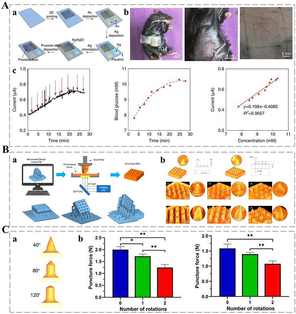 |
Figure 6 Continuous glucose monitoring applications. A (a) Schematic of the preparation process of MNs for biosensors. (b) MNAs biosensors applied to pinhole magnification images of mice, their abdomens, and their skin. (c) Current time-dependent curves and correlations of biosensing devices used to detect subcutaneous glucose after glucose injection in normal mice. Adapted From Liu Y, Yu Q, Luo X, Yang L, Cui Y. Continuous monitoring of diabetes with an integrated microneedle biosensing device through 3D printing. Microsyst Nanoeng. 2021;7:75. Copyright © 2021, The Author(s). Creative Commons CC BY license.130 B (a) DLP Schematic with MNAs and MNAs Tilt Print Angle Variations. (b) 3D model of solid and hollow MNAs with different tilt angles. C (a) Schematic of different needle angles. (b) Puncture force of solid MNAs 3D-printed without a printing tilt angle and with one or two tilting angles. (*): p-value < 0.05, and (**): p-value < 0.01. Adapted from Razzaghi M, Akbari M. The effect of 3D printing tilt angle on the penetration of 3D-printed microneedle arrays. Micromachines. 2023;14(6):1157. © 2023 by the authors. Licensee MDPI, Basel, Switzerland. This article is an open access article distributed under the terms and conditions of the Creative Commons Attribution (CC BY) license (https://creativecommons.org/licenses/by/4.0/).131 |
As shown in Figure 6B and C, the model structure of 3D printed MNAs with different tilt angles could be demonstrated (including the 3D model and size comparison of solid and hollow MNAs), as well as the physical printing results of the two types under different tilt parameters; here, the solid MNA exhibited no tilt, uniaxial 45° tilt, and biaxial 45° tilt states, whereas the hollow MNA achieved structural optimization through multi-axial adjustment of the same tilt angle (D), verifying the adaptability of tilt printing to complex geometries.131
Combination Therapy
MNs Electrical Stimulation
MNs electrodes can deliver electrical currents into the skin tissue. This electrostimulation can modulate the immune system and endogenous cytokine production, eliminating the need for external immunosuppressive drugs.132 Electrostimulation stimulates the contraction of skin cells, nerve endings, and muscles, promoting local blood circulation and skin metabolism. Moreover, it enhances the oxygen and nutrient delivery capability of skin cells, accelerates the elimination of waste products, stimulates the activity of fibroblasts, promotes the production of collagen and elastin, tightens the skin, and reduces wrinkles. It can also regulate skin physiology, reduce early inflammation, enhance immune cell and cytokine recruitment, and accelerate wound healing,133 offering a non-drug approach to skin treatment. Future research could focus on optimizing stimulation parameters for better efficacy, exploring its long-term safety, and investigating its potential in treating more complex skin conditions.
MNs Light Stimulation
Using near-infrared (NIR)-responsive MNs photothermolysis, soluble MNs work with an NIR laser to ablate diseased cells in a minimally invasive manner while releasing medication to protect the epidermis.134 The blue-light-activated MNs system targets the 400–470 nm band to effectively inhibit Propionibacterium acnes and melanoma, with the MNs also enhancing light penetration.135 There are also MNs-assisted light delivery applications, with micro-channels that enhance light absorption in the deeper layers of the skin to precisely treat scarring, hyperpigmentation, and hair follicle regeneration. Building on this, Yue et al developed a bilayer MNs system activated by NIR light for photothermal/chemotherapeutic synergistic treatment of melanoma. By integrating the photothermal agent IR820 and CUR nanodrugs (Cur NDs) into the HA-MNs, the system achieved efficient photothermal conversion and drug release under 808 nm NIR light irradiation. Compared with the blue light-activated MNs system, the NIR light has deeper tissue penetration and can more efficiently convert light energy into heat energy for the precise treatment of melanoma. In addition, this MN system treats tumors while its biodegradable back layer promotes skin regeneration and accelerates wound healing. As shown in Figure 7A, the bifunctional MN system consists of CUR nanomedicine/IR820/HA-MN and a sodium alginate/gelatin/HA backlayer, providing both photothermal/chemotherapeutic and skin repair functions. As shown in Figure 7B, IR820 significantly enhanced the photothermal conversion efficiency of the MNs, allowing it to heat rapidly under near-infrared light irradiation. H&E staining results showed that the MNs system not only effectively treated melanoma, but also promoted skin tissue regeneration and accelerated skin repair. The live/dead cell staining experiments (Figure 7C) showed that the tumor cell mortality rate was the highest in the Cur NDs/IR820/HA MNs + laser group, demonstrating its efficient anti-cancer ability. These results suggest that this dual-function MN system provides a new solution for melanoma treatment and postoperative skin repair (Figure 7).136
 |
Figure 7 Light stimulation. A (a) Schematic showing the preparation of Cur NDs/IR820/HA MNs. (b) Schematic of Cur NDs/IR820/HA MNs for tumor chemotherapy photothermal therapy, and skin rejuvenation. B (a) Near-infrared thermography of MNs under laser irradiation. (b). H&E coloring schematic. (C) Fluorogram of live/dead staining of B16F10 cells after 2 h of different MNs treatments. Adapted from Shan Y, Tan B, Zhang M, Xie X, Liao J. Restorative biodegradable two-layered hybrid microneedles for melanoma photothermal/chemo co-therapy and wound healing. J Nanobiotechnology. 2022;20(1):238. Copyright © 2022, The Author(s) Creative Commons CC BY license.136 |
The clinical value of this technology is immeasurable. It breaks through the bottleneck of traditional phototherapy applications, which exhibit poor skin penetration and are prone to off-target damage, whereas MNs photostimulation technology realizes “light-pharmaceutical synergism”, allowing for precise treatment in time and space.137 It has enormous potential in oncology dermatology, such as melanoma treatment, acne and pigment management in medical aesthetics, and hair growth in regenerative medicine.
Looking ahead, MNs photostimulation technology reveals clear development trends. In terms of material innovation, the development of composite systems combining degradable MNs and photosensitive nanomaterials is primary. Intelligence is also an important trend, and closed-loop therapeutic devices that integrate biosensing and adaptive light regulation are being developed. Clinical transformation is also essential, and long-term safety verification, personalized treatment parameter optimization, and breakthroughs in large-scale production processes should bring enormous benefits to human healthcare.
Tissue Engineering and Regenerative Medicine
Skin Tissue
The application of MNs in skin tissue needs to be precisely regulated based on the thickness of the epidermis and dermis, and studies have shown that the depth of the MNs should be controlled at 1.5 to 2.5 mm to effectively act on the reticular dermis,138 giving it a unique advantage in the medical field. Recent MNs advances have revolutionized cutaneous wound management and melanoma therapy.139 For chronic wounds, MNs integrated with bioactive components like HUVEC-derived exosomes,140 tazarotene,141 insulin, or oxygen-carrying hemoglobin within GelMA hydrogels—have demonstrated enhanced angiogenesis, collagen deposition, and hypoxia alleviation, accelerating diabetic wound healing considerably.142
In melanoma treatment, smart MNs systems, such as the D/I@PATC system that combines thermo-responsive fluorescence monitoring with light-controlled doxorubicin release, enable real-time drug tracking and potent tumor suppression via chemo-photothermal synergy.143 Similarly, the CaO2@Mn-PDA NF-loaded MNs address tumor hypoxia by synergizing photothermal conversion and chemodynamic therapy, offering minimally invasive, on-demand NIR therapeutic strategies. Wang et al developed a self-monitoring MN patch for the light-controlled synergistic treatment of melanoma. The MNs patch integrated dissolved MN and PATC particles with aggregation-induced emission properties, enabling light-controlled pulsed chemotherapy-photothermal synergistic therapy. The MNs could deliver the drug directly to the tumor site by penetrating the skin with efficient and controllable drug release, and it could be monitored in real time via fluorescence changes. In-vivo experiments showed that the MNs patch exhibited excellent anti-tumor effects in melanoma mouse models, with a tumor inhibition rate as high as 97%, and did not cause notable systemic toxicity, demonstrating good biological safety and clinical application potential.144
These innovations highlight the MNs’ potential for personalized, controlled drug delivery and real-time feedback. Future research should prioritize clinical translation, long-term biocompatibility studies, and scalable manufacturing techniques to address challenges such as batch consistency and deep tissue penetration.145
Cartilage and Bone
Osteoarthritis (OA), caused by aging and obesity, is currently treated using biomaterials with limitations of their own. The new needle-delivered therapeutic dopamine exosome (PDA@Exo MNs) can protect cartilage, promote bone regeneration, balance cartilage processes, and enhance drug delivery, exhibiting enormous clinical potential for OA treatment. As shown in Figure 8, it acted through the following mechanisms—specifically, its antioxidant capacity reduced the oxidative stress-induced apoptosis of chondrocytes; it promoted the formation of calcium nodules in bone marrow MSCs; it inhibited the expression of degradative enzymes by up-regulating genes related to cartilage matrix synthesis; and it achieved efficient drug delivery and increased local drug concentration. These results suggest that PDA@Exo MNs have considerable therapeutic potential.146 Grogan et al used MNs-loaded microspheres to form new cartilage tissues, promoting in-vivo regeneration of human OA cartilage and rabbit osteochondral defects.147
 |
Figure 8 MNs-assisted dual delivery. (A) Schematic of the regulation of PDA@Exo MN through signaling pathways. Adapted from oLi Z, Lu H, Fan L, et al. Microneedle-delivered PDA@Exo for multifaceted osteoarthritis treatment via PI3K-Akt-mTOR pathway. Adv Sci. 2024;11(42):e2406942. © 2024 The Author(s). Advanced Science published by Wiley‐VCH GmbH. Creative Commons Attribution (CC BY) license146 (B) Diagram showing the polarization of macrophages from M1 to M2 phenotype after microneedle treatment, with corresponding changes in cytokine levels. Adapted from Xia T, Zhu Y, Li K, et al. Microneedles loaded with cerium-manganese oxide nanoparticles for targeting macrophages in the treatment of rheumatoid arthritis. J Nanobiotechnology. 2024;22(1):103. Copyright © 2024, The Author(s). Creative Commons CC BY license.148 |
Osteoporosis (OP) features bone mass loss and increased fracture risk. Existing drugs have side effects and dosage-form adherence problems.149 Oh et al used coated MNs to optimize OP drug pharmacokinetics via the transdermal route. Sucrose-coated teriparatide MNs dissolve rapidly, whereas CMC-Na-coated MNs offer sustained release, suggesting MNs as a future option for peptide drug delivery in OP treatment.150
Rheumatoid arthritis (RA) is an autoimmune disease that causes synovial inflammation and bone lesions.151 Peng Hua et al developed HA-based soluble MNs for transdermal delivery of bionic nanocomplexes to modulate fibroblast-like synoviocytes (FLS) and RA macrophages, restoring synovial homeostasis for RA therapy. The dual effect of FLS apoptosis and inflammation inhibition at the localized RA joints was achieved using soluble MNs by delivering a nanocomplex (TPH/CH) of the PUMA gene and tretinoin lactone alcohol (celastrol). The ability of MNs to efficiently penetrate the skin and rapidly release the drug reduced the joint inflammation and bone erosion considerably, validating its therapeutic potential in the treatment of RA.18,152
Tendon regeneration is slow, and traditional drug treatments have side effects with insufficient angiogenesis being a major hurdle. Liu et al proposed an MNs patch with platelet-derived growth factors (PDGFs), achieving sustained and systemic PDGF administration and improving tendon healing efficiency.153 These MNs-based technologies offer innovative solutions for musculoskeletal diseases, and future research should focus on further optimizing their performance, exploring new drug combinations, and expanding their clinical applications.
Muscles and Nerves
Muscle atrophy owing to peripheral nerve injuries can restrict patient mobility and sensitivity, with no effective inhibition method yet available. Inspired by sea cucumbers, researchers have combined MNs and microchannel technology to develop a bionic MN nerve-guiding catheter, promoting Schwann cell migration, myelin sheath regeneration, axon growth, and neuromuscular function recovery, ultimately suppressing muscle atrophy and restoring neural functions.154
Myocardial infarction (MI) threatens human health owing to excessive cardiac fibrosis. A biocompatible gelatin-based MN patch with miR-29b exosomes was prepared to prevent excessive fibrosis after MI. It increased exosome retention, reduced inflammation and infarct size, inhibited fibrosis, and improved cardiac function, showing enormous clinical potential.155 For peripheral nerve damage, an enzymatic MN nerve catheter was designed. It encapsulated GOx and horseradish peroxidase in ZIF-8 nanoparticles, generating a microcurrent to promote the regeneration of muscles, vessels, and nerve fibers, revolutionizing sciatic nerve injury treatment.156
The blood-brain barrier can hinder central nervous system (CNS) drug delivery. Li et al prepared PLGA nanoparticle-based MNs (Rh-NP-DMNs) using an emulsification-solvent evaporation and a multistep decompression-free diffusion method.157 MNs electrodes have been successfully used for electrophysiological monitoring of facial muscles. These MNs electrodes can penetrate the stratum corneum of the skin and establish a low-impedance electrode-skin interface with the muscle tissue, enabling high-precision detection of muscle electrical signals. Compared with conventional needle electrodes, MNs electrodes not only reduce patient pain and tissue damage but also improve signal quality and stability. This technology shows great potential for neuromuscular function assessment, intraoperative neuromonitoring, and nerve regeneration research. With MNs technology, neuromuscular activity can be monitored more accurately, providing strong support for the diagnosis and treatment of central nervous system diseases.158 These advanced MNs-related technologies can have a considerable impact on treating various diseases and hold enormous promise for future clinical research.
Brain–Computer Interface Innovations
Microneedle Neural Probes
BCIs measure brain activity and translate it into control signals for external devices, showing enormous potential in disease treatment, motor recovery, cognitive enhancement, and augmented/virtual reality applications. Current BCIs can be classified into non-invasive and implantable devices. Wang et al developed a novel in-ear BCI, the SpiralE, which was electrically actuated to fit the inner wall of the ear canal, achieving 95% classification accuracy for visual signals and 84% for auditory signals, demonstrating its potential for efficient, covert brain–computer interaction.159 Mahmood et al developed a BCI system based on MNs electrodes for the detection of motor imagery signals. The MNs electrodes reduced the contact impedance considerably by penetrating the skin surface, increased the signal-to-noise ratio (SNR), and improved the spatial resolution of the electroencephalogram (EEG) signals. Compared to conventional gel electrodes, the MNs electrode excelled in signal quality and provided hardware support for efficient and stable BCIs (Figure 9A).160
 |
Figure 9 Brain–computer interface innovation. A (a) Wireless scalp system for motion picture brain signal detection. (b) Electroencephalogram recordings goal-directed control in virtual reality. (c) Schematic of real-time control of motion picture training and video game presentation. (d) MN electrodes vs conventional electrodes. Adapted fromMahmood M, Kwon S, Kim H, et al. Wireless soft scalp electronics and virtual reality system for motor imagery-based brain-machine interfaces. Adv Sci. 2021;8(19):e2101129. © 2021 The Authors. Advanced Science published by Wiley‐VCH GmbH. Creative Commons CC BY license.160 B (a) Schematic diagram of flexible MAE fabrication process. (b). ECG signals are recorded by the Ag/AgCl electrode, FDE and flexible MAE. (a) in the static state, and (b) in the dynamic state. (c) EMG signals of three thumb motions recorded by Ag/AgCl electrode, FDE and flexible MAE. Adapted fromRen L, Xu S, Gao J, et al. Fabrication of flexible microneedle array electrodes for wearable bio-signal recording. Sensors. 2018;18(4):1191. © 2018 by the authors. Licensee MDPI, Basel, Switzerland. This article is an open access article distributed under the terms and conditions of the Creative Commons Attribution (CC BY) license (http://creativecommons.org/licenses/by/4.0/).161 |
For example, researchers from the University of California, San Diego, and Boston University developed a new BCI system using a scalable and flexible 1024-channel per-Si MNAs with a transparent, flexible backing, the first of its kind.162 Srikantharajah et al proposed a minimally invasive flexible intracortical probe with a small cross-section per electrode, which could detect long-term neural activity and was useful for understanding brain functions and studying neurodegenerative diseases.163
Conductive polymer ink-printed MNs microelectrode arrays (MEAs) can perform single-unit recordings and are suitable for in-vitro electrophysiological and neuromodulation studies.164 In clinical applications, MEAs can diagnose and treat neurological diseases. Li et al developed a novel flexible Si MNA (SiMNA) for multimodal brain activity recording in BCIs. The technology enabled high-density MNAs through micro-nanofabrication, which could penetrate the cerebral cortex and provide high-resolution electrophysiological signals. As shown in Figure 9B(a), the flexible microneedle array electrode features a design that conforms to brain movements, reducing tissue damage. The transparent substrate facilitates the integration of optical stimulation and electrophysiological recording. Figure 9B(b) and (c) display the voltage signals recorded over time, demonstrating the electrode’s sensitivity and performance. Experiments indicate that this design performs well in chronic implantation, offering new possibilities for neuroscience research and clinical applications.161,165
Yan et al developed a novel MN brain-machine interface device (MINA) for high-resolution recording of peripheral nerve signals. It consisted of an array of axon-sized Si MNs electrodes embedded in low-modulus Si, which was ultra-flexible and stretchable. In rat experiments, the MINA successfully recorded neural signals and enabled cuffless, sutureless fixation using a photoactivated adhesive. The results demonstrated that the MINA reduced tissue encapsulation thickness and did not affect nerve health after six weeks, demonstrating its potential in the field of neuromodulation and repair.166
Challenges and Innovations
MNs in BCI applications can encounter challenges. Yet, exploring new, effective stimulation paradigms to simulate sensory inputs and decode neural activity remains crucial. This can be achieved by combining electrophysiological and electrochemical signals with high-performance BCIs,164 which can enhance the sensitivity and resolution of neural-activity monitoring applications, provide richer information, and enable a single BCI system to adapt to more scenarios. However, for the seamless integration of biological and electronic systems, efficient interface technologies are needed. These technologies should reduce environmental impacts, adapt to complex environments, and meet the demands for maintaining neural network balance.
Owing to the nonlinear nature, noise interference, and non-stationarity of brain signals, nonlinear methods should be used for signal processing to capture subtle changes. Introducing nanomaterials can improve MEA performance by enhancing the SNR. Although invasive BCIs have drawbacks—including nerve cell and blood vessel damage, infection risk, and implant rejection,167 combining MNs-based microelectrode technology with microfluidics allows for novel implantable devices. These devices with integrated microfluidic chambers and tubing can release neurotrophic factors post-implantation to repair damaged tissue and reduce the adverse effects of implantation. Future studies should focus on optimizing these technologies to further improve BCI performance and expand their application scope.
Future research should focus on optimizing these technologies to enhance BCI performance and applications. In neural tissue engineering, MNs can create 3D neural tissue models for drug screening, toxicity tests, and tissue development studies. MNs-based platforms show potential in neural tissue repair by delivering cells and growth factors. In neural prosthetics, MNs can be stable brain-computer interface connectors, helping disabled people regain motor and sensory functions. In neuro-oncology, MNs can target brain tumor drug delivery and be used in implantable treatment devices like drug delivery systems and neural tissue scaffolds. In neuropsychiatry, MNs enable neural tissue activity monitoring and deep brain stimulation recording, offering solutions for treating mental disorders like depression and Parkinson’s disease.
MNs have been extensively applied in BME fields, and their innovations continue to emerge. Table 3 provides a summary of relevant patents, clinical trials, and commercial products related to MNs in BME.
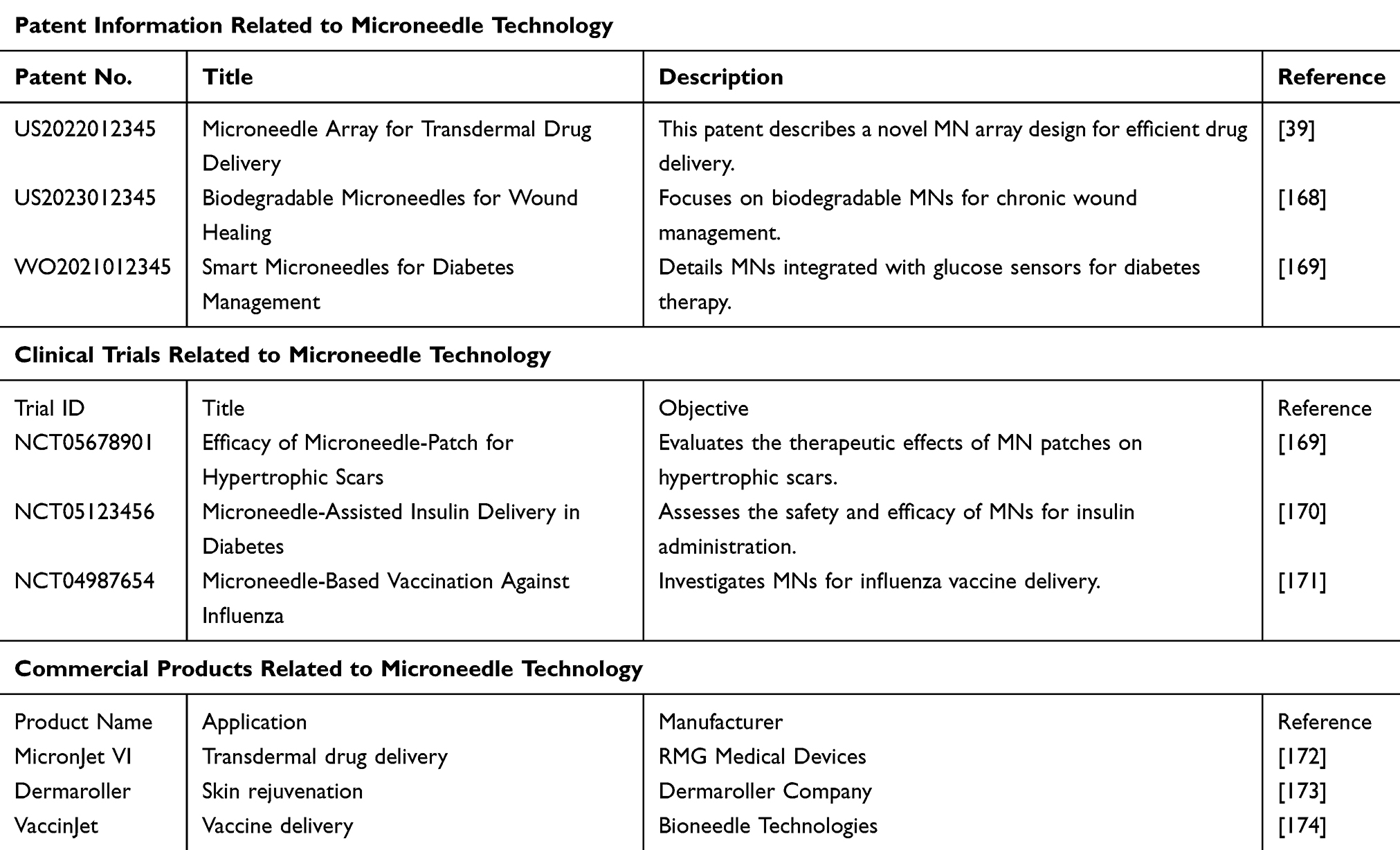 |
Table 3 Summary of Microneedle Technology: Patents, Clinical Trials, and Commercial Products |
Clinical Translation of MNs in BME
The clinical translation of MNs in BME presents challenges and opportunities. MNs have shown great potential in biomedical applications, such as drug delivery, biomarker monitoring, and tissue repair. However, clinical translation requires addressing multiple technical issues. One key challenge is ensuring consistency and reliability of MNs in clinical settings. During manufacturing, strict quality control is essential to produce MNs with uniform geometry and mechanical properties. The integration of MNs with other medical devices must be carefully considered to ensure compatibility and performance.175
In addition, demonstrating the safety and efficacy of MNs through rigorous clinical trials is critical for translation. This involves conducting studies to evaluate therapeutic effects, side effects, and long-term outcomes of MN treatments. It requires comparing MNs with existing treatments to establish their advantages and cost-effectiveness. The training of healthcare professionals in proper MN use and application is crucial for clinical implementation. A multidisciplinary approach involving engineers, clinicians, and regulatory experts is necessary to overcome challenges and accelerate clinical translation of MNs in BME.176
Conclusions and Outlook
MNs, applied transdermally, can effectively and painlessly penetrate the stratum corneum and release therapeutically active substances directly into the dermis. Proper design of MNs systems allows sustained or responsive drug delivery to meet different therapeutic needs. Such systems are minimally invasive, safe, effective, easy to use, require lower drug doses, and enable accurate dose delivery. Moreover, they can deliver drugs of different molecular weights, including protein-based biologics that are particularly important in oral transmucosal delivery.
Despite the remarkable development of MNs technology in recent years, currently approved systems are primarily used in the cosmetic field rather than in pharmacological treatments. For long-term treatment of chronic diseases, the safety and side effects of multiple-use MNs have not yet been fully validated, although several clinical trials have been conducted in areas such as diabetes management. However, more research is required to clarify their effectiveness. The large-scale production of MNs poses several challenges including cost control and scalability. The registration process for the use of MNs as commercial drugs has also not yet been widely accepted. Despite related progress in materials science, 3D printing, and molding technology, the application of these innovations in clinical studies of MNs remains limited, and further clinical validation is urgently required.
This in-depth review explores the application of MNs in BME, as well as the problems they face. MNs can achieve sustained and stable drug release in vivo to meet specific therapeutic needs. Diagnostically, MNs technology can enable the noninvasive and real-time monitoring of biomarkers. They have also been used to promote the repair and regeneration of various tissues—such as skin, cartilage, muscles, bones, and nerves—particularly related to the precise delivery of cells and growth factors. As materials science continues to improve, this technology will become more efficient and deliver more accurate therapeutic results. For example, nanotechnology allows MNs to penetrate the skin in smaller and finer forms, thereby reducing pain and injury.177 Moreover, 3D printing technology has enhanced the stability and permeability of MNs, providing new possibilities for their application.
Finally, several challenges in the development and manufacturing of MNs remain, including the precise control of delivered drug doses and medical device compliance with good manufacturing practices. In clinical applications that require small and frequent dosages—such as vaccinations and insulin therapy for managing diabetes—reliable performance can only be achieved through intensive manufacturing management and rigorous training in the dosing process. These critical steps should be elaborated upon in clinical study protocols, as it is important to develop an exhaustive set of guidelines for the clinical study of MNs. Thoughtful application of MNs from the users’ perspective remains essential to maximize their efficacy and safety.178
The future of MNs is promising because with advances in research, innovations in MN design and fabrication are expected to lead to the development of effective systems. The potential for MNs in personalized medicine is exciting, where they could be tailored to patient needs for optimal therapeutic outcomes. Additionally, integrating MNs with smart technologies may enable real-time monitoring and responsive treatments. While challenges remain, efforts to overcome manufacturing hurdles and expand clinical validation will be crucial in translating these technologies into widespread clinical use, ultimately enhancing patient care and efficacy.
Ethics Approval and Consent to Participate
There are no human and animal subjects in this review and informed consent is not applicable.
Acknowledgments
This work was supported by the Natural Science Foundation of Zhejiang Province, China (No. MS25H090029), the medical and health research project of Zhejiang Province (No. 2022KY354, 2024KY399), and Wenzhou science and technology project of China (No. Y20220935).
Disclosure
The authors report no conflicts of interest in this work.
References
1. Li G, Zheng T, Wu L, et al. Bionic microenvironment-inspired synergistic effect of anisotropic micro-nanocomposite topology and biology cues on peripheral nerve regeneration. Am Assoc Adv Sci. 2021;28.
2. Wang H, Tian J, Jiang Y, et al. A 3D biomimetic optoelectronic scaffold repairs cranial defects. Sci Adv. 2023;9(7):eabq7750. doi:10.1126/sciadv.abq7750
3. Sun M, Han K, Hu R, Liu D, Fu W, Liu W. Advances in micro/nanoporous membranes for biomedical engineering. Adv Healthc Mater. 2021;10(7):e2001545. doi:10.1002/adhm.202001545
4. Razavi Z, Soltani M, Souri M, van Wijnen AJ. CRISPR innovations in tissue engineering and gene editing. Life Sci. 2024;358:123120. doi:10.1016/j.lfs.2024.123120
5. Yi SA, Zhang Y, Rathnam C, Pongkulapa T, Lee KB. Bioengineering approaches for the advanced organoid research. Adv Mater. 2021;33(45):e2007949. doi:10.1002/adma.202007949
6. Robin M, Mouloungui E, Dali GC, et al. Mineralized collagen plywood contributes to bone autograft performance. Nature. 2024;1–8.
7. van Haasteren J, Li J, Scheideler OJ, Murthy N, Schaffer DV. The delivery challenge: fulfilling the promise of therapeutic genome editing. Nat Biotechnol. 2020;38(7):845–855. doi:10.1038/s41587-020-0565-5
8. Mdanda S, Ubanako P, Kondiah PPD, Kumar P, Choonara YE. Recent advances in microneedle platforms for transdermal drug delivery technologies. Polymers. 2021;13(15):2405. doi:10.3390/polym13152405
9. Yang J, Liu X, Fu Y, Song Y. Recent advances of microneedles for biomedical applications: drug delivery and beyond. Acta Pharm Sin B. 2019;9(3):469–483. doi:10.1016/j.apsb.2019.03.007
10. Chien MN, Chen YJ, Bai CH, Huang JT. Continuous glucose monitoring system based on percutaneous microneedle array. Micromachines. 2022;13(3):478. doi:10.3390/mi13030478
11. Himawan A, Vora LK, Permana AD, et al. Where microneedle meets biomarkers: futuristic application for diagnosing and monitoring localized external organ diseases. Adv Healthc Mater. 2023;12(5):e2202066. doi:10.1002/adhm.202202066
12. Spataro EA, Dierks K, Carniol PJ. Microneedling-associated procedures to enhance facial rejuvenation. Clin Plast Surg. 2023;50(3):479–488. doi:10.1016/j.cps.2022.12.012
13. Chilicka K, Rusztowicz M, Szyguła R, Nowicka D. Methods for the improvement of acne scars used in dermatology and cosmetology: a review. J Clin Med. 2022;11(10):2744. doi:10.3390/jcm11102744
14. Tan MG, Jo CE, Chapas A, Khetarpal S, Dover JS. Radiofrequency microneedling: a comprehensive and critical review. Dermatol Surg. 2021;47(6):755–761. doi:10.1097/DSS.0000000000002972
15. Olowe M, Parupelli SK, Desai S. A review of 3D-printing of microneedles. Pharmaceutics. 2022;14(12):2693. doi:10.3390/pharmaceutics14122693
16. O’Mahony C, Sebastian R, Tjulkins F, et al. Hollow silicon microneedles, fabricated using combined wet and dry etching techniques, for transdermal delivery and diagnostics. Int J Pharm. 2023;637:122888. doi:10.1016/j.ijpharm.2023.122888
17. Mamun AA, Zhao F. In-plane Si microneedles: fabrication, characterization, modeling and applications. Micromachines. 2022;13(5):657. doi:10.3390/mi13050657
18. Cheng W, Wang X, Zou S, et al. Fabrication of black silicon microneedle arrays for high drug loading. J Funct Biomater. 2023;14(5):245. doi:10.3390/jfb14050245
19. Huang X, Chang Q, Gao JH, Lu F. Sustained release microneedles: materials and applications in facial rejuvenation. Tissue Eng Part B Rev. 2023;29(3):190–202. doi:10.1089/ten.teb.2022.0131
20. Rad ZF, Prewett PD, Davies GJ. An overview of microneedle applications, materials, and fabrication methods. Beilstein J Nanotechnol. 2021;12:1034–1046. doi:10.3762/bjnano.12.77
21. Jung JH, Jin SG. Microneedle for transdermal drug delivery: current trends and fabrication. J Pharm Investig. 2021;51(5):503–517. doi:10.1007/s40005-021-00512-4
22. Liu J, Zhang Z, Lin X, et al. Magnesium metal–organic framework microneedles loaded with curcumin for accelerating oral ulcer healing. J Nanobiotechnol. 2024;22(1):594. doi:10.1186/s12951-024-02873-y
23. Kalyani P, Khandelwal M. Drug release kinetics from in-situ modulated agar/chitosan-bacterial cellulose patches for differently soluble drugs. Int J Biol Macromol. 2024;283(Pt 3):137602. doi:10.1016/j.ijbiomac.2024.137602
24. Omidian H, Chowdhury SD. Multifunctional Hydrogel Microneedles (HMNs) in drug delivery and diagnostics. Gels. 2025;11(3):206. doi:10.3390/gels11030206
25. Lu G, Li B, Lin L, Li X, Ban J. Mechanical strength affecting the penetration in microneedles and PLGA nanoparticle-assisted drug delivery: importance of preparation and formulation. Biomed Pharmacother. 2024;173:116339. doi:10.1016/j.biopha.2024.116339
26. Rajendran J, K J, M SS, Alluri LD, Giri J. Bioinspired silk protein modification to develop instant dissolvable microneedles with superior mechanical properties and long-term biomolecule stabilization. J Mater Chem B. 2025;13(24):7072–7089. doi:10.1039/D4TB02836H
27. Liu T, Song G, Zeng Z, Zhang X, Zhang M, Jiang G. Microneedles in diagnosis and treatment: a review. Sheng Wu Gong Cheng Xue Bao. 2021;37(4):1139–1154. doi:10.13345/j.cjb.200363
28. Xiang M, Yang C, Zhang L, Wang S, Ren Y, Gou M. Dissolving microneedles for transdermal drug delivery in cancer immunotherapy. J Mater Chem B. 2024;12(24):5812–5822. doi:10.1039/D4TB00659C
29. Sartawi Z, Blackshields C, Faisal W. Dissolving microneedles: applications and growing therapeutic potential. J Control Release. 2022;348:186–205. doi:10.1016/j.jconrel.2022.05.045
30. Wu Y, Tang Z, Ma S, Du L. The promising application of hydrogel microneedles in medical application. J Pharm Pharmacol. 2023;75(8):1011–1020. doi:10.1093/jpp/rgad058
31. Paredes AJ, McKenna PE, Ramöller IK, et al. Microarray patches: poking a hole in the challenges faced when delivering poorly soluble drugs. Adv Funct Mater. 2020;31.
32. Al-Kasasbeh R, Brady AJ, Courtenay AJ, et al. Evaluation of the clinical impact of repeat application of hydrogel-forming microneedle array patches. Drug Deliv Transl Res. 2020;10(3):690–705. doi:10.1007/s13346-020-00727-2
33. Vora LK, Sabri AH, Naser Y, et al. Long-acting microneedle formulations. Adv Drug Deliv Rev. 2023;201:115055. doi:10.1016/j.addr.2023.115055
34. Baek S-H, Shin J-H, Kim Y-C. Drug-coated microneedles for rapid and painless local anesthesia. Biomed Microdevices. 2017;19(1):2. doi:10.1007/s10544-016-0144-1
35. Caudill CL, Perry JL, Tian S, Luft JC, DeSimone JM. Spatially controlled coating of continuous liquid interface production microneedles for transdermal protein delivery. J Control Release. 2018;284:122–132. doi:10.1016/j.jconrel.2018.05.042
36. Shakya AK, Lee CH, Gill HS. Cutaneous vaccination with coated microneedles prevents development of airway allergy. J Control Release. 2017;265:75–82. doi:10.1016/j.jconrel.2017.08.012
37. Chen BZ, He MC, Zhang XP, Fei WM, Cui Y, Guo XD. A novel method for fabrication of coated microneedles with homogeneous and controllable drug dosage for transdermal drug delivery. Drug Deliv Transl Res. 2022;12(11):2730–2739. doi:10.1007/s13346-022-01123-8
38. Xu J, Xu D, Xuan X, He H. Advances of microneedles in biomedical applications. Molecules. 2021;26(19):5912. doi:10.3390/molecules26195912
39. Al-Japairai KAS, Mahmood S, Almurisi SH, et al. Current trends in polymer microneedle for transdermal drug delivery. Int J Pharm. 2020;587:119673. doi:10.1016/j.ijpharm.2020.119673
40. Abd-El-Azim HM, Tekko I, Ali AA, et al. Hollow microneedle assisted intradermal delivery of hypericin lipid nanocapsules with light enabled photodynamic therapy against skin cancer. J Control Release. 2022;348:849–869. doi:10.1016/j.jconrel.2022.06.027
41. Waghule T, Singhvi G, Dubey SK, et al. Microneedles: a smart approach and increasing potential for transdermal drug delivery system. Biomed Pharmacother. 2019;109:1249–1258. doi:10.1016/j.biopha.2018.10.078
42. Gorantla S, Dabholkar N, Sharma S, Rapalli VK, Alexander A, Singhvi G. Chitosan-based microneedles as a potential platform for drug delivery through the skin: trends and regulatory aspects. Int J Biol Macromol. 2021;184:438–453. doi:10.1016/j.ijbiomac.2021.06.059
43. Damiri F, Kommineni N, Ebhodaghe SO, et al. Microneedle-based natural polysaccharide for Drug Delivery Systems (DDS): progress and challenges. Pharmaceuticals. 2022;15(2):190. doi:10.3390/ph15020190
44. Lu H, Wang J, Li J, Gao B, He B. Advanced Silk Fibroin Biomaterials-Based Microneedles for Healthcare. Macromol Biosci. 2023;23(11):e2300141. doi:10.1002/mabi.202300141
45. You Y, Tian Y, Yang Z, et al. Intradermally delivered mRNA-encapsulating extracellular vesicles for collagen-replacement therapy. Nat Biomed Eng. 2023;7(7):887–900. doi:10.1038/s41551-022-00989-w
46. Wang P, Pu Y, Ren Y, et al. Enzyme-regulated NO programmed to release from hydrogel-forming microneedles with endogenous/photodynamic synergistic antibacterial for diabetic wound healing. Int J Biol Macromol. 2023;226:813–822. doi:10.1016/j.ijbiomac.2022.12.063
47. Ren Y, Li J, Chen Y, et al. Customized flexible hollow microneedles for psoriasis treatment with reduced-dose drug. Bioeng Transl Med. 2023;8(4):e10530. doi:10.1002/btm2.10530
48. Wu L, Park J, Kamaki Y, Kim B. Optimization of the fused deposition modeling-based fabrication process for polylactic acid microneedles. Microsyst Nanoeng. 2021;7:58. doi:10.1038/s41378-021-00284-9
49. Alperovitz CH, David NB, Ramot Y, Gross A, Mizrahi B. Living microneedles for intradermal delivery of beneficial bacteria. ACS Biomater Sci Eng. 2025;11(2):1232–1241. doi:10.1021/acsbiomaterials.4c02230
50. Zheng B, Li Q, Fang L, et al. Microorganism microneedle micro-engine depth drug delivery. Nat Commun. 2024;15(1):8947. doi:10.1038/s41467-024-53280-8
51. Onesto V, Di Natale C, Profeta M, Netti PA, Vecchione R. Engineered PLGA-PVP/VA based formulations to produce electro-drawn fast biodegradable microneedles for labile biomolecule delivery. Prog Biomater. 2020;9(4):203–217. doi:10.1007/s40204-020-00143-2
52. Yao W, Li D, Zhao Y, et al. 3D printed multi-functional hydrogel microneedles based on high-precision digital light processing. Micromachines. 2019;11(1):17. doi:10.3390/mi11010017
53. Kim MJ, Park SC, Rizal B, et al. Fabrication of circular obelisk-type multilayer microneedles using micro-milling and spray deposition. Front Bioeng Biotechnol. 2018;6:54. doi:10.3389/fbioe.2018.00054
54. Nejad HR, Sadeqi A, Kiaee G, Sonkusale S. Low-cost and cleanroom-free fabrication of microneedles. Microsyst Nanoengin. 2018;4(1):17073. doi:10.1038/micronano.2017.73
55. Sun Y, Wu J, Zhou L, et al. Genetically engineered electrospinning contributes to spinal cord injury repair by regulating the immune microenvironment. Front Bioeng Biotechnol. 2024;12:1415527. doi:10.3389/fbioe.2024.1415527
56. Weimer P, Rossi RC, Koester LS. Dissolving microneedles developed in association with nanosystems: a scoping review on the quality parameters of these emerging systems for drug or protein transdermal delivery. Pharmaceutics. 2021;13(10):1601. doi:10.3390/pharmaceutics13101601
57. Zhang L, Chen Y, Tan J, Feng S, Xie Y, Li L. Performance enhancement of PLA-based blend microneedle arrays through Shish-Kebab structuring strategy in microinjection molding. Polymers. 2023;15(10).
58. Chen YH, Lin DC, Chern E, Huang YY. The use of micro-needle arrays to deliver cells for cellular therapies. Biomed Microdevices. 2020;22(4):63. doi:10.1007/s10544-020-00518-z
59. Liu X, Chen X, Fei Y, et al. Locally injectable, ROS-scavenging, and ROS-/pH-responsive polymeric-micelles-embedded hydrogels for precise minimally invasive and long-lasting rheumatoid therapy. Adv Healthc Mater. 2025;14(3):e2403579. doi:10.1002/adhm.202403579
60. Bedir T, Kadian S, Shukla S, Gunduz O, Narayan R. Additive manufacturing of microneedles for sensing and drug delivery. Expert Opin Drug Deliv. 2024;21(7):1053–1068. doi:10.1080/17425247.2024.2384696
61. Sarabi MR, Alseed MM, Karagoz AA, Tasoglu S. Machine learning-enabled prediction of 3D-printed microneedle features. Biosensors. 2022;12(7).
62. Gillani M, Pollastri G. Impact of alignments on the accuracy of protein subcellular localization predictions. Proteins. 2025;93(3):745–759. doi:10.1002/prot.26767
63. Yan Q, Weng J, Shen S, et al. Finite element analysis for biodegradable dissolving microneedle materials on skin puncture and mechanical performance evaluation. Polymers. 2021;13(18):3043. doi:10.3390/polym13183043
64. Shin S, Kim J. Modeling highly efficient femtosecond laser ablation of aluminum for cutting. Sci Rep. 2025;15(1):5418. doi:10.1038/s41598-025-89493-0
65. Zapico P, Rodríguez-González P, Robles-Valero P, Fernández-Abia AI, Barreiro J. Influence of post-processing on the properties of multi-material parts obtained by material projection AM. Polymers. 2023;15(9):2089. doi:10.3390/polym15092089
66. Ma Y, Cai K, Xu G, et al. Large-scale and highly efficient production of ultrafine PVA fibers by electro-centrifugal spinning for NH(3) adsorption. Materials. 2023;16(7).
67. Tan JY, Li Y, Chamani F, et al. Experimental validation of diffraction lithography for fabrication of solid microneedles. Materials. 2022;15(24):8934. doi:10.3390/ma15248934
68. Ramadan M, Alghamdi AS, Subhani T, Halim KSA. Fabrication and characterization of Sn-based babbitt alloy nanocomposite reinforced with Al(2)O(3) nanoparticles/Carbon steel bimetallic material. Materials. 2020;13(12):2759. doi:10.3390/ma13122759
69. Kang S, Song JE, Jun SH, Park SG, Kang NG. Sugar-triggered burst drug releasing Poly-Lactic Acid (PLA) microneedles and its fabrication based on solvent-casting approach. Pharmaceutics. 2022;14(9):1758. doi:10.3390/pharmaceutics14091758
70. S BVJC, Mannayee G. Structural analysis and simulation of solid microneedle array for vaccine delivery applications. Mater Today Proc. 2022;65:3774–3779. doi:10.1016/j.matpr.2022.06.483
71. Pourmansouri Z, Malekkhatabi A, Toolabi M, Akbari M, Shahbazi MA, Rostami A. Anti-nociceptive effect of sufentanil polymeric dissolving microneedle on male mice by hot plate technique. Iran Biomed J. 2024;28(4):192–205. doi:10.61186/ibj.4062
72. Du G, Zhang Z, He P, Zhang Z, Sun X. Determination of the mechanical properties of polymeric microneedles by micromanipulation. J Mech Behav Biomed Mater. 2021;117:104384. doi:10.1016/j.jmbbm.2021.104384
73. Luo X, Yang L, Cui Y. Microneedles: materials, fabrication, and biomedical applications. Biomed Microdevices. 2023;25(3):20. doi:10.1007/s10544-023-00658-y
74. Chen J, Garcia ES, Zimmerman SC. Intramolecularly cross-linked polymers: from structure to function with applications as artificial antibodies and artificial enzymes. Acc Chem Res. 2020;53(6):1244–1256. doi:10.1021/acs.accounts.0c00178
75. Makvandi P, Kirkby M, Hutton ARJ, et al. Engineering microneedle patches for improved penetration: analysis, skin models and factors affecting needle insertion. Nano-Micro Letters. 2021;13(1):93. doi:10.1007/s40820-021-00611-9
76. Hong W, Chen IC, Su CY, Perng CK, Ma H, Fang HW. Evaluating pull-out strength of barbed suture in vitro by using porcine tissue and Polydimethylsiloxane (PDMS). Polymers. 2022;14(11):2170. doi:10.3390/polym14112170
77. Lin Z, Li Y, Meng G, et al. Reinforcement of silk microneedle patches for accurate transdermal delivery. Biomacromolecules. 2021;22(12):5319–5326. doi:10.1021/acs.biomac.1c01213
78. Juhasz MLW, Cohen JL. Microneedling for the treatment of scars: an update for clinicians. Clin Cosmet Investig Dermatol. 2020;13:997–1003. doi:10.2147/CCID.S267192
79. Paris JL, Vora LK, Torres MJ, Mayorga C, Donnelly RF. Microneedle array patches for allergen-specific immunotherapy. Drug Discov Today. 2023;28(5):103556. doi:10.1016/j.drudis.2023.103556
80. Zhang X, Zhou C, Chen T, et al. State-of-the-art strategies to enhance the mechanical properties of microneedles. Int J Pharm. 2024;663:124547. doi:10.1016/j.ijpharm.2024.124547
81. Wei C, Fu D, Ma T, et al. Sensing patches for biomarker identification in skin-derived biofluids. Biosens Bioelectron. 2024;258:116326. doi:10.1016/j.bios.2024.116326
82. Xu Y, Bian Q, Zhang Y, et al. Single-dose of integrated bilayer microneedles for enhanced hypertrophic scar therapy with rapid anti-inflammatory and sustained inhibition of myofibroblasts. Biomaterials. 2025;312:122742. doi:10.1016/j.biomaterials.2024.122742
83. Hao YY, Yang Y, Li QY, et al. Effect of polymer microneedle pre-treatment on drug distributions in the skin in vivo. J Drug Target. 2020;28(7–8):811–817. doi:10.1080/1061186X.2020.1757101
84. Ziaeifar E, Ziaeifar F, Mozafarpoor S, Goodarzi A. Applications of microneedling for various dermatologic indications with a special focus on pigmentary disorders: a comprehensive review study. Dermatol Ther. 2021;34(6):e15159. doi:10.1111/dth.15159
85. Fu X, Xian H, Xia C, et al. Polymer homologue-mediated formation of hydrogel microneedles for controllable transdermal drug delivery. Int J Pharm. 2024;666:124768. doi:10.1016/j.ijpharm.2024.124768
86. Detamornrat U, McAlister E, Hutton ARJ, Larrañeta E, Donnelly RF. The role of 3D printing technology in microengineering of microneedles. Small. 2022;18(18):e2106392. doi:10.1002/smll.202106392
87. Pere CPP, Economidou SN, Lall G, et al. 3D printed microneedles for insulin skin delivery. Int J Pharm. 2018;544(2):425–432. doi:10.1016/j.ijpharm.2018.03.031
88. Amani H, Shahbazi M-A, D’Amico C, Fontana F, Abbaszadeh S, Santos HA. Microneedles for painless transdermal immunotherapeutic applications. J Control Release. 2021;330:185–217. doi:10.1016/j.jconrel.2020.12.019
89. Wang QL, Zhu DD, Liu XB, Chen BZ, Guo XD. Microneedles with controlled bubble sizes and drug distributions for efficient transdermal drug delivery. Scientific Reports. 2016;6(1):38755. doi:10.1038/srep38755
90. Avcil M, Çelik A. Microneedles in drug delivery: progress and challenges. Micromachines. 2021;12(11). doi:10.3390/mi12111321
91. Chen BZ, He YT, Zhao ZQ, et al. Strategies to develop polymeric microneedles for controlled drug release. Adv Drug Deliv Rev. 2023;203:115109. doi:10.1016/j.addr.2023.115109
92. Shende P, Sardesai M, Gaud RS. Micro to nanoneedles: a trend of modernized transepidermal drug delivery system. Artif Cells Nanomed Biotechnol. 2018;46(1):19–25. doi:10.1080/21691401.2017.1304409
93. Bok M, Zhao Z-J, Jeon S, Jeong J-H, Lim E. Ultrasonically and iontophoretically enhanced drug-delivery system based on dissolving microneedle patches. Scientific Reports. 2020;10(1):2027. doi:10.1038/s41598-020-58822-w
94. Shen J, Wang J, Wu M, et al. Matrine-loaded self-adhesive swelling microneedle for inflammation regulation to improve eczema treatment. Mar Life Sci Technol. 2024;6(3):535–546. doi:10.1007/s42995-024-00235-z
95. Ziesmer J, Tajpara P, Hempel NJ, et al. Vancomycin-loaded microneedle arrays against Methicillin-resistant Staphylococcus Aureus skin infections. Adv Mater Technol. 2021;6(7):2001307. doi:10.1002/admt.202001307
96. Sheng T, Luo B, Zhang W, et al. Microneedle-mediated vaccination: innovation and translation. Adv Drug Deliv Rev. 2021;179:113919. doi:10.1016/j.addr.2021.113919
97. Yang Y, Wang P, Gong Y, et al. Curcumin-zinc framework encapsulated microneedle patch for promoting hair growth. Theranostics. 2023;13(11):3675–3688. doi:10.7150/thno.84118
98. Yang ZR, Suo H, Fan JW, et al. Endogenous stimuli-responsive separating microneedles to inhibit hypertrophic scar through remodeling the pathological microenvironment. Nat Commun. 2024;15(1):2038. doi:10.1038/s41467-024-46328-2
99. Duarah S, Sharma M, Wen J. Recent advances in microneedle-based drug delivery: special emphasis on its use in paediatric population. Eur J Pharm Biopharm. 2019;136:48–69. doi:10.1016/j.ejpb.2019.01.005
100. Hu W, Peng T, Huang Y, et al. Hyaluronidase-powered microneedles for significantly enhanced transdermal delivery efficiency. J Control Release. 2023;353:380–390. doi:10.1016/j.jconrel.2022.11.046
101. Aich K, Singh T, Dang S. Advances in microneedle-based transdermal delivery for drugs and peptides. Drug Deliv Transl Res. 2022;12(7):1556–1568. doi:10.1007/s13346-021-01056-8
102. Ma W, Zhang X, Liu Y, et al. Polydopamine decorated microneedles with Fe-MSC-derived nanovesicles encapsulation for wound healing. Adv Sci. 2022;9(13):e2103317. doi:10.1002/advs.202103317
103. Li S, Wang X, Yan Z, et al. Microneedle patches with antimicrobial and immunomodulating properties for infected wound healing. Adv Sci. 2023;10(22):e2300576. doi:10.1002/advs.202300576
104. Haghniaz R, Kim HJ, Montazerian H, et al. Tissue adhesive hemostatic microneedle arrays for rapid hemorrhage treatment. Bioact Mater. 2023;23:314–327. doi:10.1016/j.bioactmat.2022.08.017
105. Moussi K, Kavaldzhiev M, Perez JE, Alsharif N, Merzaban J, Kosel J. 3D printed microneedle array for electroporation. Annu Int Conf IEEE Eng Med Biol Soc. 2020;2202–2205. doi:10.1109/EMBC44109.2020.9175748
106. Dervisevic M, Dervisevic E, Esser L, Easton CD, Cadarso VJ, Voelcker NH. Wearable microneedle array-based sensor for transdermal monitoring of pH levels in interstitial fluid. Biosens Bioelectron. 2023;222:114955. doi:10.1016/j.bios.2022.114955
107. Pazyar N, Hatami M, Yaghoobi R, Parvar SY, Radmanesh M, Hadibarhaghtalab M. The efficacy of adding topical 5-fluorouracil to micro-needling in the treatment of vitiligo: a randomized controlled trial. J Cosmet Dermatol. 2023;22(5):1513–1520. doi:10.1111/jocd.15616
108. Sondermann S, Boppel T, Fieseler K, Schramm P, Bäumer T, Trillenberg P. Needle electromyography does not meaningfully impact findings in MR-neurography/-myography. Muscle Nerve. 2024;69(4):409–415. doi:10.1002/mus.28049
109. Gheisari M, Ghalamkarpour F, Haghighi SM, et al. The effect of topical tranexamic acid with micro-needling and micro-needling alone in treatment of macular amyloidosis. J Cosmet Dermatol. 2024;23(12):3968–3973. doi:10.1111/jocd.16517
110. Dydyk AM, Negrete G, Sarwan G, Cascella M. Median Nerve Injury, StatPearls. Treasure Island (FL): StatPearls Publishing; 2024.
111. Ventura N, Soti V. Integrating osteopathic principles for enhanced dermatological outcomes: a literature review. Cureus. 2024;16(1):e52665. doi:10.7759/cureus.52665
112. Chen Y, Feng X. Gold nanoparticles for skin drug delivery. Int J Pharm. 2022;625:122122. doi:10.1016/j.ijpharm.2022.122122
113. Long L, Ji D, Hu C, Yang L, Tang S, Wang Y. Microneedles for in situ tissue regeneration. Mater Today Bio. 2023;19:100579. doi:10.1016/j.mtbio.2023.100579
114. Zhang T, Sun B, Guo J, et al. Active pharmaceutical ingredient poly(ionic liquid)-based microneedles for the treatment of skin acne infection. Acta Biomater. 2020;115:136–147. doi:10.1016/j.actbio.2020.08.023
115. Aitchison AH, Allen NB, Mitra K, et al. Tunable alginate-polyvinyl alcohol bioinks for 3D printing in cartilage tissue engineering. Gels. 2024;10(12):829. doi:10.3390/gels10120829
116. Li C, Zhang W, Nie Y, et al. Time-sequential and multi-functional 3D printed MgO(2)/PLGA Scaffold developed as a novel biodegradable and bioactive bone substitute for challenging postsurgical osteosarcoma treatment. Adv Mater. 2024;36(34):e2308875. doi:10.1002/adma.202308875
117. Hadavi E, de Vries RHW, Smink AM, et al. In vitro degradation profiles and in vivo biomaterial-tissue interactions of microwell array delivery devices. J Biomed Mater Res B Appl Biomater. 2021;109(1):117–127. doi:10.1002/jbm.b.34686
118. Xiao M, Tang Q, Zeng S, et al. Emerging biomaterials for tumor immunotherapy. Biomater Res. 2023;27(1):47. doi:10.1186/s40824-023-00369-8
119. Sabbagh F, Kim BS. Recent advances in polymeric transdermal drug delivery systems. J Control Release. 2022;341:132–146. doi:10.1016/j.jconrel.2021.11.025
120. Qu F, Geng R, Liu Y, Zhu J. Advanced nanocarrier- and microneedle-based transdermal drug delivery strategies for skin diseases treatment. Theranostics. 2022;12(7):3372–3406. doi:10.7150/thno.69999
121. Bilal M, Mehmood S, Raza A, Hayat U, Rasheed T, Iqbal HMN. Microneedles in Smart Drug Delivery. Adv Wound Care. 2021;10(4):204–219. doi:10.1089/wound.2019.1122
122. Huang S, Ding X. Precise design strategies of nanotechnologies for controlled drug delivery. J Funct Biomater. 2022;13(4):188. doi:10.3390/jfb13040188
123. Martínez-Navarrete M, Pérez-López A, Guillot AJ, Cordeiro AS, Melero A, Aparicio-Blanco J. Latest advances in glucose-responsive microneedle-based systems for transdermal insulin delivery. Int J Biol Macromol. 2024;263(Pt 2):130301. doi:10.1016/j.ijbiomac.2024.130301
124. Peng S, Xiao F, Chen M, Gao H. Tumor-microenvironment-responsive nanomedicine for enhanced cancer immunotherapy. Adv Sci. 2022;9(1):e2103836. doi:10.1002/advs.202103836
125. Samant PP, Niedzwiecki MM, Raviele N, et al. Sampling interstitial fluid from human skin using a microneedle patch. Sci Transl Med. 2020;12(571). doi:10.1126/scitranslmed.aaw0285
126. Huang X, Yao C, Huang S, et al. Technological advances of wearable device for continuous monitoring of in vivo glucose. ACS Sens. 2024;9(3):1065–1088. doi:10.1021/acssensors.3c01947
127. Sarabi MR, Nakhjavani SA, Tasoglu S. 3D-printed microneedles for point-of-care biosensing applications. Micromachines. 2022;13(7).
128. Zheng M, Zhang Y, Hu T, Xu C. A skin patch integrating swellable microneedles and electrochemical test strips for glucose and alcohol measurement in skin interstitial fluid. Bioeng Transl Med. 2023;8(5):e10413. doi:10.1002/btm2.10413
129. Chen W, Wang Z, Wang L, Chen X. Smart chemical engineering-based lightweight and miniaturized attachable systems for advanced drug delivery and diagnostics. Adv Mater. 2022;34(6):e2106701. doi:10.1002/adma.202106701
130. Liu Y, Yu Q, Luo X, Yang L, Cui Y. Continuous monitoring of diabetes with an integrated microneedle biosensing device through 3D printing. Microsyst Nanoeng. 2021;7:75. doi:10.1038/s41378-021-00302-w
131. Razzaghi M, Akbari M. The effect of 3D printing tilt angle on the penetration of 3D-printed microneedle arrays. Micromachines. 2023;14(6):1157. doi:10.3390/mi14061157
132. Pan Y, Zhang Y, Shi X, et al. Electrical stimulation induces anti-tumor immunomodulation via a flexible microneedle-array-integrated interdigital electrode. Sci Bull. 2023;68(22):2779–2792. doi:10.1016/j.scib.2023.10.004
133. Luo R, Dai J, Zhang J, Li Z. Accelerated skin wound healing by electrical stimulation. Adv Healthc Mater. 2021;10(16):e2100557. doi:10.1002/adhm.202100557
134. Li Y, Zheng L, Cao W, et al. 5-aminolevulinic acid-loaded dissolving microneedle array for photodynamic therapy of rheumatoid arthritis on rats. Biomed Pharmacother. 2023;162:114684. doi:10.1016/j.biopha.2023.114684
135. Zhi D, Yang T, O’Hagan J, Zhang S, Donnelly RF. Photothermal therapy. J Control Release. 2020;325:52–71. doi:10.1016/j.jconrel.2020.06.032
136. Shan Y, Tan B, Zhang M, Xie X, Liao J. Restorative biodegradable two-layered hybrid microneedles for melanoma photothermal/chemo co-therapy and wound healing. J Nanobiotechnology. 2022;20(1):238. doi:10.1186/s12951-022-01426-5
137. Zhou Y, Chen C, Qu Q, et al. The effectiveness of combination therapies for androgenetic alopecia: a systematic review and meta-analysis. Dermatol Ther. 2020;33(4):e13741. doi:10.1111/dth.13741
138. Shauly O, Marxen T, Menon A, Gould DJ, Miller LB, Losken A. Radiofrequency microneedling: technology, devices, and indications in the modern plastic surgery practice. Aesthet Surg J Open Forum. 2023;5:ojad100. doi:10.1093/asjof/ojad100
139. Han C, Yang Y, Sheng Y, et al. The mechanism of lncRNA-CRNDE in regulating tumour-associated macrophage M2 polarization and promoting tumour angiogenesis. J Cell Mol Med. 2021;25(9):4235–4247. doi:10.1111/jcmm.16477
140. Yuan M, Liu K, Jiang T, et al. GelMA/PEGDA microneedles patch loaded with HUVECs-derived exosomes and Tazarotene promote diabetic wound healing. J Nanobiotechnology. 2022;20(1):147. doi:10.1186/s12951-022-01354-4
141. Guo Z, Liu H, Shi Z, et al. Responsive hydrogel-based microneedle dressing for diabetic wound healing. J Mater Chem B. 2022;10(18):3501–3511. doi:10.1039/D2TB00126H
142. Zhang X, Chen G, Liu Y, Sun L, Sun L, Zhao Y. Black phosphorus-loaded separable microneedles as responsive oxygen delivery carriers for wound healing. ACS Nano. 2020;14(5):5901–5908. doi:10.1021/acsnano.0c01059
143. Cai L, Zhao C, Chen H, et al. Suction‐cup‐inspired adhesive micromotors for drug delivery. Advanced Science. 2022;9(1):2103384. doi:10.1002/advs.202103384
144. Wang C, Zeng Y, Chen KF, et al. A self-monitoring microneedle patch for light-controlled synergistic treatment of melanoma. Bioact Mater. 2023;27:58–71. doi:10.1016/j.bioactmat.2023.03.016
145. Ruan L, Song G, Zhang X, et al. Transdermal delivery of multifunctional CaO(2)@Mn-PDA nanoformulations by microneedles for NIR-induced synergistic therapy against skin melanoma. Biomater Sci. 2021;9(20):6830–6841. doi:10.1039/D1BM01117K
146. Li Z, Lu H, Fan L, et al. Microneedle-delivered PDA@Exo for multifaceted osteoarthritis treatment via PI3K-Akt-mTOR pathway. Adv Sci. 2024;11(42):e2406942. doi:10.1002/advs.202406942
147. Grogan SP, Dorthé EW, Glembotski NE, Gaul F, D’Lima DD. Cartilage tissue engineering combining microspheroid building blocks and microneedle arrays. Connect Tissue Res. 2020;61(2):229–243. doi:10.1080/03008207.2019.1617280
148. Xia T, Zhu Y, Li K, et al. Microneedles loaded with cerium-manganese oxide nanoparticles for targeting macrophages in the treatment of rheumatoid arthritis. J Nanobiotechnology. 2024;22(1):103. doi:10.1186/s12951-024-02374-y
149. Poorirani S, Taheri SL, Mostafavi SA. Scaffolds: a biomaterial engineering in targeted drug delivery for osteoporosis. Osteoporos Int. 2023;34(2):255–267. doi:10.1007/s00198-022-06543-3
150. Oh Y-J, Kang N-W, Jeong H-R, et al. The relationship between the drug delivery properties of a formulation of teriparatide microneedles and the pharmacokinetic evaluation of teriparatide administration in rats. Pharm Res. 2022;39(5):989–999. doi:10.1007/s11095-022-03254-6
151. Lin Z, Zheng K, Zhong J, Zheng X. Advances in microneedle-based therapy for bone disorders. Biomed Pharmacother. 2023;165:115013. doi:10.1016/j.biopha.2023.115013
152. Fang L, Liu Z, Wang C, et al. Vascular restoration through local delivery of angiogenic factors stimulates bone regeneration in critical size defects. Bioactive Mater. 2024;36:580–594. doi:10.1016/j.bioactmat.2024.07.003
153. Liu X, Li Y, Wang S, et al. PDGF-loaded microneedles promote tendon healing through p38/cyclin D1 pathway mediated angiogenesis. Mater Today Bio. 2022;16:100428. doi:10.1016/j.mtbio.2022.100428
154. Hu C, Liu B, Huang X, et al. Sea cucumber-inspired microneedle nerve guidance conduit for synergistically inhibiting muscle atrophy and promoting nerve regeneration. ACS Nano. 2024;18(22):14427–14440. doi:10.1021/acsnano.4c00794
155. Yuan J, Yang H, Liu C, et al. Microneedle patch loaded with exosomes containing microRNA-29b prevents cardiac fibrosis after myocardial infarction. Adv Healthc Mater. 2023;12(13):e2202959. doi:10.1002/adhm.202202959
156. Zhang X, Ma Y, Chen Z, Jiang H, Fan Z. Implantable nerve conduit made of a self-powered microneedle patch for sciatic nerve repair. Adv Healthc Mater. 2023;12(31):e2301729. doi:10.1002/adhm.202301729
157. Li B, Lu G, Liu W, Liao L, Ban J, Lu Z. Formulation and evaluation of PLGA nanoparticulate-based microneedle system for potential treatment of neurological diseases. Int J Nanomedicine. 2023;18:3745–3760. doi:10.2147/IJN.S415728
158. Zhou W, Wang Z, Xu Q, et al. Wireless facial biosensing system for monitoring facial palsy with flexible microneedle electrode arrays. NPJ Digit Med. 2024;7(1):13. doi:10.1038/s41746-024-01002-1
159. Wang Z, Shi N, Zhang Y, et al. Conformal in-ear bioelectronics for visual and auditory brain-computer interfaces. Nat Commun. 2023;14(1):4213. doi:10.1038/s41467-023-39814-6
160. Mahmood M, Kwon S, Kim H, et al. Wireless soft scalp electronics and virtual reality system for motor imagery-based brain-machine interfaces. Adv Sci. 2021;8(19):e2101129. doi:10.1002/advs.202101129
161. Ren L, Xu S, Gao J, et al. Fabrication of flexible microneedle array electrodes for wearable bio-signal recording. Sensors. 2018;18(4):1191. doi:10.3390/s18041191
162. Lee SH, Thunemann M, Lee K, et al. Scalable thousand channel penetrating microneedle arrays on flex for multimodal and large area coverage brainmachine interfaces. Adv Funct Mater. 2022;32.
163. Srikantharajah K, Quintela RM, Doerenkamp K, et al. Minimally-invasive insertion strategy and in vivo evaluation of multi-shank flexible intracortical probes. Sci Rep. 2021;11(1):18920. doi:10.1038/s41598-021-97940-x
164. Liu Y, Xu S, Yang Y, et al. Nanomaterial-based microelectrode arrays for in vitro bidirectional brain-computer interfaces: a review. Microsyst Nanoeng. 2023;9:13. doi:10.1038/s41378-022-00479-8
165. Lee SH, Thunemann M, Lee K, et al. Scalable thousand channel penetrating microneedle arrays on flex for multimodal and large area coverage BrainMachine interfaces. Adv Funct Mater. 2022;32(25).
166. Yan D, Jiman AA, Bottorff EC, et al. Ultraflexible and stretchable intrafascicular peripheral nerve recording device with axon-dimension, cuff-less microneedle electrode array. Small. 2022;18(21):e2200311. doi:10.1002/smll.202200311
167. Maiseli B, Abdalla AT, Massawe LV, et al. Brain-computer interface: trend, challenges, and threats. Brain Inform. 2023;10(1):20. doi:10.1186/s40708-023-00199-3
168. Luo X, Yu Q, Yang L, Cui Y, Wearable S-C. Ultrasound-based microneedle smart system for diabetes management. ACS Sens. 2023;8(4):1710–1722. doi:10.1021/acssensors.2c02863
169. Disphanurat W, Sivapornpan N, Srisantithum B, Leelawattanachai J. Efficacy of a triamcinolone acetonide-loaded dissolving microneedle patch for the treatment of hypertrophic scars and keloids: a randomized, double-blinded, placebo-controlled split-scar study. Arch Dermatol Res. 2023;315(4):989–997. doi:10.1007/s00403-022-02473-6
170. Wu M, Zhang Y, Huang H, et al. Assisted 3D printing of microneedle patches for minimally invasive glucose control in diabetes. Mater Sci Eng C Mater Biol Appl. 2020;117:111299. doi:10.1016/j.msec.2020.111299
171. Norman JJ, Arya JM, McClain MA, Frew PM, Meltzer MI, Prausnitz MR. Microneedle patches: usability and acceptability for self-vaccination against influenza. Vaccine. 2014;32(16):1856–1862. doi:10.1016/j.vaccine.2014.01.076
172. Li Z, Zhao P, Ling Z, Zheng Y, Qu F, Chang H. An ultraswelling microneedle device for facile and efficient drug loading and transdermal delivery. Adv Healthc Mater. 2024;13(2):e2302406. doi:10.1002/adhm.202302406
173. Yuksel EP, Sahin G, Aydin F, Senturk N, Turanli AY. Evaluation of effects of platelet-rich plasma on human facial skin. J Cosmet Laser Ther. 2014;16(5):206–208. doi:10.3109/14764172.2014.949274
174. McNamee M, Wong S, Guy O, Sharma S. Microneedle technology for potential SARS-CoV-2 vaccine delivery. Expert Opin Drug Deliv. 2023;20(6):799–814. doi:10.1080/17425247.2023.2209718
175. Kulkarni D, Damiri F, Rojekar S, et al. Recent advancements in microneedle technology for multifaceted biomedical applications. Pharmaceutics. 2022;14(5):1097. doi:10.3390/pharmaceutics14051097
176. Kulkarni D, Gadade D, Chapaitkar N, et al. Polymeric microneedles: an emerging paradigm for advanced biomedical applications. Scientia Pharmaceutica. 2023;91(2):27. doi:10.3390/scipharm91020027
177. Yu X, Zhao J, Fan D. The progress in the application of dissolving microneedles in biomedicine. Polymers. 2023;15(20):4059. doi:10.3390/polym15204059
178. Jeong SY, Park JH, Lee YS, Kim YS, Park JY, Kim SY. The current status of clinical research involving microneedles: a systematic review. Pharmaceutics. 2020;12(11):1113. doi:10.3390/pharmaceutics12111113
 © 2025 The Author(s). This work is published and licensed by Dove Medical Press Limited. The
full terms of this license are available at https://www.dovepress.com/terms
and incorporate the Creative Commons Attribution
- Non Commercial (unported, 4.0) License.
By accessing the work you hereby accept the Terms. Non-commercial uses of the work are permitted
without any further permission from Dove Medical Press Limited, provided the work is properly
attributed. For permission for commercial use of this work, please see paragraphs 4.2 and 5 of our Terms.
© 2025 The Author(s). This work is published and licensed by Dove Medical Press Limited. The
full terms of this license are available at https://www.dovepress.com/terms
and incorporate the Creative Commons Attribution
- Non Commercial (unported, 4.0) License.
By accessing the work you hereby accept the Terms. Non-commercial uses of the work are permitted
without any further permission from Dove Medical Press Limited, provided the work is properly
attributed. For permission for commercial use of this work, please see paragraphs 4.2 and 5 of our Terms.