Back to Journals » Drug Design, Development and Therapy » Volume 15
Design, Synthesis, Molecular Modelling, and Biological Evaluation of Oleanolic Acid-Arylidene Derivatives as Potential Anti-Inflammatory Agents
Authors Hassan Mir R ![]() , Godavari G, Siddiqui NA, Ahmad B
, Godavari G, Siddiqui NA, Ahmad B ![]() , Mothana RA
, Mothana RA ![]() , Ullah R, Almarfadi OM, Jachak SM, Masoodi MH
, Ullah R, Almarfadi OM, Jachak SM, Masoodi MH ![]()
Received 12 November 2020
Accepted for publication 30 December 2020
Published 4 February 2021 Volume 2021:15 Pages 385—397
DOI https://doi.org/10.2147/DDDT.S291784
Checked for plagiarism Yes
Review by Single anonymous peer review
Peer reviewer comments 3
Editor who approved publication: Professor Anastasios Lymperopoulos
Reyaz Hassan Mir,1 Goutami Godavari,2 Nasir Ali Siddiqui,3 Bilal Ahmad,4 Ramzi A Mothana,3 Riaz Ullah,3 Omer M Almarfadi,3 Sanjay M Jachak,2 Mubashir Hussain Masoodi1
1Pharmaceutical Chemistry Division, Department of Pharmaceutical Sciences, University of Kashmir, Srinagar, India; 2Department of Natural Products, National Institute of Pharmaceutical Education and Research (NIPER), Sahibzada Ajit Singh Nagar, India; 3Department of Pharmacognosy, College of Pharmacy, King Saud University, Riyadh, Kingdom of Saudi Arabia; 4Department of Molecular Science and Technology, Ajou University, Suwon, South Korea
Correspondence: Mubashir Hussain Masoodi
Pharmaceutical Chemistry Division, Department of Pharmaceutical Sciences, University of Kashmir, Hazratbal, Srinagar, Kashmir, 190006, India
Tel +919419076525
Email [email protected]
Introduction: Oleanolic acid, a pentacyclic triterpenic acid, is widely distributed in medicinal plants and is the most commonly studied triterpene for various biological activities, including anti-allergic, anti-cancer, and anti-inflammatory.
Methods: The present study was carried out to synthesize arylidene derivatives of oleanolic acid at the C-2 position by Claisen Schmidt condensation to develop more effective anti-inflammatory agents. The derivatives were screened for anti-inflammatory activity by scrutinizing NO production inhibition in RAW 264.7 cells induced by LPS and their cytotoxicity. The potential candidates were further screened for inhibition of LPS-induced interleukin (IL-6) and tumour necrosis factor-alpha (TNF-α) production in RAW 264.7 cells.
Results: The results of in vitro studies revealed that derivatives 3d, 3e, 3L, and 3o are comparable to that of the oleanolic acid on the inhibition of TNF-α and IL-6 release. However, derivative 3L was identified as the most potent inhibitor of IL-6 (77.2%) and TNF-α (75.4%) when compared to parent compound, and compounds 3a (77.18%), 3d (71.5%), and 3e (68.8%) showed potent inhibition of NO than oleanolic acid (65.22%) at 10μM. Besides, from docking score and Cyscore analysis analogs (3e, 3L, 3n) showed greater affinity towards TNF-α and IL-1β than dexamethasone.
Conclusion: Herein, we report a series of 15 new arylidene derivatives of oleanolic acid by Claisen Schmidt condensation reaction. All the compounds synthesized were screened for their anti-inflammatory activity against NO, TNF-α and IL-6. From the data, it was evident that most of the compounds exhibited better anti-inflammatory activity.
Keywords: LPS, natural products, IL-1β, IL-6, inflammation, RAW 264.7 cells
Introduction
Inflammation is a combination of highly regulated sequences of events provoked by a variety of stimuli which include microbial, allergic, metabolic, autoimmune, constitutive and physical factors. The series of events are distinguished by five classical inflammatory signs, including redness, pain, swelling, and heat as described by Celsius. Virchow, later on, added the fifth sign loss of function during the 19th century. The response to inflammatory stimulus includes a vascular response (dilation and increased permeability) and a cellular response (leukocytes migration, cellular activation), the whole process is regulated by inflammatory mediators. First, there is an increase in the pro-inflammatory mediators and then of the anti-inflammatory mediators. Depending on the etiological factor and the balance between the inflammatory mediators will be the progression towards healing or injury.1–8 Moreover, inflammation is the body’s primary response to different harmful stimuli involving the innate and adaptive immune system. However, this physiological side of inflammation relies on the presence of endogenous suppressors of pro-inflammatory signalling pathways.9
Regardless of the fact that drug design and discovery has a high reliance on synthetic chemistry, contribution of natural products cannot be ignored as they played a prominent role in the discovery of various leads for drug development to treat various human diseases. Moreover, WHO list of essential drugs consists of 252 drugs, of which 11% are of plant origin.10–22 Proper screening of biologically active natural products results in the identification of various bioactive molecules as well as drugs.23–30 One such example is the discovery of the first analgesic, anti-inflammatory drug aspirin from the bark of willow tree by Greeks and Romans since 400 BC. In 1989 aspirin (acetylsalicylic acid) was approved as the first drug for the treatment of rheumatic disease.31 Triterpenoids represent a group of C30 compounds which are biosynthetically derived from the cyclization of squalene.32 Due to the ubiquitous nature and diverse biological activities of triterpenoids, they have been the target of interest worldwide for both chemists as well as biologists. Triterpenoids like ursolic acid, oleanolic acid, betulinic acid, and moronic acid (Figure 1) have been reported to exhibit anti-inflammatory, anti-cancer, anti-allergic, and hepatoprotective properties.12,33–42 The presence of the carboxyl group at C-17 position, hydroxyl group at the C-3 position and double bond at C-12 position in pentacyclic triterpenoids make these natural scaffolds susceptible to a variety of chemical transformations.43 Oleanolic acid, a pentacyclic triterpenic acid, is widely distributed in medicinal plants and food and is the most commonly studied triterpene for various biological activities.37,38,44 On oleanolic acid, substantial structural modifications have been executed to uncover more potent anti-inflammatory derivatives.45–47
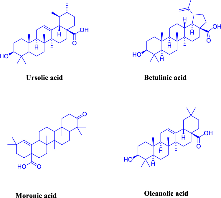 |
Figure 1 Some triterpenes with potential biological activity as an anti-inflammatory agent. |
The present study was carried out to synthesize arylidene derivatives of oleanolic acid at the C-2 position to develop more effective anti-inflammatory agents. To synthesize its various derivatives, oleanolic acid was isolated from Plectranthus rugosus by column chromatography of ethyl acetate fraction followed by recrystallization from methanol. Nmr and mass spectrometry confirmed the structures. The derivatives were screened for anti-inflammatory activity by scrutinizing the NO production inhibition in RAW 264.7 cells induced by LPS and their cytotoxicity. The potential candidates were further screened for inhibition of LPS-induced interleukin (IL-6) and tumour necrosis factor-alpha (TNF-α) production in RAW 264.7 cells.
Experimental Section
Materials and Methods
All the chemicals, as well as reagents, are of high purity. Dulbecco’s eagle medium (DMEM) and phosphate buffer saline (PBS) were purchased from Sigma, UK. Elisa kits, IL-6, Human TNF-α, were purchased from Invitrogen (USA). From Gibco USA, fetal bovine serum was obtained and lipopolysaccharide (LPS) E. Coli from Calbiochem (USA). Griess reagent was purchased from Promega (USA) and MTT from Calbiochem (San Diego CA). Besides, other chemicals that were used were of research-grade. From the central drug house (CDH) Jammu, ferric chloride, glacial acetic acid, chloroform was purchased. Ascorbic acid, ethanol, methanol, picric acid, calcium chloride, sodium chloride, 2,2 diphenyl picryl hyradyzl (DPPH), sucrose, trichloroacetic acid were procured from Merck. From Qualigens, potassium dihydrogen phosphate, hydrogen peroxide and hydrochloric acid were purchased. Ferric nitrate, ethylene diamine tetraacetate, potassium chloride, sodium hydroxide, dimethyl sulfoxide, sodium dihydrogen monophosphate was purchased from HiMedia. From sisco research laboratories (SRL) butylated hydroxytoluene was bought and from Rankem ethyl acetate and hexane was purchased.
Chemistry
In the present study, oleanolic acid (1) was isolated from leaf extract of Plectranthus rugosus by column chromatography in 60:40 hexane:ethyl-acetate followed by recrystallization from methanol. The solvents were distilled before using in the reaction. All the reactions were monitored by using 0.2 mm-thick, aluminum-backed TLC plates and were visualized under UV light at 254 nm. The structure was confirmed by nmr and mass spectrometry to give pure white amorphous powder having percentage purity more than 95% mp: 281–283°C[α]D21 +81.3° (CHCl3; c = 0.6).1H NMR (400 MHz, CDCl3): δ 5.27 (1H, s, H-12), 3.21 (IH, d, J=7.2, H-3), 2.83 (1H, d, J=11.3, H-12), 1.97 (2H, m, H-2), 0.91 (3H, s, H-23), 0.72 (3H, s, CH3-24), 0.76 (3H, s, CH3-25), 0.88 (3H, s, CH3-26), 1.14 (3H, s, CH3-27),0.92 (3H, s, CH3-29), 0.96 (3H, s, CH3-30). 13C NMR (100 MHz, CDCl3): δ 183.22 (C-28), 143.62 (C-13), 122.62 (C-12), 79.12 (C-3), 55.24 (C-5), 47.68 (C-9), 46.55 (C-17), 45.7 (C-19), 41.65 (C-14), 40.96 (C-18), 39.12 (C-8), 38.8 (C-1), 37.12 (C-10), 33.86 (C-21), 33.09 (C-29), 32.55 (C-22), 32.59 (C-7), 30.66 (C-20), 28.77 (C-23), 27.9 (C-15), 27.8 (C-2), 25.49 (C-27), 23.89 (C-30), 23.44 (C-16), 22.98 (C-11), 18.33 (C-6), 17.38 (C-26), 15.55 (C-25), 15.38 (C-24). ESI-MS m/z 456 [M+H]+
Synthesis of Arylidene-Oleanolic Acid Derivatives
The protocol for synthesis of oleanolic acid-arylidene derivatives 3 from the parent compound oleanolic acid 1 involves two steps which includes oxidation with pyridinium chlorochromate (PCC) reagent followed by Claisen Schmidt condensation (Scheme 1). So far, many synthetic strategies for the preparation of oleanolic acid analogs have been reported.48–52 In a typical procedure, to a solution of compound 1 (400 mg, 0.87 mmol) in dichloromethane (DCM) at room temperature (rt) few drops of PCC reagent were added. The reaction was monitored by TLC till its completion in 4 h. The reaction mixture was filtered to remove insoluble residue after quenching with cold water. The filtrate was extracted with ethyl-acetate and the organic layer was finally dried over sodium sulphate, concentrated in vaccum and purified by column chromatography to finally give compound 2 (365 mg 91% yield) as pure white amorphous solid; mp 179–183°C; 1H NMR (400MHz, CDCl3): δ 5.27 (1H, s,H-12), 3.22 (1H, d, J =12.0 Hz,H-18), 1.90 (2H, m), 1.82 (4H, m),1.55 (12H, m), 1.33 (4H, m) 1.05 (3H, s, CH3-23), 0.94 (3H, s, CH3-24), 0.86 (3H, s,CH3-25), 0.84 (3H, s,CH3-26), 0.82 (3H, s,CH3-27), 0.79 (3H, s,CH3-29), 0.65 (3H, s,CH3-30); 13C NMR (100 MHz, CdCl3): δ 215.7,181.25,151.29,123.32, 53.07, 52.19, 51.18, 50.07, 49.09, 47.29, 46.48, 41.34, 40.22, 39.55, 39.29, 39.18, 39.15, 39.12, 39.08, 38.39, 38.24, 38.18, 36.33, 28.19, 23.37, 21.19, 17.44, 17.78, 16.59, 15.24 ESI-MS m/z 455 [M+H]+
To the solution of compound 2 (365 mg, 0.8 mmol), in ethanol at room temperature lithium hydroxide LiOH (1.2 Eq) was added. To this mixture, various aromatic aldehydes were added (Table 1) by Claisen Schmidt condensation reaction (Scheme 1). The crude mixture was extracted with ethyl-acetate, and the organic layer was dried over sodium sulphate, concentrated in vacuum and purified by column chromatography to give pure 3a-3o in 80–90% yield
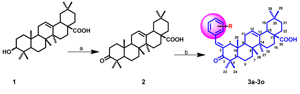 |
Scheme 1 Reagents and conditions (a) PCC, DCMrt, 90% (b) Ar-CHO, LiOH, Ethanol, rt, 80%. |
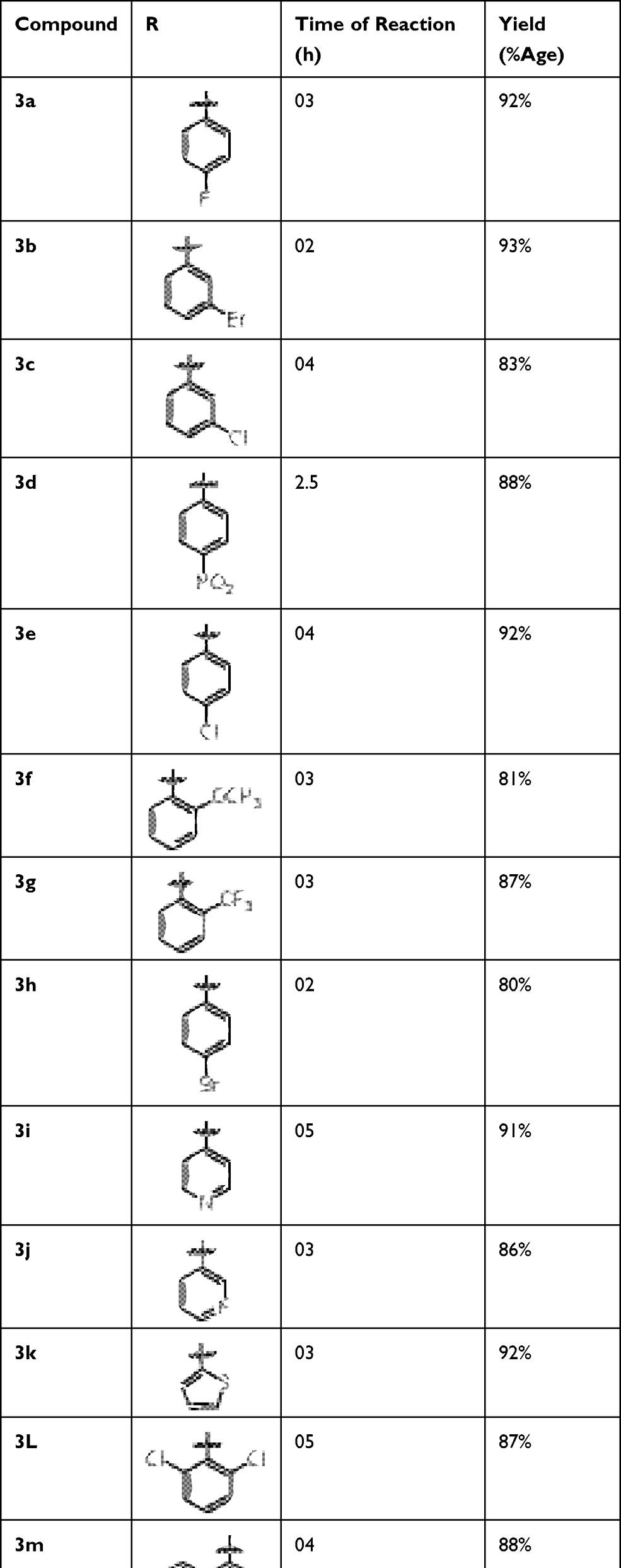 |
Table 1 Oleanolic Acid-Arylidene Derivatives Varying at the Aromatic Ring at the C-2 Position |
3-Oxo-2-[4-Fluorobenzylidenyl]-Olean-12-En-28-Oic Acid (3a)
Yield 92%; mp 159–161; 1H NMR (400 MHz, CDCl3): δ 7.57 (1H, s), 7.39 (2H, m), 7.37 (2H, m), 5.28 (1H, s), 2.35 (2H, m), 2.27 (1H, d, j = 8.0 Hz), 1.93 (4H, m), 1.78 (3H, s, CH3-23), 1.63 (3H, s, CH3-24), 1.45 (3H, s, CH3-25), 1.39 (3H, s, CH3-26). 1.09 (3H, s, CH3-27), 0.85 (3H, s, CH3-29), 0.48 (3H, s, CH3-30).13C NMR (100 MHz, CDCl3): δ 207.56, 181.08, 142.24, 138.66, 135.31, 132.19, 132.19, 130.55, 129.22,129.22, 123.87, 123.13, 55.22, 51.49, 48.89, 47.14, 46.29, 42.33, 40.87, 39.66, 39.25, 38.98, 37.46, 34.22, 33.68, 30.45, 29.18, 26.34, 24.45, 23.97, 23.77, 20.21, 19.34, 18.92, 18.23, 16.45. ESI-MS m/z 561 [M+H]+
3-Oxo-2-[3-Bromobenzylidenyl]-Olean-12-En-28-Oic Acid (3b)
Yield 93%; mp 148–151; 1HNMR (400 MHz, CDCl3): δ 7.48 (1H, s), 7.36 (2H, m), 7.22 (2H, m), 5.32 (1H, s), 2.45 (1H, m), 2.42 (2H, m), 2.23 (1H, j= 8.0 Hz), 1.92 (4H, m), 1.72 (3H, s, CH3-23), 1.62 (3H, s, CH3-24), 1.42 (3H, s, CH3-25), 1.33 (3H, s, CH3-26). 1.08 (3H, s, CH3-27), 0.80 (3H, s, CH3-29), 0.44 (3H, s, CH3-30).13C NMR (100 MHz, CDCl3): δ 207.91,183.84, 143.09, 138.43, 137.97, 136.18, 135.34, 134.67, 130.29, 129.96, 128.66, 128.19, 127.97, 127.18, 125.55, 125.08, 64.78, 53.33, 52.88, 48.28, 45.55, 43.94, 42.33, 39.66, 39.34, 39.08, 36.87, 36.64, 32.28,30.85, 29.86, 29.67, 28.17, 24.22, 23.77, 22.94, 21.32, 20.44,17.27. ESI-MS m/z 622 [M+H]+
3-Oxo-2-[3-Chlorobenzylidenyl]-Olean-12-En-28-Oic Acid (3c)
Yield 83%; mp 178–181;1H NMR (400 MHz, CDCl3): δ 7.50 (1H, s), 7.31 (2H, m), 7.25 (2H, m), 5.31 (1H, s), 2.43 (1H, m), 2.41 (2H, m), 2.27 (1H, d, j=8.0 Hz), 1.96 (4H, m), 1.76 (3H, s, CH3-23), 1.61 (3H, s, CH3-24), 1.46 (3H, s, CH3-25), 1.38 (3H, s, CH3-26). 1.04 (3H, s, CH3-27), 0.65 (3H, s, CH3-29), 0.58 (3H, s, CH3-30). 13C NMR (100 MHz, CDCl3): δ 207.94, 183.86, 143.04, 138.35, 137.97, 136.15, 135.34, 134.65, 130.23, 129.98, 128.63, 128.16, 127.93, 127.93, 127.17, 125.60, 125.05, 64.71, 53.37, 52.81, 48.27, 45.54, 43.95, 42.39, 39.64, 39.33, 39.05, 36.87, 36.64, 32.28, 30.86, 29.88, 29.66, 28.18, 24.27, 23.77, 22.91, 21.37, 20.48, 17.26. ESI-MS m/z 578 [M+H]+
3-Oxo-2-[4-Nitrobenzylidenyl]-Olean-12-En-28-Oic Acid (3d)
Yield 88%; mp 154–156; 1H NMR (400 MHz, CDCl3): δ 7.62 (1H, s), 7.45 (2H, m), 7.31 (1H, m), 7.22 (1H, m), 5.31 (1H, s), 2.41 (2H, m), 2.27 (1H, d, j= 8.0 Hz), 1.94 (4H, m), 1.79 (3H, s, CH3-23), 1.66 (3H, s, CH3-24), 1.49 (3H, s, CH3-25), 1.29 (3H, s, CH3-26). 1.04 (3H, s, CH3-27), 0.80 (3H, s, CH3-29), 0.43 (3H, s, CH3-30).13C NMR (100 MHz, CDCl3): δ 207.53, 181.04, 142.22, 137.45, 134.81, 133.44, 129.77, 129.53, 129.42, 128.13, 126.28, 123.02, 55.23, 51.97, 48.92, 47.19, 46.23, 42.77, 40.75, 39.66, 39.38, 38.97, 37.55, 34.44, 33.91, 30.22, 29.18, 26.62, 24.28, 23.91, 23.77, 20.02, 19.44, 18.66, 18.19, 16.82. ESI-MS m/z 588 [M+H]+
3-Oxo-2-[4-Chlorobenzylidenyl]-Olean-12-En-28-Oic Acid (3e)
Yield 92%; mp 183–185; 1H NMR (400 MHz, CDCl3): δ 7.52 (1H, s), 7.33 (2H, m), 7.21 (1H, m), 7.12 (1H, m), 5.41 (1H, s), 2.42 (1H, m), 2.41 (2H, m), 2.28 (1H, d, j = 8.0 Hz), 1.92 (4H, m), 1.72 (3H, s, CH3-23), 1.60 (3H, s, CH3-24), 1.48 (3H, s, CH3-25), 1.32 (3H, s, CH3-26). 1.12 (3H, s, CH3-27), 0.88 (3H, s, CH3-29), 0.44 (3H, s, CH3-30).13C NMR (100 MHz, CDCl3) δ 207.52, 181.05, 142.21, 138.66, 135.56, 134.14, 130.52, 129.30, 129.28, 123.09, 55.28, 51.92, 48.94, 47.59, 46.22, 42.77, 40.79, 39.38, 39.33, 38.97, 37.55, 34.44, 33.92, 30.29, 29.19, 26.66, 24.28, 23.92, 23.71, 20.09, 19.66, 18.63, 18.15, 16.89. ESI-MS m/z 578 [M+H]+
3-Oxo-2-[2-Methoxybenzylidenyl]-Olean-12-En-28-Oic Acid (3f)
Yield 81%; mp 171–174; 1H NMR (400 MHz, CDCl3): δ 7.46 (1H, s), 7.32 (2H, m), 7.12 (1H, m), 6.96 (1H, m), 5.32 (1H, s), 3.83 (3H, s), 2.88 (1H, m), 2.43 (2H, m), 2.28 (1H, d, j= 8.0 Hz), 1.98 (4H, m), 1.79 (3H, s, CH3-23), 1.65 (3H, s, CH3-24), 1.48 (3H, s, CH3-25), 1.39 (3H, s, CH3-26). 1.09 (3H, s, CH3-27), 0.85 (3H, s, CH3-29), 0.48 (3H, s, CH3-30). 13C NMR (100 MHz, CDCl3): δ 207.91, 183.88, 159.71, 138.27, 137.44, 134.33, 129.49, 125.88, 122.82, 116.04, 114.19, 55.49, 53.48, 52.98, 48.29, 45.42, 44.28, 42.43, 39.59, 39.49, 39.07, 36.98, 36.56, 32.31, 30.77, 29.88, 28.27, 24.39, 23.77, 22.99, 21.44, 20.55, 17.33, 17.09, 15.66. ESI-MS m/z 573 [M+H]+
3-Oxo-2-[2-Trifluoromethylbenzylidenyl]-Olean-12-En-28-Oic Acid (3g)
Yield 87%; mp 177–179; 1H NMR (400 MHz, CDCl3): δ 7.58 (1H, s), 7.52 (2H, m), 7.41 (1H, m), 7.32 (1H, m), 5.28 (1H, s), 2.48 (1H, m), 2.46 (2H, m), 2.26 (1H, d, j = 8.0 Hz), 1.94 (4H, m), 1.75 (3H, s, CH3-23), 1.62 (3H, s, CH3-24), 1.44 (3H, s, CH3-25), 1.30 (3H, s, CH3-26). 1.19 (3H, s, CH3-27), 0.95 (3H, s, CH3-29), 0.38 (3H, s, CH3-30).13C NMR (100 MHz, CDCl3): δ 207.92, 183.84, 159.77, 138.29, 137.47, 134.36, 129.47, 125.86, 122.85, 116.09, 114.16, 55.47, 53.44, 52.96, 48.24, 45.44, 44.27, 42.42, 39.53, 39.45, 39.09, 36.93, 36.52, 32.37, 30.79, 29.82, 28.24, 24.32, 23.79, 22.91, 21.42, 20.59, 17.32, 17.07, 15.61 ESI-MS m/z 611 [M+H]+
3-Oxo-2-[4-Bromobenzylidenyl]-Olean-12-En-28-Oic Acid (3h)
Yield 80%; mp 163–166; 1H NMR (400 MHz, CDCl3): δ 7.54 (1H, s), 7.32 (2H, m), 7.22 (1H, m), 7.14 (1H, m), 5.43 (1H, s), 2.45 (1H, m), 2.42 (2H, m), 2.26 (1H, d, j = 8.0 Hz), 1.94 (4H, m), 1.74 (3H, s, CH3-23), 1.64 (3H, s, CH3-24), 1.42 (3H, s, CH3-25), 1.34 (3H, s, CH3-26). 1.14 (3H, s, CH3-27), 0.82 (3H, s, CH3-29), 0.42 (3H, s, CH3-30) 13C NMR (100 MHz, CDCl3): δ 207.51, 181.06, 142.22, 138.68, 135.57, 134.11, 130.53, 129.35, 129.23, 123.04, 55.23, 51.96, 48.93, 47.54, 46.21, 42.78, 40.73, 39.34, 39.34, 38.96, 37.56, 34.47, 33.95, 30.28, 29.11, 26.63, 24.24, 23.95, 23.78, 20.04, 19.68, 18.67, 18.13, 16.82 ESI-MS m/z 622[M+H]+
3-Oxo-2-[Pyridine-4-Ylmethylene]-Olean-12-En-28-Oic Acid (3i)
Yield 91%; mp 138–141; 1HNMR (400 MHz, CDCl3): δ 8.57 (1H, s), 8.39 (H, m), 7.48 (2H, m), 7.27 (1H, m) 5.24 (1H, s), 2.39 (2H, m), 2.28 (1H, d, j = 8.0 Hz), 1.98 (4H, m), 1.75 (3H, s, CH3-23), 1.63 (3H, s, CH3-24), 1.45 (3H, s, CH3-25), 1.39 (3H, s, CH3-26). 1.09 (3H, s, CH3-27), 0.85 (3H, s, CH3-29), 0.48 (3H, s, CH3-30).13C NMR (100 MHz, CDCl3): δ 205.56, 180.07, 143.27, 139.67, 136.32, 134.18, 134.18, 131.54, 128.21,128.21, 124.88, 124.15, 56.21, 52.48, 49.88, 48.18, 47.26, 43.33, 41.88, 38.65, 38.23, 36.99, 35.43, 33.24, 32.69, 30.46, 28.19, 27.39, 25.47, 22.99, 23.76, 21.25, 18.36, 17.94, 17.26, 16.43 ESI-MS m/z 544 [M+H]+
3-Oxo-2-[Pyridine-3-Ylmethylene]-Olean-12-En-28-Oic Acid (3j)
Yield 93%; mp 148–151; 1H NMR (400 MHz, CDCl3): δ 8.77 (1H, s), 8.44 (H, m), 7.22 (2H, m), 7.12 (1H, m) 5.33 (1H, s), 2.35 (2H, m), 2.25 (1H, d, j = 8.0Hz), 1.91 (4H, m), 1.71 (3H, s, CH3-23), 1.61 (3H, s, CH3-24), 1.40 (3H, s, CH3-25), 1.32 (3H, s, CH3-26). 1.02 (3H, s, CH3-27), 0.91 (3H, s, CH3-29), 0.41 (3H, s, CH3-30).13C NMR (100MHz, CDCl3): δ 205.46, 180.22, 143.77, 139.66, 136.43, 134.88, 134.88, 131.87, 128.93,128.93, 124.32, 124.32, 56.44, 52.77, 49.84, 48.39, 47.39, 43.28, 41.89, 38.56, 38.45, 36.89, 35.34, 33.58, 32.79, 30.63, 28.91, 27.28, 25.78, 22.30, 23.69, 21.59, 18.29, 17.92, 17.19, 16.54 ESI-MS m/z 544 [M+H]+
3-Oxo-2-[Thiophene-2-Ylmethylene]-Olean-12-En-28-Oic Acid (3k)
Yield 92%; mp 184–186; (thiophen-2-ylmethylene)1H NMR (400 MHz, CDCl3): δ 8.13 (1H, s), 7.22 (2H, m), 7.12 (1H, m) 5.23 (1H, s), 2.32 (2H, m), 2.20 (1H, d, j = 8.0Hz), 1.92 (4H, m), 1.78 (3H, s, CH3-23), 1.68 (3H, s, CH3-24), 1.48 (3H, s, CH3-25), 1.37 (3H, s, CH3-26). 1.01 (3H, s, CH3-27), 0.82 (3H, s, CH3-29), 0.49 (3H, s, CH3-30).13C NMR (100 MHz, CDCl3): δ 204.49, 180.28, 148.79, 142.68, 136.48, 134.80, 134.83, 131.77, 128.85,128.23, 124.56, 124.39, 54.48, 52.57, 49.36, 48.74, 47.92, 43.93, 41.49, 38.29, 38.19, 36.19, 35.78, 33.49, 32.49, 30.49, 28.29, 27.48, 25.36, 22.22, 23.82, 21.57, 18.29, 17.74, 17.19, 16.49 ESI-MS m/z 549 [M+H]+
3-Oxo-2-[2,6-Dichlorobenzylidenyl]-Olean-12-En-28-Oic Acid (3L)
Yield 87%; mp 172–174; 1H NMR (400 MHz, CDCl3): δ 7.53 (1H, s), 7.31 (2H, m), 7.12 (1H, m) 5.28 (1H, s), 2.36 (2H, m), 2.26 (1H, d, j = 8.0 Hz), 1.97 (4H, m), 1.75 (3H, s, CH3-23), 1.64 (3H, s, CH3-24), 1.45 (3H, s, CH3-25), 1.32 (3H, s, CH3-26). 1.13 (3H, s, CH3-27), 0.89 (3H, s, CH3-29), 0.40 (3H, s, CH3-30).13C NMR (100 MHz, CDCl3): δ 207.59, 181.03, 142.29, 138.56, 135.38, 132.12, 132.10, 130.59, 129.24,129.24, 123.86, 123.43, 55.58, 51.42, 48.81, 47.19, 46.25, 42.35, 40.81, 39.64, 39.21, 38.94, 37.42, 34.27, 33.63, 30.47, 29.16, 26.32, 24.48, 23.90, 23.79, 20.22, 19.37, 18.97, 18.21, 16.48 ESI-MS m/z 612 [M+H]+
3-Oxo-2-[Naphthalene-1-Ylmethylene]-Olean-12-En-28-Oic Acid (3m)
Yield 88%; mp 185–187; 1H NMR (400 MHz, CDCl3): δ 8.17 (2 H, m), 7.59 (2H, m), 7.47 (2H, m), 7.29 (1H, m), 7.13 (1H, m) 5.24 (1H, s), 2.31 (2H, m), 2.20 (1H, d, j = 8.0Hz), 1.90 (4H, m), 1.71 (3H, s, CH3-23), 1.68 (3H, s, CH3-24), 1.47 (3H, s, CH3-25), 1.32 (3H, s, CH3-26). 1.19 (3H, s, CH3-27), 0.86 (3H, s, CH3-29), 0.44 (3H, s, CH3-30).13C NMR (100MHz, CDCl3): δ 207.87, 181.11, 142.29, 138.78, 135.42, 132.17, 132.11, 130.46, 129.31,129.31, 123.78, 123.33, 55.48, 51.41, 48.82, 47.64, 46.82, 42.59, 40.57, 39.33, 39.28, 38.79, 37.29, 34.19, 33.85, 30.29, 29.18, 26.59, 24.51, 23.75, 23.59, 20.29, 19.29, 18.89, 18.69, 16.54 ESI-MS m/z 593 [M+H]+
3-Oxo-2-[Benzylidenyl]-Olean-12-En-28-Oic Acid (3n)
Yield 81%; mp 144–146; 1H NMR (400 MHz, CDCl3): δ 7.58 (2H, m), 7.39 (2H, m), 7.37 (1H, m), 7.13 (IH, m), 5.26 (1H, s), 2.38 (2H, m), 2.21 (1H, d, j = 8.0Hz), 1.94 (4H, m), 1.77 (3H, s, CH3-23), 1.67 (3H, s, CH3-24), 1.46 (3H, s, CH3-25), 1.37 (3H, s, CH3-26). 1.09 (3H, s, CH3-27), 0.89 (3H, s, CH3-29), 0.47 (3H, s, CH3-30).13C NMR (100MHz, CDCl3): δ 207.51, 181.04, 142.28, 138.64, 135.39, 132.15, 132.14, 130.59, 129.28,129.28, 123.89, 123.18, 55.23, 51.41, 48.85, 47.13, 46.27, 42.39, 40.81, 39.63, 39.26, 38.91, 37.45, 34.29, 33.64, 30.43, 29.16, 26.33, 24.48, 23.92, 23.73, 20.28, 19.33, 18.99, 18.28, 16.43 ESI-MS m/z 543 [M+H]+
3-Oxo-2-[3-Nitrobenzylidenyl]-Olean-12-En-28-Oic Acid (3o)
Yield 83%; mp 161–163; 1H NMR (400 MHz, CDCl3): δ 8.30 (1H, s), 8.13 (1H, m), 7.91 (2H, 1), 7.61 (1H, m), 7.25 (1H, m), 5.33 (1H, s), 2.46 (1H, m), 2.40 (2H, m), 2.24 (1H, d, j=8.0Hz), 1.91 (4H, m), 1.79 (3H, s, CH3-23), 1.63 (3H, s, CH3-24), 1.42 (3H, s, CH3-25), 1.31 (3H, s, CH3-26). 1.02 (3H, s, CH3-27), 0.81 (3H, s, CH3-29), 0.42 (3H, s, CH3-30).13C NMR (100 MHz, CDCl3): δ 207.98, 183.85, 143.05, 138.39, 137.97, 136.19, 135.32, 134.61, 130.29, 129.96, 128.66, 128.18, 127.92, 127.95, 127.13, 125.69, 125.05, 64.79, 53.32, 52.89, 48.22, 45.58, 43.91, 42.35, 39.65, 39.31, 39.07, 36.82, 36.67, 32.27, 30.83, 29.85, 29.62, 28.17, 24.27, 23.75, 22.93, 21.38, 20.42, 17.28 ESI-MS m/z 588 [M+H]+
Biological Activity
Evaluation of Cell Viability by MTT Assay
MTT (3-[4,5-dimethylthiazol-2-yl]-2,5 diphenyltetrazolium bromide) assay was used to study the cytotoxicity of all synthetic compounds against cultured RAW 264.7 cells (purchased commercially from ATCC, Manassas, VA, USA).53 Camptothecin was taken as a reference standard in this study. In brief RAW 264.7 cells at a density of 16,000 cells/well were seeded into 96-well plates and were allowed to adhere in a CO2 incubator at 37 ° C for a time period of 24 h. After 24 h of incubation, the cells were treated with different concentrations of oleanolic derivatives (0–10 µM/mL) for another 24 h. Afterwards, 20 µL of MTT (0.5mg/mL in PBS, PH 7.4) was added and kept for incubation for another 4 h at 37°C. Finally, the supernatant was removed, and 100 µL of dimethyl sulfoxide (DMSO) was added to each well to dissolve the formazan crystals which are formed after the addition of MTT. The absorbance was measured by using the synergy Mx plate reader at 570 nm. The results were expressed as a percentage of cell viability by using LPS-induced group as a control group. Three replicates were carried for each treatment.
NO Assay
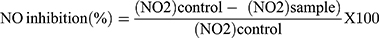
For the evaluation of NO production in RAW 264.7 cells, the concentration of nitrite was measured in the supernatant as an indicator using Griess reaction. In brief, RAW 264.7 macrophage cells (2 x 10⁵ cells/well) were treated with different concentrations of synthetic derivatives in the presence or absence of LPS (1µg/mL) 1 h before LPS treatment and then kept in incubation for 24 h. Dexamethasone in different concentrations was used as a positive control. After an incubation period, supernatant (100 µL) was collected by centrifugation at 1000 rpm and mixed with Griess reagent (0.1% N-1-naphthyl ethylenediamine dihydrochloride and 1% sulphanilamide in 5% phosphoric acid) and kept for incubation for 10 min at room temperature in dark. By using the synergy Mx Plate reader, absorbance was measured at 540 nm. With respect to the standard concentration curve of sodium nitrite (NaNo2), concentration of nitrite was calculated.54
The percentage inhibition of NO was calculated with the following formula
Measurement of Cytokine Production in RAW 264.7 Cells
The inhibitory effect of all synthetic derivatives on the production of cytokines (TNF-α and IL-6) was determined by enzyme-linked immunosorbent assay (ELISA) kit. In 96-well plate RAW 264.7 cells were seeded at a density of 2 x 10⁵ cells/well and left for incubation for overnight. The cells were then pretreated with synthetic derivatives for 1hr before stimulation with LPS for 24 h to induce inflammation. To collect the supernatant, the culture plate was then centrifuged at 1500 rpm and assayed according to the protocol of the manufacturer (Invitrogen) to measure the amount of TNF-α and IL-6 produced in each sample. The whole experiment was carried out in triplicate.
Molecular Docking and Selection of Predicted Interaction Pose
Docking of all the synthetic derivatives of oleanolic acid on TNF-α (PDB ID: 2AZ5) and IL-1β (PDB ID: 3040) was performed with molecular operating environment (MOE)2019.01 program.55 Structure breaks of TNF-α and IL-1β after retrieved from the RCBS Protein database56 were fixed using MOE2019.01. Partial charges and hydrogen atoms were added to both proteins after removing the water molecules. By using the optimized potential for liquid simulations (OPLS) force field in MOE2019.01, the protein structures were minimized. The 3D structure of all the synthetic derivatives of oleanolic acid was prepared and minimized using the MMFF94x force field in MOE2019.57 After structure preparation of synthetic derivatives of oleanolic acid and proteins, by using induced-fit docking protocol, the ligands were docked on co-crystallized ligand site in TNF-α and site predicted by MOE and meta pocket 2.058 in case of IL-1β. All the docked solutions were clustered, and representative solutions of the most populated cluster were selected based on score and subjected to manual selection, based on suitability and binding. The highest scoring solution among manually selected groups of experimentally active compounds were considered as the representative pose. The binding affinity of representative docked pose was calculated using Cyscore.59,60
Statistical Analysis
All the results were expressed as (mean ± of SD). Graphpad prism eight and microsoft excel was used to calculate SD. P-value of less than 0.5 was considered significant. All the experiments were carried out in triplicates.
Results
Effect on Cell Viability
To find the safety level of all the five potent analogs (3d, 3e, 3L, 3n, 3o), MTT assay was performed. Raw cells were subjected to different concentrations (1–100 µM) of (3d, 3e, 3L, 3n, 3o), for a time period of 48 hours. At low concentrations (1–10 µM), we observed that cell viability did not get affected and was more than 80% at 10µM (Figure 2). Therefore, to investigate the anti-inflammatory of (3d, 3e, 3L, 3n, 3o), on RAW 264.7 cells, our data is non-toxic at a concentration of (1–10 µM).
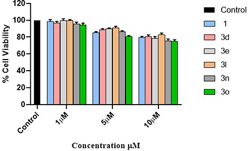 |
Figure 2 Cytotoxicity of compounds 1, 3d, 3e, 3L, 3n and 3o: RAW 264.7 cells were treated up to 10 µM concentrations with 1, 3d, 3e, 3L, 3n and 3o for 48 h, and cell viability checked by MTT assay. The data represent mean ± SD (n = 3) of the representative experiment. |
Effect of Derivatives on NO Production in LPS Stimulated RAW 264.7 Cells
Among 15 new derivatives, compounds 3a (77.18%), 3d (71.5%) and 3e (68.8%) showed more potent inhibition of NO than oleanolic acid (65.22%) at 10µM which is comparable to dexamethasone (85.7%) and L-NAME (80.5). Analogs 3c, 3k, 3m, 3g, and 3L showed moderate inhibition of NO, respectively. However, analogs 3b, 3f, 3h, 3i and 3j, showed poor inhibitory activity (Table 2).
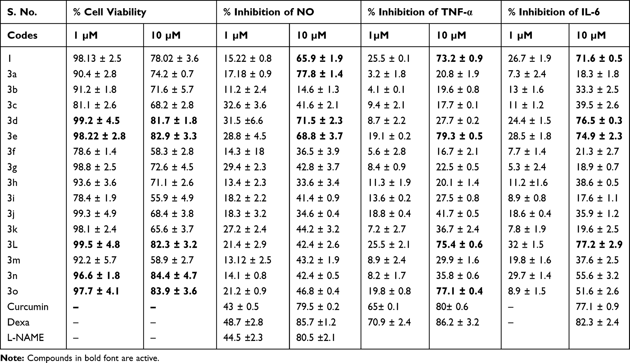 |
Table 2 The % Cell Viability and Percentage (%) Inhibition of Compounds 1–15 Against NO, IL-6, and TNF-α in LPS (1 µg/mL) Stimulated RAW 246.7 Cells |
Measurement of Pro-Inflammatory Cytokines (TNF-α and IL-6) in RAW 264.7 Cells
The synthetic analogs were observed for IL-6 and TNF-α inhibition. Compounds 3d, 3e, and 3L showed maximum IL-6 inhibition of 76.5%,74.9% and 77.2% respectively, which is comparable with oleanolic acid (71.6%) at 10µM concentration. Analogs 3b, 3c, 3h, 3j, 3m, 3n, and 3o exhibit moderate activity (Table 2), whereas, analogs 3a, 3f, 3g, 3i, 3k showed weak IL-6 inhibitory activity. In the case of TNF-α, analogs 3e, 3L and 3o showed maximum inhibitions against TNF-α of 79.3%, 75.4%and 77.1%, which are more potent than oleanolic acid (73.2%). Others exhibited either moderate or weak activity than oleanolic acid (Table 2). In LPS untreated cells, the levels of TNF-α and IL-6 were undetectable and served as a control. At 10µM concentration, there were no significant changes in cell viability on the treatment of RAW 264.7 cells. Analogs that exhibit more than 50% inhibition of TNF-α and IL-6 are considered more potent.61,62
Molecular Docking Studies
All the synthetic derivatives were docked on TNF-α and IL-1β (Figure 3A and B). The top-scoring solutions have been analyzed for the Cyscore score, and ADME properties are mentioned in (Table 3). The docking pose shows the interaction of (3d, 3e, 3L, 3n, 3o) and dexamethasone with TNF-α and IL-β. The binding affinity of analogs and dexamethasone with TNF-α and IL-1β was calculated using Cyscore 2.60,63 From docking score and Cyscore analysis, it shows that the analogs (3e, 3L, 3n) are having a greater affinity towards TNF-α and IL-1β than dexamethasone which was also verified from in vitro results.
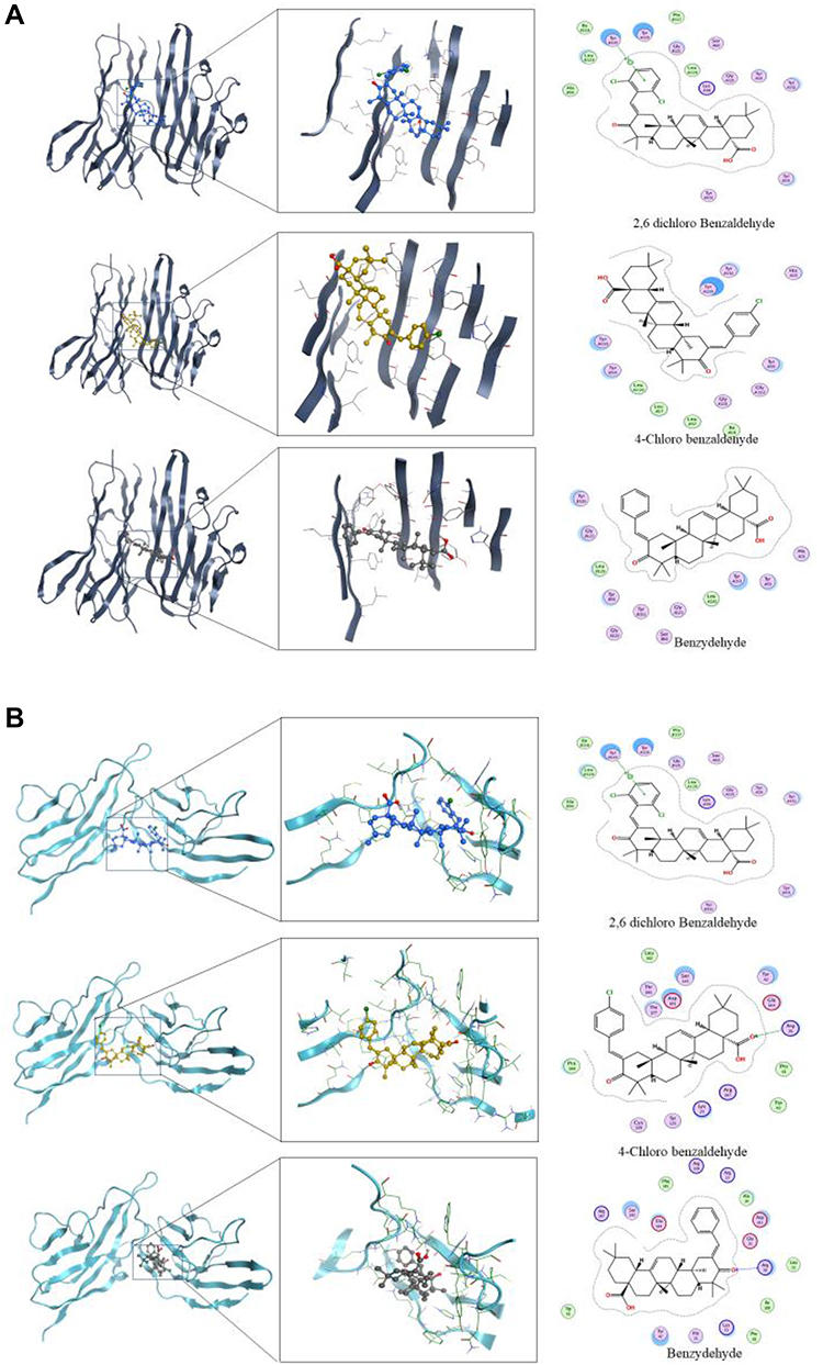 |
Figure 3 Interaction of active compounds with TNF-α (A) and IL-1β (B). |
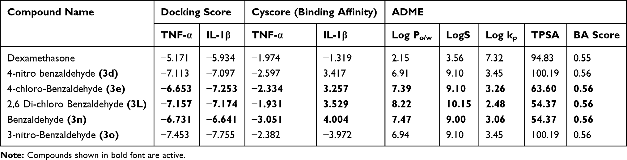 |
Table 3 Docking and Cyscore Score Along with ADME Properties |
Discussion
In the current study, we investigated the various derivatives of oleanolic acid on the production of pro-inflammatory mediators in RAW 264.7 mouse macrophages stimulated by LPS. In RAW 264.7 mouse macrophages, the analogs were evaluated for their potential to reduce lipopolysaccharide (LPS) induced TNF-α and IL-6 production. RAW 264.7 cells upon activation with LPS releases TNF-α and IL-6. LPS, a component of a gram-negative bacterial cell wall, induces macrophages and monocytes activation which is having a pivotal role in the innate immune response. Stimulation of RAW 264.7 cells with LPS leads to a series of intracellular events which results in the secretion of cytokines as well as other mediators of inflammation that eventually constitute the pro-inflammatory response. RAW 264.7 cells on pretreatment with the analogs (3d, 3e, 3L, 3o) at various concentrations followed by LPS treatment for 24 h results in the downregulation of pro-inflammatory cytokines (IL-6 and TNF-α). At 10µg/mL, the analogs showed potent inhibition of cytokines. In LPS untreated cells, the levels of IL-6 and TNF-α were not detectable and served as a control. MTT assay also showed that up to 10µg/mL concentration, the analogs (3d, 3e, 3L, 3n, 3o) did not affect the viability of RAW 264.7 cells. So the binding of potent analogs ((3d, 3e, 3L, 3n, 3o) results in the downregulation of pro-inflammatory cytokines (TNF-α and IL-6) and upregulated the secretion of anti-inflammatory cytokine IL-10.
Moreover, the analogs (3a, 3d, 3e) also inhibit the production of NO also, which is also released during the process of inflammation by activated phagocytes or resident cells to activate various macrophages. With reference to standard drug dexamethasone, the inhibitory potential of analogs was studied. Besides, from docking score and Cyscore analysis analogs (3e, 3L, 3n) showed greater affinity towards TNF-α and IL-1β than dexamethasone, which was also verified from our in vitro results.
So we have undertaken a research program directed toward the structural modifications of (1) at C-2 position to fine tune its biological potential as an anti-inflammatory agent. Compound (1) was subjected to oxidation by pyridinium chlorochromate (PCC) reagent at room temperature (rt) that resulted in the formation of (C-3) oxidized derivative (2) in almost quantitative yield. To the solution of compound (2) (365 mg, 0.8 mmol), in ethanol at room temperature lithium hydroxide LiOH (1.2 Eq) was added. To this mixture, various aromatic aldehydes were added by Claisen Schmidt condensation reaction. The crude mixture was extracted with ethyl-acetate, and the organic layer was dried over sodium sulphate, concentrated in vacuum and purified by column chromatography to give pure 3a-3o in 80–90% yield. The products were confirmed by 1H NMR, 13C NMR, and MS analysis. By employing the above reaction conditions, a series of oleanolic acid-arylidene derivatives that vary at substitutions on aromatic ring has been synthesized.
Conclusion
Herein, we report a series of 15 new arylidene derivatives of oleanolic acid by Claisen Schmidt condensation reaction. All the compounds synthesized were screened for their anti-inflammatory activity against NO, TNF-α and IL-6. From the data, it was evident that most of the compounds exhibited better anti-inflammatory activity. Molecular docking studies further confirm the anti-inflammatory activity of the derivatives (3d, 3e, 3L, 3n, 3o). Compounds (3d, 3e, 3L, 3n, and 3o) with o-nitro, o-chloro and p-chloro substitutions were found to be the most promising derivatives. The study also encouraged to study the molecular mechanisms involved, which further demonstrates its anti-inflammatory action at the molecular level.
Acknowledgment
The authors of this study extend their appreciation to the Research Supporting Project, King Saud University, Saudia Arabia, for supporting this study (RSP-2020/119) and for funding this work. The authors would also like to extend their sincere appreciation to the University of Kashmir, for providing necessary facilities to carry out this work.
Disclosure
The authors declare no conflicts of interest in this work.
References
1. Stankov SV. Definition of inflammation, causes of inflammation and possible anti-inflammatory strategies. Open Inflamm J. 2012;5(1):1–9. doi:10.2174/1875041901205010001
2. Tracy RP. The five cardinal signs of inflammation: calor, dolor, rubor, tumor … and penuria (apologies to Aulus Cornelius Celsus, De medicina, c. AD 25). J Gerontol a Biol Sci Med Sci. 2006;61(10):1051–1052. doi:10.1093/gerona/61.10.1051
3. Rather L. Disturbance of function (functio laesa): the legendary fifth cardinal sign of inflammation, added by Galen to the four cardinal signs of celsus. Bull N Y Acad Med. 1971;47(3):303.
4. Ferrero‐Miliani L, Nielsen O, Andersen P, Girardin S. Chronic inflammation: importance of NOD2 and NALP3 in interleukin‐1β generation. Clin Exp Immunol. 2007;147(2):227–235.
5. Hawiger J, Zienkiewicz J. Decoding inflammation, its causes, genomic responses, and emerging countermeasures. Scand J Immunol. 2019;90(6):e12812. doi:10.1111/sji.12812
6. Taams LS. Inflammation and immune resolution. Clin Exp Immunol. 2018;193(1):1–2. doi:10.1111/cei.13155
7. Koller GM, Schafer C, Kemp SS, et al. Pro-inflammatory mediators, IL (interleukin)-1β, TNF (tumor necrosis factor) α, and thrombin directly induce capillary tube regression. Arterioscler Thromb Vasc Biol. 2020;40(2):365–377. doi:10.1161/ATVBAHA.119.313536
8. Zanoli L, Briet M, Empana JP, et al. Vascular consequences of inflammation: a position statement from the ESH working group on vascular structure and function and the ARTERY society. J Hypertens. 2020;38(9):1682–1698. doi:10.1097/HJH.0000000000002508
9. Medzhitov R. Origin and physiological roles of inflammation. Nature. 2008;454(7203):428. doi:10.1038/nature07201
10. Rates SMK. Plants as source of drugs. Toxicon. 2001;39(5):603–613. doi:10.1016/S0041-0101(00)00154-9
11. Newman DJ, Cragg GM. Natural products as sources of new drugs over the last 25 years. J Nat Prod. 2007;70(3):461–477. doi:10.1021/np068054v
12. Gosslau A, Li S, Ho CT, Chen KY, Rawson NE. The importance of natural product characterization in studies of their anti‐inflammatory activity. Mol Nutr Food Res. 2011;55(1):74–82. doi:10.1002/mnfr.201000455
13. Lee KH, Itokawa H, Akiyama T, Morris‐Natschke SL. Plant-derived natural products research in drug discovery. Nat Prod Chem Biol. 2012;65:351–388.
14. Spainhour CB. Natural products. Pharm Sci Encycl. 2010;1–62.
15. Cragg GM, Grothaus PG, Newman DJ. Natural products in drug discovery: recent advances. In: Cechinel-Filho V, editor. Plant Bioactives and Drug Discovery: Principles, Practice, and Perspectives.
16. Strömstedt AA, Felth J, Bohlin L. Bioassays in natural product research–strategies and methods in the search for anti‐inflammatory and antimicrobial activity. Phytochem Anal. 2014;25(1):13–28. doi:10.1002/pca.2468
17. Lee KH, Itokawa H, Akiyama T, Morris‐Natschke SL. Plant‐derived natural products research: recent progress in drug discovery. In: Wiley Encyclopedia of Chemical Biology. Hoboken: Wiley; 2007:1–22.
18. Lachance H, Wetzel S, Waldmann H. Role of Natural Products in Drug Discovery. Wiley Online Library; 2010:187–229.
19. Nunes C, Barreto Arantes M, Menezes de Faria Pereira S, et al. Plants as sources of anti-inflammatory agents. Molecules. 2020;25(16):3726. doi:10.3390/molecules25163726
20. Mir RH, Shah AJ, Mohi-Ud-Din R, et al. Natural Anti-inflammatory compounds as drug candidates in alzheimer’s disease. Current Medicinal Chemistry. In press 2020.
21. Hassan R, Masoodi MH. Saussurea lappa: a comprehensive review on its pharmacological activity and phytochemistry. Curr Tradit Med. 2020;6(1):13–23. doi:10.2174/2215083805666190626144909
22. Mir RH, Masoodi MH. Anti-inflammatory plant polyphenolics and cellular action mechanisms. Curr Bioact Compd. 2020;16(6):809–817. doi:10.2174/1573407215666190419205317
23. Grabley S, Sattler I. Natural products for lead identification: nature is a valuable resource for providing tools. In: Hillisch A, Hilgenfeld R, editors. Modern Methods of Drug Discovery. Springer; 2003:87–107.
24. Lee K-H. Discovery and development of natural product-derived chemotherapeutic agents based on a medicinal chemistry approach. J Nat Prod. 2010;73(3):500–516. doi:10.1021/np900821e
25. Gautam R, Jachak SM. Recent developments in anti‐inflammatory natural products. Med Res Rev. 2009;29(5):767–820.
26. Kobyliak N, Falalyeyeva T, Mykhalchyshyn G, Kyriienko D, Komissarenko I. Effect of alive probiotic on insulin resistance in type 2 diabetes patients: randomized clinical trial. Diabetes Metab Syndr. 2018;12(5):617–624. doi:10.1016/j.dsx.2018.04.015
27. Nance CL. Clinical efficacy trials with natural products and herbal medicines. In: Ramzan I, editor. Phytotherapies: Efficacy, Safety and Regulation. Hoboken: Wiley; 2015:65–88.
28. Freire MO, Van Dyke TE. Natural resolution of inflammation. Periodontology. 2013;63(1):149–164. doi:10.1111/prd.12034
29. Mir RH, Sawhney G, Verma R, et al. Oreganum vulgare: in-vitro assessment of cytotoxicity, molecular docking studies, antioxidant, and evaluation of anti-inflammatory activity in LPS stimulated RAW 264.7 cells. Med Chem. 2020. doi:10.2174/1573406416666200904110828
30. Bhat MF, Hassan R, Masoodi MH. Nuclear magnetic resonance (NMR) for plant profiling and disease metabolomics-fast tracking plant based drug discovery from northern India. Nucl Magn Reson. 2018;2:1.
31. Vane JR. Inhibition of prostaglandin synthesis as a mechanism of action for aspirin-like drugs. Nat New Biol. 1971;231(25):232–235. doi:10.1038/newbio231232a0
32. Prestwich GD, Abe I, Zheng YF, Robustell BJ, Dang T. Enzymatic cyclization of squalene analogs. Pure Appl Chem. 1999;71(6):1127–1131. doi:10.1351/pac199971061127
33. Jäger S, Trojan H, Kopp T, Laszczyk M, Scheffler A. Pentacyclic triterpene distribution in various plants – rich sources for a new group of multi-potent plant extracts. Molecules. 2009;14(6):2016–2031. doi:10.3390/molecules14062016
34. Meng Y-Q, Liu D, Cai -L-L, Chen H, Cao B, Wang Y-Z. The synthesis of ursolic acid derivatives with cytotoxic activity and the investigation of their preliminary mechanism of action. Bioorg Med Chem. 2009;17(2):848–854. doi:10.1016/j.bmc.2008.11.036
35. Kwon T-H, Lee B-M, Chung S-H, Kim D-H, Lee Y-S. Synthesis and NO production inhibitory activities of ursolic acid and oleanolic acid derivatives. Bull Korean Chem Soc. 2009;30(1):119–123.
36. Ikeda Y, Murakami A, Ohigashi H. Ursolic acid: an anti‐and pro‐inflammatory triterpenoid. Mol Nutr Food Res. 2008;52(1):26–42. doi:10.1002/mnfr.200700389
37. Liu J. Oleanolic acid and ursolic acid: research perspectives. J Ethnopharmacol. 2005;100(1–2):92–94. doi:10.1016/j.jep.2005.05.024
38. Tian Z, Lin G, Zheng R-X, Huang F, Yang M-S, Xiao P-G. Anti-hepatoma activity and mechanism of ursolic acid and its derivatives isolated from Aralia decaisneana. World J Gastroenterol. 2006;12(6):874. doi:10.3748/wjg.v12.i6.874
39. Shyu M-H, Kao T-C, Yen G-C. Oleanolic acid and ursolic acid induce apoptosis in HuH7 human hepatocellular carcinoma cells through a mitochondrial-dependent pathway and downregulation of XIAP. J Agric Food Chem. 2010;58(10):6110–6118. doi:10.1021/jf100574j
40. Ma C, Nakamura N, Miyashiro H, Hattori M, Shimotohno K. Inhibitory effects of ursolic acid derivatives from Cynomorium songaricum, and related triterpenes on human immunodeficiency viral protease. Phytother Res. 1998;12(S1):S138–S142.
41. Cos P, Maes L, Vanden Berghe D, Hermans N, Pieters L, Vlietinck A. Plant substances as anti-HIV agents selected according to their putative mechanism of action. J Nat Prod. 2004;67(2):284–293. doi:10.1021/np034016p
42. Yu D, Sakurai Y, Chen C-H, et al. Anti-AIDS agents 69. moronic acid and other triterpene derivatives as novel potent anti-HIV agents. J Med Chem. 2006;49(18):5462–5469. doi:10.1021/jm0601912
43. Pathak A, Singh S, Biabani M, et al. Synthesis of combinatorial libraries based on terpenoid scaffolds. Comb Chem High Throughput Screen. 2002;5(3):241–248. doi:10.2174/1386207024607275
44. Liu J. Pharmacology of oleanolic acid and ursolic acid. J Ethnopharmacol. 1995;49(2):57–68. doi:10.1016/0378-8741(95)90032-2
45. Patel NK, Pulipaka S, Dubey SP, Bhutani KK. Pro-inflammatory cytokines and nitric oxide inhibitory constituents from Cassia occidentalis roots. Nat Prod Commun. 2014;9(5):1934578X1400900519.
46. Patel NK, Bhutani KK. Pinostrobin and Cajanus lactone isolated from Cajanus cajan (L.) leaves inhibits TNF-α and IL-1β production: in vitro and in vivo experimentation. Phytomedicine. 2014;21(7):946–953. doi:10.1016/j.phymed.2014.02.011
47. Bhandari P, Patel NK, Bhutani KK. Synthesis of new heterocyclic lupeol derivatives as nitric oxide and pro-inflammatory cytokine inhibitors. Bioorg Med Chem Lett. 2014;24(15):3596–3599. doi:10.1016/j.bmcl.2014.05.032
48. Ngoc TD, Moons N, Kim Y, et al. Synthesis of triterpenoid triazine derivatives from allobetulone and betulonic acid with biological activities. Bioorg Med Chem. 2014;22(13):3292–3300. doi:10.1016/j.bmc.2014.04.061
49. Urban M, Vlk M, Dzubak P, Hajduch M, Sarek J. Cytotoxic heterocyclic triterpenoids derived from betulin and betulinic acid. Bioorg Med Chem. 2012;20(11):3666–3674. doi:10.1016/j.bmc.2012.03.066
50. Haavikko R, Nasereddin A, Sacerdoti-Sierra N, et al. Heterocycle-fused lupane triterpenoids inhibit Leishmania donovani amastigotes. MedChemComm. 2014;5(4):445–451. doi:10.1039/C3MD00282A
51. Rashid S, Dar BA, Majeed R, Hamid A, Bhat BA. Synthesis and biological evaluation of ursolic acid-triazolyl derivatives as potential anti-cancer agents. Eur J Med Chem. 2013;66:238–245. doi:10.1016/j.ejmech.2013.05.029
52. Tang C, Zhu L, Chen Y, et al. Synthesis and biological evaluation of oleanolic acid derivative–chalcone conjugates as α-glucosidase inhibitors. RSC Adv. 2014;4(21):10862–10874. doi:10.1039/C3RA46492J
53. Xu X, Yin P, Wan C, et al. Punicalagin inhibits inflammation in LPS-induced RAW264. 7 macrophages via the suppression of TLR4-mediated MAPKs and NF-κB activation. Inflammation. 2014;37(3):956–965. doi:10.1007/s10753-014-9816-2
54. Joo T, Sowndhararajan K, Hong S, et al. inhibition of nitric oxide production in LPS-stimulated RAW 264.7 cells by stem bark of Ulmus pumila L. Saudi J Biol Sci. 2014;21(5):427–435. doi:10.1016/j.sjbs.2014.04.003
55. Deeth RJ, Fey N, Williams-Hubbard B. DommiMOE: an implementation of ligand field molecular mechanics in the molecular operating environment. J Comput Chem. 2005;26(2):123–130. doi:10.1002/jcc.20137
56. Pappalardo E, Cantone D. Database systems in biology. In: Barolli L, editor. Enterprise Business Modeling, Optimization Techniques, and Flexible Information Systems. IGI Global; 2013:80–96.
57. Heinzerling L, Klein R, Rarey M. Fast force field‐based optimization of protein–ligand complexes with graphics processor. J Comput Chem. 2012;33(32):2554–2565. doi:10.1002/jcc.23094
58. Zhang Z, Li Y, Lin B, Schroeder M, Huang B. Identification of cavities on protein surface using multiple computational approaches for drug binding site prediction. Bioinformatics. 2011;27(15):2083–2088. doi:10.1093/bioinformatics/btr331
59. Cao Y, Dai W, Miao Z. Evaluation of protein–ligand docking by Cyscore. In: Gore M, Jagtap UB, editor. Computational Drug Discovery and Design. Springer; 2018:233–243.
60. Li H, Leung K-S, Wong M-H, Ballester PJ. Substituting random forest for multiple linear regression improves binding affinity prediction of scoring functions: Cyscore as a case study. BMC Bioinform. 2014;15(1):291. doi:10.1186/1471-2105-15-291
61. Ammon H. Modulation of the immune system by Boswellia serrata extracts and boswellic acids. Phytomedicine. 2010;17(11):862–867. doi:10.1016/j.phymed.2010.03.003
62. Lin W-C, Lin J-Y. Five bitter compounds display different anti-inflammatory effects through modulating cytokine secretion using mouse primary splenocytes in vitro. J Agric Food Chem. 2010;59(1):184–192. doi:10.1021/jf103581r
63. Li H, Sze KH, Lu G, Ballester PJ. Machine‐learning scoring functions for structure‐based drug lead optimization. Wiley Interdiscip Rev Comput Mol Sci. 2020;e1465.
 © 2021 The Author(s). This work is published and licensed by Dove Medical Press Limited. The
full terms of this license are available at https://www.dovepress.com/terms
and incorporate the Creative Commons Attribution
- Non Commercial (unported, 3.0) License.
By accessing the work you hereby accept the Terms. Non-commercial uses of the work are permitted
without any further permission from Dove Medical Press Limited, provided the work is properly
attributed. For permission for commercial use of this work, please see paragraphs 4.2 and 5 of our Terms.
© 2021 The Author(s). This work is published and licensed by Dove Medical Press Limited. The
full terms of this license are available at https://www.dovepress.com/terms
and incorporate the Creative Commons Attribution
- Non Commercial (unported, 3.0) License.
By accessing the work you hereby accept the Terms. Non-commercial uses of the work are permitted
without any further permission from Dove Medical Press Limited, provided the work is properly
attributed. For permission for commercial use of this work, please see paragraphs 4.2 and 5 of our Terms.