Back to Journals » Drug Design, Development and Therapy » Volume 10
Design, development, and characterization of lipid nanocarriers-based epigallocatechin gallate delivery system for preventive and therapeutic supplementation
Authors Frias I, Neves AR, Pinheiro M ![]() , Reis S
, Reis S
Received 31 March 2016
Accepted for publication 9 June 2016
Published 31 October 2016 Volume 2016:10 Pages 3519—3528
DOI https://doi.org/10.2147/DDDT.S109589
Checked for plagiarism Yes
Review by Single anonymous peer review
Peer reviewer comments 2
Editor who approved publication: Prof. Dr. Wei Duan
Iúri Frias, Ana Rute Neves, Marina Pinheiro, Salette Reis
Research Unit on Applied Molecular Biosciences, Rede de Química e Tecnologia, Departamento de Ciências Químicas, Faculdade de Farmácia, Universidade do Porto, Porto, Portugal
Abstract: Green tea is manufactured from the leaves of Camellia sinensis and has been shown to possess, among other properties, anticancer, antiobesity, antiatherosclerotic, antidiabetic, antibacterial, and antiviral effects. The beneficial effects of green tea are related to the activities of (-)-epigallocatechin gallate (EGCG). This catechin is very unstable, undergoing degradation and epimerization, which is responsible for the loss of its health benefits. Encapsulation in nanoparticles (NPs) is an effective method to protect EGCG from adverse environmental conditions. In this work, solid lipid NPs (SLN) and nanostructured lipid carriers (NLC) were successfully developed to be used as biocompatible nanocarriers, enhancing the stability of EGCG. The mean diameter of the NPs was found to be around 300–400 nm, which is suitable for oral administration. Moreover, EGCG was effectively encapsulated with a remarkable efficiency of encapsulation of 80% and 90% for SLN and NLC, respectively. In addition, high storage stability of the formulations is expected as they maintain the initial characteristics for 3 months. Limited release of EGCG from the NPs was observed in simulated gastric and intestinal fluids. MTT and lactate dehydrogenase (LDH) assays demonstrated that NPs possess low toxicity, and so have potential to be used for preventive and therapeutic EGCG supplementation.
Keywords: EGCG, lipid nanoparticles, nanoparticles, nutraceuticals, supplementation
Introduction
Green tea is an infusion of the tea plant Camellia sinensis, consumed for centuries in People’s Republic of China, associated with many health benefits. This beverage has high concentrations of antioxidants, namely, polyphenols, such as epigallocatechin, epicatechin, epicatechin gallate, and epigallocatechin gallate (EGCG).1 Recent studies found that these polyphenols have numerous benefits in the prevention and treatment of cancer, vascular and degenerative diseases, diabetes, obesity, and other health concerns. Of the former compounds, EGCG is the most abundant and therapeutically active.2,3 The worldwide population consumption of EGCG would be of high interest as this compound may prevent the appearance of severe health concerns. Notwithstanding, EGCG has an extremely low intestinal absorbance and a high degradation rate in the gastrointestinal environment.4,5 In fact, the oral bioavailability of EGCG after drinking tea containing catechins is only approximately 0.1% in humans, with a peak plasma EGCG concentration of 0.15 μM after consumption of two cups of green tea.1 For these reasons, the use of natural sources of EGCG in the dietary nutrition seems to be insufficient to reach therapeutic concentrations of EGCG and consequently health benefits. The use of nanotechnology in medicine, more specifically the use of nanocarrier systems to incorporate EGCG, is well known, and currently some nanoformulations are being developed.1–5 The aim of this study was to develop an inexpensive, biocompatible nanosystem to improve the stability of EGCG in the gastrointestinal tract to enhance its bioavailability. The chosen nanocarriers were lipid nanoparticles (NPs), more specifically solid lipid nanoparticles (SLN) and nanostructured lipid carriers (NLC). In comparison with other NPs (eg, liposomes and polymeric NPs), SLN and NLC are much more economical to produce, show high drug-loading capacity (LC), do not require the use of organic solvents, and are easy to scale-up.6,7 The matrix of SLN consists of only solid lipids, presenting perfect crystallinity.8 This results in lower drug encapsulation efficiency (EE) because there are very few empty spaces in which the drug can be accommodated.7 NLC are a new generation of lipid NPs that have been shown to overcome the shortcomings of SLN.7 NLC consist of both solid and liquid lipids, resulting in lower crystallinity, a higher incidence of defects in the matrix, and a less denser lipid packaging over time.9 Thus, higher drug EE and stability during long-term storage are achieved when compared with SLN.7,9
The developed lipid NPs were characterized according to their average diameter, polydispersity index (PDI), zeta (ζ) potential, surface morphology, EE, LC, in vitro release, and cell viability studies. The stability of the formulations was also verified during a period of 3 months by periodical measurements of particle size, ζ potential, and EE. Moreover, the structural stability of EGCG was maintained because the EE was constant and showed a similar absorption profile with a maximum at the wavelength of 273 nm, characteristic of EGCG. In addition, recently, prepared NPs and stored NPs used in the MTT and LDH assays showed similar results, which confirmed that EGCG conserved its biological function. The overall results suggest a physical and chemical protection conferred to EGCG by the lipid NPs, which may help to improve the stability and oral bioavailability of EGCG.
Materials and methods
Materials
For the NP synthesis, Precirol® ATO 5 was provided by Gattefossé (Nanterre, France), polysorbate 60 was supplied by Merck (Darmstadt, Germany), and miglyol-812 from Acofarma (Madrid, Spain). (−)-EGCG (≥80%) was obtained from Sigma-Aldrich (St Louis, MO, USA). Fasted state simulated gastric fluid (FaSSGF) and fasted state simulated intestinal fluid (FaSSIF) were prepared by using the SIF® instant powder (Phares Drug Delivery AG, Muttenz, Switzerland) according to the manufacturer’s instructions. For the bioavailability assays, MTT was purchased from Sigma-Aldrich, while the LDH cytotoxicity detection kit was obtained from Takara Bio Inc. (Shiga, Japan). Double-deionized water used was obtained from a MilliQ-water system (Millipore, Bedford, MA, USA) with a conductivity of <0.1 μS cm−1.
Methods
Preparation of SLN and NLC
The method chosen for the preparation of SLN and NLC was the high-shear homogenization and ultrasonication technique.10 For the SLN, Precirol® ATO 5 and polysorbate 60 were used (Table 1). In the case of NLC, Precirol® ATO 5, polysorbate 60, and the liquid lipid miglyol-812 were added (Table 1). The lipid phase, containing Precirol® ATO 5, miglyol-812, and the stabilizer polysorbate 60, was melted at 70°C, approximately 10°C above the lipid’s melting point. The molten lipid was then dispersed in Milli-Q water and EGCG was added in different amounts (10, 20, 30, and 50 mg) (Table 1) at the same temperature. The mixture was then passed through an Ultra-Turrax T25 (Janke and Kunkel IKA-Labortechnik, Staufen, Germany) at 7,000 rpm for 30 seconds to produce an emulsion, followed by sonication using Sonics and Materials Vibra-Cell™ CV18 (Newtown, CT, USA) at 70% power for 30 seconds in the case of SLN and 5 minutes in the case of NLC to reduce the diameter of the lipid particles. The chosen formulations were NPs with 30 mg of EGCG because this amount achieved homogenous formulations with high EE and high drug-to-lipid ratio, which will reduce the formulation cost and the risk of lipid-induced toxicity. The formulations with 50 mg of EGCG were not stable and resulted in catechin precipitation. The chosen formulations appeared white and milky and had low viscosity. Finally, the nanoemulsions were left to cool and stored at room temperature (25°C).
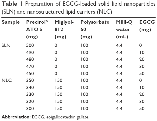 | Table 1 Preparation of EGCG-loaded solid lipid nanoparticles (SLN) and nanostructured lipid carriers (NLC) |
Characterization of the formulations
EGCG, EE, and LC
The EGCG, EE, and LC of the lipid NPs were evaluated by measuring absorption at the wavelength of 273 nm using a UV/Vis spectrophotometry (Jasco, Easton, MD, USA). The lipid NPs with EGCG were diluted in double-deionized water (1:40) and centrifuged. To separate the lipid NPs from their aqueous medium, the diluted samples were centrifuged in Ultrafree® Centrifugal Amicon® Ultra-4 Centrifugal Filter Devices, with nominal molecular weight cutoff 50,000 kDa molecular weight cut-off (MWCO) (Millipore, Billerica, MA, USA) at 25°C for 10 minutes or until complete separation. The supernatant with the dissolved EGCG was collected and measured. The EE of EGCG was defined by the ratio of the amount of EGCG measured (in mg) in the supernatant to the initial amount of EGCG added (in mg) to the formulation, while the LC of EGCG was determined by calculating the ratio between the amount of EGCG measured in the NPs and the total amount of EGCG and lipids present in the formulations.11
|

|

Particle size measurement
Particle size was measured by dynamic light scattering (DLS) using a BI-MAS DLS instrument (Brookhaven Instruments, Holtsville, NY, USA), operating at a scattering angle of 90°. Prior to the measurements, samples were diluted (1:100) in Milli-Q water and filtered with a syringe filter (800 nm). DLS data were analyzed at a temperature of 37°C with a dust cutoff set to 30. The mean hydrodynamic diameter (Z-average) and the PDI were determined as measures of the width of the particle size distributions. At each measurement, six runs of 2 minutes each were performed. The measurements were performed in triplicate.
ζ potential measurement
The ζ-potential of the particles was determined by measuring the electrophoretic mobility using a BI-MAS DLS instrument (Brookhaven Instruments). Prior to the measurements, samples were diluted (1:100) in Milli-Q water and filtered with a syringe filter (800 nm). For each measurement, ten runs (each one with ten cycles) were performed at a temperature of 37°C. The measurements were performed in triplicate.
Morphology determination
To characterize the morphology of the SLN and NLC, the NPs were visualized by scanning electron microscopy (Cryo-SEM) using a JEOL JSM-6301F instrument (Tokyo, Japan) with an Oxford Instruments INCA Energy 350 energy source (Abingdon, UK) and a Gatan Alto 2500 cryo transfer system (Pleasanton, CA, USA). The NPs dispersions were dropped on a grid, rapidly cooled in a liquid nitrogen slush (−210°C), and transferred under vacuum to the cold stage of the preparation chamber. The samples were fractured, sublimated (4 minutes, −90°C) to reveal greater detail, and coated with a gold–palladium alloy. Finally, the specimens were moved under vacuum into the SEM chamber where they were observed at −150°C.
In vitro drug release study
The in vitro drug release study was performed using a cellulose dialysis bag diffusion technique (Cellu.Sep® T1 with a nominal molecular weight cutoff of 3,500 [Frilabo, Milheiros, Maia, Portugal]) filled with 2 mL of the samples: EGCG-loaded SLN (EGCG-SLN) and EGCG-loaded NLC (EGCG-NLC).
An in vitro drug release study was performed using a direct dispersion method with appropriate buffers at the pH 1.2 and 6.5 in order to simulate the release of the drug following the gastrointestinal tract. To this end, the samples were incubated for 4 hours in simulated gastric fluid (pH=1.2 with SIF® powder) before placing them in simulated intestinal fluid (pH=6.5 with FaSSIF: buffer solution containing potassium dihydrogen phosphate) for up to 24 hours, at the simulated body temperature 37°C, while being stirred at 100 rpm. At regular intervals, 200 μL of the solution was collected to quantify the EGCG release, and the same amount was replaced with the same volume of buffer to maintain sink conditions. The EGCG release was quantified using a plate reader at 273 nm. The cumulative percentage of the release compound was determined using the average of the triplicates.
Caco-2 cell culture
Caco-2 cell line is a human epithelial colorectal adenocarcinoma cell line widely used as an in vitro model of the human small intestinal barrier to predict the absorption of drugs because it forms confluent and differentiated monolayers with microvilli, tight junctions, and transport systems.12–15 Caco-2 cells (passages 23–32) from the American Type Culture Collection (ATCC® HTB-37™, Rockville, MD, USA) were cultured in Dulbecco’s Modified Eagle’s Medium supplemented with 10% fetal bovine serum, 1% Fungizone, and 1% Pen–Strep at 37°C and 5% CO2. Cells were supplied with fresh medium every 2 days and subcultured by treatment with 0.25% trypsin–EDTA when 80%–90% confluency was reached, followed by counting in a Neubauer chamber with trypan blue solution (0.4%, w/v). Cells were seeded in a 96-well plate (104 cells per well) and grown at 37°C in an atmosphere of 5% CO2 in supplemented EBM-2 medium.
Effect of NPs on cell viability and cytotoxicity
Cells were incubated with different concentrations of the formulations SLN, NLC, and free EGCG (corresponding to 5, 10, 50, 100, and 500 μM of EGCG) for 4 hours. The medium of each well was separated from the cells and stored for the LDH assay, and cells were treated with 0.5 mg/mL of MTT for 3 hours at 37°C, 5% CO2. Finally, dimethyl sulfoxide was added to dissolve MTT formazan and incubated for 15 minutes at 37°C, followed by measurement of absorbance at 550 and 690 nm. The medium resulting from the incubation of SLN and NLC with cells was centrifuged (250× g, 10 minutes, at room temperature). The LDH released into culture supernatants was detected with the LDH cytotoxicity detection kit according to the manufacturer’s instructions (Takara Bio, Inc., Otsu, Japan).
Statistical analysis
Statistical analysis was performed using IBM® SPSS® Statistics version 21.0 (IBM Corporation, Armonk, NY, USA). The results are reported as mean ± standard deviation for a minimum of three independent experiments. Two-tailed Student’s t-test and one-way analysis of variance (ANOVA) were performed to compare two and multiple independent groups, respectively. When the group was significantly different (P<0.05), differences between groups were compared with the Tukey post hoc test. Paired samples were analyzed with the paired-samples two-tailed Student’s t-test. Differences were considered to be statistically significant at P<0.05.
Results and discussion
Particle-size measurement
The mean hydrodynamic particle sizes for SLN and NLC (both unloaded and EGCG-loaded NPs) are presented in Table 2. The mean particle size of SLN- and NLC-unloaded NPs was found to be 206±3 and 216±4 nm, respectively (Table 2). The entrapment of EGCG showed size distributions with mean diameters of 364±11 and 300±7 nm, respectively, for SLN and NLC. Thus, statistically significant differences were observed (P<0.05), suggesting that the incorporation of EGCG strongly influenced the size of NPs. The incorporation of EGCG in the NPs changes the arrangement of the lipid matrix, leading to a much higher diameter. In addition, PDI was found to be <0.2 for all formulations (Table 2), which confirmed that the NPs were in a state of acceptable monodispersity distribution, with low variability and no aggregation.10
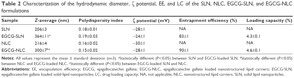 | Table 2 Characterization of the hydrodynamic diameter, ζ potential, EE, and LC of the SLN, NLC, EGCG-SLN, and EGCG-NLC formulations |
ζ potential measurement
As shown in Table 2, all the formulations showed a pronounced negative average ζ potential of approximately −30 mV. The incorporation of EGCG did not significantly change (P>0.05) the surface charge of NLC and SLN. Moreover, SLN and NLC formulations are considered physically stable because of the electrostatic repulsions between particles as both possess a high absolute ζ potential of approximately |30| mV.16
EE and LC
Table 2 summarizes the EE and LC obtained for EGCG in both SLN and NLC formulations. The EE of EGCG in SLN and NLC was found to be 83% and 90%, respectively. The LC of EGCG obtained for SLN and NLC was 4.3% and 4.6%, respectively. The high percentage of entrapment obtained confirms that both lipid NPs constitute suitable nanocarriers for EGCG entrapment despite its hydrophilic character. These high encapsulation rates can be explained by the capacity of EGCG to complex with lipids, as previously described.17 In addition, no statistically significant variations (P>0.05) were observed in the EE and LC between SLN and NLC; thus, both lipid nanocarriers are appropriate systems for EGCG incorporation.
Morphology evaluation
Cryo-SEM imaging revealed spherical particles, uniform in shape with smooth surfaces for all the formulations (Figure 1). The mean diameter was in the range of 100–300 nm, and there was no visible aggregation of the particles. These observations validate the aforementioned results obtained with DLS. As we can see in Figure 1, both unloaded (Figure 1A and C) and loaded (Figure 1B and D) NPs exhibit similar morphologies, indicating that the incorporation of EGCG does not have an effect on the particles’ morphology.
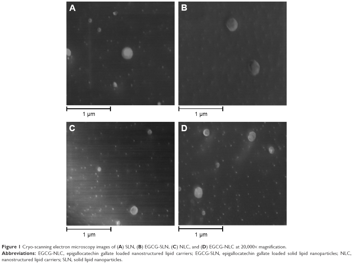 | Figure 1 Cryo-scanning electron microscopy images of (A) SLN, (B) EGCG-SLN, (C) NLC, and (D) EGCG-NLC at 20,000× magnification. |
In vitro EGCG release kinetics
The in vitro release of EGCG from SLN and NLC was measured in simulated physiological dissolution mediums to simulate the passage of the NPs through the gastrointestinal tract at body temperature (37°C) to predict the in vivo kinetics. A slow EGCG release profile was observed for SLN and NLC in the simulated gastric medium (pH=1.2), with only 10% of EGCG released from both NPs within the first 4 hours under gastric conditions (Figure 2). At the intestinal conditions (pH=6.5), the EGCG release follows double kinetics characterized by an initial fast and then a slower release (Figure 2). Thus, a burst release can be observed during the initial 4 hours, followed by a slower and sustained release of EGCG, reaching a maximum cumulative release of approximately 40% in both NPs (Figure 2). These results show that the majority of EGCG (>60%) remained entrapped in the lipid NPs after their contact with the simulated digestive fluids. Therefore, the majority of the EGCG is available for absorption by the intestinal mucosa and distribution throughout the body, reinforcing that both nanocarriers are a suitable platform for the oral delivery of EGCG, conferring protection from the degradation, and ultimately enhancing EGCG’s bioavailability.
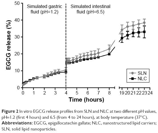 | Figure 2 In vitro EGCG release profiles from SLN and NLC at two different pH values, pH=1.2 (first 4 hours) and 6.5 (from 4 to 24 hours), at body temperature (37°C). |
Furthermore, the obtained results were analyzed using mathematical models for drug release kinetics, which are detailed in the Supplementary materials. The EGCG release from SLN and NLC occurs by diffusion controlled described by zero-order and Korsmeyer–Peppas models as the plots of the amount of EGCG released versus time were found to be linear, using these models with correlation coefficients between 0.981 and 0.996, for both pH studied and both SLN and NLC formulations, respectively (Tables S1 and S2).
Effect of SLN and NLC on cell viability and membrane integrity
The effect of the lipid NPs on intestinal cell viability and membrane integrity after 4 hours of incubation was studied in vitro on Caco-2 cells, through MTT and LDH assays, respectively. From Figure 3, it can be observed that free EGCG in the range of concentrations used (0–500 μM) did not reveal any cytotoxic effect, showing a cell viability of around 100%. SLN and NLC by themselves exhibited some level of toxicity for concentrations >50 μM. A similar effect was verified for EGCG-loaded SLN and NLC, which can be attributed to the lipid moiety of the NPs’ composition and not to the encapsulated EGCG molecules. When cells were exposed to 5 and 10 μM of all types of SLN and NLC for 4 hours, no changes were observed in MTT metabolization, indicating that both EGCG-loaded SLN and NLC did not affect the metabolic activity of cells. Only for concentrations higher than 50 μM, a significant reduction in the cell metabolic activity was observed. From Figure 4, it can be noted that both SLN and NLC interfere with the membrane integrity at all the concentrations studied, this effect being independent of the concentration. The presence of EGCG in the formulations seems to reduce the cytotoxicity in comparison with the unloaded formulations in a concentration-dependent manner. The IC50 obtained through nonlinear regression of the mean percentage cytotoxicity values versus concentration of formulation for EGCG, EGCG-SLN, and EGCG-NLC was 16, 55, and 35 μM, respectively. Therefore, the lipid NPs seem to decrease the toxicity of free EGCG in terms of membrane integrity.
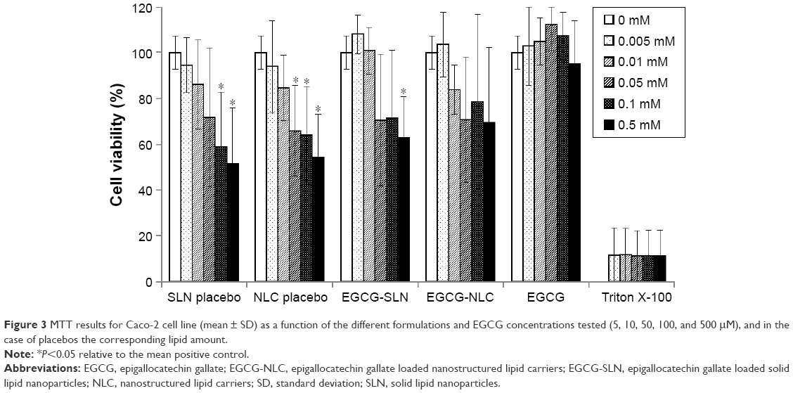 | Figure 3 MTT results for Caco-2 cell line (mean ± SD) as a function of the different formulations and EGCG concentrations tested (5, 10, 50, 100, and 500 μM), and in the case of placebos the corresponding lipid amount. |
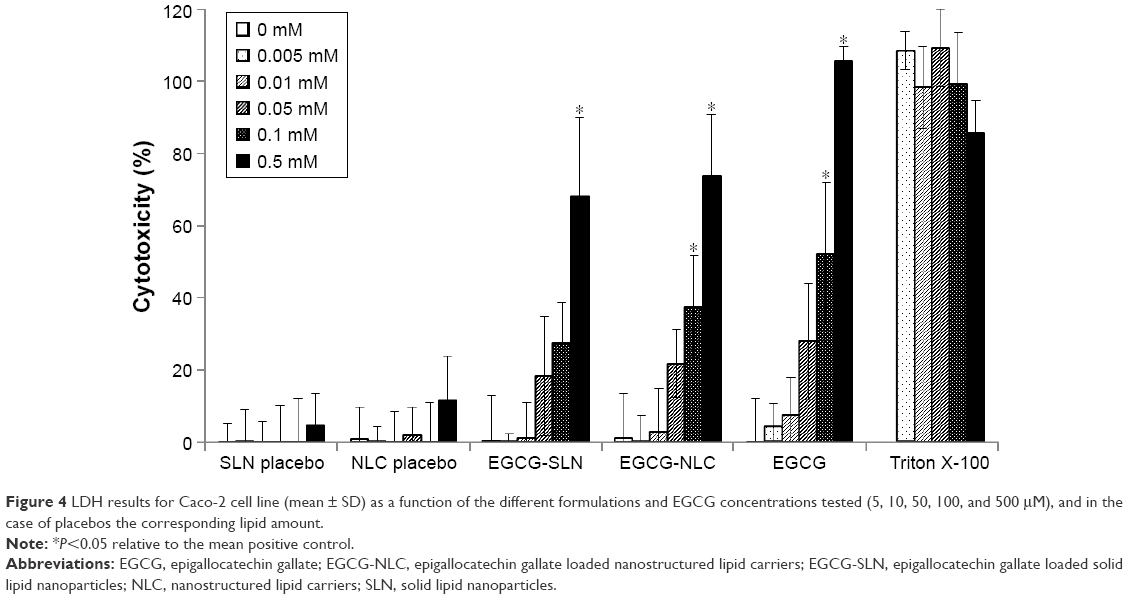 | Figure 4 LDH results for Caco-2 cell line (mean ± SD) as a function of the different formulations and EGCG concentrations tested (5, 10, 50, 100, and 500 μM), and in the case of placebos the corresponding lipid amount. |
Long-term stability of SLN and NLC
The physical stability of the lipid NPs was also verified periodically by analyzing the variation of the hydrodynamic diameter, polydispersion, ζ potential, and EE during storage conditions for 3 months at room temperature (Figure 5). From Figure 5A, it can be observed that there is a decrease in the hydrodynamic diameter of both EGCG-loaded SLN and NLC with storage. On the other hand, from Figure 5B, a slight decrease in the ζ potential for all the formulations with storage can be seen. As shown in Figure 5C, no tendency to EE variations was observed for both SLN and NLC with and without EGCG after 3 months, reinforcing the long-term stability of the developed NPs. Moreover, no statistically significant differences were observed between SLN and NLC (P>0.05), suggesting that both lipid nanosystems are suitable for EGCG incorporation and for release prevention during shelf conditions of storage at room temperature.
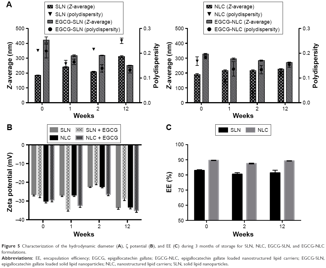 | Figure 5 Characterization of the hydrodynamic diameter (A), ζ potential (B), and EE (C) during 3 months of storage for SLN, NLC, EGCG-SLN, and EGCG-NLC formulations. |
Conclusion
This work focused on the development of lipid NPs for the incorporation of EGCG in order to enhance the oral bioavailability of this poorly absorbed hydrophilic compound,18 leading to the creation of dietary supplements, nutraceuticals, and functional food for health promotion and disease risk reduction.19 EGCG-loaded lipid NPs (SLN and NLC) were successfully produced by a modified hot homogenization technique with high encapsulating rates. SLN and NLC possess high EEs of approximately 80% and 90% and LCs of approximately 4.3% and 4.6%, respectively. Both formulations show an average hydrodynamic diameter of 300–400 nm, and both unloaded and loaded EGCG NPs showed a narrow-size distribution, suggesting that the incorporation of the catechin does not influence the size and the polydispersity of the NPs. In addition, the high negative ζ potential of around −30 mV suggests physical stability of both formulations.10 Morphological studies showed spherical and uniform NPs with a smooth surface. The stability study demonstrated physical stability during a period of at least 3 months. Moreover, the in vitro release study revealed a high stability and a slower release of EGCG in the simulated gastric environment with a small increase in the release of EGCG in the simulated intestinal medium. The cumulative release over 24 hours revealed that less than ~40% of the total catechin incorporated was released. This permits the conclusion that both lipid NPs are highly stable systems and can be considered suitable carriers for the oral administration of EGCG, avoiding the premature release of EGCG in simulated gastric and intestinal conditions and allowing a controlled release after the absorbance into the bloodstream. Finally, the in vitro viability and cytotoxicity studies revealed that the formulations were biocompatible, and the obtained IC50 is far superior to the reported therapeutic concentrations.20–22
In summary, SLN and NLC were successfully developed for EGCG protection and stabilization and can be a useful platform for the enhancement of EGCG bioavailability. Both lipid NPs are suitable for EGCG delivery after oral administration, representing a promising strategy for enhancing its in vivo efficacy.
Acknowledgments
This work received financial support from the European Union (FEDER funds) and National Funds (FCT/MEC, Fundação para a Ciência e a Tecnologia and Ministério da Educação e Ciência) under the Partnership Agreement PT2020 UID/MULTI/04378/2013–POCI/01/0145/FEDER/007728. ARN also thanks her postdoctoral grant under the project NORTE-01-0145-FEDER-000011. MP thanks FCT for the postdoctoral fellowship SFRH/BPD/99124/2013. The authors also thank Dr Daniela Silva (CEMUP, UP) for expert help with SEM experiments.
The abstract of this paper was presented at the NanoPT 2015 conference on February 11–13 in Porto as an oral presentation with interim findings. This paper includes work based on the master’s thesis submitted by Iúri Frias while at University of Porto. The authors declare that the manuscript has been submitted solely to the Drug Design, Development and Therapy journal and is not published, in press, or submitted elsewhere; and consists of an original work developed in the authors’ group that was written solely to be published in this journal.
Disclosure
The authors report no conflicts of interest in this work.
References
Zhang J, Nie S, Wang S. Nanoencapsulation enhances epigallocatechin-3-gallate stability and its antiatherogenic bioactivities in macrophages. J Agric Food Chem. 2013;61(38):9200–9209. | ||
Fangueiro JF, Andreani T, Fernandes L, et al. Physicochemical characterization of epigallocatechin gallate lipid nanoparticles (EGCG-LNs) for ocular instillation. Colloids Surf B Biointerfaces. 2014;123: 452–460. | ||
Leu JG, Chen SA, Chen HM, et al. The effects of gold nanoparticles in wound healing with antioxidant epigallocatechin gallate and α-lipoic acid. Nanomedicine. 2012;8(5):767–775. | ||
Smith A, Giunta B, Bickford PC, Fountain M, Tan J, Shytle RD. Nanolipidic particles improve the bioavailability and α-secretase inducing ability of epigallocatechin-3-gallate (EGCG) for the treatment of Alzheimer’s disease. Int J Pharm. 2010;389(1–2):207–212. | ||
Wang D, Taylor EW, Wang Y, Wan X, Zhang J. Encapsulated nanoepigallocatechin-3-gallate and elemental selenium nanoparticles as paradigms for nanochemoprevention. Int J Nanomedicine. 2012;7:1711–1721. | ||
Doktorovova S, Souto EB, Silva AM. Nanotoxicology applied to solid lipid nanoparticles and nanostructured lipid carriers – a systematic review of in vitro data. Eur J Pharm Biopharm. 2014;87(1):1–18. | ||
Muller RH, Radtke M, Wissing SA. Solid lipid nanoparticles (SLN) and nanostructured lipid carriers (NLC) in cosmetic and dermatological preparations. Adv Drug Deliv Rev. 2002;54(Suppl 1):S131–S155. | ||
Muller RH, Mader K, Gohla S. Solid lipid nanoparticles (SLN) for controlled drug delivery – a review of the state of the art. Eur J Pharm Biopharm. 2000;50(1):161–177. | ||
Muller RH, Keck CM. Challenges and solutions for the delivery of biotech drugs – a review of drug nanocrystal technology and lipid nanoparticles. J Biotechnol. 2004;113(1–3):151–170. | ||
Neves AR, Lucio M, Martins S, Lima JL, Reis S. Novel resveratrol nanodelivery systems based on lipid nanoparticles to enhance its oral bioavailability. Int J Nanomedicine. 2013;8:177–187. | ||
Danhier F, Lecouturier N, Vroman B, et al. Paclitaxel-loaded PEGylated PLGA-based nanoparticles: in vitro and in vivo evaluation. J Control Release. 2009;133(1):11–17. | ||
Artursson P, Karlsson J. Correlation between oral drug absorption in humans and apparent drug permeability coefficients in human intestinal epithelial (Caco-2) cells. Biochem Biophys Res Commun. 1991;175(3):880–885. | ||
Artursson P, Palm K, Luthman K. Caco-2 monolayers in experimental and theoretical predictions of drug transport. Adv Drug Deliv Rev. 2001;46(1–3):27–43. | ||
Shah P, Jogani V, Bagchi T, Misra A. Role of Caco-2 cell monolayers in prediction of intestinal drug absorption. Biotechnol Prog. 2006;22(1):186–198. | ||
Corti G, Maestrelli F, Cirri M, Zerrouk N, Mura P. Development and evaluation of an in vitro method for prediction of human drug absorption II. Demonstration of the method suitability. Eur J Pharm Sci. 2006;27(4):354–362. | ||
Müller H, Jacobs C, Kayser O. Nanosuspensions as particulate drug formulations in therapy. Rationale for development and what we can expect for the future. Adv Drug Deliv Rev. 2001;47(1):3–19. | ||
Sun Y, Hung WC, Chen FY, Lee CC, Huang HW. Interaction of tea catechin (-)-epigallocatechin gallate with lipid bilayers. Biophys J. 2009;96(3):1026–1035. | ||
Smith TJ. Green Tea Polyphenols in drug discovery – a success or failure? Expert Opin Drug Discov. 2011;6(6):589–595. | ||
Shahidi H. Nutraceuticals, functional foods and dietary supplements in health and disease. J Food Drug Anal. 2012;20:226–230. | ||
Mereles D, Hunstein W. Epigallocatechin-3-gallate (EGCG) for clinical trials: more pitfalls than promises? Int J Mol Sci. 2011;12(9):5592–5603. | ||
Yang CS, Wang H. Cancer therapy combination: green tea and a phosphodiesterase 5 inhibitor? J Clin Invest. 2013;123(2):556–558. | ||
Feng W, Hwang HS, Kryshtal DO, et al. Coordinated regulation of murine cardiomyocyte contractility by nanomolar (-)-epigallocatechin-3-gallate, the major green tea catechin. Mol Pharmacol. 2012;82(5):993–1000. |
Supplementary materials
 | Table S1 Value of R2 obtained from the release of EGCG from SLN for different models of mechanism of drug release |
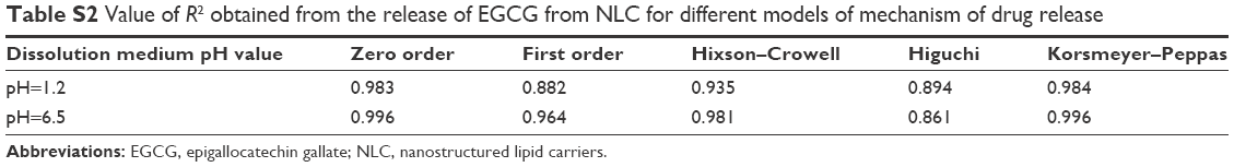 | Table S2 Value of R2 obtained from the release of EGCG from NLC for different models of mechanism of drug release |
 © 2016 The Author(s). This work is published and licensed by Dove Medical Press Limited. The
full terms of this license are available at https://www.dovepress.com/terms
and incorporate the Creative Commons Attribution
- Non Commercial (unported, 3.0) License.
By accessing the work you hereby accept the Terms. Non-commercial uses of the work are permitted
without any further permission from Dove Medical Press Limited, provided the work is properly
attributed. For permission for commercial use of this work, please see paragraphs 4.2 and 5 of our Terms.
© 2016 The Author(s). This work is published and licensed by Dove Medical Press Limited. The
full terms of this license are available at https://www.dovepress.com/terms
and incorporate the Creative Commons Attribution
- Non Commercial (unported, 3.0) License.
By accessing the work you hereby accept the Terms. Non-commercial uses of the work are permitted
without any further permission from Dove Medical Press Limited, provided the work is properly
attributed. For permission for commercial use of this work, please see paragraphs 4.2 and 5 of our Terms.