Back to Journals » International Journal of Nanomedicine » Volume 18
Denture Base Resin Coated with Titanium Dioxide (TiO2): A Systematic Review
Authors Yadfout A ![]() , Asri Y, Merzouk N, Regragui A
, Asri Y, Merzouk N, Regragui A ![]()
Received 11 August 2023
Accepted for publication 23 October 2023
Published 22 November 2023 Volume 2023:18 Pages 6941—6953
DOI https://doi.org/10.2147/IJN.S425702
Checked for plagiarism Yes
Review by Single anonymous peer review
Peer reviewer comments 2
Editor who approved publication: Prof. Dr. RDK Misra
Asmae Yadfout, Yousra Asri, Nadia Merzouk, Anissa Regragui
Department of Removable Prosthodontics, Faculty of Dentistry, Mohammed V University, Rabat, Morocco
Correspondence: Asmae Yadfout, Email [email protected]
Background: The main objective of this systematic review was to evaluate the effect of coating with titanium dioxide nanoparticles (TiO2 nanoparticle) on the surface condition of removable acrylic resin prosthetic base materials.
Methods: Our review is registered in the PROSPERO database under the identification code CRD42023397170. Electronic database searches of PubMed, Scopus and Science Direct including studies from January 2009 to January 2023 were conducted and supplemented with manual searches. Research questions were generated in accordance with the PICO strategy. The modified Consolidated Standards of Reporting Trials (CONSORT) checklist was used to evaluate the quality of the selected studies.
Results: Since the included studies were variable in design, a meta-analysis was not performed. The electronic searches retrieved 29 references that met the eligibility criteria, among which 5 studies matched the inclusion criteria for this review. Significant differences were detected between the TiO2 NP-coated and uncoated groups. The available data indicate that TiO2 NP coating elicits antimicrobial activity and improves the wear resistance of polymethylmethacrylate (PMMA) surfaces. Moreover, the nanoparticles provide high levels of glossiness and decelerate the process of color change of heat-cured acrylic resin, thus increasing the lifespan of dentures.
Conclusion: The collective results clearly indicate that TiO2 nanoparticle coating induces alterations in the surface properties of pure PMMA, enhancing the mechanical, physical and biological characteristics of the denture base material. Further studies are essential to identify the optimal thickness of coating and concentrations of nanoparticles for clinical applications.
Keywords: resin, denture base, polymethylmethacrylate, titanium dioxide, coating
Introduction
Dental prosthesis materials need to possess favorable mechanical and biological properties to ensure their safe and optimal use. However, due to the nature of these materials and their prolonged exposure to the moist oral cavity environment, various problems remain to be resolved.1
Polymethylmethacrylate (PMMA) resins, classified as non-resorbable polymers, are extensively used for denture base fabrication owing to their simplicity of use and ease of processing and repair, biocompatibility, satisfactory mechanical and biological properties, low cost and good aesthetic results. However, following long periods of intraoral use, PMMAs are susceptible to discoloration, deterioration, fatigue, surface roughness (porosity), loss of elasticity and flexural strength, lack of radio-opacity, abrasion, fracture, and microbial colonization from exposure to the contaminated oral environment.2–8
To enhance the properties of denture base resins, various reinforcing agents, such as rubbers, macrofibers, and fillers, have been explored.9 With advances in nanodentistry, fillers in the form of nanoparticles, nanofibers or nanotubes have been increasingly utilized to reinforce dental biomaterials, in particular, dental resins.10 Nanoparticle fillers, such as alumina (Al203), zirconia (ZrO2), titania (TiO2), silver (Ag), gold (Au), platinum (Pt), hyaluronic acid (HA), silicon dioxide (SiO2), amorphous nano-hydroxyapatite, and lanthanum and cerium-doped hydroxyapatite, have been developed over recent years with the aim of improving the mechanical properties of denture base acrylics.9,11–18
TiO2 nanoparticle remains one of the preferred alternatives due to their ease of availability, biocompatibility, chemical stability, physical properties, low weight, antimicrobial activity and cost-effectiveness.9,19,20 Among the three naturally occuring phases of nanocrystalline TiO2 (anatase, brookite and rutile), anatase is the most comprehensively studied form due to its superior photocatalytic performance.21
Two methods have been proposed for incorporation of TiO2 nanoparticle onto denture base resin. TiO2 nanoparticle can either be mixed with the resin or coated using brushing, dipping or spraying techniques. In the process of reinforcing heat-cured denture base resins with TiO2 nanoparticle, a range of concentrations (1wt% to 5wt%) of TiO2 nanoparticle is required to obtain an antimicrobial effect. However, according to earlier studies, these concentrations could negatively impact the mechanical strength of dentures owing to internal decomposition induced by the photocatalytic effect of TiO2 nanoparticles, in addition to causing considerable whitening of PMMA resin.2,22 As shown by Abdelraouf et al, these negative effects on denture material properties were minimized for self/chemical-cured resins.22,23
The TiO2 nanoparticle coating method appears to have minimal influence on the mechanical strength of dentures and allows the denture base to maintain a considerable thickness (>1 mm), since the coat only involves the 2 µm external surface. Thus, mechanical strength is not compromised.24,25
Numerous studies to date have investigated the effects of TiO2 nanoparticle on the mechanical, biological, and physical properties of PMMA. The impact of added nanoparticles appears to be dependent on the concentrations used. To our knowledge, no systematic reviews have been conducted to establish the effects of TiO2 nanoparticle coating on the surface condition of removable acrylic resin prosthetic bases. In the present review, we collated studies in the literature investigating the impacts of TiO2 nanoparticle coating on denture base acrylic resin and summarized the findings in the context of potential clinical applications.
Materials and Methods
Focused Question
This systematic review adhered to PRISMA guidelines and was recorded in the PROSPERO database under the identification code CRD42023397170.
The review was designed according to the PICO format as follows:
Population: PMMA denture resin specimens
Intervention: Coating with TiO2 nanoparticle
Comparison: PMMA denture resin specimens without TiO2 nanoparticle coating
Outcome: The effect of TiO2 nanoparticle coating on the surface condition of removable acrylic resin prosthetic bases
The following question was posed: “Does TiO2 nanoparticle improve the surface properties of acrylic resin removable denture bases”.
Search Strategy
An electronic search was conducted using the PubMed, Science Direct, and Scopus databases and supplemented with a manual search of different combinations of Medical Subject Headings (MeSH) terms as follows: resin, denture base, polymethylmethacrylate, titanium dioxide and coating. Using this strategy, all relevant articles published between January 2009 and January 2023 were located. The articles were imported into the Zotero desktop reference management software (version 5.0.96.3) for removal of duplicates and evaluation of titles and abstracts.
The studies were independently analyzed by two investigators (YA and AR), who excluded reports that did not meet the inclusion criteria. After screening, the selected articles and those that did not clearly describe the characteristics of the study in the title and abstract were comprehensively read. Any discrepancies during the screening and selection process were resolved by discussion, and a third reviewer (AY) was consulted to reach a consensus.
Eligibility Criteria
The inclusion criteria were as follows: articles published between 2009 and 2023, English as the language of publication, studies concerning heat-curing acrylic denture base resin, studies assessing denture base resins coated with titanium dioxide, and in vitro studies.
The exclusion criteria were as follows: narrative reviews, opinion papers, case reports, in vivo studies, duplicate articles, studies on incorporation of TiO2 nanoparticle into resin acrylic, reports on TiO2 nanoparticle mixed with other types of particles, and articles focusing on the biocompatibility of TiO2 nanoparticle coats.
Data Extraction
Data were independently extracted from the full text of included studies by two investigators (YA and AR) using a standard form. In case of any doubt, a third evaluator (AY) was consulted. The following information was collected: authors, year of publication, study design, sample characteristics (number of specimens, particle size and percentage of TiO2 nanoparticle, type of denture base resin), objective of the study, and outcome.
Quality Assessment of Included Studies
The modified CONSORT checklist26 including 14 items was used for reporting in vitro studies on dental materials and adapted to assess the quality of the included articles. If parameters related to each item were reported, the article was marked “Y” (yes), whereas if the information could not be located, the article was marked “N” (no).
The Cochrane risk of bias tool was adapted. Risk of bias was evaluated after verifying the following criteria: explanation of sample size calculation, sample allocation and concealment, blinding of the operator, and whether the preparation of materials was conducted according to the manufacturer’s instructions and selective outcomes were reported. If the criterion was clearly described, the article was assigned a score of 0. In cases where the approach used was considered inadequate, the score was 1, and where a specific setting was not divulged, the score was 2. Articles assigned a total score of 0 to 3 were graded as low risk, 4 to 7 as moderate risk, and 8 to 10 as high risk of bias.19 The assessment was performed individually by two authors (AY, AR), while the third author (YA) resolved any disagreements after a reasonable debate.
Results
Search Results
A total of 241 articles were initially identified. After elimination of duplicate articles, the number of articles that remained was 232. Following a comprehensive review of the titles and abstracts, 29 articles were retained for examination of the full text. Among these reports, 5 met the inclusion criteria and were subsequently included for qualitative synthesis and data extraction24,27–30 (Figure 1).
 |
Figure 1 Prisma Flow Chart for the Study. Notes: PRISMA figure adapted from Page MJ, McKenzie JE, Bossuyt PM, Boutron I, Hoffmann TC, Mulrow CD, et al. The PRISMA 2020 statement: an updated guideline for reporting systematic reviews. BMJ. 2021;372:n71. Creative Commons. |
General Characteristics of Included Studies
The characteristics of the included ayrticles and data extraction are listed in (Table 1).
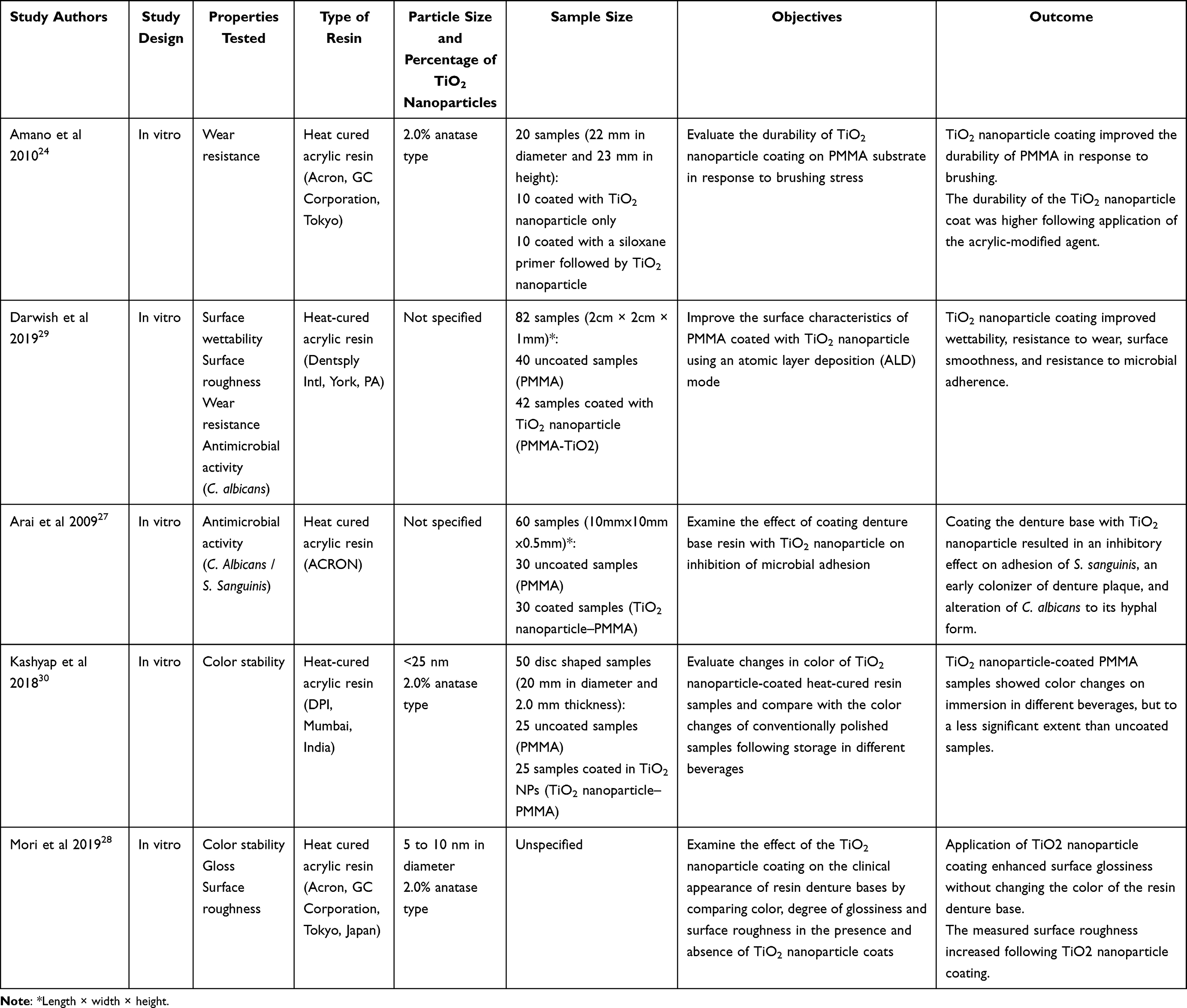 |
Table 1 Characteristics of the Included Studies |
General Outcomes of Included Studies
Wear Resistance
Two of the included studies evaluated the durability of TiO2 nanoparticle coating on PMMA substrate in response to brushing. The group of Amano24 demonstrated that the TiO2 nanoparticle coating on PMMA substrate remained in place, with loss of the nanoparticles only after exposure to mechanical stress comprising 1 × 10⁵ brushing cycles and after 2 × 10⁵ brushing cycles in cases of prior treatment with an acryloxypropyl trimethoxysilane-based agent preceding the coating step. In this study, the coating was sprayed on to the substrate. However, information regarding the thickness of the coating layer was not provided.24
The surface roughness changes of PMMA and TiO2 nanoparticle–PMMA were measured by the group of Darwish29 after exposure to 6000 linear brushing cycles. Notably, the mean surface roughness (µm) of PMMA samples increased significantly (*p < 0.05) from 0.12 ± 0.02 to 3.23 ± 0.95 after 6000 brushing strokes. In contrast, the mean surface roughness (µm) of TiO2 nanoparticle–PMMA specimens was 0.08 ± 0.03 before and 0.17 ± 0.03 after brushing, indicating no significant changes in the coated material. In this study, the atomic layer deposition (ADL) technique was used, and the thickness of TiO2 nanoparticle film on PMMA specimens was ~30 nm.29
Antimicrobial Activity
Darwish et al29 reported a statistically significant difference in the mean number of viable attached cells of C. albicans between TiO2 nanoparticle–PMMA and PMMA (control) groups (p = 0.0001). Higher antimicrobial activity was observed with TiO2 nanoparticle–PMMA compared with PMMA. In the same study, the mean water contact angle of uncoated PMMA specimens (67°) was shown to be reduced to less than 5° after coating, leading to a super-hydrophilic surface.29
The group of Arai explored the effect of TiO2 nanoparticle coating on microbial adhesion, with particular focus on S. sanguinis, a known early colonizer of denture plaque, and C. albicans, a pathogen causing oral mycosis that strongly adheres to denture base resin.27 The investigators used the spray-on method for coating the denture base acrylic resin and adenosine triphosphate (ATP) analysis to measure active microorganisms. However, no detailed information regarding the thickness of the coating layer was provided. The mean ATP contents for S. sanguinis on TiO2 NP-coated and uncoated resin plate surfaces were 4.67 and 7.24 ± 1.44 log RLU, respectively. The mean ATP contents for C. albicans on TiO2 nanoparticle-coated and uncoated resin plate surfaces were 5.33 ± 0.05 and 7.03 ±0.13 log RLU, respectively. Significant differences were observed between the TiO2 nanoparticle-coated and uncoated groups for both species (p = 0.000). Although C. albicans was altered to the hyphal form in plates of the TiO2 nanoparticle-coated group, biofilm formation was inhibited on plate surfaces.
Surface Appearance27
To explore the changes in color of TiO2 nanoparticle–PMMA-coated resin samples relative to color modifications of non-coated samples after storage in different beverages, including artificial saliva (control), coffee, cola, alcohol, and turmeric solution, Kashyap et al30 obtained light-specific L*a*b* values for all samples using a Datacolor SPECTRUM 650 spectrophotometer. The light was blocked by a black background, and all measurements were performed in triplicate. Calculation of mean values of L*a*and b* and color value differences (ΔE) showed higher color change levels for non-coated than coated samples, with significant differences between the two groups (p < 0.05) following storage in coffee, cola, alcohol, and turmeric solution. In contrast, no significant differences between the coated and non-coated samples were observed in artificial saliva (p = 0.1137). The solution with the maximum staining potential was turmeric, followed by alcohol, cola, and coffee, with clinically non-significant changes (ΔE > 6).30
Mori et al28 investigated the effects of TiO2 nanoparticle coating on the appearance of denture base resin by comparing color and degree of glossiness in the presence and absence of TiO2 nanoparticle. To this end, L*a*b* values were obtained using a reflective colorimeter. All measurements were performed in triplicate with blocking of light on a black background and mean values calculated. The glossiness of each specimen was measured with a glossmeter at an angle of 60° from the normal surface with blocking of light on a black background, from which mean values were calculated. The surface roughness of each specimen was evaluated with a profilometer. Surface morphology was assessed using field-emission scanning electron microscopy (FE-SEM).28 The results revealed no significant color variations between the coated and uncoated samples and a lower ΔE value (1.2) that was not easily perceptible by the human eye.28 Significant differences between PMMA and TiO2 nanoparticle-coated specimens were detected in terms of glossiness and surfaces roughness (p < 0.05). The glossiness levels of pure PMMA and TiO2 nanoparticle-coated specimens were determined as 3.06 ± 0.89 and 12.8 ±0.73 and surface roughness as 0.30 ±0.02, and 0.35± 0.01 mm, respectively.28
Results of Quality Assessment
Methodological Quality
The studies on methodological quality included for review are summarized in (Table 2).
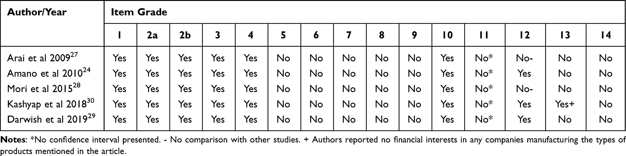 |
Table 2 Results of the Assessment from Selected in vitro Studies Using the Modified CONSORT Checklist |
Risk of Bias
(Table 3) presents risk of bias for all the studies included in this systematic review. The articles included showed moderate risk of bias. The risk of bias values generated were principally attributed to sample size estimation, allocation concealment of specimens, and blinding of the machinist. None of the authors reported blinding in the included studies. The concealment and allocation methods employed to divide samples among diverse groups were not clearly described in all the studies.
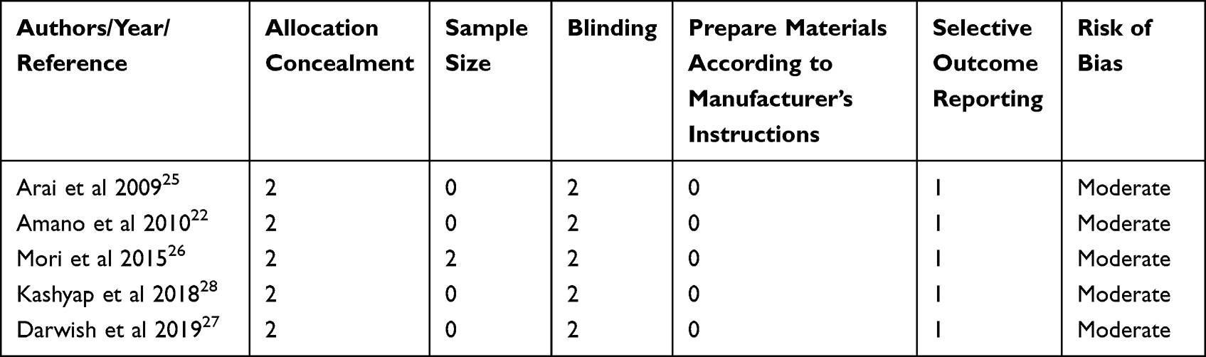 |
Table 3 Risk of Bias Tool (Adapted and Modified from Cochrane Risk of Bias Tool) |
Discussion
This systematic review was performed to establish the effects of TiO2 nanoparticle coating on denture acrylic resin. Based on analysis of the outcomes from the included studies, we conclude that the mechanical, physical and biological properties of denture PMMA undergo changes following nanoparticle coating.
Several studies in the literature have assessed the flexural strength variations of heat-cured PMMA denture bases modified with TiO2 nanoparticle. A concentration of 1wt% of TiO2 nanoparticle was determined as the most effective for increasing the flexural strength of resin in studies by Tandra et al and Karci et al. The lowest flexural strength value was observed with concentrations >5wt%.16,31–33 For self-cured acrylic resin, Abdelraouf et al23 demonstrated that the addition of 5wt% TiO2 nanoparticle improved flexural strength.
One theory to explain the reduction in heat-cured denture base resin is that increased concentrations of TiO2 nanoparticle (>5wt%) act as an impurity, which disrupts the polymerization process. High concentrations of fillers additionally reduce the mobility of polymer chains, leading to brittle behavior and early failure. By acting as a plasticizer at high concentrations, TiO2 nanoparticle increases the levels of residual monomers, resulting in decreased flexural strength of resin. The inhomogeneous distribution of particles causes agglomeration, which may also account for this reduction. Specifically, agglomerates act as weak sites within the matrix that negatively affect the mechanical properties of the polymerized material. Due to their large surface area-to-volume ratio, nanoparticles are only required in a small quantity for modification of polymer properties. Upon penetration of the matrix, TiO2 nanoparticles are distributed uniformly without agglomeration. In addition, the establishment of strong interactions between filler and PMMA matrix reduces the mobility of the polymer chain, thus enhancing the strength of the resin.19,32–34
Since TiO2 nanoparticle coating affects only 2 µm of the upper surface, the thickness of the denture base remains more than 1 mm. Therefore, the coating method does not appear to compromise the flexural strength of denture resin. Data from studies within the current review confirmed that differences in flexural strength between unmodified PMMA and TiO2 nanoparticle-coated PMMA groups were not statistically significant (p = 0.0995). The process used for coating was ALD, which provides a thin TiO2 nanoparticle film, as shown by Darwish et al.29 These findings were similar to those reported by Amirabad et al, who worked on generating a thin film of TiO2 nanoparticle. According to their report, a thick layer of TiO2 nanoparticle is composed of particles that do not strongly interact with each other. Furthermore, certain parts of TiO2 particles penetrate the PMMA, leading to removal of PMMA in some areas and a consequent decrease in flexural strength of resin.35
The group of Amano24 showed that the TiO2 nanoparticle coat has sufficient durability against friction caused by brushing. Application of an acrylate-modified agent further improved the longevity of the TiO2 nanoparticle coating. Mechanistically, to ensure high resistance to friction, the TiO2 nanoparticle layer needs to be strongly bonded to its substrate. The authors suggested that acrylate-modified siloxane agent could elevate the binding of TiO2 nanoparticle to PMMA substrate via chemical bonding. The flexural bond strength of TiO2 nanoparticle coating may therefore rely on the strength of the layer formed by siloxane primer (SP) treatment.24,29
According to the groups of Azmy and Zhong, increase of the wear resistance of PMMA heat-cured acrylic resin could be attributed to the strong bond between nanoparticles and resin matrix (chemical interactions), which is regarded as the primary reason of wear resistance in reinforced groups. Moreover, the strong bond decreases the incidence of nanoparticle exfoliation during abrasion.4,33 Further studies are warranted to establish the relationships among thickness of coating, flexural strength of the substrate and bond strength to the substrate.
Restorative materials with high surface roughness serve as a favorable substrate for attachment of microorganisms. A surface roughness value of 0.2 µm is considered the threshold for bacterial adhesion above which the aggregation of microorganisms increases significantly, leading to stomatitis, peri-implantitis and other systemic fatal infections, such as aspiration pneumonia and systemic candidiasis.2,36–41 According to several earlier studies, laboratory and chairside polishing techniques could achieve a surface roughness below the 0.2 µm threshold for surfaces of removable dentures composed of PMMA. However, this is not the case for the fitting surfaces of denture base materials, which still show Ra measurements between 3.4 and 7.6 µm, even upon processing under ideal laboratory conditions, and therefore remain vulnerable to food adhesion and microbial colonization.38 Conventional denture cleaning methods, such as mechanical and chemical cleansing, are ineffective in completely removing denture plaque. Furthermore, these methods are often complicated, particularly for the elderly and individuals requiring nursing care or with a low ability to carry out normal daily life activities.2,35
TiO2 nanoparticle displays a large spectrum of activity against microorganisms, including bacteria and fungi, in particular, Candida species. This antimicrobial effect of TiO2 nanoparticle is mainly attributed to their antiadhesion activity in the mouth that impedes the adhesion of foods, microbes and plaques as well as their photocatalytic activity and superhydrophilic properties.35,39 In the presence of water and under UV irradiation of acrylic resin containing TiO2 nanoparticle, reactive oxygen species (ROS), including hydroxyl (OH−), superoxide free radicals (O2−), and hydrogen peroxide (H2O2), are generated by photocatalysis of the nanoparticles.10,22,42–44 These aggressive oxidative species damage the cell surface and destroy the organic materials within cells, leading to inactivation of microorganisms.40,41,45
The antimicrobial effect of TiO2 nanoparticle coating may further be attributed to superior wear resistance.46,47 Moreover, even after the appearance of surface defects following exposure to a particular number of brushing cycles, antifouling and antimicrobial properties could be maintained as long as the defect remained surrounded by a considerable coating of TiO2 nanoparticle.20,24,28
In reports by Arai et al and Darwish et al, coating a denture base acrylic resin with TiO2 nanoparticle resulted in an inhibitory effect on adhesion of S. sanguinis and alteration of C. albicans to its hyphal form.27,29 The study of Darwish29 further showed that the mean water contact angle of uncoated PMMA specimens (67°) was decreased to <5° after TiO2 nanoparticle coating, resulting in a super-hydrophilic surface. Identical results were obtained by the groups of Amirabad and Mutter.35,48 Enhancement of hydrophilicity of the denture surface could facilitate removal of contaminants with water, prevent hydrophobic adhesion of bacterial species such as S sanguinis, an early colonizer in denture plaque, and suppress subsequent adhesion of other microbes.49
According to reports by Sawada et al (2010, 2014), fluorapatite-titania (FApTiO2) exerts potent antifungal effects due to its ability to produce large amounts of hydroxyl radicals throughout photocatalysis and provide higher hydrophilicity to resin than that offered by TiO2 nanoparticle,46,50 supporting its potential utility as a good alternative functional coating material.
Many polymers possess functional groups in their molecular chains that are able to absorb ultraviolet light. The energy absorbed from UV light leads to increased instability of the polymer. To generate a more stable structure, the excited molecules tend to disperse excess energy. This dispersion event causes rupture and photochemical degradation of the molecule, which contributes to material color or brightness changes, loss of opacity, cracks, and increased stiffness. These intrinsic factors are not in the control of the dentist and cannot be easily prevented.51,52 However, changes due to extrinsic factors (such as thermal alterations, exposure to cleansers, prolonged water exposure, artificial dyes and cleaning procedures) can be avoided using an impervious self-cleaning and resistant surface coating such as TiO2.49,51,53
Acrylic resin can absorb liquids owing to the polarity of the PMMA molecules.13 After absorption, the solution disperses into the polymer network, thereby causing hydrolysis and development of aberrant optical properties in acrylic, which may result in color changes.54
The group of Kashiyap30 attempted to place prosthesis in the conditions of daily use. In their research, TiO2 nanoparticle coating slowed down the process of color change of heat-cured acrylic resin caused by extrinsic factors. Based on the data, the authors suggested that TiO2 nanoparticle could prevent color changes to some extent and increase the lifespan of the prosthesis. The spray method was used with a solution containing 2.0% anatase TiO2, but the thickness of the coating was not specified.30 Their findings conform with the results of other studies on the color stability of acrylic resin following the addition of TiO2 nanoparticle.52,54
Application of coating may create another problem related to the possibility of discoloration of dentures due to the white color of TiO2 nanoparticle materials. This risk was analyzed by Mori et al, who showed no significant differences in a* and b* values between coated and uncoated samples. The ΔE values obtained between PMMA and TiO2 nanoparticle-coated specimens were <3, which is considered below the level of macroscopic discrimination and clinically acceptable according to the National Bureau of Standards.52 Consequently, color changes were regarded as absent.28 This finding may be attributable to the thickness of the coating and the small quantity of TiO2 nanoparticle employed.
Other factors that potentially influence the appearance of dentures include surface roughness, glossiness, and accumulation of stains.51
The group of Mori additionally reported significant differences in surface roughness between PMMA and TiO2 nanoparticle-coated specimens (p < 0.05). TiO2 nanoparticle coating enhanced the surface roughness of the substrate by about 0.05 µm, in keeping with data obtained by Amirabad et al showing an increase in roughness of PMMA after coating with TiO2 nanoparticle.28,35 This could be explained by the lack of polish recommended to increase the surface area of coating, and, as a result, we assume that the esthetic features of the denture base are decreased.
The surface roughness of PMMA reinforced by the addition of TiO2 nanoparticle was evaluated in a recent study by Zore et al. Their data suggest that the surface roughness of PMMA coated with TiO2 nanoparticle depends on the concentration of added nanoparticles.34 A series of experiments further demonstrated that surface roughness decreased with increasing TiO2 nanoparticle concentrations. With the addition of 1% TiO2 nanoparticle, surface roughness was higher compared to pure PMMA, which decreased with 5% TiO2 nanoparticle but remained higher than that of pure PMMA.34
Mori et al28 additionally reported that application of TiO2 NP coatings afforded high levels of glossiness to dentures. Based on these results, automatic polishing achieved by TiO2 nanoparticle coating may serve as an effective method for removable dentures, saving considerable time and effort.
The heterogeneity of published data is a major limitation of the current systematic review. Moreover, the thickness of coating was not described in all the included studies, and therefore, the influence of TiO2 nanoparticle of varying thickness on the properties tested remains unknown. Moreover, the confidence interval was not specified in all studies, highlighting the need to interpret the results with caution. We considered a confidence interval of 95%, since it is the most commonly used level in research and in the included studies, p-value was always compared against the 0.05 level of significance (α). Furthermore, the specific statistical tests used were not reported in several studies. The sample size was not specified in one study, and in all studies, data were not presented in tables.
Conclusions
To our knowledge, the current study is unique in that no similar systematic reviews have been conducted in this area. The collective in vitro results obtained suggest that TiO2 nanoparticle coating of resin dentures improves the surface properties of PMMA and provides an alternative nanocomposite that avoids the disadvantages of this denture base material. However, further studies with comparable rigorous methodologies are warranted to optimize the utility of nanoparticles as dental coating materials, such as standardization of the coating method, TiO2 nanoparticle concentrations, coat thickness, and specimen measurements.
Abbreviations
ALD, Atomic layer deposition; CONSORT, Consolidated Standards of Reporting Trials; HA, Hyaluronic acid; MeSH, Medical Subject Headings; ROS, Reactive oxygen species; SP, Siloxane primer; PMMA, Polymethylmethacrylate; TiO2, Titanium dioxide.
Author Contributions
All authors made a significant contribution to the work reported, whether that is in the conception, study design, execution, acquisition of data, analysis and interpretation, or in all these areas; took part in drafting, revising or critically reviewing the article; gave final approval of the version to be published; have agreed on the journal to which the article has been submitted; and agree to be accountable for all aspects of the work.
Funding
This research received no external funding.
Disclosure
The authors report no conflicts of interest in this work.
References
1. Wang Q, Huang JY, Li HQ, et al. Recent advances on smart TiO(2) nanotube platforms for sustainable drug delivery applications. Int J Nanomedicine. 2017;12:151–165. doi:10.2147/IJN.S117498
2. Tsuji M, Ueda T, Sawaki K, Kawaguchi M, Sakurai K. Biocompatibility of a titanium dioxide-coating method for denture base acrylic resin. Gerodontology. 2016;33:539–544. doi:10.1111/ger.12204
3. Dikbas I, Gurbuz O, Unalan F, Koksal T. Impact strength of denture polymethyl methacrylate reinforced with different forms of E-glass fibers. Acta Odontol Scand. 2013;71:727–732. doi:10.3109/00016357.2012.715198
4. Zhang X, Zhang X, Zhu B, Lin K, Chang J. Mechanical and thermal properties of denture PMMA reinforced with silanized aluminum borate whiskers. Dent Mater J. 2012;31:903–908. doi:10.4012/dmj.2012-016
5. Silva Cde S, Machado AL, Chaves Cde AL, Pavarina AC, Vergani CE. Effect of thermal cycling on denture base and autopolymerizing reline resins. J Appl Oral Sci. 2013;21:219–224. doi:10.1590/1679-775720130061
6. Chandu GS, Asnani P, Gupta S, Khan FM. Comparative evaluation of effect of water absorption on the surface properties of heat cure acrylic: an in vitro study. J Int Oral Health. 2015;7:63–68.
7. Neppelenbroek KH, Pavarina AC, Vergani CE, Giampaolo ET. Hardness of heat-polymerized acrylic resins after disinfection and long-term water immersion. J Prosthet Dent. 2005;93:171–176. doi:10.1016/j.prosdent.2004.10.020
8. Chau VB, Saunders TR, Pimsler M, Elfring DR. In-depth disinfection of acrylic resins. J Prosthet Dent. 1995;74:309–313. doi:10.1016/s0022-3913(05)80140-4
9. Abdulrazzaq Naji S, Jafarzadeh Kashi TS, Behroozibakhsh M, Hajizamani H, Habibzadeh S. Recent advances and future perspectives for reinforcement of poly(methyl methacrylate) denture base materials: a literature review. J Dent Educ. 2018;5:490–502.
10. Li X, Liu W, Sun L, et al. Resin composites reinforced by nanoscaled fibers or tubes for dental regeneration. BioMed Res Int. 2014;2014:542958. doi:10.1155/2014/542958
11. Waly GH. Effect of incorporating undoped or silver-doped photocatalytic titanium dioxide on the antifungal effect and dynamic viscoelastic properties of long-term acrylic denture liners. Future Dent J. 2018;4:8–15. doi:10.1016/j.fdj.2018.03.002
12. Vuorinen AM, Dyer SR, Lassila LVJ, Vallittu PK. Effect of rigid rod polymer filler on mechanical properties of poly-methyl methacrylate denture base material. Dent Mater. 2008;24:708–713. doi:10.1016/j.dental.2007.07.003
13. Karacaer O, Polat TN, Tezvergil A, Lassila LVJ, Vallittu PK. The effect of length and concentration of glass fibers on the mechanical properties of an injection- and a compression-molded denture base polymer. J Prosthet Dent. 2003;90:385–393. doi:10.1016/S0022391303005183
14. Bourlidi S, Qureshi J, Soo S, Petridis H. Effect of different initial finishes and parylene coating thickness on the surface properties of coated PMMA. J Prosthet Dent. 2016;115:363–370. doi:10.1016/j.prosdent.2015.08.019
15. Casaletto MP, Ingo GM, Kaciulis S, Mattogno G, Pandolfi L, Scavia G. Surface studies of in vitro biocompatibility of titanium oxide coatings. Appl Surf Sci. 2001;172:167–177. doi:10.1016/S0169-4332(00)00844-8
16. Harini P, Mohamed K, Padmanabhan TV. Effect of titanium dioxide nanoparticles on the flexural strength of polymethylmethacrylate: an in vitro study. Indian J Dent Res. 2014;25:459–463. doi:10.4103/0970-9290.142531
17. Hamdy T, Mousa S, Sherief M. Effect of incorporation of lanthanum and cerium-doped hydroxyapatite on acrylic bone cement produced from phosphogypsum waste. Egypt J Chem. 2019. doi:10.21608/ejchem.2019.17446.2069
18. Hamdy T, Saniour S, Sherief M, Zaki D. Effect of incorporation of 20 wt% amorphous nano-hydroxyapatite fillers in polymethyl methacrylate composite on the compressive strength. Int J Biol Chem Sci. 2015;6:8585.
19. Bangera MK, Kotian R, Ravishankar N. Effect of titanium dioxide nanoparticle reinforcement on flexural strength of denture base resin: a systematic review and meta-analysis. Jpn Dent Sci Rev. 2020;56:68–76. doi:10.1016/j.jdsr.2020.01.001
20. Alrahlah A, Fouad H, Hashem M, Niazy AA, AlBadah A. Titanium oxide (TiO₂)/polymethylmethacrylate (PMMA) denture base nanocomposites: mechanical, viscoelastic and antibacterial behavior. Materials (Basel). 2018;11:1096. doi:10.3390/ma11071096
21. Miah AT. Chapter 11. Visible light responsive titania-based nanostructures for photocatalytic reduction of carbon dioxide. In: Nguyen Tri P, Wu H, Nguyen TA, Barnabé S, Bénard P, editors. Micro and Nano Technologies. Nanomaterials for CO2 Capture, Storage, Conversion and Utilization. Amsterdam, the Netherlands: Elsevier; 2021:239–266. doi:10.1016/B978-0-12-822894-4.00009-5
22. Cierech M, Szerszeń M, Wojnarowicz J, Łojkowski W, Kostrzewa-Janicka J, Mierzwińska-Nastalska E. Preparation and characterisation of poly(methyl Metacrylate)-titanium dioxide nanocomposites for denture bases. Polymers (Basel). 2020;12:2655. doi:10.3390/polym12112655
23. Abdelraouf RM, Bayoumi RE, Hamdy TM. Influence of incorporating 5% weight titanium oxide nanoparticles on flexural strength, micro-hardness, surface roughness and water sorption of dental self-cured acrylic resin. Polymers. 2022;14:3767. doi:10.3390/polym14183767
24. Amano D, Ueda T, Sugiyama T, Takemoto S, Oda Y, Sakurai K. Improved brushing durability of titanium dioxide coating on polymethylmethacrylate substrate by prior treatment with Acryloxypropyl trimethoxysilane-based agent for denture application. Dent Mater J. 2010;29:97–103. doi:10.4012/dmj.2009-073
25. Pulker HK, Paesold G, Ritter E. Refractive indices of TiO(2) films produced by reactive evaporation of various titanium-oxygen phases. Appl Opt. 1976;15:2986–2991. doi:10.1364/AO.15.002986
26. Faggion CM. Guidelines for reporting pre-clinical in vitro studies on dental materials. J Evid Based Dent Pract. 2012;12:182–189. doi:10.1016/j.jebdp.2012.10.001
27. Arai T, Ueda T, Sugiyama T, Sakurai K. Inhibiting microbial adhesion to denture base acrylic resin by titanium dioxide coating. J Oral Rehabil. 2009;36:902–908. doi:10.1111/j.1365-2842.2009.02012.x
28. Mori K, Tsuji M, Ueda T, Sakurai K. Color and gloss evaluation of titanium dioxide coating for acrylic resin denture base. J Prosthodont Res. 2015;59:249–253. doi:10.1016/j.jpor.2015.06.001
29. Darwish G, Huang S, Knoernschild K, et al. Improving polymethyl methacrylate resin using a novel titanium dioxide coating. J Prosthodont. 2019;28:1011–1017. doi:10.1111/jopr.13032
30. Kashyap RS, Nalinakshamma M, Shetty S, Rao S. Color stability of heat-cured polymethyl methacrylate denture base resin coated with titanium dioxide upon storage in different beverages. J Interdiscip Dent. 2018;8:87. doi:10.4103/jid.jid_85_17
31. Ahmad N, Jafri Z, Khan ZH. Evaluation of nanomaterials to prevent oral candidiasis in PMMA based denture wearing patients. A systematic analysis. J Oral Biol Craniofac Res. 2020;10:189–193. doi:10.1016/j.jobcr.2020.04.012
32. Tandra E, Wahyuningtyas E, Sugiatno E. The effect of nanoparticles TiO2 on the flexural strength of acrylic resin denture plate. Padjadjaran J Dent. 2018;30:35–40. doi:10.24198/pjd.vol30no1.16110
33. Azmy E, Al-Kholy MRZ, Al-Thobity AM, Gad MM, Helal MA. Comparative effect of incorporation of ZrO2, TiO2, and SiO2 nanoparticles on the strength and surface properties of PMMA denture base material: an in vitro study. Int J Biomater. 2022;2022:5856545. doi:10.1155/2022/5856545
34. Zore A, Abram A, Učakar A, et al. Antibacterial effect of polymethyl methacrylate resin base containing TiO2 nanoparticles. Coatings. 2022;12:1757. doi:10.3390/coatings12111757
35. Amirabad LM, Tahriri M, Zarrintaj P, Ghaffari R, Tayebi L. Preparation and characterization of TiO2-coated polymerization of methyl methacrylate (PMMA) for biomedical applications: in vitro study. Asia Pac J Chem Eng. 2022;17:e2761. doi:10.1002/apj.2761
36. Bollen CM, Lambrechts P, Quirynen M. Comparison of surface roughness of oral hard materials to the threshold surface roughness for bacterial plaque retention: a review of the literature. Dent Mater. 1997;13:258–269. doi:10.1016/s0109-5641(97)80038-3
37. Bollen CM, Papaioanno W, Van Eldere J, Schepers E, Quirynen M, van Steenberghe D. The influence of abutment surface roughness on plaque accumulation and peri-implant mucositis. Clin Oral Implants Res. 1996;7:201–211. doi:10.1034/j.1600-0501.1996.070302.x
38. Zissis AJ, Polyzois GL, Yannikakis SA, Harrison A. Roughness of denture materials: a comparative study. Int J Prosthodont. 2000;13:136–140.
39. Totu EE, Nechifor AC, Nechifor G, Aboul-Enein HY, Cristache CM. Poly(methyl methacrylate) with TiO2 nanoparticles inclusion for Stereolitographic complete denture manufacturing – the Future in dental care for elderly edentulous patients? J Dent. 2017;59:68–77. doi:10.1016/j.jdent.2017.02.012
40. Ireland JC, Klostermann P, Rice EW, Clark RM. Inactivation of Escherichia coli by titanium dioxide photocatalytic oxidation. Appl Environ Microbiol. 1993;59:1668–1670. doi:10.1128/aem.59.5.1668-1670.1993
41. Cho M, Chung H, Choi W, Yoon J. Linear correlation between inactivation of E coli and OH radical concentration in TiO2 photocatalytic disinfection. Water Res. 2004;38:1069–1077. doi:10.1016/j.watres.2003.10.029
42. Iesalnieks M, Eglītis R, Juhna T, Šmits K, Šutka A. Photocatalytic activity of TiO(2) coatings obtained at room temperature on a polymethyl methacrylate substrate. Int J Mol Sci. 2022;23. doi:10.3390/ijms232112936
43. Lee HS, Woo CS, Youn BK, et al. Bandgap modulation of TiO2 and its effect on the activity in photocatalytic oxidation of 2-Isopropyl-6-Methyl-4-Pyrimidinol. Top Catal. 2005;35:255–260. doi:10.1007/s11244-005-3832-2
44. Watté J, Van Gompel W, Lommens P, De Buysser K, Van Driessche I. Titania nanocrystal surface functionalization through Silane chemistry for low temperature deposition on polymers. ACS Appl Mater Interfaces. 2016;8:29759–29769. doi:10.1021/acsami.6b08931
45. Ono Y, Iwahashi H. Titanium dioxide nanoparticles impart protection from ultraviolet irradiation to fermenting yeast cells. Biochem Biophys Rep. 2022;30:101221. doi:10.1016/j.bbrep.2022.101221
46. Sawada T, Yoshino F, Kimoto K, et al. ESR detection of ROS Generated by TiO2 coated with fluoridated apatite. J Dent Res. 2010;89:848–853. doi:10.1177/0022034510370806
47. Kado D, Sakurai K, Sugiyama T, Ueda T. Evaluation of cleanability of a titanium dioxide (TiO2)-coated acrylic resin denture base. Prosthodont Res Pract. 2005;4:69–76. doi:10.2186/prp.4.69
48. Mutter MM, Khalil SG, Ismael ME, Jabbar RH. Preparation of TiO2:PMMA:PVA nanocomposite thin film as a smart coating as the self-cleaning application. J Phys Conf Ser. 2022;2322:012072. doi:10.1088/1742-6596/2322/1/012072
49. Smith DC. The cleansing of dentures. Dent Pract Dent Rec. 1966;17:39–43.
50. Sawada T, Sawada T, Kumasaka T, et al. Self-cleaning effects of acrylic resin containing fluoridated apatite-coated titanium dioxide. Gerodontology. 2014;31:68–75. doi:10.1111/ger.12052
51. Goiato MC, Dos Santos DM, Souza JF, Moreno A, Pesqueira AA. Chromatic stability of acrylic resins of artificial eyes submitted to accelerated aging and polishing. J Appl Oral Sci. 2010;18:641–645. doi:10.1590/s1678-77572010000600018
52. Andreotti AM, Goiato MC, Moreno A, Nobrega AS, Pesqueira AA, Dos Santos DM. Influence of nanoparticles on color stability, microhardness, and flexural strength of acrylic resins specific for ocular prosthesis. Int J Nanomedicine. 2014;9:5779–5787. doi:10.2147/IJN.S71533
53. Purnaveja S, Fletcher AM, Ritchie GM, Amin WM, Moradians S, Dodd AW. Colour stability of two self curing denture base materials. Biomaterials. 1982;3:249–250. doi:10.1016/0142-9612(82)90029-1
54. Alhotan A, Elraggal A, Yates J, Haider J, Jurado CA, Silikas N. Effect of different solutions on the colour stability of nanoparticles or fibre reinforced PMMA. Polymers (Basel). 2022;14:1521. doi:10.3390/polym14081521
 © 2023 The Author(s). This work is published and licensed by Dove Medical Press Limited. The
full terms of this license are available at https://www.dovepress.com/terms
and incorporate the Creative Commons Attribution
- Non Commercial (unported, 3.0) License.
By accessing the work you hereby accept the Terms. Non-commercial uses of the work are permitted
without any further permission from Dove Medical Press Limited, provided the work is properly
attributed. For permission for commercial use of this work, please see paragraphs 4.2 and 5 of our Terms.
© 2023 The Author(s). This work is published and licensed by Dove Medical Press Limited. The
full terms of this license are available at https://www.dovepress.com/terms
and incorporate the Creative Commons Attribution
- Non Commercial (unported, 3.0) License.
By accessing the work you hereby accept the Terms. Non-commercial uses of the work are permitted
without any further permission from Dove Medical Press Limited, provided the work is properly
attributed. For permission for commercial use of this work, please see paragraphs 4.2 and 5 of our Terms.