Back to Journals » Breast Cancer: Targets and Therapy » Volume 17
Demonstrating the Absence of Correlation Between Molecular Docking and in vitro Cytotoxicity in Anti-Breast Cancer Research: Root Causes and Practical Resolutions
Authors Megantara S ![]() , Rusdin A, Budiman A, Puluhulawa LE, Ikram NKBK, Muchtaridi M
, Rusdin A, Budiman A, Puluhulawa LE, Ikram NKBK, Muchtaridi M ![]()
Received 25 June 2025
Accepted for publication 15 October 2025
Published 5 November 2025 Volume 2025:17 Pages 1005—1023
DOI https://doi.org/10.2147/BCTT.S549682
Checked for plagiarism Yes
Review by Single anonymous peer review
Peer reviewer comments 3
Editor who approved publication: Professor Pranela Rameshwar
Sandra Megantara,1 Agus Rusdin,2 Arif Budiman,2 Lisa Efriani Puluhulawa,3 Nur Kusaira Binti Khairul Ikram,4 Muchtaridi Muchtaridi1
1Department of Pharmacy Analysis and Medicinal Chemistry, Faculty of Pharmacy, Universitas Padjadjaran, Sumedang, 45363, Indonesia; 2Department of Pharmaceutical and Technology Pharmaceutics, Faculty of Pharmacy, Universitas Padjadjaran, Sumedang, 45363, Indonesia; 3Department Pharmacy, Faculty Sport and Health, Universitas Negeri Gorontalo, Gorontalo, 96128, Indonesia; 4Institute of Biological Sciences, Faculty of Science, Universiti Malaya, Kuala Lumpur, 50603, Malaysia
Correspondence: Sandra Megantara, Email [email protected] Agus Rusdin, Email [email protected]
Introduction: In silico methods have significantly transformed the landscape of drug discovery by enabling rapid and cost-effective screening of prospective therapeutic compounds. However, these computational techniques remain limited in their ability to fully predict complex biological behavior, particularly within the constraints of quantum level interactions and simplified receptor-ligand models. As such, validation through experimental data remains critical.
Purpose: This review aims to critically evaluate the correlation between molecular docking predictions specifically Gibbs free energy (ΔG) and in vitro cytotoxicity data (IC50 values) obtained from MCF-7 breast cancer cell studies.
Methodology: A structured methodology was employed, applying predefined inclusion and exclusion criteria to identify studies reporting both in silico molecular docking results and in vitro cytotoxicity data on the MCF-7 cell line, with a focus on compounds targeting breast cancer–related proteins.
Results: Findings demonstrated that, contrary to theoretical expectations, no consistent linear correlation was observed between ΔG values and IC50 across the analyzed compounds and targets. This discrepancy arises from several intertwined factors, including variability in protein expression within cell-based systems, compound-specific characteristics such as permeability and metabolic stability, and methodological limitations of docking approaches that rely on rigid receptor conformations and simplified scoring functions. In addition, the chemical diversity of the evaluated compounds further contributes to the inconsistency of cytotoxic outcomes. Nevertheless, when experimental and computational systems are uniformly controlled, a measurable and meaningful correlation between ΔG and IC50 can be demonstrated.
Conclusion: This review underscores the need to move beyond single parameter docking predictions and adopt integrated strategies that combine computational models with empirical validations. Future studies should emphasize the use of standardized in vitro conditions, rational target selection, and complementary techniques such as molecular dynamics simulations, intracellular exposure assessment, and target engagement validation. These integrative approaches will enhance the predictive power of in silico methods and foster a more reliable foundation for anti-breast cancer drug development.
Keywords: molecular docking, in vitro cytotoxicity, MCF-7 cell line, Gibbs free energy, anti-breast cancer drug discovery
Introduction
Computational chemistry has become powerful tools in the field of drug discovery and development, fundamentally transforming the process by which researchers find and enhance prospective medicines. Computational techniques enable the simulation and study of drug-receptor interactions, making it possible to screen large chemical libraries quickly and efficiently1 In silico techniques greatly accelerate the discovery of new drugs by accurately predicting the binding affinity and stability of drug candidates, hence reducing the time and money required for traditional experimental procedures.2–4
In silico approaches possess impressive capabilities, yet they are inherently limited in terms of their accuracy in predicting. The complicated principles of quantum physics, which drug molecules work inside due to their small dimensions, are not fully captured by most computational tools.5 Despite the use of modern techniques to make predictions at the quantum level, the quantum realm remains inherently unpredictable, which introduces a level of uncertainty to the simulation findings. The unpredictability of drug candidates’ behavior in biological systems poses a barrier to the reliability of in silico approaches in accurately predicting their outcomes.6,7
In order to improve the reliability and strength of in silico approaches, it is crucial to conduct thorough data verification and validation. The integration of empirical data with computational predictions is essential for enhancing these models and ensuring their predictive capability.8 Through a thorough comparison of computer-generated results with actual experimental results, researchers can detect inconsistencies, enhance algorithms, and ultimately create more precise and dependable computational tools for the process of discovering new drugs.9,10
Several published research have examined the relationship between in silico (computer-based) and in vitro (laboratory-based) outcomes, although they frequently report a lack of consistent correlation. These findings emphasize the complex relationships and difficulties involved in converting computer predictions into biological actualities. This discrepancy highlights the need for further focused research to better comprehend the processes that contribute to the observed differences and to improve the accuracy of prediction of in silico approaches.9,11,12
This work is driven by the necessity for more accurate and dependable associations, and it specifically focuses on the development of drugs that combat breast cancer. Our study intends to reduce variations by focusing primarily on the MCF-7 cell line in laboratory conditions and precisely investigating protein targets that are expressed or dysregulated in breast cancer. This approach is important due to the widespread usage of in silico technologies in breast cancer research. We utilize IC50 values obtained from in vitro experiments to directly quantify the cytotoxic doses of the test compounds and examine their relationship with Gibbs energy values derived from in silico investigations. The IC50 value indicates the concentration of a compound required to inhibit a biological response by 50%, serving as a standard index of cytotoxic potency in cell-based assays. In contrast, Gibbs free energy (ΔG) from molecular docking reflects the predicted binding affinity between a ligand and its target protein, where more negative values denote stronger interactions. Together, these two parameters allow a complementary assessment linking computational affinity predictions with experimental pharmacological outcomes. This approach is founded on the theoretical assumption that there should be a direct link between IC50 and Gibbs energy values. This correlation allows for a more nuanced comprehension of the connection between computational predictions and experimental results in the development of drugs for breast cancer.
Methodology
This review employed a structured and systematic approach to collect and evaluate relevant literature, ensuring a focused discussion on the correlation between IC50 values and Gibbs free energy (ΔG) in MCF-7 cell line studies. The literature search was conducted across internationally recognized databases, including Scopus, PubMed, and Clarivate Web of Science, to guarantee comprehensive coverage and high-quality sources. Specific keywords such as “in silico”, “in vitro”, “MCF-7”, “breast cancer”, and “molecular docking” were used in various combinations to identify pertinent studies.
A set of inclusion criteria was applied to maintain consistency and relevance. Studies were required to: (i) report both in vitro cytotoxicity data (IC50 values) and in silico docking results (ΔG values); (ii) perform cytotoxicity assays specifically on the MCF-7 breast cancer cell line; (iii) conduct molecular docking against protein targets relevant to breast cancer; and (iv) focus on single chemical compounds rather than multi-compound extracts or mixtures. Exclusion criteria included non-English publications, conference abstracts without full data, and studies lacking either in vitro or in silico results. This filtering ensured that only high-quality, comparable research articles were retained for further analysis.
To enhance comparability, data transformation was performed. IC50 values reported in various units (eg, µM, nM, mg/mL) were standardized into µg/mL, while Gibbs free energy values (ΔG) were consistently extracted as reported (kcal/mol) without modification. This normalization process minimized heterogeneity and allowed a rational comparison between cytotoxic potency and docking predictions across different studies. While the review followed a systematic methodology for data collection, screening, and extraction, the presentation emphasizes a narrative storyline rather than a protocol-driven systematic review format. This allowed for both critical evaluation and interpretative discussion of the relationship between IC50 and ΔG, highlighting not only the observed discrepancies but also the underlying methodological, biological, and computational factors that shape these outcomes. The consistent use of the MCF-7 cell line across selected studies provided a robust framework for identifying trends and limitations, and for proposing strategies to improve the translational value of docking studies in breast cancer drug discovery. The flowchart of the methodology can be seen in (Figure 1) This review article employs a systematic methodology to gather rele-vant literature, ensuring a focused discussion on the correlation between IC50 values and Gibbs energy (ΔG) in MCF7 cell line studies. A structured search strategy using specific keywords such as “In Silico”, “In Vitro”, “MCF7”, and “breast cancer” was applied to identify pertinent studies. In-clusion criteria mandated that studies provide both in vitro and in silico data, perform cytotoxicity assays on MCF7 cell lines, and conduct molecular docking studies targeting proteins relevant to breast cancer. Only studies focusing on single compounds were included, excluding those on mul-ti-compound extracts. This approach ensured the collection of high-quality, relevant research papers, all of which provided comprehensive in vitro cytotoxicity data and in silico molecular docking studies. The consistent use of MCF7 cell lines across the selected studies facilitated a detailed analysis of the correlation between IC50 values and ΔG, providing a robust founda-tion for this review.
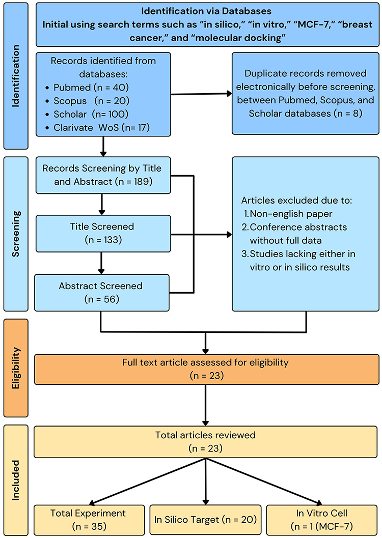 |
Figure 1 Flowchart of the methodology applied in this review. The schematic outlines the systematic approach used for literature identification, screening, eligibility assessment, and inclusion, focusing on studies that report both in vitro IC50 values in MCF-7 breast cancer cells and in silico docking-derived Gibbs free energy (ΔG). |
Molecular Docking Simulation in Drug Discovery
Molecular Docking Simulations and Their Role in Drug Design
Molecular docking simulations have emerged as a fundamental methodology in drug design, providing a computational approach to forecast the atomic-level interactions between tiny chemicals (ligands) and biological molecules, typically proteins. This enables researchers to virtually evaluate the manner in which a potential drug candidate could attach itself to a particular target protein that is linked to a certain disease. Important factors involve the prediction of binding affinity, where docking software calculates the strength of the contact between a ligand and a protein, aiding in the identification of molecules with strong affinity that are essential for the efficiency of drugs. In addition, docking simulations offer valuable information about the binding modes, which unveil the precise orientation and conformation of the ligand when it is attached to the protein. This knowledge is helpful in the process of lead optimization. Virtual screening relies on this methodology to efficiently screen large collections of prospective drug candidates using computer simulations, resulting in a substantial reduction in time and resources compared to traditional high-throughput screening methods. After identifying promising drug candidates, docking simulations can be used to enhance their structure in order to improve their ability to bind to specific target proteins. This process involves iteratively refining the ligand’s fit for the target protein, hence increasing its binding affinity and selectivity. In addition, docking simulations provide valuable insights into the mechanism of action by illustrating the precise atomic-level interactions between a drug candidate and the target protein. This understanding is essential for comprehending the medication’s mechanism and possible adverse effects. Nevertheless, there are certain constraints to consider. One such limitation is the issue of accuracy, as the reliability of predictions relies heavily on the quality of both the protein structure and the docking software employed. Additionally, there is a limitation in terms of dynamics, as the process of docking often portrays an interaction in a fixed state, when in reality, proteins and ligands are capable of flexibility and shape alteration. Although there are limitations, molecular docking simulations are a potent tool that has transformed the field of drug discovery. They provide a rapid and cost-efficient method to evaluate potential drug candidates, thereby expediting the development of novel therapeutics for diverse ailments.13–15
Overview of How Simulations Provide Insights into Molecular Behavior
The realm of molecules, characterized by complicated interplay of forces and interactions, is frequently too little and delicate to be directly observed. Computer simulations are effective instruments for revealing the intricacies of molecular behavior.16 They function as virtual laboratories, enabling researchers to modify and examine molecules at the atomic scale. There are two main categories of simulations: Molecular Dynamics (MD) simulations and Monte Carlo (MC) simulations. Molecular dynamics (MD) simulations are akin to microscopic films, as they trace the paths of individual atoms throughout time, unveiling alterations in molecular structure, protein folding routes, and the kinetics of chemical reactions. On the other hand, Monte Carlo simulations employ a statistical methodology by randomly selecting various arrangements and determining their energies in order to investigate thermodynamic characteristics, phase changes, and the binding of ligands. Simulations surpass the constraints of experiments by investigating severe situations, offering intricate information at the atomic level, and demonstrating the ability to forecast material design and drug development. Nevertheless, these models have several drawbacks such as substantial computing expenses, reliance on model precision, and frequently shorter timeframes in comparison to real-world phenomena. Simulations have the ability to alter our comprehension of molecular behavior, providing distinctive perspectives on the dynamics, interactions, and characteristics of molecules. They are highly valuable tools in various do-mains such as medicine, materials science, chemistry, and physics.17
The Use of Molecular Docking Simulation on Anti Breast Cancer Drug Design and Development
Molecular docking simulations have emerged as a helpful tool in the battle against breast cancer, expediting the process of designing and developing novel anti-cancer medications.18,19 The progression of breast cancer is frequently dependent on the presence of the estrogen receptor (ER) protein. Molecular docking is a vital technique used to identify prospective ER ligands by virtually screening collections of candidate molecules. This process aims to identify compounds that are anticipated to have a strong affinity for the ER binding pocket. This enables the rapid identification of potential medication candidates that have the ability to disrupt estrogen signaling and hinder the proliferation of cancer cells. In addition, docking simulations have the ability to specifically target different subtypes of the estrogen receptor, such as ERα and Erβ.20 This enables the development of more precise medicines that have fewer adverse effects. In addition to ER-positive breast cancer, docking simulations can be used to target other signaling pathways that are utilized by cancer cells and to block enzymes that are essential for cancer growth.21 These simulations enhance drug design by improving the structure of drug candidates to better match the binding pocket of the target protein. This results in more powerful and specific medications. Additionally, the simulations predict any unintended side effects to prevent unpleasant consequences. Nevertheless, docking simulations are subject to some constraints, including their ability to only pro-vide a fixed representation of interactions and their reliance on the accuracy of protein structures and docking software. Molecular docking simulations are transforming the field of anti-breast cancer drug design by facilitating virtual screening, identifying targets, and optimizing leads. This provides a potent tool in the continuous fight against breast cancer, despite the obstacles faced.22–25
In vitro Studies in Drug Discovery
Explanation of in vitro Experiments and Their Significance
In vitro investigations are essential in the initial stages of drug discovery as they provide a controlled setting to investigate the interaction between prospective therapeutic compounds and biological targets. These studies include a wide variety of tests aimed at evaluating the effectiveness, strength, specificity, harmfulness, and pharmacokinetic characteristics of a potential medicine. Enzyme inhibition assays can be used to quantify a compound’s capacity to bind to and hinder the activity of an enzyme linked with a disease. On the other hand, receptor binding assays assess the compound’s attraction to a particular cellular receptor. Utilizing cultured cells or isolated tissues in in vitro models allows researchers to examine the effects of a medicine on cellular viability, signal transduction pathways, and specific disease characteristics. Moreover, in vitro ADME (absorption, distribution, metabolism, and excretion) studies have the ability to forecast a drug’s bioavailability, likelihood of drug-drug interactions, and elimination from the body. The importance of in vitro research rests in their capacity to efficiently evaluate a large number of therapeutic candidates, pick promising leads for further advancement, and detect potential safety issues before moving on to more intricate in vivo trials. Although in vitro models may not possess the complete intricacy of a living organism, they offer essential insights that direct the drug discovery process and facilitate the creation of safe and effective treatments.26–28
Common Methodologies Used in in vitro Studies
Drug molecules are analyzed using a wide range of methods in laboratory investigations to examine their potential therapeutic effects. Cell-based assays are commonly used to evaluate several aspects of drug activity by exploiting cultivated cells. The methods used to assess the effectiveness of drugs include: (1) Cytotoxicity assays, which measure cell death or proliferation when exposed to the drug; (2) Enzyme activity assays, which quantify the drug’s ability to inhibit or activate a specific enzyme target; (3) Receptor binding assays, which determine the drug’s affinity and selectivity for a specific cellular receptor; (4) Reporter gene assays, which monitor changes in gene expression caused by the drug; and (5) Cellular signaling assays, which evaluate the drug’s impact on signal transduction pathways. Surface plasmon resonance (SPR) and isothermal titration calorimetry (ITC) are biophysical techniques that accurately determine the binding affinity of medicines and their targets. In addition, cell-free assays that utilize pure enzymes or separated proteins provide a quick evaluation of a drug’s interaction with its specific molecular target. Moreover, in vitro ADME (absorption, distribution, metabolism, and excretion) experiments employ specialized methodologies to forecast the pharmacokinetic characteristics of a medicine. The choice of these approaches relies on the particular drug target, intended result, and stage of drug discovery, collectively offering a thorough assessment of a drug molecule’s potential as a therapeutic agent.29–31
Importance of Reliable in vitro Data for Predicting Drug Behavior
Accurate in vitro data plays a crucial role in forecasting how drugs will behave in living organisms, serving as a necessary link between initial discovery and successful clinical trials. Firstly, the use of strong and reliable in vitro data enables the rapid screening of large collections of prospective drug candidates, effectively identifying those that have the desired ability to bind to and selectively interact with the target molecule. This greatly decreases the amount of effort and money allocated to compounds that are unlikely to be successful. Furthermore, in vitro experiments offer significant insights into the mechanism of action of a drug, clarifying its contact with the target and the subsequent cellular response. An essential aspect of this process is to comprehend the underlying mechanisms, which is vital for enhancing the effectiveness of primary chemicals and anticipating any possible unintended consequences. Moreover, obtaining dependable in vitro data regarding the ADME (absorption, distribution, metabolism, and excretion) properties of a drug aids in evaluating its bioavailability, potential for drug-drug interactions, and elimination from the body. This information is crucial for making informed decisions about the drug’s formulation and dosage strategies. Although in vitro models can-not completely mimic the intricacies of a living organism, they provide a regulated and repeatable setting to produce statistically meaningful data. This data may then be used to make more confident predictions about how drugs would behave in a living organism. This ultimately enhances the rate of success in drug discovery, resulting in the creation of safer and more effective treatments.32–34
In vitro Cytotoxicity Study in Anti Breast Cancer Drug Discovery and Development
In the relentless quest for novel weapons against breast cancer, In Vitro cytotoxicity assays play a crucial role in the initial phases of drug discovery and development. These assays provide a controlled setting to evaluate the potential efficacy of new drug candidates by examining their ability to kill breast cancer cells. These assays assess the capacity of a potential treatment to hinder the development or survival of breast cancer cells by employing laboratory-grown cultures of human breast cancer cell lines that reflect several subtypes of the disease. The medication candidate is administered to the grown cells at different concentrations. Following incubation, cell viability is assessed using techniques such as the MTT test, XTT assay, cell counting, and Annexin V/PI staining. The advantages of these assays encompass their capacity for high-throughput analysis, cost-effectiveness, and ability to generate consistent outcomes in a controlled setting, enabling the early detection of potential drug candidates. Nevertheless, there are certain constraints such as the restricted intricacy and physiological significance of cell lines, as well as apprehensions regarding the drug’s specificity towards cancer cells as opposed to healthy cells. Although there are difficulties, In Vitro cytotoxicity assays play a vital role in drug development by offering initial information on a medication’s capacity to specifically attack and destroy cancer cells. This, in turn, facilitates subsequent In Vivo investigations and clinical trials. In addition to their role in initial screening, these assays are valuable for investigating pharmacological mechanisms of action, assessing the effectiveness of combination therapy, and finding potential candidates capable of overcoming drug resistance. In Vitro cytotoxicity tests play a crucial role in the process of discovering drugs to combat breast cancer, making a substantial contribution to the battle against this malignant diseases.35–38
Basic Theory of the Correlation Between in vitro and in silico Study
Theoretical Correlation Between Gibbs Energy and IC50 Values
In the field of drug development, the Gibbs free energy (ΔG) derived from molecular docking simulations is frequently employed as a measure of the strength of interaction between a ligand (such as a drug molecule) and its target protein. The fundamental concept states that a decrease in ΔG value is indicative of a higher binding affinity. The reason for this is that the Gibbs free energy shift indicates the spontaneity of the binding process. A greater negative ΔG value indicates a more favorable interaction.
Conversely, the half-maximal inhibitory concentration (IC50) quantifies the effectiveness of a molecule in suppressing a certain biological or metabolic activity. The IC50 value denotes the concentration of the drug necessary to hinder 50% of the target activity. In theory, a drug that has a high binding affinity (shown by a larger negative ΔG) should need a smaller dose to accomplish the same degree of inhibition, leading to a lower IC50 number.
The correlation between ΔG and IC50 can be estimated using the following equation: The change in Gibbs free energy (ΔG) can be calculated using the equation ΔG = RT ln (IC50), where R is the gas constant and T is the temperature. R represents the gas constant, whereas T represents the temperature measured in Kelvin. This equation demonstrates a negative correlation: if the value of ΔG decreases, the IC50 values should also fall, indicating a higher level of potency.39–43
Development of Anti Breast Cancer Agent: in silico and in vitro Studies
MCF-7 Cell Line: A Key Model for Anti-Breast Cancer Drug Development
The MCF7 cell line, which first emerged in the 1970s, is a crucial resource in breast cancer research, playing a fundamental role in the study of prospective anti-cancer substances. MCF7 cells are derived from a pleural effusion of a woman with metastatic breast cancer and they represent an invasive ductal carcinoma, which is the most prevalent form of breast cancer. These cells display the traits of specialized mammary epithelium, including the presence of specific markers like E-cadherin, β-catenin, and cytokeratin 18 (CK18), while not showing markers associated with mesenchymal cells such vimentin and smooth muscle actin (SMA). Significantly, MCF7 cells have the ability to generate dome-like structures in laboratory conditions, closely resembling the structures found in mammary glands.44–48
MCF7 cells have strong expression of estrogen receptor (ER) and progesterone receptor (PR) at the molecular level. This characteristic makes them well-suited for investigating estrogen-dependent breast cancers, which are the most common form of the disease. The tumors do not have HER2 gene amplification, indicating that they belong to the luminal A subtype of breast cancer. This subtype is known to have a favorable prognosis. In addition, MCF7 cells exhibit characteristics similar to stem cells, allowing them to generate mammospheres (3D clusters) in a laboratory setting and develop tumors in mice. In addition, they also exhibit the presence of additional receptors, such as the androgen receptor, which may serve as prospective targets for innovative treatments.44–48
The MCF7 cell line has numerous benefits for cytotoxicity investigations. Their proliferation and accessibility from commercial sources render them suitable for ex-tensive-scale experimentation. The fact that they are ER positive enables researchers to examine the impact of anti-estrogen treatments on the proliferation and viability of cells. MCF7 cells serve as a typical model for luminal A breast cancer, enabling the evaluation of medication effectiveness against this prevalent subtype. The biology of MCF7 cells has been widely studied for many decades, establishing a strong basis for future research.
Nevertheless, it is crucial to acknowledge the constraints of MCF7 cells. They do not cover all types of breast cancer, so other cell lines must be used to provide a more complete understanding. Furthermore, the restricted ability of these malignancies to spread to other parts of the body may not accurately represent the very aggressive nature of metastatic cancers. Although MCF7 cells have the ability to generate tumors in mice, this poses ethical concerns in the context of animal experimentation.
To summarize, the MCF7 cell line is an effective tool for early drug development and comprehending the mechanisms of anti-cancer drugs, namely those that target estrogen receptor-positive breast tumors. Researchers must recognize the constraints of this cell line and employ it in conjunction with other cell lines and research methodologies to get a comprehensive comprehension of breast cancer biology and formulate efficacious treatments. MCF7 cells are commonly used in cytotoxic studies due to their convenient proliferation, wide availability, and their ability to represent luminal A breast cancer. These cells have been extensively studied and characterized, making them highly valuable for breast cancer research.
Focusing on MCF7 provides methodological strength, as it is the most widely studied breast cancer cell line with abundant cytotoxicity and docking data available in the literature. This ensures greater consistency in data interpretation, reduces inter-cell-line variability, and minimizes bias when correlating IC50 values with docking parameters. Consequently, MCF7 serves as a robust and reliable reference model for this review.
Protein, Receptor and Enzymes Used as Target for Development of Anti Breast Cancer Agent
The subsequent targets are frequently employed in molecular docking investigations in this study owing to their pivotal functions in cancer advancement and therapy:
Maintenance of DNA Integrity and Preservation of Genomic Stability
BRCA1, also known as Breast Cancer Type 1 Susceptibility Protein, plays a vital role in the process of DNA repair and the preservation of genomic stability. BRCA1 mutations greatly enhance the probability of developing breast and ovarian cancer, making it a crucial focus for medicines aimed at leveraging its involvement in DNA repair processes to specifically eliminate cancer cells.49,50
The Process of Cell Division and the Movement of Microtubules
Tubulin, an essential protein for the production of microtubules and cell division, represents another crucial target. By impeding the process of tubulin polymerization, the normal functioning of microtubules is disrupted, resulting in the halting of the cell cycle and the initiation of programmed cell death. This property makes it a viable approach in the treatment of cancer. Aurora Kinase, which regulates chromosome segregation and cytokinesis during cell division, is a potential therapeutic target for triggering mitotic arrest and death in cancer cells.51
Receptor Tyrosine Kinases
Receptor tyrosine kinase a type of protein that play a role in cell signaling by transferring phosphate groups to tyrosine residues. EGFR, also known as Epidermal Growth Factor Receptor, and its tyrosine kinase domain, referred to as EGFR-TK, have significant functions in the processes of cell proliferation, survival, and differentiation. EGFR is frequently overexpressed or mutated in various types of cancer, such as breast cancer. By specifically targeting EGFR, it is possible to impede the growth and advancement of tumors. Likewise, HER2 (Human Epidermal Growth Factor Receptor 2), a different type of protein that triggers cell growth, is excessively produced in highly aggressive breast tumors. By specifically focusing on HER2, it is possible to greatly enhance the prognosis and overall well-being of patients. The Insulin-like Growth Factor 1 Receptor (IGF1R) is responsible for regulating cell growth and promoting cell survival. By blocking IGF1R, cancer cell proliferation can be reduced and the effective-ness of other treatments can be enhanced.52,53
Estrogen Receptors
Estrogen Receptor Alpha (ER Alpha) plays a vital role in controlling the activation of genes in response to estrogen. A significant number of breast malignancies have ER-positive characteristics, and the inhibition of ER Alpha can effectively suppress the development and proliferation of cancer cells driven by estrogen. Exemestane, an aromatase inhibitor, reduces the levels of estrogen and is employed in the treatment of estrogen-dependent breast cancer. ER Beta (Estrogen Receptor Beta) provides alternate approaches for the treatment of estrogen-dependent breast tumors.54,55
Immune Response and Cellular Proliferation
TNFRSF5 (CD40) and Ki-67 (MK167) play crucial roles in immune responses and cellular proliferation, respectively. Therefore, they are significant targets for manipulating immune responses and evaluating the efficacy of anti-cancer therapies.56
Regulation of Apoptosis
Proteins like cIAP1 (Cellular Inhibitor of Apoptosis Protein 1) and xIAP (X-linked Inhibitor of Apoptosis Protein) prevent cell death by attaching to and deactivating caspases, which are enzymes involved in the process of apoptosis. By specifically targeting these inhibitors, it is possible to induce apoptosis (cell death) in cancer cells, hence improving the effectiveness of cancer treatments. BCL proteins, which control programmed cell death (apoptosis), are important targets for cancer treatment.57,58
Cellular Metabolism and Telomere Maintenance
NUDT5, a Nudix Hydrolase 5 enzyme, is involved in cellular metabolism. Inhibiting NUDT5 can interfere with crucial metabolic processes in cancer cells, resulting in decreased cell growth and survival. Telomerase, an enzyme that preserves the length of telomeres and safeguards chromosomes from deterioration, is frequently overexpressed in cancer cells, making it a possible target for restricting their ability to replicate.59
DNA Transcription and RNA Processing
Topoisomerase-IIβ, which plays a role in DNA transcription, is specifically targeted to inhibit DNA replication in cancer cells, ultimately resulting in cell death. CLK-3, also known as CDC-like Kinase 3, plays a role in controlling RNA splicing. If CLK-3 is inhibited, it can interfere with the processing of RNA in cancer cells. This disruption has the potential to cause the cells to stop dividing and undergo programmed cell death, known as apoptosis.60,61
The Cytoskeleton and Cellular Movement
ARPBCC, also known as Actin-Related Protein Binding Complex Component, controls the structure of the actin cytoskeleton and the shape of cells. Directing efforts against ARPBCC can impact the movement of cancer cells and potentially hinder the spread of cancer to other parts of the body.
Signal Transduction
Signal transduction refers to the process by which cells communicate and transmit signals inside the body. C-ABL, also known as ABL Proto-Oncogene 1, Non-Receptor Tyrosine Kinase, plays a crucial role in many communication pathways. As a result, it is an important target for cancer treatment.62
Correlation Between in vitro Cytotoxicity Study (IC50) and Molecular Docking Study (Gibbs Energy)
Several important insights are revealed when examining the dynamics of IC50 values and Gibbs energy (ΔG) in MCF7 cell investigations. IC50 values vary greatly, ranging from very strong (eg, 0.01 µM) to weaker (eg, 82.87 µM) (Table 1). These values represent the different abilities of drugs to inhibit specific protein targets in MCF7 cells. These values are crucial in clinical contexts, providing guidance for deter-mining appropriate dosage strategies and assessing the effectiveness of treatments.63–66
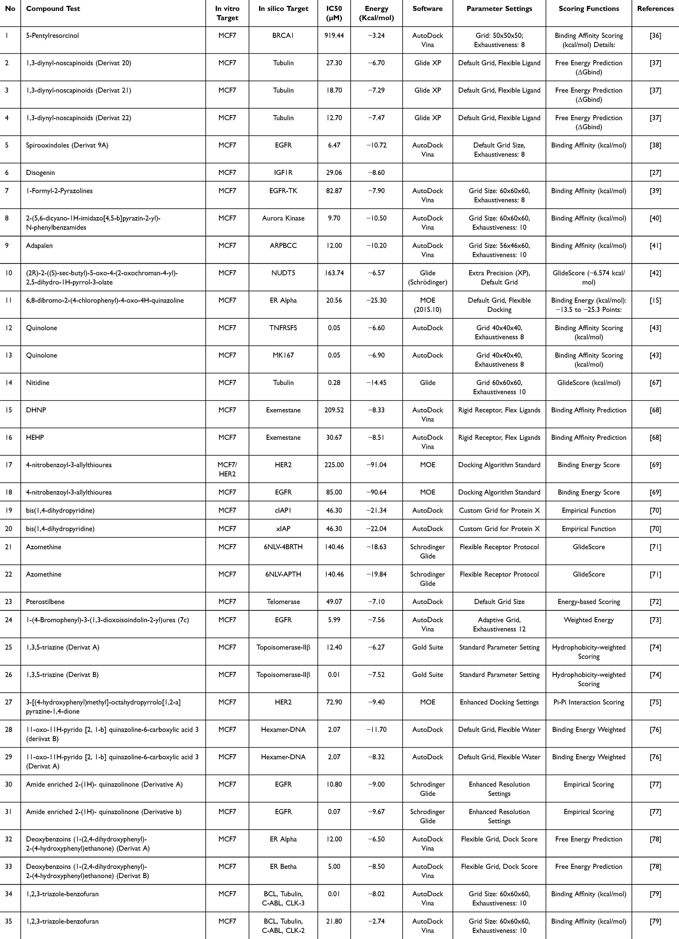 |
Table 1 In vitro Cytotoxic Studies of Several Compounds on the MCF-7 Cell Line and Molecular Docking Studies on Several Proteins, Receptors, and Enzymes with Up-Down Regulation on Breast Cancer |
Theoretical frameworks of ΔG emphasize its function in assessing the thermodynamic favorability of compound-protein interactions. Lower ΔG values (−14.45 kcal/mol to −2.74 kcal/mol) suggest higher binding affinities (Table 1), which are essential for optimal therapeutic activity. ΔG predictions are used as prognostic markers in the field of drug discovery, helping to prioritize compounds for further research based on their capacity to bind.
Although theoretical predictions indicate a clear relationship between ΔG and IC50 values, where a lower ΔG corresponds to better potency (lower IC50),80,81 findings in this review from MCF7 cell research reveal a more intricate situation. Although many compounds display predictable patterns, the dataset demonstrates a degree of unpredictability (Figure 2). Main Discovery in this study revealed an important finding regarding the correlation. While theoretical expectations indicate a direct correlation between ΔG and IC50 values, the dataset shows that there is no consistent linear correlation across all compounds and protein targets tested (Figure 2). Simply Despite at-tempting a thorough analysis by categorizing the target receptor, we have not discovered any linear correlation between the Gibbs energy value and the IC50 value. This disparity highlights the intricacies involved in transforming theoretical predictions into practical results in the field of pharmaceutical research.
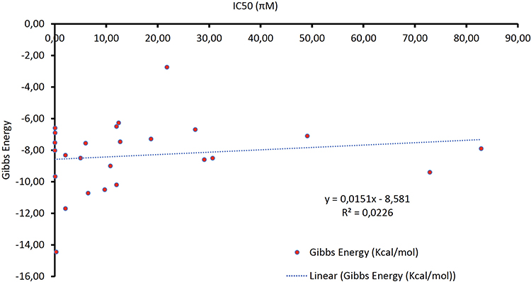 |
Figure 2 Correlation analysis between Gibbs free energy (ΔG) values obtained from molecular docking and IC50 values from in vitro cytotoxicity assays in MCF-7 breast cancer cells. The dataset illustrates the absence of a consistent linear correlation (R² = 0.0226), despite theoretical expectations of an inverse relationship between ΔG and IC50. This highlights the influence of biological variability, structural diversity, and methodological differences in experimental and computational setups. |
However, when all methodological and biological systems are carefully controlled such as harmonizing protein targets, docking parameters, and IC50 unit conversions a strong linear correlation can indeed be observed, as demonstrated in the additional dataset presented in Figure 3.
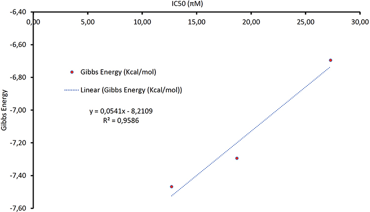 |
Figure 3 Correlation analysis between Gibbs free energy (ΔG) values and IC50 data under controlled methodological conditions. The figure demonstrates a strong linear correlation (R² = 0.9586) when protein targets, docking parameters, IC50 unit conversions, and assay conditions are harmonized, using derivatives of 1,3-diynyl-noscapinoids against tubulin and a spirooxindole derivative against EGFR. |
The correlation analysis between Gibbs free energy (ΔG) derived from molecular docking and IC50 values obtained from in vitro assays underlines the significance of conducting studies within a controlled and standardized system. In the dataset presented, three derivatives of 1,3-diynyl-noscapinoids (Derivat 20, 21, and 22) were evaluated against the MCF-7 breast cancer cell line using tubulin as the target protein, along with one spirooxindole derivative (Derivat 9A) evaluated against EGFR. When the analysis was performed with harmonized conditions same cell line, clearly defined protein targets, consistent preparation of ligands, standardized docking protocols, and normalized IC50 units the results exhibited a strong linear correlation (R² = 0.9586). This finding demonstrates that when methodological and biological variations are minimized, the theoretical assumption of an inverse relationship between ΔG and IC50 can indeed be observed in practice.
The noscapinoid derivatives displayed a clear trend: as ΔG values became more negative, indicating stronger predicted binding affinity, the corresponding IC50 values decreased, reflecting higher cytotoxic potency. This alignment supports the validity of docking predictions when contextualized under carefully controlled conditions. The spirooxindole derivative (Derivat 9A), with a much stronger ΔG of –10.72 kcal/mol, also showed superior IC50 potency (6.47 µM), reinforcing this pattern. The linear regression equation (y = 0.0541x – 8.2109) not only confirms the positive correlation but also quantitatively demonstrates the predictive capacity of docking when variability in experimental and computational setups is reduced (Figure 3).
This result emphasizes that discrepancies previously reported in the literature where no or weak correlations were observed are largely attributable to uncontrolled differences in experimental design, protein selection, assay types, and docking protocols. By carefully aligning these factors, the correlation between computational and experimental outcomes becomes more robust, offering a reliable predictive framework for drug discovery. Therefore, this study highlights the importance of systematic harmonization of both in vitro and in silico methodologies as a prerequisite for meaningful interpretation of docking outcomes in the context of cytotoxicity data.
Biological intricacies inside cellular contexts have a significant impact, affecting the effectiveness of compounds beyond what can be predicted by ΔG. Divergent IC50 values might arise due to several factors, including drug absorption, metabolism, and unique interactions with physio-logical pathways, despite the indication of great binding affinity based on ΔG.80,81
In addition, the presence of target-specific variability adds more complexity to the interaction. Protein targets have distinct interactions with substances because of differences in their structure and function.82,83 Differences in the amounts of target expression, alterations that occur after protein synthesis, and the specific location inside the cell all have a major effect on the measured IC50 values, regardless of the predictions of thermodynamic free energy change (ΔG). Various factors, such as different methods used in experiments, specific details of cell culture, and the sensitivity of the assay, can cause variations in IC50 results. These variations may hide any connections between ΔG and IC50 values that are seen in real-world situations.
In the future, the combination of computational ΔG estimates with empirical IC50 data shows potential when used together. Nevertheless, comprehensive assessments that span the metabolism of compounds, the permeability of cells, and the selectivity of targets are essential in order to improve the accuracy of predictions and optimize tactics for discovering drugs. Precision medicine strategies, customized for individual protein targets and biological circumstances, provide chances to discover potent and selective treatment options.
To summarize, the complex connection between IC50 values and ΔG in MCF7 cell research emphasizes the difficulties of converting theoretical projections into practical results in the field of drug development. Researchers can improve the reliability of preclinical assessments and advance personalized medicine by addressing biological complexities and experimental factors.
Root Causes of the Absence of Correlation Between Molecular Docking and in vitro Cytotoxicity in Anti-Breast Cancer Research
The theoretical expectation of an inverse relationship between Gibbs free energy (ΔG), as predicted by molecular docking, and in vitro cytotoxicity (IC50) arises from thermodynamic principles: a more negative ΔG implies stronger binding affinity between a ligand and its protein target, which should, in principle, translate to greater biological potency and hence a lower IC50. However, empirical observations in this study reveal a conspicuous absence of such a linear correlation across various compounds tested on the MCF-7 breast cancer cell line. This discrepancy is not incidental but rather emerges from a confluence of mechanistic, structural, and methodological root causes that collectively undermine the assumed predictability of docking outcomes.
Biologically, even though in vitro systems eliminate many of the systemic complexities inherent to in vivo models, they are not devoid of influential variables that directly affect cellular responses. The MCF-7 cell line, although standardized to some extent, may exhibit variation in the expression of specific protein targets due to culture conditions, passage number, or intrinsic heterogeneity. This variation means that a compound predicted to bind strongly to a certain receptor may not manifest cytotoxicity if that receptor is minimally expressed or functionally inactive within the cellular context. Furthermore, the assumption in docking simulations that a ligand reaches and binds its target does not necessarily hold true in vitro. Factors such as poor membrane permeability, low cellular uptake, or active efflux mechanisms can significantly diminish intracellular concentrations of a compound, rendering it biologically inactive despite its favorable binding score. Additionally, metabolic processes albeit limited compared to in vivo still operate in cell-based systems. Enzymes present in MCF-7 cells may rapidly degrade or modify the compound, especially if the structure includes labile moieties like esters, amides, or nitro groups, thereby preventing interaction with the intended target.
Equally critical is the substantial structural diversity of the compounds analyzed in this study. The chemical library includes ligands with vastly different physicochemical properties, molecular weights, lipophilicities, and conformational flexibilities. These differences introduce a wide range of behaviors not accounted for in docking scores. For instance, compounds with high polarity or extreme hydrophobicity may not distribute uniformly in the culture medium or may fail to permeate the cell membrane effectively, resulting in limited intracellular exposure. Molecules that are highly rigid might fit perfectly into a docking model due to reduced entropic penalties, leading to exaggerated ΔG predictions, while more flexible molecules might be penalized in silico but adopt bioactive conformations in a cellular environment. Furthermore, certain functional groups in some compounds may trigger nonspecific reactivity or aggregation under cell culture conditions, which alters their effective cytotoxic profiles independently of the specific target interaction modeled in silico. The diversity in scaffolds also implies differential target engagement and potential polypharmacology, which are not reflected in single-target docking simulations.
Adding to these confounding variables are the methodological limitations inherent to molecular docking itself. Most docking protocols treat the protein receptor as a rigid structure, overlooking the dynamic conformational changes that naturally occur upon ligand binding a phenomenon known as induced fit. This rigid approximation can lead to misrepresentation of both the binding pose and the affinity. Additionally, standard scoring functions primarily evaluate enthalpic interactions such as hydrogen bonding, hydrophobic contacts, and van der Waals forces, while largely neglecting entropic contributions, solvation effects, or the thermodynamic cost of desolvating the ligand and receptor. These simplifications can result in docking scores that do not accurately reflect the true binding energetics under physiological or even in vitro conditions. Moreover, molecular docking is typically performed on a single protein target, often assuming that it is the principal driver of a compound’s activity. This assumption fails to capture the biological complexity observed in cell-based assays, where ligands may exert cytotoxicity through off-target interactions, synergistic effects, or network-level disruptions.
The divergence is further exacerbated by variability in the execution and interpretation of in vitro assays themselves. Differences in assay type, such as MTT versus Annexin V staining, or variations in incubation times, cell densities, and serum concentrations, can all influence IC50 readouts. The manner in which compounds are prepared, solubilized, and administered also affects their bioavailability and stability in culture. In some instances, compounds with excellent docking profiles might precipitate or degrade during the assay period, leading to underestimation of their potency. Inconsistencies in data reporting such as rounded IC50 values, single-replicate experiments, or lack of proper controls further contribute to noise within the dataset. Moreover, publication bias toward compounds that show extreme activity (either very high or very low IC50) may skew the available literature and limit the robustness of comparative analyses.
Collectively, these factors illustrate that the observed lack of correlation between docking predictions and cytotoxic outcomes is not merely an anomaly but the natural consequence of layered and intersecting limitations across biological, structural, computational, and experimental domains. The discrepancy is particularly evident when structurally diverse compounds are compared against a variety of targets under non-uniform assay conditions. This multifactorial disjunction underscores the limitations of using ΔG values as standalone predictors of bioactivity and highlights the necessity of contextualizing in silico findings within the biological and physicochemical realities of in vitro testing. Ultimately, acknowledging and dissecting these root causes is essential not only for interpreting current findings but also for guiding the development of more integrated and predictive models in future drug discovery efforts.
Practical Resolutions to Align Molecular Docking Predictions with in vitro Cytotoxicity Outcomes
Bridging the persistent gap between molecular docking-derived binding affinities and in vitro cytotoxic responses necessitates the development of a translationally integrated framework one that reconciles computational predictions with experimental realities through methodological augmentation and data contextualization. Central to this approach is the transition from using ΔG values as solitary predictors to embedding them within multi parametric models. These refined frameworks incorporate permeability, metabolic stability, and compound specific pharmacokinetic behaviors. Recent developments in machine learning guided scoring functions have demonstrated that when ADMET descriptors and molecular descriptors are incorporated alongside ΔG, prediction accuracy for in vitro potency improves significantly compared to traditional docking alone.1
Structure-based techniques can also be refined through the adoption of ensemble docking methodologies. Rather than relying on a single rigid receptor structure, ensemble docking utilizes multiple receptor conformations derived from molecular dynamics (MD) simulations. This accounts for target flexibility and induced-fit interactions, thereby improving both docking accuracy and biological relevance. Studies employing MD-derived conformational libraries have reported enhanced alignment between predicted binding energies and experimental affinities, especially for targets with flexible or disordered binding pockets.2
Another key consideration lies in the chemical diversity of the compound library itself. The broad structural variance among tested ligands introduces inconsistencies that obscure global correlations. Stratifying compounds into structurally homogeneous chemotypes prior to analysis allows for the identification of scaffold specific structure activity relationships. Within such clusters, local correlations between ΔG and IC50 can be recovered, especially when QSAR descriptors such as polar surface area, hydrogen bond donors, and electronic features are taken into account. This approach yields more precise insight than global models that ignore scaffold-specific effects.3
Addressing intracellular exposure is equally crucial. While docking assumes ligand access to its binding site, in vitro potency is fundamentally governed by the amount of free drug available at the site of action. Advanced LC-MS techniques now permit quantification of the intracellular free fraction (F_ic), a parameter encompassing cellular uptake, metabolism, and sequestration. When F_ic is integrated into potency analysis, many false-negative results where compounds fail to exhibit cytotoxicity despite high docking scores are resolved, as the discrepancy often stems from poor intracellular bioavailability.4
To confirm target engagement under cellular conditions, complementary biophysical techniques should be employed. The Cellular Thermal Shift Assay (CETSA), for example, allows direct observation of ligand-induced stabilization of the target protein within the cell. This technique distinguishes between ligands that bind and exert biological effects versus those that merely show theoretical affinity in silico. When combined with viability assays, CETSA data validate or refute docking predictions in real-time.5
On the experimental side, variability in assay protocols is a known confounder in correlational studies. Standardization of experimental parameters including cell density, incubation time, and serum composition has been shown to reduce inter-laboratory IC50 variability significantly. Moreover, incorporating orthogonal readouts such as ATP-luminescence assays or annexin V flow cytometry can further refine the interpretation of cytotoxic responses, clarifying whether observed effects stem from true cell death or merely growth arrest.6
Additionally, phenotypic screening frameworks that integrate omics-level data provide broader insight into compound behavior. For instance, metabolomic profiling of drug-treated and resistant MCF-7 cell lines has revealed adaptive pathway rewiring that affects compound sensitivity regardless of predicted binding affinity. Such systems biology perspectives can inform combinatorial design strategies and highlight context-specific vulnerabilities.7
When these resolutions are combined into an iterative screening pipeline starting with data-augmented docking, followed by chemotype-specific modeling, intracellular exposure validation, and real-time target engagement assessment they create a predictive continuum that aligns theoretical affinity with biological efficacy. This translational strategy not only enhances the accuracy of preclinical assessments but also accelerates the discovery of therapeutically viable anti-breast-cancer candidates.
Conclusion
In conclusion, the examination of IC50 values and Gibbs free energy (ΔG) in MCF-7 cell research underscores the inherent complexity and multidimensionality of the drug discovery process. This review confirms that, despite theoretical expectations, there is no consistent or immediate correlation between ΔG values derived from molecular docking and cytotoxic potency observed in in vitro assays. The absence of this correlation arises from an interplay of biological, structural, and methodological factors, including limited target expression, intracellular bioavailability, structural diversity of ligands, and the simplifications inherent in rigid docking protocols.
To improve predictive accuracy and ensure translational relevance, it is no longer sufficient to rely solely on ΔG scores as proxies for biological activity. Instead, future research should integrate advanced computational approaches such as ensemble docking, machine-learning-guided scoring functions, and molecular dynamics simulations with experimental validation tools like intracellular exposure quantification and cellular thermal shift assays. These complementary strategies enable the verification of target engagement and help contextualize docking results within the biological behavior of compounds in a cellular environment.
Moreover, refining the selection of cell lines for cytotoxic assays, along with targeting well-characterized receptors and maintaining rigorous standardization of in vitro conditions, will significantly enhance data reliability. Stratification of chemical scaffolds, incorporation of ADMET profiles, and scaffold-specific SAR modeling are also crucial in mitigating the noise introduced by chemical heterogeneity. Collectively, these advancements form a robust and iterative pipeline that bridges theoretical affinity and practical efficacy. In addition, future studies should incorporate positive and negative control experiments, redocking validation, and multiparametric comparisons to strengthen the correlation between docking parameters and biological outcomes.
Importantly, strengthening the integration between in vitro and in silico studies will enhance the predictability of in vivo outcomes, thereby bridging computational predictions with translational pharmacology. Therefore, the future of computational-experimental pharmacology lies in an integrated, interdisciplinary model one that balances mechanistic precision with biological complexity. By aligning in silico predictions more closely with empirical outcomes, the drug discovery process can be both accelerated and refined, ultimately improving the development of effective and selective anti-breast cancer agents.
Funding
The authors thank to Rector of Universitas Padjadjaran for funding this study with review article grant (2025).
Disclosure
The authors repot no conflicts of interest in this work.
This paper has been uploaded to Pre Prints.org as a preprint: https://www.preprints.org/manuscript/202412.0797/v1
References
1. Noor F, Junaid M, Almalki AH, Almaghrabi M, Ghazanfar S, Tahir Ul Qamar M. Deep learning pipeline for accelerating virtual screening in drug discovery. Sci Rep. 2024;14(1):1–13. doi:10.1038/s41598-024-79799-w
2. Hillisch A, Heinrich N, Wild H. Computational chemistry in the pharmaceutical industry: from childhood to adolescence. ChemMedChem. 2015;10(12):1958–1962. doi:10.1002/cmdc.201500346
3. Miteva MA, Villoutreix BO. Computational biology and chemistry in MTi: emphasis on the prediction of some ADMET properties. Mol Inform. 2017;36(10). doi:10.1002/minf.201700008
4. Hu Y, Stumpfe D, Bajorath J. Computational exploration of molecular scaffolds in medicinal chemistry. J Med Chem. 2016;59(9):4062–4076. doi:10.1021/acs.jmedchem.5b01746
5. Salmaso V, Moro S. Bridging molecular docking to molecular dynamics in exploring ligand-protein recognition process: an overview. Front Pharmacol. 2018;9(AUG). doi:10.3389/fphar.2018.00923
6. Wan S, Sinclair RC, Coveney PV. Uncertainty quantification in classical molecular dynamics. Philos Trans R Soc a Math Phys Eng Sci. 2021;379(2197):20200082. doi:10.1098/rsta.2020.0082
7. Niazi SK. Quantum mechanics in drug discovery: a comprehensive review of methods, applications, and future directions. Int J Mol Sci. 2025;26(13):1–20. doi:10.3390/ijms26136325
8. Guéniche N, Lakehal Z, Habauzit D, et al. Combined in silico and in vitro approaches to identify P-glycoprotein-inhibiting pesticides. J Biochem Mol Toxicol. 2024;38(1). doi:10.1002/jbt.23588
9. Razzak Mahmood Kubba AA, Shihab WA, Al-Shawi NN. In silico and in vitro approach for design, synthesis, and anti-proliferative activity of novel derivatives of 5-(4-aminophenyl)-4-substituted phenyl-2, 4-dihydro-3h-1, 2, 4-triazole-3-thione. Res J Pharm Technol. 2020;13(6):3329–3339. doi:10.5958/0974-360X.2020.00591.0
10. Andrade EL, Bento AF, Cavalli J, et al. Non-clinical studies required for new drug development – part I: early in silico and in vitro studies, new target discovery and validation, proof of principles and robustness of animal studies. Braz J Med Biol Res. 2016;49(11). doi:10.1590/1414-431X20165644
11. Huang F, Zhu Q, Zhou X, et al. Role of CFD based in silico modelling in establishing an in vitro-in vivo correlation of aerosol deposition in the respiratory tract. Adv Drug Deliv Rev. 2021;170:369–385. doi:10.1016/j.addr.2020.09.007
12. Choi S-M, Kang C-Y, Lee B-J, Park J-B. In vitro-in vivo correlation using in silico modeling of physiological properties, metabolites, and intestinal metabolism. Curr Drug Metab. 2017;18(11):973–982. doi:10.2174/1389200218666171031124347
13. Santos LHS, Ferreira RS, Caffarena ER. Integrating molecular docking and molecular dynamics simulations. In: Docking Screens for Drug Discovery. Springer; 2019:13–34.
14. Saikia S, Bordoloi M. Molecular docking: challenges, advances and its use in drug discovery perspective. Curr Drug Targets. 2019;20(5):501–521. doi:10.2174/1389450119666181022153016
15. Chen G, Seukep AJ, Guo M. Recent advances in molecular docking for the research and discovery of potential marine drugs. Mar Drugs. 2020;18(11):545. doi:10.3390/md18110545
16. Mateus A, Gordon LJ, Wayne GJ, et al. Prediction of intracellular exposure bridges the gap between target- and cell-based drug discovery. Proc Natl Acad Sci U S A. 2017;114(30). doi:10.1073/pnas.1701848114
17. Shah A, Jain M. Limitations and future challenges of computer-aided drug design methods. In: Computer Aided Drug Design (CADD): From Ligand-Based Methods to Structure-Based Approaches. 2022. doi:10.1016/B978-0-323-90608-1.00006-X
18. Larsson P, Engqvist H, Biermann J, et al. Optimization of cell viability assays to improve replicability and reproducibility of cancer drug sensitivity screens. Sci Rep. 2020;10(1). doi:10.1038/s41598-020-62848-5
19. Marusyk A, Janiszewska M, Polyak K. Intratumor heterogeneity: the rosetta stone of therapy resistance. Cancer Cell. 2020;37(4):471–484. doi:10.1016/j.ccell.2020.03.007
20. Arifian H, Maharani R, Megantara S, Ikram NKK, Muchtaridi M. Glycine-conjugated α-mangostins as potential estrogen receptor alpha (ERα) antagonists through pharmacophore modeling, docking analysis, and molecular dynamics simulations. Appl Sci. 2024;14(13):5549. doi:10.3390/app14135549
21. Huang X, Hu J, Bailey WM. In Silico discovery of novel androgen receptor inhibitors for prostate cancer therapy using virtual screening, molecular docking, and molecular dynamics simulations. Sci Rep. 2025;15(1):1–19. doi:10.1038/s41598-025-15038-0
22. Komura H, Watanabe R, Mizuguchi K. The trends and future prospective of in silico models from the viewpoint of ADME evaluation in drug discovery. Pharmaceutics. 2023;15(11):2619. doi:10.3390/pharmaceutics15112619
23. Ahmed MF, Youns M, Belal A. Design, synthesis, molecular docking and anti-breast cancer activity of novel quinazolinones targeting estrogen receptor α. Acta Pol Pharm - Drug Res. 2016;73(1):115–127.
24. Gnanaselvan S, Yadav SA, Manoharan SP. Structure-based virtual screening of anti-breast cancer compounds from Artemisia absinthium—insights through molecular docking, pharmacokinetics, and molecular dynamic simulations. J Biomol Struct Dyn. 2024;42(6):3267–3285. doi:10.1080/07391102.2023.2212805
25. El Rhabori S, Alaqarbeh M, El Aissouq A, Bouachrine M, Chtita S, Khalil F. Design, 3D-QSAR, molecular docking, ADMET, molecular dynamics and MM-PBSA simulations for new anti-breast cancer agents. Chem Phys Impact. 2024;8:100455. doi:10.1016/j.chphi.2023.100455
26. Chung TDY, Terry DB, Smith LH. In Vitro and in Vivo Assessment of ADME and PK Properties During Lead Selection and Lead Optimization – Guidelines. Benchmarks and Rules of Thumb.; 2004.
27. Nair DG, Weiskirchen R. Advanced in vitro models for preclinical drug safety: recent progress and prospects. Curr Issues Mol Biol. 2025;47(1):1–18. doi:10.3390/cimb47010007
28. Stielow M, Witczyńska A, Kubryń N, Fijałkowski Ł, Nowaczyk J, Nowaczyk A. The Bioavailability of Drugs—The Current State of Knowledge. Molecules. 2023;28(24):8038. doi:10.3390/molecules28248038
29. Kumar V, Chunchagatta Lakshman PK, Prasad TK, et al. Target-based drug discovery: applications of fluorescence techniques in high throughput and fragment-based screening. Heliyon. 2024;10(1):e23864. doi:10.1016/j.heliyon.2023.e23864
30. Perez JJ, Perez RA, Perez A. Computational modeling as a tool to investigate PPI: from drug design to tissue engineering. Front Mol Biosci. 2021;8. doi:10.3389/fmolb.2021.681617
31. Nierode G, Kwon PS, Dordick JS, Kwon SJ. Cell-based assay design for high-content screening of drug candidates. J Microbiol Biotechnol. 2015;26(2):213. doi:10.4014/jmb.1508.08007
32. Freiberger EC, Thompson MP, Zhang X, et al. Utility of common in vitro systems for predicting circulating metabolites. Drug Metab Dispos. 2024;52(12):1373–1378. doi:10.1124/dmd.124.001732
33. Bouhaddou M, Yu LJ, Lunardi S, et al. Predicting in vivo efficacy from in vitro data: quantitative systems pharmacology modeling for an epigenetic modifier drug in cancer. Clin Transl Sci. 2020;13(2):419–429. doi:10.1111/cts.12727
34. Lai Y, Chu X, Di L, et al. Recent advances in the translation of drug metabolism and pharmacokinetics science for drug discovery and development. Acta Pharm Sin B. 2022;12(6):2751–2777. doi:10.1016/j.apsb.2022.03.009
35. Khanal P, Patil VS, Bhandare VV, et al. Systems and in vitro pharmacology profiling of diosgenin against breast cancer. Front Pharmacol. 2023;13. doi:10.3389/fphar.2022.1052849
36. Kaur K, Verma H, Gangwar P, Dhiman M, Jaitak V. Design, synthesis, in vitro and in silico evaluation of indole-based tetrazole derivatives as putative anti-breast cancer agents. RSC Med Chem. 2024;15(4):1329–1347. doi:10.1039/d3md00730h
37. Mozafarinia M, Karimi S, Farrokhnia M, Esfandiari J. In vitro breast cancer targeting using Trastuzumab-conjugated mesoporous silica nanoparticles: towards the new strategy for decreasing size and high drug loading capacity for drug delivery purposes in MSN synthesis. Microporous Mesoporous Mater. 2021;316:110950. doi:10.1016/j.micromeso.2021.110950
38. Zhou Y, Yu F, Guo M, Tang Y, Xu Q. Bridging the gap: the role of 3D cell cultures in mimicking tumor microenvironment for enhanced drug testing accuracy. Front Bioeng Biotechnol. 2025;13(August):1–16. doi:10.3389/fbioe.2025.1498141
39. Goettig P, Chen X, Harris JM. Correlation of experimental and calculated inhibition constants of protease inhibitor complexes. Int J Mol Sci. 2024;25(4):2429. doi:10.3390/ijms25042429
40. Tonge PJ. Drug-target kinetics in drug discovery. ACS Chem Neurosci. 2018;9(1):29–39. doi:10.1021/acschemneuro.7b00185
41. Sumalapao DEP, Janairo JIB, Gloriani NG. Dipole moment, solvation energy, and ovality account for the variations in the biological activity of HIV-1 reverse transcriptase inhibitor fragments. Annu Res Rev Biol. 2018;22(5):1–8. doi:10.9734/ARRB/2018/38945
42. Jiang M, Lu S, Telu S, Pike VW. An empirical quantitative structure-activity relationship equation assists the discovery of high-affinity phosphodiesterase 4D inhibitors as leads to PET radioligands. J Med Chem. 2023;66(2):1543–1561. doi:10.1021/acs.jmedchem.2c01745
43. Negami T, Terada T. Calculations of the binding free energies of the comprehensive in vitro proarrhythmia assay (CiPA) reference drugs to cardiac ion channels. Biophys Physicobiol. 2023;20(2). doi:10.2142/biophysico.bppb-v20.0016
44. Alsharif S, Sharma P, Bursch K, et al. Keratin 19 maintains E-cadherin localization at the cell surface and stabilizes cell-cell adhesion of MCF7 cells. Cell Adhes Migr. 2021;15(1):1–17. doi:10.1080/19336918.2020.1868694
45. Comşa Ş, Cimpean AM, Raica M. The story of MCF-7 breast cancer cell line: 40 years of experience in research. Anticancer Res. 2015;35(6):3147–3154.
46. Krause S, Maffini MV, Soto AM, Sonnenschein C. The microenvironment determines the breast cancer cells’ phenotype: organization of MCF7 cells in 3D cultures. BMC Cancer. 2010;10(1). doi:10.1186/1471-2407-10-263
47. Novak DD, Troitskaya OS, Nushtaeva AA, et al. EGFR suppression inhibits the sphere formation of MCF7 cells overexpressing EGFR. Acta Naturae. 2023;15(2(57)):59–69. doi:10.32607/actanaturae.17857
48. Pulze L, Congiu T, Brevini TAL, et al. Mcf7 spheroid development: new insight about spatio/temporal arrangements of TNTS, amyloid fibrils, cell connections, and cellular bridges. Int J Mol Sci. 2020;21(15):5400. doi:10.3390/ijms21155400
49. Kornepati AVR, Boyd JT, Murray CE, et al. Tumor intrinsic PD-L1 promotes DNA repair in distinct cancers and suppresses PARP inhibitor-induced synthetic lethality. Cancer Res. 2022;82(11):2156–2170. doi:10.1158/0008-5472.CAN-21-2076
50. Tarsounas M, Sung P. The antitumorigenic roles of BRCA1–BARD1 in DNA repair and replication. Nat Rev Mol Cell Biol. 2020;21(5):284–299. doi:10.1038/s41580-020-0218-z
51. Liu H, Cali Daylan AE, Yang J, et al. Aurora kinase A inhibition potentiates platinum and radiation cytotoxicity in non-small-cell lung cancer cells and induces expression of alternative immune checkpoints. Cancers. 2024;16(16):2805. doi:10.3390/cancers16162805
52. Bobin C, Iddir Y, Butterworth C, et al. Sequential analysis of cfDNA reveals clonal evolution in patients with neuroblastoma receiving ALK-targeted therapy. Clin Cancer Res. 2024;30(15):3298–3315. doi:10.1158/1078-0432.CCR-23-3110
53. Yam C, Patel M, Hill HA, et al. Targeting the epidermal growth factor receptor pathway in chemotherapy-resistant triple-negative breast cancer: a Phase II study. Cancer Res Commun. 2024;4(10):2823–2834. doi:10.1158/2767-9764.CRC-24-0255
54. Wang Z-Y, Yin L. Estrogen receptor alpha-36 (ER-α36): a new player in human breast cancer. Mol Cell Endocrinol. 2015;418:193–206. doi:10.1016/j.mce.2015.04.017
55. Jia M, Dahlman-Wright K, Gustafsson J-Å. Estrogen receptor alpha and beta in health and disease. Best Pract Res Clin Endocrinol Metab. 2015;29(4):557–568. doi:10.1016/j.beem.2015.04.008
56. Zhang W, Xu Y, Wang Y, et al. Prognostic analysis of three forms of Ki-67 in patients with breast cancer with non-pathological complete response before and after neoadjuvant systemic treatment. Cancer Med. 2023;12(8):9363–9372. doi:10.1002/cam4.5693
57. Sun H, Lu J, Liu L, Yang CY, Wang S. Potent and selective small-molecule inhibitors of cIAP1/2 proteins reveal that the binding of Smac mimetics to XIAP BIR3 is not required for their effective induction of cell death in tumor cells. ACS Chem Biol. 2014;9(4):994–1002. doi:10.1021/cb400889a
58. Valentini E, Di Martile M, Brignone M, et al. Bcl-2 family inhibitors sensitize human cancer models to therapy. Cell Death Dis. 2023;14(7). doi:10.1038/s41419-023-05963-1
59. Page BDG, Valerie NCK, Wright RHG, et al. Targeted NUDT5 inhibitors block hormone signaling in breast cancer cells. Nat Commun. 2018;9(1):250. doi:10.1038/s41467-017-02293-7
60. Madabhushi R. The roles of DNA topoisomerase IIβ in transcription. Int J Mol Sci. 2018;19(7):1917. doi:10.3390/ijms19071917
61. Yoshida T, Kim JH, Carver K, et al. CLK2 is an oncogenic kinase and splicing regulator in breast cancer. Cancer Res. 2015;75(7):1516–1526. doi:10.1158/0008-5472.CAN-14-2443
62. Zhang F, Liu X. Inhibition of c-Abl suppresses the proliferation, invasion and migration of glioma cells. BMC Cancer. 2025;25(1). doi:10.1186/s12885-025-14764-y
63. Zaharieva MM, Trochopoulos A, Dimitrova L, et al. New insights in Routine procedure for mathematical evaluation of in vitro cytotoxicity data from cancer cell lines. Int J Bioautomation. 2018;22(2):87. doi:10.7546/ijba.2018.22.2.87-106
64. Damiani E, Solorio JA, Doyle AP, Wallace HM. How reliable are in vitro IC50 values? Values vary with cytotoxicity assays in human glioblastoma cells. Toxicol Lett. 2019;302:28–34. doi:10.1016/j.toxlet.2018.12.004
65. Madden SF, Cremona M, Farrelly AM, Low WH, McBryan J. Proteomic time course of breast cancer cells highlights enhanced sensitivity to Stat3 and Src inhibitors prior to endocrine resistance development. Cancer Gene Ther. 2023;30(2):324–334. doi:10.1038/s41417-022-00548-0
66. Radi MH, El-Shiekh RA, El-Halawany AM, Al-Abd AM, Abdel-Sattar E. In vitro cytotoxic study of euphorbia grantii Oliv. Aerial Parts against MCF-7 and MCF-7ADR breast cancer cell lines: a bioactivity-guided isolation. ACS Omega. 2023;8(20):18299–18305. doi:10.1021/acsomega.3c02091
67. Yang X, Zhao Z, Zhao C, Li Y, El-Kott AF, Bani-Fwaz MZ. Anti-breast adenocarcinoma and anti-urease anti-tyrosinase properties of 5-pentylresorcinol as natural compound with molecular docking studies. J Oleo Sci. 2022;71(7):1031–1038. doi:10.5650/jos.ess22024
68. Pragyandipta P, Meher RK, Naik MR, et al. In silico-inspired design of 1,3-diynyl congeners of noscapine as promising tubulin-binding anticancer agent: chemical synthesis and cellular activity with breast cancer cell lines. ChemistrySelect. 2021;6(14):3500–3511. doi:10.1002/slct.202004723
69. Nishtala VB, Gandamalla D, Yellu NR, Basavoju S. Synthesis of spirooxindoles promoted by the deep eutectic solvent, ZnCl2+urea via the pseudo four-component reaction: anticancer, antioxidant, and molecular docking studies. Synth Commun. 2019;49(20):2671–2682. doi:10.1080/00397911.2019.1639193
70. Tri Suma AA, Wahyuningsih TD. Study of 1-formyl-2-pyrazolines as anticancer drug candidates. Indones J Pharm. 2023;34(4):630–639. doi:10.22146/ijp.7188
71. Raju VRK, Jha A. Access to imidazopyrazine conjugated benzamides as potential anticancer agents. Russ J Gen Chem. 2023;93(10):2717–2725. doi:10.1134/S1070363223100262
72. Dutta P, Sen P, Kandasamy T, Ghosh SS. Targeting AR-positive breast cancer cells via drug repurposing approach. Comput Biol Chem. 2024;108:108007. doi:10.1016/j.compbiolchem.2023.108007
73. Niranjan V, Jayaprasad S, Uttarkar A, Kusanur R, Kumar J. Design of novel coumarin derivatives as NUDT5 antagonists that act by restricting ATP synthesis in breast cancer cells. Molecules. 2023;28(1):89. doi:10.3390/molecules28010089
74. Lawal B, Kuo Y-C, Rachmawati Sumitra M, Wu ATH, Huang H-S. Identification of a novel immune-inflammatory signature of COVID-19 infections, and evaluation of pharmacokinetics and therapeutic potential of RXn-02, a novel small-molecule derivative of quinolone. Comput Biol Med. 2022;148:105814. doi:10.1016/j.compbiomed.2022.105814
75. Tuyen TT, Quan PM, Nghi DH, et al. Nitidine from Zanthoxylum rhetsa and its cytotoxic activities in vitro and in silico ADMET properties. Vietnam J Chem. 2024;62(S1):124–129. doi:10.1002/vjch.202300278
76. Vasanthi R, Reuben Jonathan D, Usha G. Anticancer and molecular docking studies of chalcone derivatives. Int J ChemTech Res. 2016;9(9):419–428.
77. Widiandani T, Purwanto BT. The potency of 4-nitrobenzoyl-3-allylthiourea as an agent of breast cancer with EGFR/HER2: in silico and in vitro study. Rasayan J Chem. 2022;15(3):2083–2088. doi:10.31788/RJC.2022.1536976
78. Ibrahim NS, Mohamed MF, Elwahy AHM, Abdelhamid IA. Biological activities and docking studies on novel bis 1,4-DHPS linked to arene core via ether or ester linkage. Lett Drug Des Discov. 2018;15(10):1036–1045. doi:10.2174/1570180815666180105162323
79. Aazam ES, Thomas R. Solution stage fluorescence and anticancer properties of azomethine compounds from sulpha drugs: synthesis, experimental and theoretical insights. J Mol Struct. 2024;1295:136669. doi:10.1016/j.molstruc.2023.136669
80. Hill AD, Reilly PJ. A Gibbs free energy correlation for automated docking of carbohydrates. J Comput Chem. 2008;29(7):1131–1141. doi:10.1002/jcc.20873
81. Bag A, Ghorai PK. Development of quantum chemical method to calculate half maximal inhibitory concentration (IC50). Mol Inform. 2016;35(5):199–206. doi:10.1002/minf.201501004
82. Kooijman JJ, van Riel WE, Dylus J, et al. Comparative kinase and cancer cell panel profiling of kinase inhibitors approved for clinical use from 2018 to 2020. Front Oncol. 2022;12. doi:10.3389/fonc.2022.953013
83. Salem ME, Mahrous EM, Ragab EA, Nafie MS, Dawood KM. Synthesis and anti-breast cancer potency of mono- and Bis-(pyrazolyl[1,2,4]triazolo[3,4-b][1,3,4]thiadiazine) derivatives as EGFR/CDK-2 target inhibitors. ACS Omega. 2023;8(38):35359–35369. doi:10.1021/acsomega.3c05309
 © 2025 The Author(s). This work is published and licensed by Dove Medical Press Limited. The
full terms of this license are available at https://www.dovepress.com/terms
and incorporate the Creative Commons Attribution
- Non Commercial (unported, 4.0) License.
By accessing the work you hereby accept the Terms. Non-commercial uses of the work are permitted
without any further permission from Dove Medical Press Limited, provided the work is properly
attributed. For permission for commercial use of this work, please see paragraphs 4.2 and 5 of our Terms.
© 2025 The Author(s). This work is published and licensed by Dove Medical Press Limited. The
full terms of this license are available at https://www.dovepress.com/terms
and incorporate the Creative Commons Attribution
- Non Commercial (unported, 4.0) License.
By accessing the work you hereby accept the Terms. Non-commercial uses of the work are permitted
without any further permission from Dove Medical Press Limited, provided the work is properly
attributed. For permission for commercial use of this work, please see paragraphs 4.2 and 5 of our Terms.