Back to Journals » Clinical, Cosmetic and Investigational Dermatology » Volume 15
Demodicosis Imitating Acne Vulgaris: A Case Report
Authors Paichitrojjana A ![]()
Received 12 January 2022
Accepted for publication 10 March 2022
Published 19 March 2022 Volume 2022:15 Pages 497—501
DOI https://doi.org/10.2147/CCID.S358000
Checked for plagiarism Yes
Review by Single anonymous peer review
Peer reviewer comments 2
Editor who approved publication: Dr Jeffrey Weinberg
Anon Paichitrojjana
School of Anti-Aging and Regenerative Medicine, Mae Fah Luang University, Bangkok, 10110, Thailand
Correspondence: Anon Paichitrojjana, School of Anti-Aging and Regenerative Medicine, Mae Fah Luang University, 36/87-88 PS Tower 25Fl, Asoke Road, Sukumvit 21, Klong Toey Nua, Wattana, Bangkok, 10110, Thailand, Tel +66819343050, Email [email protected]
Abstract: Demodicosis is caused by Demodex mite infestation and can present with a variety of clinical manifestations, including pityriasis folliculorum type, rosacea-like type, folliculitis-like type and perioral dermatitis-like type. Therefore, this skin condition is often misdiagnosed or underdiagnosed. This report presents a 19-year-old woman with a history of pityriasis folliculorum type demodicosis and successful treatment with oral ivermectin. After one year of remission, the patient began to develop a dry, itchy rash on her face for one month before multiple small edematous papules and pustules gradually appeared on both cheeks. The patient was first diagnosed as acne vulgaris and treated with doxycycline for 2 weeks, but the clinical symptoms did not show any signs of improvement. After reassessment based on clinical presentation and laboratory examination that found multiple Demodex mites from pustules and rash on both cheeks, the patient was diagnosed with folliculitis-like type demodicosis. However, this patient still had a very good response to oral ivermectin and metronidazole gel, and all clinical symptoms disappeared within 4 weeks after treatment. This is a case report of demodicosis imitating acne vulgaris and the first report demonstrating a change in clinical manifestations of demodicosis from pityriasis folliculorum type to folliculitis-like type.
Keywords: Demodex mite, demodicosis, acne vulgaris, pityriasis folliculorum, folliculitis-like demodicosis
Introduction
Demodex is a genus of mites that live in the pilosebaceous unit of mammals as a normal flora. In special circumstances, Demodex mite can cause multiple skin disorders grouped under the term demodicosis or demodicidosis, which has various clinical features based on the literature. For example, pityriasis folliculorum type manifests as erythematous patch with dry, sandpaper-like rough skin due to the increased scale within hair follicles.1,2 Rosacea-like type consists of itchy, burning, erythematous skin with papulopustular lesions involving the face of patients, with or without pre-existing rosacea.3,4 Folliculitis-like type is described as localized follicular pustules that mimic acne vulgaris or folliculitis.2,5 Perioral dermatitis-like type has the appearance of papulopustular lesions involving the perioral area like perioral dermatitis.6 This report describes a young woman who initially developed clinical symptoms of pityriasis folliculorum type demodicosis which turned into folliculitis-like type demodicosis, imitating acne vulgaris.
Case Report
A healthy 19-year-old woman presented with dry, itchy rash with multiple small edematous papules and pustules on both cheeks for four weeks. One year ago, this patient had a history of recurrent unexplained eczematous rash on her face that had a good response to topical 0.1% triamcinolone cream and relapsed immediately after treatment. Skin examination revealed dry erythematous patch, rough skin with “sandpaper-like” texture on her face. An abnormally high density of Demodex mites was detected with more than 5 Demodex mites/cm2 from rash on her cheeks by standardized skin surface biopsy. Based on clinical symptoms and laboratory test, the patient was diagnosed with pityriasis folliculorum type demodicosis and treated with two doses of oral ivermectin (200 μg/kg per dose, 1 week apart) and topical 0.75% metronidazole gel for 4 weeks. After the treatment, all clinical symptoms disappeared without using any other topical medications. Six weeks ago, the patient began to develop dry itchy rash on her face (Figure 1), and two weeks later multiple small erythematous papules and pustules gradually appeared on both cheeks. The patient did not have a history of using new cosmetic products or any other topical treatment before the symptoms manifested. Physical examination revealed dry, patchy erythematous patch on face and upper part of the neck combined with multiple small edematous erythematous papules and minute whitish papules, size 3–5 mm, discrete on both cheeks (Figure 2). The patient was first diagnosed as acne vulgaris and treated with doxycycline 100 mg twice a day for 2 weeks, but the clinical symptoms did not show any signs of improvement. Laboratory investigations were performed to determine the cause of rash and pustules on the face. Standardized skin surface biopsy from the rash with microscopic examination found 8 Demodex mites/cm2 of sampling area (Figure 3). Superficial needle-scraping with methylene blue staining was used to determine the cause of pustules. Five Demodex mites were detected from five pustular lesions on both cheeks (Figure 4A–C). Based on clinical symptoms and laboratory findings, the patient was diagnosed with folliculitis-like type demodicosis and was treated with 2 doses of oral ivermectin (200 μg/kg, 1 week apart) in combination with moisturizer and topical 0.75% metronidazole gel. Most of the erythematous papules, pustules and dry erythematous patch gradually resolved in 2 weeks after treatment had started. All clinical symptoms disappeared in an additional two weeks with topical 0.75% metronidazole gel treatment.
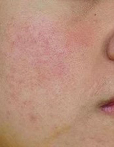 |
Figure 1 Dry erythematous patch, follicular scales and rough skin with “sandpaper-like” texture on the right cheek. |
 |
Figure 2 Dry, patchy erythema on face and upper part of the neck combined with multiple small edematous erythematous papules and minute whitish papules, size 3–5 mm, discrete on the right cheek. |
 |
Figure 3 Multiple Demodex mites are detected by standardized skin surface biopsy from the rash on the right cheek. |
 |
Figure 4 (A–C) Demodex mites are detected by superficial needle-scraping with methylene blue staining from pustular lesions on both cheeks. |
Discussion
Demodex mites are common ectoparasites in human skin. Studies have shown that the number of mites increases with age and is found to be significantly higher in people with obesity, high blood sugar levels, end-stage chronic renal failure and immunodeficiency.7–9 In addition, an abnormal increase in the number of Demodex mites has been reported following repeated facial application of topical steroids and other immunomodulators.10 Changing in facial skin microenvironment, moisture, pH levels, types and quantities of skin surface lipids and epidermal barrier function may facilitate Demodex mite proliferation.11 The Demodex mite can cause multiple skin disorders, which are grouped under the term demodicosis or demodicidosis. Some studies have shown an association between acne vulgaris and Demodex mite infestation, suggesting that when conventional treatments for acne vulgaris are ineffective, examination of Demodex mites and acaricidal therapies should be considered.2,12 This report demonstrates a case of demodicosis presenting with clinical symptoms that looked like acne vulgaris, including erythematous edematous papules and pustules on both cheeks, leading to the difficulty in differential diagnosis. Therefore, the method used to determine the mite density per square centimeter is important for diagnosing demodicosis. Standardized skin surface biopsy (SSSB) is commonly used to determine Demodex mite density from skin of the patients. Demodicosis is diagnosed when there is a high density of Demodex mites (>5 Demodex mites/cm2).13 A method for assessing Demodex mite density in papulopustular lesions, called “superficial needle-scraping” (SNS), is performed by scraping off 5 small pustules with the tip of a needle for microscopic examination. Demodex mite density was reported as “mites per 5 pustules” for SNS. Demodicosis is diagnosed when there is a high density of Demodex mites, at ≥3 Demodex mites per 5 pustules.14 In this patient, the results of microscopic examination revealed an abnormal increase in the number of Demodex mites from both SSSB and SNS methods and, combined with the relevant clinical presentations, led to the diagnosis of folliculitis-like demodicosis. Treatment with acaricides for demodicosis is expected to reduce the excessive number of Demodex mites and recover the patients from clinical symptoms. The following topical medications have been reported to be effective for demodicosis: metronidazole, permethrin, benzoyl benzoate, crotamiton, lindane, tea tree oil and ivermectin. All topical treatments should be used with caution, because of the sensitive skin in demodicosis patients, and many topical therapies have also been associated with mild irritation.15 Oral medication should be considered in patients with severe symptoms or not responding to topical therapy. Oral metronidazole has shown efficacy in reducing Demodex mite density and improving clinical symptoms of demodicosis.16 Ivermectin and fluralaner can inhibit overlapping molecular pathways regulating neuron activity, causing paralysis, lack of feeding and indirect killing effect on the Demodex mite, so they can be used to treat demodicosis without side effects to the human body.17,18 Last year, this patient had a history of pityriasis folliculorum type demodicosis and successful treatment with oral ivermectin. This time, the patient still responded well to oral ivermectin and metronidazole gel treatment despite the changes in clinical type of demodicosis from pityriasis folliculorum type to folliculitis-like type, which imitated acne vulgaris.
Conclusion
Demodicosis is not an uncommon skin disease and can show a variety of clinical symptoms that dermatologists are not familiar with, making it often misdiagnosed or underdiagnosed. This is a case report of demodicosis presenting with multiple edematous erythematous papules and pustules on both cheeks, misdiagnosed as acne vulgaris. After reassessment based on clinical presentation and laboratory examination that found multiple Demodex mites from pustules and rash on both cheeks, the patient was diagnosed with folliculitis-like demodicosis imitating acne vulgaris. Demodicosis can occur whenever there is an imbalance between microenvironment, density of Demodex mites and host immunity. Moreover, the clinical presentation of demodicosis can change from one type to another, as in this patient, changing from pityriasis folliculorum type to folliculitis-like type.
Statement of Ethics
The author states that the patient gave written informed consent for the case to be published (including publication of images). This research complies with all Ethical Guidelines for human studies in accordance with the World Medical Association Declaration of Helsinki. This paper is exempt from The Mae Fah Luang Ethics Committee on Human Research approval, with reference number COE 224/2021. Since it is a case report with no more than three cases, the report is derived from a review of medical records and cannot be linked to an individual unless the written consent of the patient is obtained.
Acknowledgments
The author would like to thank School of AntiAging and Regenerative Medicine, Mae Fah Luang University for research facilities.
Funding
This study did not receive any funding.
Disclosure
The author reports no conflicts of interest in this work.
References
1. Karincaoglu Y, Bayram N, Aycan O, Esrefoglu M. The clinical importance of Demodex folliculorum presenting with nonspecific facial signs and symptoms. J Dermatol. 2004;31(8):618–626. doi:10.1111/j.1346-8138.2004.tb00567.x
2. Aktaş Karabay E, Aksu Çerman A. Demodex folliculorum infestations in common facial dermatoses: acne vulgaris, rosacea, seborrheic dermatitis. An Bras Dermatol. 2020;95(2):187–193. doi:10.1016/j.abd.2019.08.023
3. Forton F, De Maertelaer V. Rosacea-like demodicosis and papulopustular rosacea may be two phenotypes of the same disease, and pityriasis folliculorum may be their precursor: response to the comment of Tatu. J Eur Acad Dermatol Venereol. 2019;33(1):e47–e48. doi:10.1111/jdv.15162
4. Hsu CK, Hsu MM, Lee JY. Demodicosis: a clinicopathological study. J Am Acad Dermatol. 2009;60(3):453–462. doi:10.1016/j.jaad.2008.10.058
5. Akçınar UG, Ünal E, Doğruman A. Demodex spp. as a possible aetiopathogenic factor of acne and relation with acne severity and type. Postepy Dermatol Alergol. 2018;35(2):174–181. doi:10.5114/ada.2018.75239
6. Yun CH, Yun JH, Baek JO, Roh JY, Lee JR. Demodex mite density determinations by standardized skin surface biopsy and direct microscopic examination and their relations with clinical types and distribution patterns. Ann Dermatol. 2017;29(2):137–142. doi:10.5021/ad.2017.29.2.137
7. Özer TT, Akyürek Ö, Durmaz S. Association between Demodex folliculorum and Metabolic syndrome. J Cosmet Dermatol. 2020;19(11):3145–3149. doi:10.1111/jocd.13721
8. Karincaoglu Y, Seyhan ME, Bayram N, Aycan O, Taskapan H. Incidence of Demodex folliculorum in patients with end stage chronic renal failure. Ren Fail. 2005;27(5):495–499. doi:10.1080/08860220500198037
9. Ivy SP, Mackall CL, Gore L, Gress RE, Hartley AH. Demodicidosis in childhood acute lymphoblastic leukemia: an opportunistic infection occurring with immunosuppression. J Pediatr. 1995;127(5):751–754. doi:10.1016/s0022-3476(95)70168-0
10. Fujiwara S, Okubo Y, Irisawa R, Tsuboi R. Rosaceiform dermatitis associated with topical tacrolimus treatment. J Am Acad Dermatol. 2010;62(6):1050–1052. doi:10.1016/j.jaad.2009.01.029
11. Demirdağ HG, Özcan H, Gürsoy Ş, Beker Akbulut GT. The effects of sebum configuration on Demodex spp. Density. Turk J Med Sci. 2016;46(5):1415–1421. doi:10.3906/sag-1504-77
12. Zhao YE, Hu L, Wu LP, Ma JX. A meta-analysis of association between acne vulgaris and Demodex infestation. J Zhejiang Univ Sci B. 2012;13(3):192–202. doi:10.1631/jzus.B1100285
13. Forton F, Seys B. Density of Demodex folliculorum in rosacea: a case-control study using standardized skin-surface biopsy. Br J Dermatol. 1993;128(6):650–659. doi:10.1111/j.1365-2133.1993.tb00261.x
14. Huang HP, Hsu CK, Lee JY. A new superficial needle-scraping method for assessing Demodex density in papulopustular rosacea. J Cosmet Dermatol. 2020;19(4):896–900. doi:10.1111/jocd.13082
15. Sarac G. A comparison of the efficacy and tolerability of topical agents used in facial Demodex treatment. J Cosmet Dermatol. 2019;18(6):1784–1787. doi:10.1111/jocd.12986
16. Salem DA, El-Shazly A, Nabih N, El-Bayoumy Y, Saleh S. Evaluation of the efficacy of oral ivermectin in comparison with ivermectin-metronidazole combined therapy in the treatment of ocular and skin lesions of Demodex folliculorum. Int J Infect Dis. 2013;17(5):e343–347. doi:10.1016/j.ijid.2012.11.022
17. da Rocha MC, Travassos AR, Uva L, Sequeira H, Filipe P. Demodicosis Treatment with Systemic Ivermectin. Skinmed. 2017;15(4):293–295.
18. Nakata Y, Fuse T, Yamato K, et al. A single amino acid substitution in the third transmembrane region has opposite impacts on the selectivity of the parasiticides fluralaner and ivermectin for ligand-gated chloride channels. Mol Pharmacol. 2017;92(5):546–555. doi:10.1124/mol.117.109413
 © 2022 The Author(s). This work is published and licensed by Dove Medical Press Limited. The
full terms of this license are available at https://www.dovepress.com/terms
and incorporate the Creative Commons Attribution
- Non Commercial (unported, 3.0) License.
By accessing the work you hereby accept the Terms. Non-commercial uses of the work are permitted
without any further permission from Dove Medical Press Limited, provided the work is properly
attributed. For permission for commercial use of this work, please see paragraphs 4.2 and 5 of our Terms.
© 2022 The Author(s). This work is published and licensed by Dove Medical Press Limited. The
full terms of this license are available at https://www.dovepress.com/terms
and incorporate the Creative Commons Attribution
- Non Commercial (unported, 3.0) License.
By accessing the work you hereby accept the Terms. Non-commercial uses of the work are permitted
without any further permission from Dove Medical Press Limited, provided the work is properly
attributed. For permission for commercial use of this work, please see paragraphs 4.2 and 5 of our Terms.