
Back to Journals » Infection and Drug Resistance » Volume 19
Deglycosylation at War: Host N-Glycoprotein Remodeling in Infection and Immunity
Authors Qian X, Wang J, Zhang Y, Lin Q, Ye Y, Lin J, Lu X, Yuan S, Chen L, Sun G
Received 5 March 2026
Accepted for publication 6 June 2026
Published 9 July 2026 Volume 2026:19 607119
DOI https://doi.org/10.2147/IDR.S607119
Checked for plagiarism Yes
Review by Single anonymous peer review
Peer reviewer comments 2
Editor who approved publication: Dr Oliver Planz
Xin Qian,1,* Jianjun Wang,2,* Yuxin Zhang,1 Qiange Lin,1 Yilin Ye,1 Junyou Lin,1 Xinrong Lu,3 Shuying Yuan,4 Li Chen,3 Guiqin Sun1
1School of Medical Technology and Information Engineering, Zhejiang Chinese Medical University, Hangzhou, People’s Republic of China; 2Department of Otorhinolaryngology, The Second People’s Hospital of Linhai, TaiZhou, People’s Republic of China; 3Department of Medical Microbiology and Parasitology, Fudan University, Shanghai, People’s Republic of China; 4Department of Clinical Laboratory, Jiaxing Maternity and Child Health Care Hospital, Jiaxing, People’s Republic of China
*These authors contributed equally to this work
Correspondence: Guiqin Sun, School of Medical Technology and Information Engineering, Zhejiang Chinese Medical University, Hangzhou, Zhejiang, People’s Republic of China, Email [email protected] Li Chen, Department of Medical Microbiology and Parasitology, Fudan University, Shanghai, People’s Republic of China, Email [email protected]
Abstract: N-glycan remodeling, including the hydrolysis of both glycopeptide linkages and glycosidic linkages, serves as a critical regulatory mechanism in biological processes including molecular recognition and immune responses. Growing evidence reveals that the dynamic balance between host protein glycosylation and N-glycan remodeling significantly influences the outcomes of infectious diseases. During infection, host N-glycoproteins remodeling can be driven either by the host as a defense strategy or by pathogens as an offensive mechanism. This review systematically examines how both host- and pathogen-induced N-glycan remodelling events affect infection outcomes. We highlight the dual functionality: host-driven remodeling often enhances immune recognition and pathogen clearance, whereas pathogen-driven remodeling facilitates adhesion, immune evasion, and nutrient acquisition. By integrating evidence from direct enzymatic N-glycan remodeling and indirect glycan-processing models, this review offers a novel perspective on glycan-mediated microbial-host interactions, highlighting how targeted modulation of these processes may advance therapeutic strategies against infectious diseases.
Keywords: deglycosylation, glycan, glycosidase, microbial-host interactions, infectious disease, immunity
Introduction
In eukaryotes, glycosylation modification represents one of the most common post-translational modifications, maintaining multicellular coordination and participates in molecular recognition, differentiation, development, signal transduction, and immune response.1–3 The reverse process, deglycosylation, which is mediated by specific glycosidases through cleavage of surface glycans, provides equally dynamic regulation. Recent advances in studies of microbial-host interaction have particularly highlighted the critical role of host N-glycoprotein deglycosylation in infection and immunity regulation.
Glycan modifications on membrane-anchored proteins and lipids serve as essential structural mediators of microbial-host interactions.4 A paradigmatic example of symbiotic glycan exploitation is observed in infant gut ecosystems: human milk oligosaccharides resist infant digestion but are selectively metabolized by Bifidobacterium longum subsp. infantis through dedicated gene clusters, while bacterial-derived short-chain fatty acids in turn promote host intestinal development.5 Glycosylation of pathogens is equally critical for their infection ability, but shifts from mutualistic to adversarial roles under pathogenic conditions.6–8 Under pathogenic conditions, this normally symbiotic relationship transforms into an adversarial dynamic where glycans become molecular weapons. Host employs glycan-recognition systems to detect microbial threats, while pathogens evolve countermeasures to subvert these defenses.9 For instance, Corynebacterium diphtheriae secretes an IgG-specific Endo-beta-N-acetylglucosaminidase (ENGase) that selectively disrupts Fcγ-mediated effector functions, actively cleaving host N-glycans to evade immunity.10 However, some commensal bacteria like Staphylococcus epidermidis require intact host surface glycans for adhesion, illustrating how the N-glycans can serve as either anchors for mutualism or targets for subversion.11 These examples collectively demonstrate how glycans serve as both bridges and battlefields in microbial-host interactions, shaping outcomes from mutualism to pathogenesis.
Notably, glycan remodeling of host N-glycoproteins occurs via two distinct mechanisms: cleavage of glycopeptide bonds (complete N-deglycosylation) and cleavage of glycosidic bonds. In these processes, glycosidases are key weapons used by both host and microbes in this glycan-remodeling battle.12 This enzymatic arms race is exploited by both sides to tilt the balance of infection. Pathogens manipulate the glycans of the host cell by secreting microbial glycosidases or interfering with the host’s glycosidases, thereby affecting critical steps such as adhesion, colonization, proliferation, toxin activity and thus influencing the progression of the infection. Concurrently, host immune responses are dynamically regulated through glycan-remodeling events, which can either enhance antimicrobial defenses or be exploited by pathogens for immune evasion.
This review systematically examines the dual roles of host cell N-glycan remodeling in infection and immunity, aiming to elucidate the molecular mechanisms by which pathogens hijack host deglycosylation process, analyze how deglycosylation dynamically modulates both protective and pathological immune responses, and explore the therapeutic potential of targeting these processes for infectious disease intervention. By integrating these perspectives, we provide a comprehensive framework for understanding deglycosylation as a critical interface in microbial-host interactions.
N‑Glycan Remodeling by Glycosidases
N‑glycan remodeling includes enzymatic cleavage of glycopeptide bonds (complete N‑deglycosylation) and cleavage of glycosidic bonds. This process is carried out by cleaving N-glycans from various glycosylated compounds, including monosaccharides, oligosaccharides, polysaccharides, saponins, and glycoproteins, resulting in the formation of monosaccharides, oligosaccharides or glycocomplexes.13,14 It occurs in a wide range of species, including humans, plants, and bacteria. The removal of glycan chains by deglycosylation regulates the function of conjugated proteins and influences various immune responses, such as receptor recognition, signal transduction, and the effects of immune molecules.5,15
Glycoside hydrolases (GH) are key enzymes involved in glycoprotein deglycosylation in almost all organisms that can hydrolyze various glycosidic bonds or glycosidic oligosaccharides.16,17 Glycosidases vary by species: for example, arabinosidase, xylanase, and thioglucosidase are found in plants; rhamnosidase and glucosidase in bacteria, and mannosidase in mammals.14 Each species produces specific glycosidases suited to its metabolic needs. Crucially, the subcellular localization of these glycosidases determines their accessibility to substrates and thus their functional impact on infection and immunity.18 For instance, bacterial endoglycosidases such as the IgG‑specific ENGase from Corynebacterium diphtheriae are secreted or surface‑associated, allowing them to directly engage host immunoglobulins in the extracellular space.10 Viral neuraminidases are embedded in the viral envelope, facilitating virion release and spread.19 Whereas host deglycosylating enzymes are tightly compartmentalized: NGLY1 acts in the cytosol, neuraminidase 1 (NEU1) resides in lysosomes and at the plasma membrane, and ER‑resident glucosidases participate in glycoprotein quality control.18–22 This compartmentalization dictates when and where a deglycosylation event occurs, profoundly shaping the outcome of microbial-host interactions.
In particular, certain glycosidases are directly involved in infection and immune responses. Based on cleavage specificities, glycosidases acting on host N‑glycans can be broadly classified into several functional groups. Examples include enzymes that remove entire N-glycans (eg, PNGase F), those that trim terminal sugars (eg, neuraminidases), and those that cleave internal glycosidic bonds such as the core chitobiose bond (eg, Endo H). Additional examples are provided in Table 1 and the enzymatic cleavage schematic are presented in Figure 1.
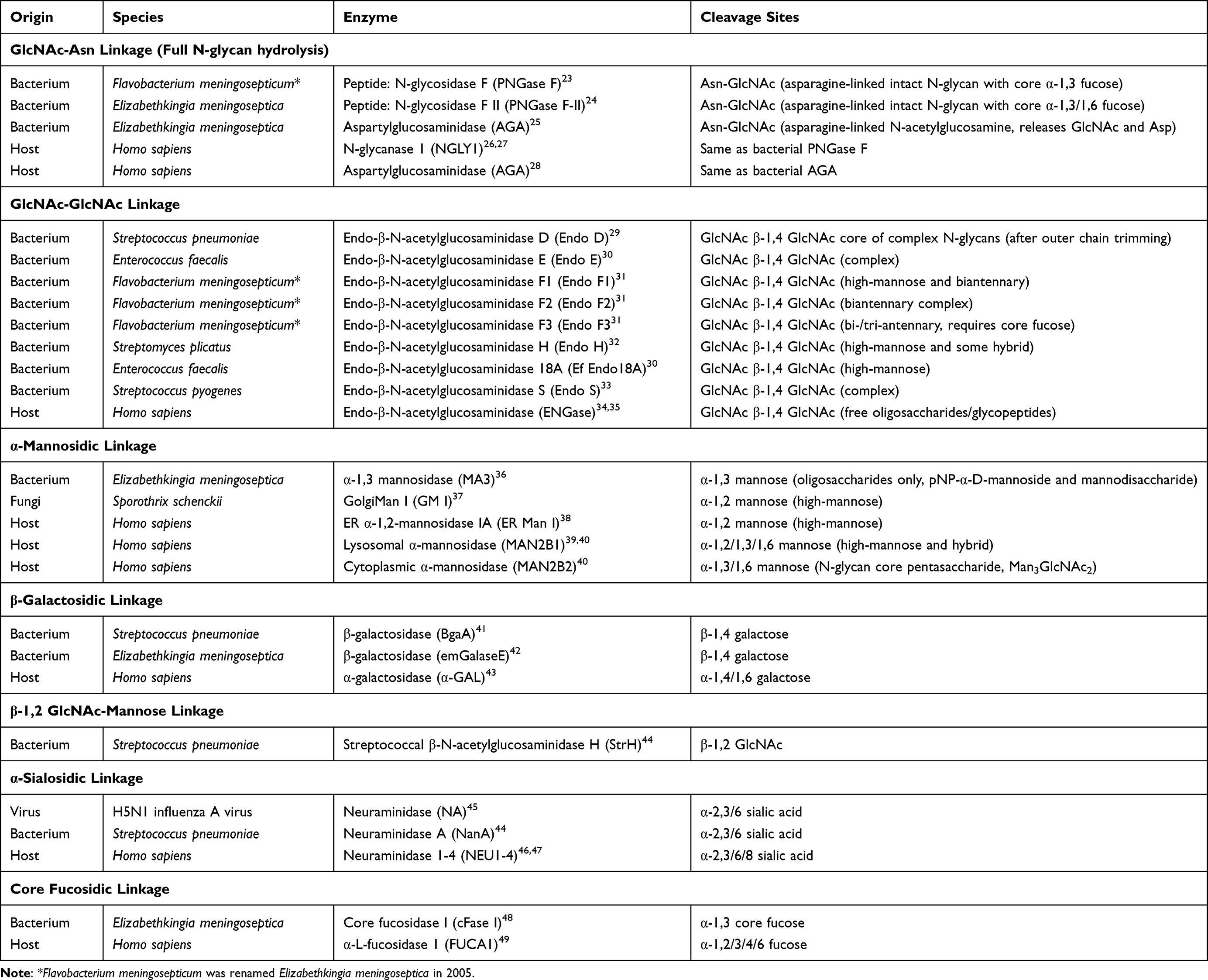 |
Table 1 Glycosidases Involved in the Hydrolysis of Host N-Glycoproteins |
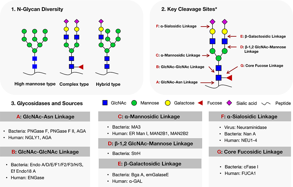 |
Figure 1 N‑glycan types, cleavage linkages, and infection‑related glycosidases. *The various types of N-glycans depicted are simplified schematics and may not fully reflect the physiological glycan structures. Core fucosylation is not present on all N-glycans; the diagram illustrates a fucosylated form for clarity. The depicted complex-type N-glycan serves solely to indicate the cleavage positions targeted by the indicated enzymes. |
Host Cell N‑Glycan Remodeling Modulates Pathogen Infection Efficiency
Pathogen infection proceeds through multiple stages, including recognition, adhesion, colonization, proliferation, and release. N-glycans in host cell surface critically modulate various normal and pathological processes between host cells, as well as between host cells and pathogens.50,51 During infection, specific N-glycans sequences on host receptor cells can serve as targets for pathogen recognition and exploitation, while also providing an energy source and facilitating the maintenance of infection. Therefore, remodeling of these N-glycans on host cells can alter the progression of infection. This section discusses how host cell N‑glycan remodeling modulates pathogen infection efficiency.
Microbial-Host Interface: N-Glycan Remodeling in Recognition, Adhesion and Colonization
Adhesion and colonization are crucial initial steps for most pathogens to successfully invade the host and establish persistent infections.52,53
Host-Mediated N-Glycan Remodeling Blocks Pathogen Adhesion and Colonization
N-glycan chains on host cells are key structures that enable pathogen adhesion and invasion.5,54 Studies have shown that host cell deglycosylation prevents viral infection by inhibiting viral recognition and binding to host cells. Zhao et al reported that in MLE‑12 and A549 cells infected with H1N1 swine influenza virus, the expression of host NEU1 was upregulated as a virus‑induced host response, leading to removal of sialic acid residues from the cell membrane and potentially reducing viral entry receptor availability.55 Bertoldi et al conducted a cross‑sectional clinical study comparing patients with Gitelman/Bartter syndrome (GS/BS) and healthy controls, finding that GS/BS patients with remarkably low SARS‑CoV‑2 susceptibility had higher levels of non‑glycosylated angiotensin-converting enzyme 2 (ACE2), suggesting an association between ACE2 deglycosylation and reduced infection risk.56 However, the study did not determine whether this increase resulted from impaired glycosylation or enhanced deglycosylation, leaving the mechanistic direction unresolved. Together, these examples indicate that reduced N‑glycans on host cell surfaces, whether mediated by upregulated host glycosidases or associated with naturally increased non‑glycosylated receptor forms, may interrupt pathogen adhesion and colonization, representing an intrinsic barrier that reduces the availability of entry receptors. However, the latter observation remains correlative and requires further experimental validation to establish causality and distinguish between impaired glycosylation versus enhanced deglycosylation.
Pathogen-Exploited N-Glycan Remodeling Promotes Host Recognition and Adhesion
Notably, pathogens employ a counter-strategy. They secrete glycosidases or modulate host glycosidases to alter the N-glycan structures on host cell surfaces, thereby strengthening their interaction with the host. Sialidases are used by influenza virus to cleave α2-3 sialic acids in mucus, promoting virus particle recognition and entry into epithelial cells.57 Host NEU1 is mobilized by Pseudomonas aeruginosa to remove sialic acids from the extremities of Mucin1 receptors, thereby enhancing the binding of bacterial flagellin to the receptors and facilitating bacterial adhesion and invasion.58 Furthermore, a sequential action of neuraminidase A (NanA), β-galactosidase A (BgaA), and Streptococcal β-N-acetylglucosaminidase H (StrH) secreted by Streptococcus pneumoniae deglycosylates host glycoproteins, thus enhancing bacterial adhesion and colonization in the host.59
Furthermore, it has been demonstrated that the removal of entire N-glycan chains from host cell surfaces using Peptide: N-glycosidase F (PNGase F) significantly reduces the adhesion and colonization capabilities of pathogens such as Staphylococcus aureus, Staphylococcus epidermidis, Salmonella typhimurium, Seneca Valley virus, Dengue virus.60–64
By expressing aberrant glycosidases, pathogens remove terminal sialic acids from host glycoproteins, thereby exposing previously hidden glycans. These pathogen‑induced N-glycan remodeling events subvert the host’s glycan‑based defensive barrier, directly promoting bacterial adhesion and colonization. Complete removal of entire N‑glycan chains from host cells’ surfaces (eg, by PNGase F) has also been linked to infection outcomes in vitro, though such treatments are experimental. Whether endogenous glycosidases achieve a similar effect during natural infection remains to be determined. Nevertheless, together with desialylation, these deglycosylation events are closely associated with bacterial adhesion and colonization.
Host Cell N-Glycan Remodeling Affects Pathogen Infection Status
Host N-Glycan Remodeling Attenuates Bacterial Toxin Binding and Cytotoxicity
N-glycans on host cell surface often serve as attachment sites for bacterial toxins. Therefore, host‑mediated deglycosylation could theoretically reduce toxin binding and subsequent cytotoxicity. Direct experimental evidence for this concept comes from in vitro enzymatic treatment models that mimic host deglycosylation. Specifically, Rahman et al demonstrated that deglycosylation on host cells using neuraminidase, PNGase F, and Endo-α-N-acetylgalactosaminidase significantly reduced the binding of Kingella kingae RtxA toxin to target cells, inhibiting toxin entry and thus decreasing target cell damage and death.65 This finding provides proof‑of‑concept that deglycosylation can attenuate toxin action, supporting the plausibility of host deglycosylation as a defense mechanism. However, it remains to be determined whether host cells naturally upregulate such deglycosylation activities during infection to achieve toxin resistance, as the current study employed exogenous glycosidases under non‑physiological conditions.
Pathogen-Exploited N-Glycan Remodeling on Host Cells Augments Viral Virulence and Replication
Deglycosylation induced by pathogens through the secretion of glycosidases during infection is crucial for their virulence and destructive capabilities.66 A direct example of pathogen‑driven deglycosylation contributing to virulence found by Ju et al is that the neuraminidase (NA) secreted by H5N1 influenza virus mediates deglycosylation of lysosome-associated membrane proteins (LAMP), destabilizing LAMP and inducing lysosomal rupture, ultimately leading to tracheal epithelial cell death.45 During influenza virus infection, the encoded NEU removes terminal N-glycan from the host cell surface, facilitating viral particle release and enhancing the virus’s spread.67 This process increases cytotoxicity, disrupts cellular homeostasis, and ultimately leads to host cell death.
During infection, pathogen-induced N-glycan remodeling may alters virus’s ability to sustain infection in the host, affecting its replication, assembly, and other processes. St Clair et al reported that in Huh7 cells, infection with dengue virus serotype 2 (DENV2) leads to increased expression and activity of human sialidases NEU1-4, as determined by enzymatic activity assays, which is crucial for DENV2 replication and release.68 Fibroblasts, the natural host cells for human cytomegalovirus (HCMV), exhibit a significant reduction in HCMV DNA following in vitro deglycosylation treatment, resulting in marked inhibition of HCMV replication and infection.69 Additionally, when host deglycosylation is inhibited, viral replication can be suppressed. For example, the glycosidase inhibitor N-butyl-deoxynojirimycin strongly inhibits envelope formation of hepatitis B virus and assembly and secretion of Ebola virus.70,71 Similarly, the α-glucosidase inhibitor, castanospermine, inhibits the assembly of measles virus and the replication of hepatitis C virus.72
Pathogen-induced N-glycan remodeling presents a key mechanism that enhances viral virulence by targeting host N-glycan structures, destabilizing critical proteins, and facilitating viral release and replication. Such sequential disruption not only amplifies cytotoxicity but also alters infection dynamics, promoting viral spread and immune evasion. Notably, inhibiting deglycosylation has shown potential as a therapeutic strategy to suppress viral replication and disrupt pathogen life cycles, offering a promising approach for antiviral intervention. Of note, while pathogen‑secreted glycosidases offer direct mechanistic evidence, many findings on N-glycan remodeling induced by both host and pathogen remain at the in vitro experimental level, warranting cautious interpretation.
Pathogen-Derived Glycosidases Liberate Host Glycans as Nutritional Sources for Proliferation
Upon entering the host, hydrolyzing host polysaccharides or oligosaccharides, which facilitates the acquisition of energy and nutrients, as well as pathogen proliferation, is a critical mechanism for pathogen survival and infection.
The survival of bacteria within the host partly depends on their capacity to utilize host metabolites, such as products derived from deglycosylated glycoprotein.73 Bacteria have evolved diverse mechanisms to induce host protein N-glycan remodeling, including direct induction, secretion of glycosidases, and utilization of starch utilization systems (SUS). Studies have demonstrated that Francisella tularensis infection of macrophages triggers the N-glycan remodeling of the glutamine transporter SLC1A5, with the resulting free oligosaccharides serving as a factor affecting its intracellular survival.5,73 Keffeler et al found that under glucose-limited conditions, the growth of Enterococcus faecalis was supported by the polysaccharides derived from the cleavage of the chitobiose core linkage of host high-mannose glycoproteins by the glycosidase EfEndo18A upregulated by E. faecalis.30 Similarly, Oral streptococci, Streptococcus pneumoniae, and Actinomyces odontolyticus have been reported to utilize their own glycosidases to deglycosylate host glycoproteins.59,74,75 Furthermore, a unique SUS was utilized by Bacteroides fragilis to deglycosylate host glycoproteins, with the complex N-glycans serving as the sole energy source for its growth.76
This nutrient-acquisition strategy supports bacterial proliferation within the host, indirectly contributing to infection persistence and severity.
Host N-Glycan Remodeling in Immune Recognition and Evasion
The microbial-host interaction represents a pivotal axis in infection dynamics, requiring host to counter pathogen invasion, persistent infection, and immune evasion strategies. The host immune defense system comprises innate and adaptive immunity, in which glycan chains on immune cells and molecules mediate pattern recognition, pathogen opsonization, and toxin neutralization.77 Consequently, host cell N-glycan remodeling carries significant immunological implications, substantially shaping immune response efficacy and ultimately determining infection outcomes.77,78
Host-Mediated N-Glycan Remodeling Activates Immunity Against Infection
Host cell N-glycan remodeling serves as a crucial regulatory mechanism in microbial-host interactions, playing a pivotal role in immunity. Through modulation of glycosidase expression, host can alter glycosylation patterns on immune cells, effector molecules, and receptors, thereby enhancing pathogen recognition and clearance.79 Ackerman et al demonstrated that HIV infection induces B cell glycosylation reprogramming, leading to increased Fc agalactosylation on HIV-specific antibodies. This modification enhances antibody binding to FCGR3A (as validated by galactosidase treatment), which in turn drives stronger antibody-dependent cell-mediated viral inhibition (ADCVI) and antibody-dependent cellular cytotoxicity (ADCC), particularly in elite controllers who display the highest levels of agalactosylated glycans.80 Supported by two evidence tiers, namely observational correlation in HIV elite controllers and causal validation through in vitro enzymatic deglycosylation with galactosidase treatment, this case showed degalactosylation directly augments antiviral immunity.
Furthermore, hosts actively reshape IgG glycosylation patterns during latent tuberculosis (TB) infection, producing afucosylated antibodies that optimize FcγRIIIa engagement and NK cell-mediated immunity. This specific removal of core fucose from the IgG Fc N‑glycan potentiates host immunity through augmented ADCC and phagocytosis, contributing to Mycobacterium tuberculosis control and immunological memory maintenance.81 In contrast, Active TB patients exhibit elevated levels of agalactosylated IgG, which impairs host defenses by diminishing phagocytic capacity and bactericidal activity against M. tuberculosis. These findings collectively demonstrate that host-specific antibody deglycosylation patterns serve as a double-edged sword in anti-infective immunity: afucosylation enhances FcγRIIIa-mediated effector functions, whereas agalactosylation may paradoxically compromise microbial clearance. This dichotomy is mechanistically supported by in vitro evidence from Van Coillie et al, who used defined glycoengineered IgG and FcγRIIIa variants in SPR binding assays to establish a direct causal link between specific deglycosylation events and altered receptor affinity, which translates into differential NK cell activation and infection outcomes.
Multiple studies have demonstrated the critical role of host glycosidases in anti-infection immunity, with direct evidence linking deglycosylation to enhanced antiviral responses.78 Specifically, patients with congenital disorders of deglycosylation (CDDG) caused by N-glycanase 1 (NGLY1) deficiency not only exhibit significantly reduced viral infection rates but also show persistently elevated expression of interferon-stimulated genes in peripheral blood mononuclear cells, indicating that deglycosylation potentiates type I interferon signaling to boost antiviral immunity.82 In Ngly1-knockout mouse models, embryonic fibroblasts display decreased susceptibility to vesicular stomatitis virus infection accompanied by significantly increased virus-induced IFN-β production, confirming that NGLY1 deficiency suppresses viral replication through activation of innate immune signaling pathways.83 Furthermore, in human rhabdomyosarcoma cells, Ngly1 knockdown not only inhibits Coxsackievirus A16 and Enterovirus 71 replication but also enhances RIG-I-like receptor-mediated viral pattern recognition, further demonstrating that deglycosylation exerts anti-infective effects by modulating innate immune sensor function.84 Thus, deglycosylation of NGLY1 directly links to augmented antiviral immunity across multiple evidence levels—from patients to mice to cells—by unleashing type I interferon and RIG‑I‑like receptor pathways.
These findings collectively reveal that N-glycan remodeling of host cells establishes effective anti-infection defense through multiple immune mechanisms including type I interferon pathway activation and Fc‑mediated effector optimization, yet the strength of causal inference varies markedly: the NGLY1 studies offer multi‑level evidence (human, mouse, cell), whereas the HIV and TB conclusions are primarily drawn from correlative patient data and in vitro glycoengineering experiments, lacking direct in vivo proof that naturally occurring deglycosylation events alone drive the observed protection or pathology. Future efforts should prioritize optimizing evidence hierarchies and leveraging robust patient cohort studies to establish causality and assess the therapeutic potential of modulating specific deglycosylation pathways in infectious diseases.
Pathogen Subversion of Host N-Glycan Remodeling Promotes Immune Evasion in the Infection
Pathogens evade immunity via two distinct deglycosylation strategies: deglycosylating host immunoglobulins to disrupt defenses, or hijacking host glycosidases to enable immune escape and infection. Specifically, pathogens manipulate host immunity through enzymatic removal of N-glycans from immunoglobulins and other immune molecules, thereby subverting antimicrobial defenses and compromising immune barriers to facilitate infection.85 Mechanistic evidence shows that the single-domain ENGase secreted by Corynebacterium diphtheriae specifically hydrolyzes the Asn297 glycan of IgG antibodies, directly blocking FcγR-mediated effector functions while preserving neutralization activity.86 Enterococcus faecalis-derived Endo-β-N- acetylglucosaminidase E (Endo E) removes glycans from lactoferrin, RNase B, and IgG, impairing lactoferrin’s antibiofilm activity while reducing IgG-Fc receptor binding and classical complement activation.87 Both bacterial sialidases (eg, from Actinomyces) and fungal α-mannosidases (eg, from Candida albicans) target host IgA: the former desialylates IgA to enhance its proteolytic degradation, while the latter cleaves IgA glycans to block FcγR binding, collectively compromising IgA-mediated mucosal immunity.51,88 Pathogens may also hijack host glycosidases: Leishmania upregulates host N-glycanase to deglycosylate and degrade ATPase Copper Transporting Alpha (ATP7A), evading copper homeostasis-mediated antimicrobial effects, while bacterial suppression of host β-galactosidase 1 attenuates pattern-triggered immunity (PTI) pathway-dependent defenses.89,90 Thus, N-glycan remodeling serves as a convergent immune evasion mechanism across diverse pathogens, with direct enzymatic evidence linking N-glycan removal to blockade of FcγR, complement, and mucosal immunity, as well as to subversion of intracellular antimicrobial pathways.
To further dissect the causal role of N-glycan remodeling in immune suppression, in vitro studies using purified glycosidases have systematically evaluated the functional consequences of removing glycans from host immune molecules. In vitro studies simulating pathogen-induced host deglycosylation have revealed suppressed innate immune responses, including impaired phagocytic activity. For instance, pulmonary surfactant protein A, which normally enhances viral phagocytosis and clearance by alveolar macrophages, exhibits reduced herpes simplex virus recognition and binding after PNGase F treatment, consequently diminishing its ability to promote macrophage phagocytosis and cytokine secretion, thereby suppressing host immune responses.91 Zhao et al demonstrated that deglycosylation of bactericidal myeloperoxidase by PNGase F, α-neuraminidase, and α1,6-fucosidase decreases its oxidative and antimicrobial activities, potentially predisposing the host to more severe infections and inflammation.92 Research indicates that glycosylation of adaptive immune effector molecules is crucial for immunological functions, and deglycosylation may lead to impaired pattern recognition and compromised opsonization.77 Raskova et al found that while deglycosylated sIgA maintains in vitro binding capacity to Escherichia coli O55, it loses protective activity.93 Koike et al reported that rabies vaccine-induced glycoform-specific IgG1 exhibits potent neutralizing activity in vitro, whereas its deglycosylated counterpart loses both lectin-binding capacity and functional activity.94 Mahant et al demonstrated that reduced Fc glycan maturity in bacterial vaginosis patients’ IgG correlated with diminished herpes simplex virus (HSV) neutralization, suggesting anaerobic bacterial glycosidases may impair IgG-HSV glycoprotein E interactions.95 Additionally, studies silencing glycosidase-encoding genes in fungi have observed attenuated host immune responses, providing indirect genetic evidence for the critical role of glycosidases in host-fungal interactions and virulence.88
Host N‑glycan remodeling undeniably modulates immune responses, yet the field is less coherent than often assumed. Well‑supported mechanisms such as pathogen‑derived ENGase, Endo E, and sialidases contrast sharply with emerging and less‑validated hypotheses like pathogen hijacking of host glycosidases. Moreover, contradictory observations abound because deglycosylation can either boost immunity as seen in NGLY1 deficiency or blunt it as demonstrated by IgG agalactosylation in active tuberculosis, indicating that the outcome is highly context dependent. Methodological limitations further complicate generalization, including the use of non‑physiological enzyme doses in vitro, inherent heterogeneity of glycan structures, and a lack of cell‑type‑specific genetic models. Recognizing these uncertainties and discordances is essential for the field to progress beyond a deceptively unified narrative.
Summary
N-glycan remodeling of host N-glycoproteins has become a common defense strategy in the microbial-host survival competition, which is a double edged sword in infection and immunity: host mediated remodeling is predominantly protective, whereas pathogen induced remodeling is predominantly pathogenic. Across the mechanisms discussed in this review, host‑driven N-glycan remodeling can block pathogen adhesion (eg, NEU1‑mediated sialic acid removal), attenuate bacterial toxin binding, and enhance immune effector functions such as opsonization, ADCC, and phagocytosis. In contrast, pathogen‑driven N-glycan remodeling promotes microbial adhesion and colonization, augments viral replication and release, liberates host N-glycans as nutrients, and subverts antibody‑mediated immunity. Based on this, we propose that N-glycan remodeling of host N-glycoproteins, shaped by microbial-host interactions, plays a crucial role in disease progression and immune regulation (Figure 2).
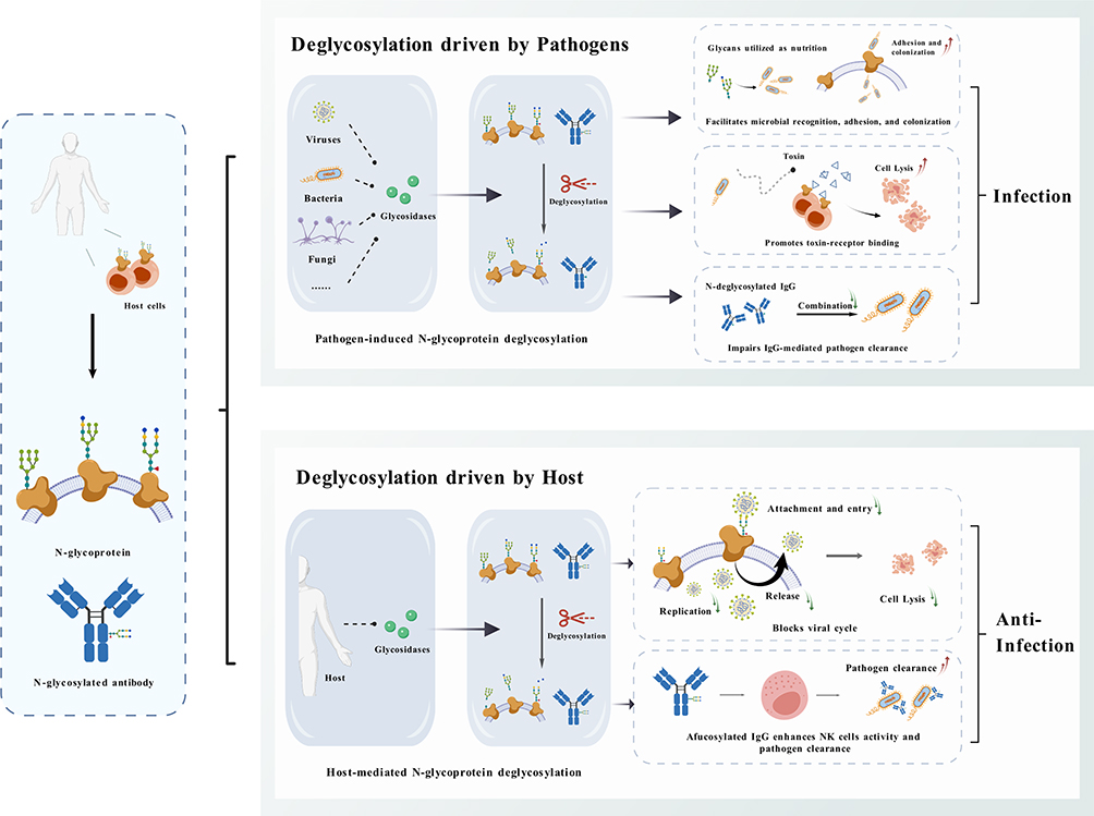 |
Figure 2 The schematic diagram of host N-glycoprotein deglycosylation in the microbial-host interaction process. Created with BioGDP.com.96 |
Outlook
Pathogen infection outcomes are critically governed by the opposing forces of host N-glycoprotein glycosylation and deglycosylation, a regulatory axis that orchestrates microbial-host interactions at the molecular level.
From the pathogen perspective, many infectious agents have evolved glycosidases to directly deglycosylate host glycoproteins or manipulate host’s glycosidases, thereby subverting immune recognition and promoting infection. Beyond well recognized bacterial and viral examples, the fungus Aspergillus fumigatus utilizes its glycosylasparaginase AspA to cleave N-glycosidic bonds (GlcNAc-Asn) of host glycoproteins, thus disrupting macrophage-mediated immune recognition and inflammatory responses to facilitate fungal immune evasion.97 Glycosidase-targeting inhibitors are emerging as promising therapeutic candidates against multiple viruses including SARS-CoV-2, hepatitis B virus, and measles virus.98,99 This finding suggests that targeting pathogen-derived glycosidases may represent a novel therapeutic strategy against infectious diseases, a concept already validated by influenza neuraminidase inhibitors.
In host systems, while N-glycoprotein deglycosylation has been well characterized as a central regulator of infection, accumulating evidence implicates O-glycoprotein deglycosylation as a comparably important player. For instance, Entamoeba histolytica induces contact-dependent O-glycoprotein deglycosylation in HepG2 cells, disrupting the balance of O-GlcNAc modification and interfering with homeostatic intracellular signaling molecules, ultimately leading to HepG2 cell death.100 Furthermore, glycolipids constitute essential membrane components involved in cell recognition and signal transduction, and their glycosylation patterns are increasingly recognized as key determinants in infection and immunity.101–103 Although existing evidence links these glycoconjugates to infection and immunity, the current understanding of O‑glycoprotein deglycosylation during infection remains limited, and the potential relationship between alterations in surface glycolipid glycosylation patterns and infectious processes has received little attention. Future systematic studies should therefore focus on fungal pathogens and broaden the scope to include O-glycoproteins and glycolipids, which may reveal previously unrecognized deglycosylation mechanisms in microbial-host interactions.
Beyond expanding the substrate repertoire, the field faces translational challenges. Most current evidence remains in vitro or correlative, with limited in vivo validation of physiological deglycosylation, and many studies fail to distinguish between impaired glycosylation and enhanced glycan removal. Consequently, their explanatory power for the “arms race” of deglycosylation in infection immunity is inherently limited. To address this gap, future efforts should establish physiologically relevant animal models that recapitulate infection‑driven deglycosylation, and, crucially, determine whether both host and pathogens actively manipulate deglycosylation to tilt the immune balance in their favor. Unraveling this bidirectional regulation will be essential for translating deglycosylation biology into effective therapeutic strategies that break the infection-immunity stalemate.
Moreover, although deglycosylation modulates infection and immune outcomes, the translational potential of targeting glycosidases remains largely unexplored. Two parallel therapeutic strategies are recommended. One is to target pathogen‑derived glycosidases, which has already proven successful for influenza virus neuraminidase (clinically approved inhibitors include oseltamivir, zanamivir, and peramivir). Expanding this concept to bacterial endoglycosidases such as Streptococcus pyogenes Endo S or Streptomyces plicatus Endo H represents a promising but underexplored direction. The other is to modulate host glycosidases. Host NEU1 inhibition reduces viral entry and bacterial adhesion, while NGLY1 inhibition enhances type I interferon responses.45,83 Experimental inhibitors exist, including C9‑BFA for NEU1 and compound 19 for NGLY1, but none have entered clinical trials for infectious diseases.26,104 Ultimately, translating current mechanistic insights into clinical applications will require exploring host glycosidase modulators and glycomics‑based biomarkers for patient stratification. Addressing these gaps will be essential to move from descriptive biology toward tangible benefits for infectious disease management.
In general, N-glycan remodeling in the host serves as a pivotal modulator of infection outcomes. With the advancement of glycobiology and glycomics, in-depth studies on the mechanism of host N-glycan remodeling in infection and immune regulation are expected to provide crucial scientific foundations and new strategies for the development of anti-infective drugs and novel vaccines. Unraveling the remaining mysteries of this glycan-based battlefield promises to refine our fundamental understanding of infectious diseases.
Abbreviations
ACE2, angiotensin-converting enzyme 2; ADCC, antibody-dependent cellular cytotoxicity; ADCVI, antibody-dependent cell-mediated viral inhibition; AGA, aspartylglucosaminidase; Asp, aspartic acid; Asn, asparagine; ATP7A, ATPase copper transporting alpha; BgaA, β-galactosidase A; CDDG, congenital disorders of deglycosylation; cFase I, core fucosidase I; DENV2, dengue virus serotype 2; Endo E, endo-β-N-acetylglucosaminidase E; ENGase, endo-β-N-acetylglucosaminidase; ER, endoplasmic reticulum; ERAD, ER-associated degradation; ERMan I, ER α-1,2-mannosidase I; FcR, Fc receptor; FUCA1, α-L-fucosidase 1; GH, glycoside hydrolase; GM I, Golgi mannosidase I; gpcXase I, glycoprotein core xylosidase I; H1N1, swine-origin influenza virus subtype H1N1; HCMV, human cytomegalovirus; HBV, hepatitis B virus; HIV, human immunodeficiency virus; HSV, herpes simplex virus; IFN-β, interferon beta; IgG, immunoglobulin G; LAMP, lysosome-associated membrane proteins; MA3, α-1,3 mannosidase; MAN2B1, lysosomal α-mannosidase; MAN2B2, cytoplasmic α-mannosidase; MANBA, β-mannosidase; MUC1, mucin 1; NA, neuraminidase; NanA, neuraminidase A; NEU1-4, neuraminidase 1-4; NGLY1, N-glycanase 1; PNGase F, peptide: N-glycosidase F; PTI, pattern-triggered immunity; RIG-I, retinoic acid-inducible gene I; SARS-CoV-2, severe acute respiratory syndrome coronavirus 2; sIgA, secretory immunoglobulin A; SLC1A5, solute carrier family 1 member 5; StrH, streptococcal β-N-acetylglucosaminidase H; SUS, starch utilization system; TB, tuberculosis; α-GAL, α-galactosidase.
Acknowledgments
We thank all the participants of this study for their support.
Funding
This work was supported by the Medical Health Science and Technology Project of Zhejiang Provincial Health Commission (No. 2024KY450), the Cooperation Project of Linhai Second People’s Hospital and Zhejiang Chinese Medical University (No. 2023-HT-1266).
Disclosure
The authors report no conflicts of interest in this work.
References
1. Arnold JN, Wormald MR, Sim RB, et al. The impact of glycosylation on the biological function and structure of human immunoglobulins. Annu Rev Immunol. 2007;25:21–14. doi:10.1146/annurev.immunol.25.022106.141702
2. Munkley J. Aberrant sialylation in cancer: therapeutic opportunities. Cancers. 2022;14:4248. doi:10.3390/cancers14174248
3. Barel M, Charbit A. Role of glycosylation/deglycolysation processes in francisella tularensis pathogenesis. Front Cell Infect Microbiol. 2017;7:71. doi:10.3389/fcimb.2017.00071
4. Hooper LV, Gordon JI. Glycans as legislators of host-microbial interactions: spanning the spectrum from symbiosis to pathogenicity. Glycobiology. 2001;11:1R–10R. doi:10.1093/glycob/11.2.1R
5. Sela DA, Chapman J, Adeuya A, et al. The genome sequence of Bifidobacterium longum subsp. infantis reveals adaptations for milk utilization within the infant microbiome. PNAS. 2008;105:18964. doi:10.1073/pnas.0809584105
6. Gorzkiewicz M, Cramer J, Xu HC, et al. The role of glycosylation patterns of viral glycoproteins and cell entry receptors in arenavirus infection. Biomed Pharmacother. 2023;166:115196. doi:10.1016/j.biopha.2023.115196
7. Rowland RR, Brandariz-Nuñez A. Role of N-linked glycosylation in porcine reproductive and respiratory syndrome virus (PRRSV) infection. J Gen Virol. 2024;105:001994. doi:10.1099/jgv.0.001994
8. Kern K, Delaroque N, Boysen A, et al. Glycosylation of bacterial antigens changes epitope patterns. Front Immunol. 2023;14:1258136. doi:10.3389/fimmu.2023.1258136
9. Tytgat HL, de Vos WM. Sugar coating the envelope: glycoconjugates for microbe-host crosstalk. Trends Microbiol. 2016;24:853–861. doi:10.1016/j.tim.2016.06.004
10. Sastre DE, Bournazos S, Du J, et al. Potent efficacy of an IgG-specific endoglycosidase against IgG-mediated pathologies. Cell. 2024;187:6994–7007. doi:10.1016/j.cell.2024.09.038
11. Roy P, Horswill AR, Fey PD. Glycan-dependent corneocyte adherence of Staphylococcus epidermidis mediated by the lectin subdomain of Aap. MBio. 2021;12:e0290820.
12. Engibarov S, Gocheva Y, Lazarkevich I, et al. Bacterial Sialidases: biological significance and application. Appl Biosci. 2025;4:17. doi:10.3390/applbiosci4020017
13. Hou B, Lim EK, Higgins GS, et al. N-glucosylation of cytokinins by glycosyltransferases of Arabidopsis thaliana. J Bio Chem. 2004;279:47822–47832. doi:10.1074/jbc.M409569200
14. Cerqueira N, Brás N, Ramos MJ, et al. Glycosidases–a mechanistic overview. In: Carbohydrates: Comprehensive Studies on Glycobiology and Glycotechnology. IntechOpen; 2012. doi:10.5772/52019
15. Lin B, Qing X, Liao J, et al. Role of protein glycosylation in host-pathogen interaction. Cells. 2020;9:1022. doi:10.3390/cells9041022
16. Sala K, Pengthaisong S, Beagbandee C, et al. Expression and characterization of a rice β-Xylosidase with Xylooligosaccharide hydrolysis and transglycosylation activities. J Agric Food Chem. 2025;73:10418–10429. doi:10.1021/acs.jafc.4c13281
17. Raba G, Luis AS. Mucin utilization by gut microbiota: recent advances on characterization of key enzymes. Essays Biochem. 2023;67:345–353. doi:10.1042/EBC20220121
18. Miyagi T, Yamaguchi K. Mammalian sialidases: physiological and pathological roles in cellular functions. Glycobiology. 2012;22:880–896. doi:10.1093/glycob/cws057
19. Sato R, Okura T, Kawahara M, et al. Apical trafficking pathways of influenza A virus HA and NA via Rab17- and Rab23-positive compartments. Front Microbiol. 2019;10:1857. doi:10.3389/fmicb.2019.01857
20. Suzuki T, Huang C, Fujihira H. The cytoplasmic peptide: N-glycanase (NGLY1) - structure, expression and cellular functions. Gene. 2016;577:1–7. doi:10.1016/j.gene.2015.11.021
21. Du J, Shui H, Chen R, et al. Neuraminidase-1 (NEU1): biological Roles and therapeutic relevance in human disease. Curr Issues Mol Biol. 2024;46:8031–8052. doi:10.3390/cimb46080475
22. Chang J, Block TM, Guo JT. Viral resistance of MOGS-CDG patients implies a broad-spectrum strategy against acute virus infections. Antivir Ther. 2015;20:257–259. doi:10.3851/IMP2907
23. Plummer TH, Elder JH, Alexander S, et al. Demonstration of peptide: n-glycosidase F activity in endo-beta-N-acetylglucosaminidase F preparations. J Biol Chem. 1984;259:10700–10704. doi:10.1016/S0021-9258(18)90568-5
24. Sun G, Yu X, Bao C, et al. Identification and characterization of a novel prokaryotic peptide: n-glycosidase from Elizabethkingia meningoseptica. J Biol Chem. 2015;290:7452–7462. doi:10.1074/jbc.M114.605493
25. Saarela J, Laine M, Oinonen C, et al. Activation and oligomerization of aspartylglucosaminidase. J Biol Chem. 1998;273:25320–25328. doi:10.1074/jbc.273.39.25320
26. Liu R, Gu J, Ye Y, et al. A Natural Compound Containing a Disaccharide Structure of Glucose and Rhamnose Identified as Potential N-Glycanase 1 (NGLY1) Inhibitors. Molecules. 2023;28:7758. doi:10.3390/molecules28237758
27. Yuan S, Chen Y, Zou L, et al. Functional prediction of the potential NGLY1 mutations associated with rare disease CDG. Heliyon. 2024;10:e28787. doi:10.1016/j.heliyon.2024.e28787
28. Banning A, König JF, Gray SJ, et al. Functional analysis of the Ser149/Thr149 variants of human aspartylglucosaminidase and optimization of the coding sequence for protein production. Int J Mol Sci. 2017;18:706. doi:10.3390/ijms18040706
29. Muramatsu H, Tachikui H, Ushida H, et al. Molecular cloning and expression of endo-β-N-acetylglucosaminidase D, which acts on the core structure of complex type asparagine-linked oligosaccharides. J Biol Chem. 2001;129:923–928.
30. Keffeler EC, Iyer VS, Henderson AJ, et al. Activity of CcpA-regulated GH18 family glycosyl hydrolases that contributes to nutrient acquisition and fitness in Enterococcus faecalis. Infect Immun. 2021;89:e0034321.
31. Elder JH, Alexander S. endo-beta-N-acetylglucosaminidase F: endoglycosidase from Flavobacterium meningosepticum that cleaves both high-mannose and complex glycoproteins. PNAS. 1982;79:4540–4544. doi:10.1073/pnas.79.15.4540
32. Magnelli PE, Bielik AM, Guthrie EP. Identification and characterization of protein glycosylation using specific endo- and exoglycosidases. J Vis Exp. 2011;58:e3749.
33. Collin M, Olsén A. EndoS, a novel secreted protein from Streptococcus pyogenes with endoglycosidase activity on human IgG. EMBO J. 2001;20:3046–3052. doi:10.1093/emboj/20.12.3046
34. Suzuki T, Yano K, Sugimoto S, et al. Endo-β-N-acetylglucosaminidase, an enzyme involved in processing of free oligosaccharides in the cytosol. PNAS. 2002;99:9691–9696. doi:10.1073/pnas.152333599
35. Lu X, Tong Y, Wu M, et al. Downregulation of Endo-Beta-N-Acetylglucosaminidase in Caenorhabditis elegans improves stress adaptivity. Cells Tissues Organs. 2026;215:153–168. doi:10.1159/000546244
36. Shen D, Lu X, Li W, et al. Identification and characterization of an α-1, 3 mannosidase from Elizabethkingia meningoseptica and its potential attenuation impact on allergy associated with cross-reactive carbohydrate determinant. Biochem Biophys Res Commun. 2023;672:17–26. doi:10.1016/j.bbrc.2023.06.035
37. Lopes-Bezerra LM, Lozoya-Perez NE, López-Ramírez LA, et al. Functional characterization of Sporothrix schenckii glycosidases involved in the N-linked glycosylation pathway. Sabouraudia. 2014;53:60–68. doi:10.1093/mmy/myu057
38. Tremblay LO, Herscovics A. Cloning and expression of a specific human α1, 2-mannosidase that trims Man9GlcNAc2 to Man8GlcNAc2 isomer B during N-glycan biosynthesis. Glycobiology. 1999;9:1073–1078. doi:10.1093/glycob/9.10.1073
39. Liao YF, Lal A, Moremen KW. Cloning, expression, purification, and characterization of the human broad specificity lysosomal acid α-mannosidase. J Biol Chem. 1996;271:28348–28358. doi:10.1074/jbc.271.45.28348
40. Park C, Meng L, Stanton LH, et al. Characterization of a human core-specific lysosomal α1, 6-mannosidase involved in N-glycan catabolism. J Biol Chem. 2005;280:37204–37216. doi:10.1074/jbc.M508930200
41. Singh AK, Pluvinage B, Higgins MA, et al. Unravelling the multiple functions of the architecturally intricate Streptococcus pneumoniae β-galactosidase, BgaA. PLoS Pathog. 2014;10:e1004364. doi:10.1371/journal.ppat.1004364
42. Tong Y, Lu X, Shen D, et al. Identification and characterization of emGalaseE, a β-1, 4 galactosidase from Elizabethkingia meningoseptica, and its application on living cell surface. Int J Biol Macromol. 2024;268:131766. doi:10.1016/j.ijbiomac.2024.131766
43. Dean KJ, Sweeley CC. Studies on human liver alpha-galactosidases.I. Purification of alpha-galactosidase A and its enzymatic properties with glycolipid and oligosaccharide substrates. J Biol Chem. 1979;254:9994–10000. doi:10.1016/S0021-9258(19)86663-2
44. Clarke VA, Platt N, Butters TD. Cloning and expression of the β-N-acetylglucosaminidase gene from Streptococcus pneumoniae: generation of truncated enzymes with modified aglycon specificity. J Biol Chem. 1995;270:8805–8814. doi:10.1074/jbc.270.15.8805
45. Ju X, Yan Y, Liu Q, et al. Neuraminidase of influenza A virus binds lysosome-associated membrane proteins directly and induces lysosome rupture. J Virol. 2015;89:10347–10358. doi:10.1128/JVI.01411-15
46. Yesilkaya H, Soma-Haddrick S, Crennell SJ, et al. Identification of amino acids essential for catalytic activity of pneumococcal neuraminidase A. Res Microbiol. 2006;157:569–574. doi:10.1016/j.resmic.2005.12.009
47. Tringali C, Papini N, Fusi P, et al. Properties of recombinant human cytosolic sialidase HsNEU2: the enzyme hydrolyzes monomerically dispersed GM1 ganglioside molecules. J Biol Chem. 2004;279:3169–3179. doi:10.1074/jbc.M308381200
48. Li T, Li M, Hou L, et al. Identification and characterization of a core fucosidase from the bacterium Elizabethkingia meningoseptica. J Biol Chem. 2018;293590:1243–1258. doi:10.1074/jbc.M117.804252
49. Liu SW, Li YK. Expression, Purification and Characterization of Human α-l-Fucosidase. J Chin Chem Soc. 2009;56:850–858. doi:10.1002/jccs.200900126
50. Dong W, Yang X, Li X, et al. Investigation of N-glycan functions in receptor for advanced glycation end products V domain through chemical glycoprotein synthesis. J Am Chem Soc. 2024;146:18270. doi:10.1021/jacs.4c01413
51. Bernard K. The genus Corynebacterium and other medically relevant coryneform-like bacteria. J Clin Microbiol. 2012;50:3152–3158. doi:10.1128/JCM.00796-12
52. An J, Cai J, Zhang B, et al. Pili Subunit PilA contributes to the cytoadhesion of glaesserella parasuis to host cells and provides immunoprotection. Appl Environ Microbiol. 2023;89:e02002–22. doi:10.1128/aem.02002-22
53. Stones DH, Krachler AM. Against the tide: the role of bacterial adhesion in host colonization. Biochem Soc Trans. 2016;44:1571–1580. doi:10.1042/BST20160186
54. Taylor SL, McGuckin MA, Wesselingh S, et al. Infection’s sweet tooth: how glycans mediate infection and disease susceptibility. Trends Microbiol. 2018;26:92–101. doi:10.1016/j.tim.2017.09.011
55. Zhao Q, Zhang J, Yang S, et al. H1N1 swine influenza viruses upregulate NEU1 expression through histone H3 acetylation regulated by HDAC2. Virology. 2025;601:110305. doi:10.1016/j.virol.2024.110305
56. Bertoldi G, Ravarotto V, Sgarabotto L, et al. Impaired ace2 glycosylation and protease activity lowers susceptibility to SARS-CoV-2 infection in Gitelman/Bartter syndromes. J Intern Med. 2022;291:522–524. doi:10.1111/joim.13426
57. Escuret V, Terrier O. Co-infection of the respiratory epithelium, scene of complex functional interactions between viral, bacterial, and human neuraminidases. Front Microbiol. 2023;14:1137336. doi:10.3389/fmicb.2023.1137336
58. Lillehoj EP, Hyun SW, Liu A, et al. NEU1 sialidase regulates membrane-tethered mucin (MUC1) ectodomain adhesiveness for Pseudomonas aeruginosa and decoy receptor release. J Biol Chem. 2015;290:18316–18331. doi:10.1074/jbc.M115.657114
59. Burnaugh AM, Frantz LJ, King SJ. Growth of Streptococcus pneumoniae on human glycoconjugates is dependent upon the sequential activity of bacterial exoglycosidases. J Bacteriol. 2008;190:221–230. doi:10.1128/JB.01251-07
60. Kappala D, Sarkhel R, Dixit SK, et al. Role of different receptors and actin filaments on Salmonella Typhimurium invasion in chicken macrophages. Immunobiology. 2018;223:501–507. doi:10.1016/j.imbio.2018.01.003
61. Wagner C, Barlag B, Gerlach RG, et al. The S almonella enterica giant adhesin SiiE binds to polarized epithelial cells in a lectin-like manner. Cell Microbiol. 2014;16:962–975. doi:10.1111/cmi.12253
62. Chessa D, Winter MG, Jakomin M, et al. Salmonella enterica serotype Typhimurium Std fimbriae bind terminal α(1,2)fucose residues in the cecal mucosa. Mol Microbiol. 2009;71:864–875. doi:10.1111/j.1365-2958.2008.06566.x
63. Jayawardena N, Miles LA, Burga LN, et al. N-linked glycosylation on anthrax toxin receptor 1 is essential for seneca valley virus infection. Viruses. 2021;13:769. doi:10.3390/v13050769
64. Diao Y, Wang N, Pan W. Effect of glycosylated modification of CHO-K1 cells on dengue virus infection. J Yangzhou Univ Agric LiCfe Sci. 2021;42:33–38. doi:10.16872/j.cnki.1671-4652.2021.05.006
65. Rahman WU, Osickova A, Klimova N, et al. Binding of Kingella kingae RtxA toxin depends on cell surface oligosaccharides, but not on β2 integrins. Int J Mol Sci. 2020;21:9092. doi:10.3390/ijms21239092
66. Combret V, Rincé I, Budin-Verneuil A, et al. Utilization of glycoprotein-derived N-acetylglucosamine-L-asparagine during Enterococcusaecalis infection depends on catabolic and transport enzymes of the glycosylasparaginase locus. Res Microbiol. 2024;175:104169. doi:10.1016/j.resmic.2023.104169
67. Yang J, Liu S, Du L, et al. A new role of neuraminidase (NA) in the influenza virus life cycle: implication for developing NA inhibitors with novel mechanism of action. Rev Med Virol. 2016;26:242–250. doi:10.1002/rmv.1879
68. St Clair LA, Pujari PG, Perera R. Human sialidase activity is vital for dengue virus serotype 2 infection. bioRxiv. 2022;2022–10.
69. Zheng L. Role of N-linked glycosylation in human cytomegalovirus infection process in human fibroblasts [PhD Thesis]. Dalian Medical University; 2019. doi:10.26994/d.cnki.gdlyu.2019.000015
70. Lazar C, Durantel D, Macovei A, et al. Treatment of hepatitis B virus-infected cells with α-glucosidase inhibitors results in production of virions with altered molecular composition and infectivity. Antiviral Research. 2007;76:30–37. doi:10.1016/j.antiviral.2007.04.004
71. Yuan S. Possible FDA-approved drugs to treat Ebola virus infection. Infect Dis Poverty. 2015;4:23. doi:10.1186/s40249-015-0055-z
72. Elbein AD, Heath EC. Inhibitors of the biosynthesis and processing of N-linked oligosaccharide. CritRev in Biochemistry. 2014;16:21–49.
73. Case EDR, Chong A, Wehrly TD, et al. The Francisella O-antigen mediates survival in the macrophage cytosol via autophagy avoidance. Cell Microbiol. 2014;16:862–877. doi:10.1111/cmi.12246
74. Byers HL, Tarelli E, Homer KA, et al. Sequential deglycosylation and utilization of the N-linked, complex-type glycans of human a1-acid glycoprotein mediates growth of Streptococcus oralis. Glycobiology. 1999;9:469–479. doi:10.1093/glycob/9.5.469
75. Renzi F, Manfredi P, Mally M, et al. The N-glycan glycoprotein deglycosylation complex (Gpd) from Capnocytophaga canimorsus deglycosylates human IgG. PLoS Pathog. 2011;7:e1002118. doi:10.1371/journal.ppat.1002118
76. Cao Y, Rocha ER, Smith CJ. Efficient utilization of complex N-linked glycans is a selective advantage for Bacteroides fragilis in extraintestinal infections. PNAS. 2014;111:12901–12906. doi:10.1073/pnas.1407344111
77. Rudd PM, Elliott T, Cresswell P, et al. Glycosylation and the immune system. Science. 2001;291:2370–2376. doi:10.1126/science.291.5512.2370
78. Qin R, Mahal LK. The host glycomic response to pathogens. Curr Opin Struct Biol. 2021;68:149–156. doi:10.1016/j.sbi.2020.12.011
79. Sayed IM, Bhattacharyya A, Das S. Editorial: microbial sensing to control host immune responses. Front Microbiol. 2022;13:1054640. doi:10.3389/fmicb.2022.1054640
80. Ackerman ME, Crispin M, Yu X, et al. Natural variation in Fc glycosylation of HIV-specific antibodies impacts antiviral activity. J Clin Invest. 2013;123:2183–2192. doi:10.1172/JCI65708
81. Van Coillie J, Schulz MA, Bentlage AE, et al. Role of N-glycosylation in FcγRIIIa interaction with IgG. Front Immunol. 2022;13:987151. doi:10.3389/fimmu.2022.987151
82. Lam C, Wolfe L, Need A, et al. NGLY1-related congenital disorder of deglycosylation. In: GeneReviews. University of Washington; 2022. PMID: 29419975.
83. Yang K, Huang R, Fujihira H, et al. N-glycanase NGLY1 regulates mitochondrial homeostasis and inflammation through NRF1. J Exp Med. 2018;215:2600–2616. doi:10.1084/jem.20180783
84. Wu KX, Phuektes P, Kumar P, et al. Human genome-wide RNAi screen reveals host factors required for enterovirus 71 replication. Nat Commun. 2016;7:13150. doi:10.1038/ncomms13150
85. Naegeli A, Bratanis E, Karlsson C, et al. Streptococcus pyogenes evades adaptive immunity through specific IgG glycan hydrolysis. J Exp Med. 2019;216:1615–1629. doi:10.1084/jem.20190293
86. Trastoy B, Du JJ, Cifuente JO, et al. Mechanism of antibody-specific deglycosylation and immune evasion by Streptococcal IgG-specific endoglycosidases. Nat Commun. 2023;14:1705. doi:10.1038/s41467-023-37215-3
87. Garbe J, Sjögren J, Cosgrave EF, et al. EndoE from Enterococcus faecalis hydrolyzes the glycans of the biofilm inhibiting protein lactoferrin and mediates growth. PLoS One. 2014;9:e91035. doi:10.1371/journal.pone.0091035
88. Ortiz-Ramírez JA, Cuéllar-Cruz M, Villagómez-Castro JC, et al. Fungal glycosidases in sporothrix species and Candida albicans. J Fungi. 2023;9:919. doi:10.3390/jof9090919
89. Paul R, Chakrabarty A, Samanta S, et al. Leishmania major-induced alteration of host cellular and systemic copper homeostasis drives the fate of infection. Commun Biol. 2024;7:1226. doi:10.1038/s42003-024-06716-2
90. Ghorashi AC, Kohler JJ. Not All Quiet on the Sugar Front: glycan Combatants in Host-Pathogen Interactions. Biochemistry. 2019;59:3061–3063. doi:10.1021/acs.biochem.9b00524
91. Van Iwaarden J, Van Strijp J, Visser H, et al. Binding of surfactant protein A (SP-A) to herpes simplex virus type 1-infected cells is mediated by the carbohydrate moiety of SP-A. J Biol Chem. 1992;267:25039–25043. doi:10.1016/S0021-9258(19)74002-2
92. Wang J, Jn L, Cui Z, et al. Deglycosylation influences the oxidation activity and antigenicity of myeloperoxidase. Nephrology. 2018;23:46–52. doi:10.1111/nep.12926
93. Raskova Kafkova L, Brokesova D, Krupka M, et al. Secretory IgA N-glycans contribute to the protection against E. coli O55 infection of germ-free piglets. Mucosal Immunol. 2021;14:511–522. doi:10.1038/s41385-020-00345-8
94. Koike G, Katz ISS, Fernandes ER, et al. Glycosylation is required for the neutralizing activity of human IgG1 antibodies against human rabies induced by pre-exposure prophylaxis. Immunobiology. 2021;226:152058. doi:10.1016/j.imbio.2021.152058
95. Mahant AM, Fong V, Gromisch M, et al. Neutralizing activity of cervicovaginal secretions against herpes simplex virus is mediated by mucosal igg and viral glycoprotein E and adversely impacted by vaginal dysbiosis. bioRxiv. 2025;2025–05.
96. Jiang S, Li H, Zhang L, et al. Generic Diagramming Platform (GDP): a comprehensive database of high-quality biomedical graphics. Nucleic Acids Res. 2025;53:D1670–D1676. doi:10.1093/nar/gkae973
97. Pinzan CF, Valero C, de Castro PA, et al. Aspergillus fumigatus conidial surface-associated proteome reveals factors for fungal evasion and host immunity modulation. Nat Microbiol. 2024;9:2710–2726. doi:10.1038/s41564-024-01782-y
98. Ferjancic Z, Bihelovic F, Vulovic B, et al. Development of iminosugar-based glycosidase inhibitors as drug candidates for SARS-CoV-2 virus via molecular modelling and in vitro studies. J Enzyme Inhib Med Chem. 2024;39:2289007. doi:10.1080/14756366.2023.2289007
99. Siastatin B, van den Nieuwendijk AMCH, Wu L. Molecular basis for inhibition of heparanases and β-glucuronidases. J Am Chem Soc. 2024;146:125–133. doi:10.1021/jacs.3c04162
100. Lee YA, Min A, Shin MH. O-deGlcNAcylation is required for Entamoeba histolytica-induced HepG2 cell death. Microb Pathog. 2018;123:285–295. doi:10.1016/j.micpath.2018.07.012
101. Iwabuchi K, Tsuji M, Kasahara K. Editorial: the role of glycolipids and sphingolipids in the differentiation and function of innate immune cells. Front Immunol. 2025;16:1758633. doi:10.3389/fimmu.2025.1758633
102. Ghorashi AC, Boucher A, Archer-Hartmann SA, et al. Fucosylation of glycoproteins and glycolipids: opposing roles in cholera intoxication. Nat Chem Biol. 2025;21:555–566. doi:10.1038/s41589-024-01748-5
103. Negi G, Pandey VK, Potharaju PS, et al. SARS-CoV-2 evolved variants bind to sialylated gangliosides and are inhibited by a tetravalent sialo-glycocluster. ACS Infect Dis. 2025;11:3036–3049. doi:10.1021/acsinfecdis.5c00143
104. Hyun SW, Imamura A, Ishida H, et al. The sialidase NEU1 directly interacts with the juxtamembranous segment of the cytoplasmic domain of mucin-1 to inhibit downstream PI3K-Akt signaling. J Biol Chem. 2021;279:101337. doi:10.1016/j.jbc.2021.101337
 © 2026 The Author(s). This work is published and licensed by Dove Medical Press Limited. The
full terms of this license are available at https://www.dovepress.com/terms
and incorporate the Creative Commons Attribution
- Non Commercial (unported, 4.0) License.
By accessing the work you hereby accept the Terms. Non-commercial uses of the work are permitted
without any further permission from Dove Medical Press Limited, provided the work is properly
attributed. For permission for commercial use of this work, please see paragraphs 4.2 and 5 of our Terms.
© 2026 The Author(s). This work is published and licensed by Dove Medical Press Limited. The
full terms of this license are available at https://www.dovepress.com/terms
and incorporate the Creative Commons Attribution
- Non Commercial (unported, 4.0) License.
By accessing the work you hereby accept the Terms. Non-commercial uses of the work are permitted
without any further permission from Dove Medical Press Limited, provided the work is properly
attributed. For permission for commercial use of this work, please see paragraphs 4.2 and 5 of our Terms.