Back to Journals » Clinical Ophthalmology » Volume 17
Current State of Knowledge in Ocular Blood Flow in Glaucoma: A Narrative Review
Authors Alasbali T ![]()
Received 12 July 2023
Accepted for publication 24 August 2023
Published 31 August 2023 Volume 2023:17 Pages 2599—2607
DOI https://doi.org/10.2147/OPTH.S426709
Checked for plagiarism Yes
Review by Single anonymous peer review
Peer reviewer comments 3
Editor who approved publication: Dr Scott Fraser
Tariq Alasbali
Department of Ophthalmology, Faculty of Medicine, College of Medicine, Imam Mohammed Ibn Saud Islamic University, Riyadh, Saudi Arabia
Correspondence: Tariq Alasbali, Department of Ophthalmology, Faculty of Medicine, College of Medicine, Imam Mohammed Ibn Saud Islamic University, Riyadh, Saudi Arabia, Email [email protected]
Abstract: Glaucoma is a multifactorial disease that is dependent on Intra Ocular Pressure (IOP) and associated with risk factors related to reduced ocular blood flow (OBF). In clinical practice, it is instrumental to update and review the considerable evidence of the current imaging technologies utilized in the investigation of OBF involved in both the onset and progression of glaucoma. Bibliographic databases, including PubMed and Google Scholar, were searched for articles on OBF techniques published between 2018 and 2023 using keywords such as “ocular blood flow”, “glaucoma”, “invasive ocular blood flow measurement”, and “non-invasive ocular blood flow measurement”. All types of methodologies were considered, except for editorials, letters to the editor, and animal studies. This review provides comprehensive information on the recent state-of-the-art imaging innovations used to monitor and measure the ocular blood flow in glaucoma.
Keywords: ocular blood flow, glaucoma, laser speckle, color Doppler, Doppler Fourier domain optical coherence tomography, optical coherence tomography
Introduction
Adequate oxygen and nutrient supply to the eye are the main functions of the ocular blood flow (OBF). Ocular blood flow related to glaucoma is a widely explored area of study.1 Owing to slow glaucomatous progression, ocular blood flow cannot be clearly linked to progression of glaucoma.2 In glaucoma eyes the OBF is lower, although raised Intra Ocular Pressure (IOP) is the most important risk factor for the development and progression of glaucoma.3 Glaucoma is known to progress in some patients in spite of a lower IOP. Upregulation of hypoxia-related factors in glaucomatous eyes indicates depletion of oxygen, and oxidative stress caused by the fluctuation of the oxygen supply, along with low perfusion pressure and disturbed autoregulation, are the major causes of tissue damage. This is identifiable as splinter hemorrhages which typically are attributed to a regional breakdown of the blood vessels and associated venous blockage due to regional venous reasons.4,5
Glaucoma identification and differentiation can be built on the understanding of its vascular dysfunctions which characterize its etiology and pathogenesis.6 Thus a direct and broad assessment of OBF calls for the assessment of OBF in diverse tissues of the eye.7 This makes the qualitative assessment of OBF important for the early diagnosis and management of glaucoma.
With the accelerated dynamics of imaging technologies, various invasive and non-invasive techniques are currently available for evaluating OBF in glaucoma.8 These technologies discussed and available for glaucomatous OBF assessment have inherent advantages and limitations in measuring the different aspects of OBF. Thus, there exists a need for continuous efforts to develop definite and dependable methods for assessing the ocular BF in glaucoma.
The purpose of this study was to comprehensively review and appraise innovative imaging technologies frequently applied to probe into ocular blood flow dynamics in glaucoma. Subsequently this review is expected to provide physicians with clinically relevant information in identifying the most beneficial imaging diagnostic modality available for evaluating OBF in glaucoma.
For the methodology of this narrative review article, a bibliographic literature search was undertaken from the online databases like MEDLINE, Embase, Cochrane Reviews, Web of Science, the Allied and Contemporary Medicine (AMED) for articles published on ocular BF techniques published from 2018 to 2023. Boolean logics with keywords such as “ocular blood flow”, “glaucoma”, “invasive ocular blood flow measurement”, “non-invasive ocular blood flow measurement” were used to guarantee the systematic searching. All types of methodologies were considered, except editorials, letters to the editor, and animal studies. The articles identified were filtered by screening their titles and abstracts, and the full-texts of those were retrieved. Eligible studies were selected from the criteria designed to match the current study’s aims and objectives. A bibliographic sampling from the articles was selected to further identify other potential sources of information related to the study. Studies were further sieved with regard to imaging mode and clinical use in glaucoma addressed. Thus, this study is presented as a narrative review briefing the capabilities of the imaging technologies in addressing the glaucomatous OBF pathology, along with the barriers characteristic to each technology discussed.
The technologies reported in this review can be classified as either non-invasive or invasive. Non-invasive techniques include Color Doppler Imaging (CDI), Laser Doppler Velocimetry (LDV), Laser Speckle Technique, Laser Doppler Flowmetry (LDF), Retinal Vessel Analyzer (RVA), retinal oximetry, and blue-field entoptic techniques. The invasive techniques discussed are scanning laser ophthalmoscopic angiography using fluorescein and/or Indocyanine Green (ICG) dye.
Innovative Diagnostic Imaging for Ocular Blood Flow
Non-Invasive Techniques
Color Doppler Imaging (CDI)
CDI is a medical imaging technique providing a comprehensive evaluation that employs ultrasound waves to measure and visualize the velocity of red blood cells in vessels.9
First, B-scan grayscale imaging allows for the visualization of anatomical structures detailing with a structural context for the blood flow analysis.10 Second, colorized representation of blood flow (BF) is achieved by utilizing the Doppler effect, where moving red blood cells alters their frequency on encountering ultrasound waves (Doppler shift).11 By assigning different colors to varying frequencies, CDI creates a color map representing blood flow patterns.12 Finally, CDI provides velocity data obtained from the Doppler shift of moving red blood cells. The velocity information allows for quantitative evaluation of blood flow characteristics, including the speed and direction of blood flow within vessels.12
CDI studies on orbital hemodynamics can serve as potential biomarkers for progression of glaucoma.13 CDI, with no ionizing radiation exposure and economic and easy accessibility, has been reported as a useful approach for diagnosing and monitoring glaucoma cases.14 Although CDI is an effective method for assessing large arteries, it does not provide quantitative information on the vessel diameter. This technique cannot measure vessel diameter; thus, volumetric blood flow calculations are not possible.15
Overall, CDI is a valuable non-invasive tool in medicine for assessing blood flow dynamics and evaluating the velocity of red blood cells, providing useful information for diagnosing various vascular conditions.
Laser Speckle Technique (LST)
Laser Speckle Flowgraphy (LSFG) is a fascinating technology based on the laser speckle phenomenon occurring on illumination of surfaces by coherent laser light.16,17 This is utilized in measuring the ocular blood flow and by analyzing the changes in the laser speckle pattern, LSFG can provide remarkable insights to glaucomatous changes.18,19
Structural and functional glaucoma metrics can be associated with the blood flow metrics identified with LST, which can be a critical tool in the identification of glaucoma severity and diagnosis of glaucoma.20,21 With faster and reliable Optic Nerve Head (ONH) perfusion assessment LST is a promising tool in the later stage management and monitoring of glaucoma.22 The limitation of LST is that it measures only the velocity and not the diameter of the vessel; hence, it cannot be used to study volumetric blood flow.23
Blue Field Entoptic Technique
An interesting technology, the blue field entoptic technique relies on the blue field entoptic phenomenon, caused by the difference in absorption properties between red blood cells and leukocytes.24 The dynamic red blood cells on the retina absorb the illumination of the 430 nm wavelength blue light, while the leukocytes appear as bright points of light against a dark background. By analysis of the attributes of these bright points, such as their number, velocity, and pulsatility, the blue field entoptic technique feeds valuable information about leukocyte dynamics in the retina.25 This is a non-invasive technique based on the blue field entoptic phenomenon, manifested due to the difference in the absorption properties of red blood cells and leukocytes.26 Blue light with a narrow optical spectrum at a wavelength of 430 nm is considered the best to view this phenomenon. When the retina is illuminated with blue light, moving red blood cells absorb light, whereas leukocytes do not absorb light.26
The blue arc entoptic phenomenon perception corresponds with the systemic and functional changes associated with glaucoma, and is utilized in this technique, making it an advantage.27 The subjective nature of the technique demands the cooperative involvement of the patient for precise measurements and presents with a setback of uncertainty as to whether leukocyte dynamics observed correlate to the glaucomatous retinal changes.28
Heidelberg Retinal Flowmetry (HRF)
HRF, an innovative non-invasive technique, is essentially a scanning version of Laser Doppler Technology, which assesses the Doppler shift in laser light.29 HRF emits low power and as the light interacts with the moving blood cells in the retinal vessels, it undergoes the well-known Doppler shift. On analyzing this shift, HRF provides precious blood flow velocity and volume information in the retinal vasculature.30 In suspected glaucoma cases, capillary density in the superior and inferior parapapillary retina can be monitored using HRF.31
Retinal Oximetry
A non-invasive technique, retinal oximetry has been used since the 1950s to measure relative oxygen saturation in retinal blood vessels.32 This technique involves capturing images at two wavelengths at a wavelength sensitive to oxyhemoglobin 600 nm, and 570 nm which is not sensitive to oxyhemoglobin.33 Comparing the brightness of the reflectance from the retinal vessels at these two different wavelengths gives an indirect evaluation of the level of oxygenation. Retinal oximetry aids in the diagnosis and monitoring of various ocular disorders, with details of retinal health and vasculature.34
Impaired blood supply to the retina and optic nerve head (ONH) in primary open-angle glaucoma (POAG) can be investigated using retinal oximetry as it can give insights to the retinal metabolic oxygen requirements.35
Retinal Vessel Analyzer (RVA)
The RVA is a non-invasive instrument used to assess the pattern of large retinal vessels based on diameter measurements, which is useful information in evaluating retinal blood dynamics.36 A valuable measurement in ophthalmology, retinal vessel diameter is a major determinant of retinal blood flow and structural changes in retinal vessels and has been linked to several vascular-related systemic pathologies.37 RVA is an amazing tool to assist in achieving precise recording of the measurements of retinal vessel diameter to evaluate the ocular blood flow and its regulators.38
RVA can detect reduced macular vessel density, which is typical in glaucoma and can detect microstructural deformations, and can be utilized to assist in glaucoma diagnosis.39 One of the limitations of the RVA instrument, however, is obtaining the right quality images of media opacities in cases such as cataracts and corneal pathologies.40 The RVA image quality is also dependent on the fixation stability and even slight eye movements; it can lead to straying away from right in measurements.41
Laser Doppler Velocimetry (LDV)
A non-invasive technique, LDV involves using two separate laser Doppler velocimeters (LDVs) to measure the blood flow velocities in both directions in the retinal arteries and veins.42 This bidirectional technique provides a comprehensive assessment of the absolute blood flow velocities in these blood vessels. Clinicians and researchers can get a better understanding of the hemodynamics of the retinal circulation using this BF assessment as it can also provide information on the resistance and pulsatility indices of the retinal blood vessels.43 This technique is helpful in studying various retinal vascular conditions such as diabetic retinopathy, retinal vein occlusion, and hypertensive retinopathy.44
The main setback of this technique is that it may be staggered by eye movements and other displacements, media opacities can give rise to blurred images and thus cannot measure circulation in the optic nerve head (ONH).45 LDV without ionizing radiation is a useful approach for diagnosing and monitoring glaucoma patients.46
Laser Doppler Flowmetry (LDF)
A laser Doppler flowmeter is a non-invasive device that works by emitting a low-power laser beam onto the surface of the retina or choroid and utilizes laser Doppler technology to measure blood flow in the retinal and choroidal capillaries. By analyzing the frequency shift, the device can quantify the velocity and volume of blood flow in the retinal and choroidal capillaries.47
It typically consists of a laser light source, a modified fundus camera to capture images of the retinal or choroidal vasculature, and a digital system to analyze the images and to calculate blood flow parameters such as blood flow velocity, blood volume, and blood flow rate.48
Laser Doppler flowmetry is a valuable tool in ophthalmology and can provide important information about the microcirculation in the retina and choroid.48 It is used in research and clinical settings to study various retinal and choroidal diseases, such as diabetic retinopathy, age-related macular degeneration, and retinal vascular occlusions.
The non-invasive nature of the laser Doppler flowmeter makes it a safe and efficient tool for assessing blood flow in the retinal and choroidal capillaries. It can aid in the diagnosis, monitoring, and treatment evaluation blood flow in the optic nerve in eyes with primary open-angle glaucoma.49
Doppler Fourier Domain Optical Coherence Tomography (Doppler FD OCT)
A non-invasive imaging technique, Doppler OCT measures blood velocity and volumetric flow rate in retinal branch vessels and is based on the principle of Doppler shift. Doppler OCT combines the principles of traditional OCT imaging with Doppler flowmetry to provide information about blood flow in the retina.50 It uses a low-coherence light source to generate interference patterns, which are then analyzed to create high-resolution cross-sectional images of the tissue.51
By analyzing the frequency shift of the backscattered light caused by moving blood cells, Doppler OCT can quantify the velocity and direction of blood flow in retinal vessels. This information can be used to assess blood flow abnormalities, such as occlusions, stenosis, or abnormal vessel dilation, which may be indicative of various retinal vascular diseases.52
Doppler OCT has become a new imaging biomarker in ophthalmology for studying and diagnosing retinal vascular disorders, by providing detailed and quantitative information about blood flow dynamics in the retina, allowing for earlier detection and more accurate monitoring of various ocular conditions especially glaucoma.53
The drawbacks include the compromised image quality due to the phase artifacts that occur in vessels with high BF velocities, and being sensitive to BF parallel to the OCT beam.54
Optical Coherence Tomography Angiography (OCTA)
OCTA uses the principles of optical coherence tomography (OCT) and works by measuring the variations in the reflectivity of different layers of the retina to capture high-resolution three-dimensional images of the retinal and choroidal blood vessels without the need for contrast agents or dye injection.55 One of the key advantages of OCTA is its ability to selectively visualize the blood vessels without the interference of other retinal structures, such as the retinal pigment epithelium (RPE) or the choroid. This allows for more accurate assessment of the characteristics of the blood vessels, such as their density, diameter, and flow velocity.56
OCTA being non-invasive with high-resolution imaging capabilities has various clinical applications, including the monitoring of treatment response and disease progression over time for ocular conditions such as diabetic retinopathy, age-related macular degeneration, and glaucoma, thus making this technique the most favored and advantageous.56 Many artifacts, such as motion, attenuation, segmentation, and projection artifacts, impose limitations on OCTA and can critically interfere with the interpretation of OCT‑A images.57
Table 1 provides a brief summary of the non-invasive techniques discussed, with glaucoma imaging being addressed.
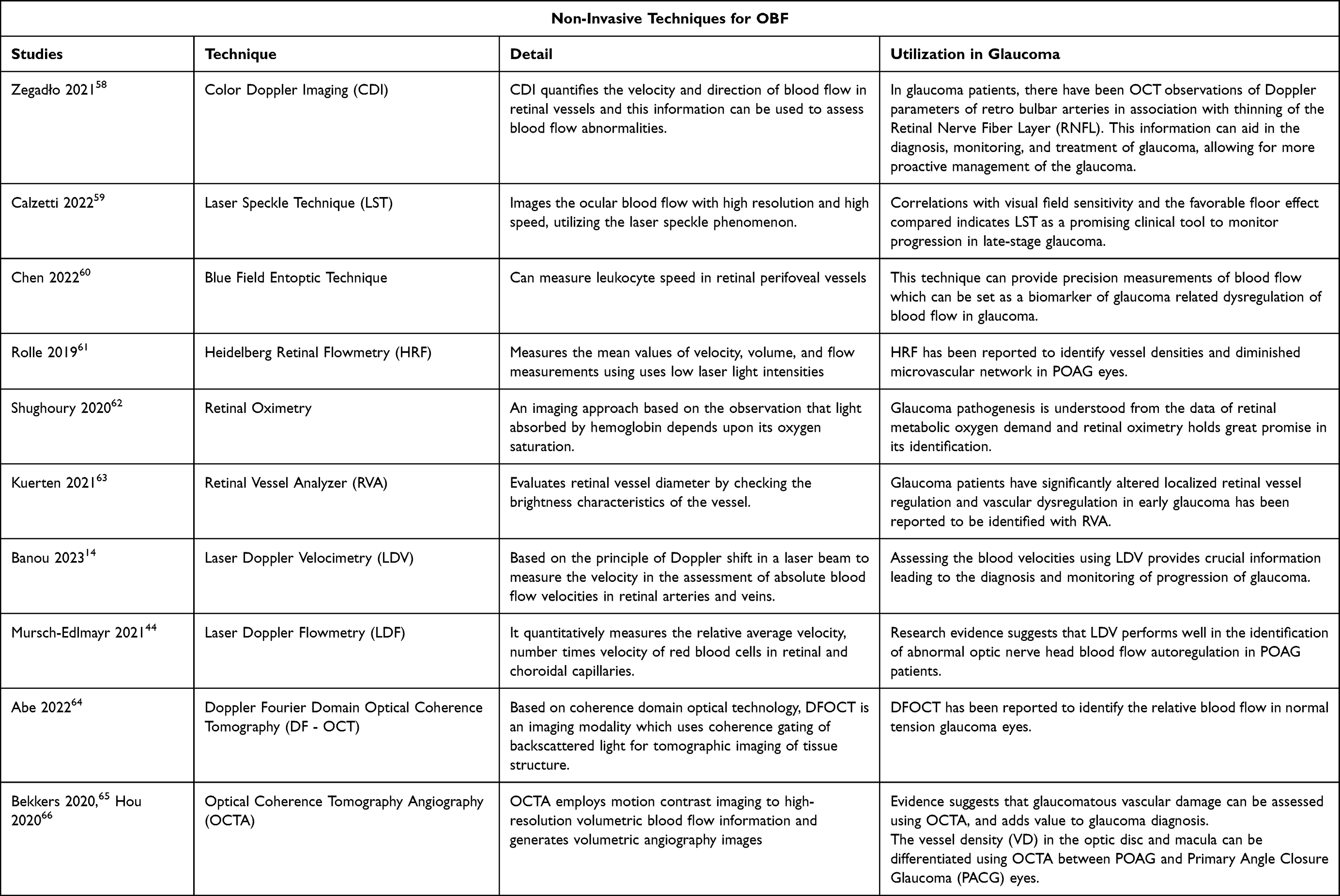 |
Table 1 Summary of the Non-Invasive Techniques Used for OBF Measurement with Mention of Glaucoma Imaging |
Invasive Techniques
Scanning Laser Ophthalmoscope Angiography (SLO)
An invasive technique, SLO produces high-resolution images of the fundus utilizing a scanning laser ophthalmoscope along with different filters to perform fluorescein angiography (FA) and indo cyanine green (ICG) angiography.58
During SLO, a contrast agent (either fluorescein or indocyanine green dye) is injected into the patient’s bloodstream and the SLO system captures images of the dye as it circulates through the blood vessels in the retina. These images are recorded on videotape or stored digitally at a rate of 30 images per second, allowing for the visualization of the dye’s movement and the assessment of blood flow within the retinal vessels.59
By analyzing the SLO images, healthcare professionals can evaluate the blood flow patterns, identify any abnormalities or blockages, and assess the health of the retinal vasculature.60 SLO is particularly useful as multicolor imaging61 and auto fluorescence imaging to identify reduced blood flow in the retina, choroid, and optic nerve head demonstrating its potential in diagnosing glaucoma.62
Scanning Laser Ophthalmoscopic Angiography with Fluorescein and/or Indocyanine Green (ICG) Dye
During digital scanning laser ophthalmoscopic angiography (SLO-A), an invasive technique, a contrast agent, either fluorescein or indo cyanine green dye, is injected into the patient’s bloodstream and to capture images of the dye as it circulates through the blood vessels in the retina and choroid.14 By quantifying various aspects of blood flow, such as vessel diameter, blood velocity, and blood volume, SLO-A provides valuable information about the perfusion of the retina and choroid.44
These techniques can help in the diagnosis and monitoring of various ocular conditions, by visualizing the retinal circulation flow index which is evidence of reduced retinal hemodynamics, indicative of glaucoma.64
Dye leakage is a drawback of this technique which can interfere with the visualization of fine retinal vascular structures, along with the side effect of invasive dyes such as nausea and local and general allergic reactions.65
Table 2 features a brief summary of the invasive techniques discussed, with glaucoma imaging addressed.
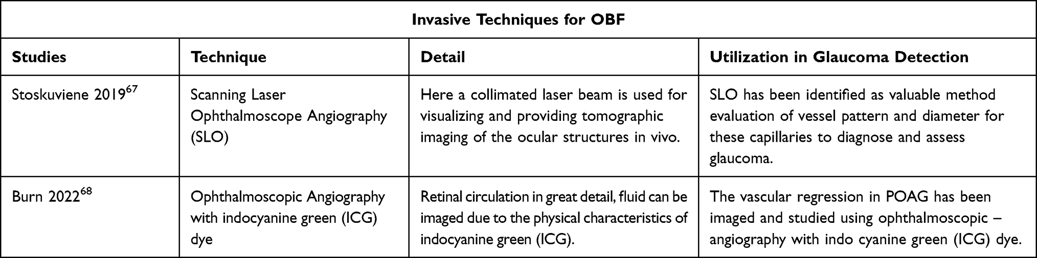 |
Table 2 Summary of the Invasive Techniques Used for OBF Measurement with Mention of Glaucoma Imaging |
Strengths and Limitations of Review
This review implements a comprehensive search through the PubMed search engine using a wide range of keywords, yielding a panoramic view of the current state-of-the-art imaging technologies used in OBF. While PubMed is a valuable resource, it is important to note that it may have limitations in terms of geographical coverage and language accessibility, and may not include all relevant studies published in non-English languages and in other countries where such technologies are not yet available. This review was also limited by the quality of the available studies, many of which were pilot trials or feasibility studies, with small sample sizes and heterogeneity in data, with no evaluation results. As seen from the limited number of studies available on ocular blood flow imaging in glaucoma cases, this field has enormous potential for improvement and evaluation.
Conclusion
Ocular blood flow is impaired in glaucoma and its clinical evaluation is complex; thus, it is of interest in ophthalmology. Many state-of-the-art technologies for evaluating OBF in glaucoma have evolved, catering to the early detection and risk of glaucoma progression.
There is a critical lack of scientific evidence from clinical and evaluation studies to support the feasibility of measuring the OBF in glaucoma. Trials should be undertaken using gold-standard normative databases for each measurement technique to correlate the relationship between ocular hemodynamics, metabolism, and glaucoma progression. The non-invasive technique of OCT angiography is currently supported as a great scientific and clinical tool for the identification of glaucoma pathogenesis, which can lead to its earlier detection.
From the discussed techniques, no single technique has become the standard for measuring ocular blood flow (OBF), and each technique has its own limitations. One of the challenges is that these techniques typically measure only one aspect of blood flow, such as velocity or volume, while blood flow involves multiple variables. However, despite these limitations, the collective application of these techniques in numerous studies has provided valuable insights into the hemodynamic changes associated with normal physiology and ocular diseases. By using a combination of techniques, researchers have been able to identify patterns and trends in OBF that can aid in understanding various conditions.
As technology continues to advance, the shortcomings of current methods are gradually being addressed. Newer techniques are being developed, and existing ones are being refined to provide more accurate and comprehensive measurements of OBF. These advancements may include improvements in resolution, increased sensitivity, and the ability to measure multiple variables simultaneously.
In the future, it is likely that physicians will have access to a wider range of OBF measurement methods in the clinical setting.
This expanded toolbox of techniques will provide more comprehensive assessments of ocular blood flow and contribute to better diagnosis, monitoring, and management of ocular diseases. It is important to note that ongoing research and technological advancements are essential in order to validate and establish the clinical utility of these emerging techniques. The field of OBF measurement is evolving, and with further advancements, more standardized and clinically applicable methods may emerge.
Disclosure
The author reports no conflicts of interest in this work.
References
1. Pandey HC, Coshic P, C CS, Arcot PJ, Kumar K. Blood supply management in times of SARS‐CoV‐2 pandemic–challenges, strategies adopted, and the lessons learned from the experience of a hospital‐based blood centre. Vox Sang. 2021;116(5):497–503.
2. Wu X, Konieczka K, Liu X, et al. Role of ocular blood flow in normal tension glaucoma. Adv Ophthal Pract Res. 2022;16:100036. doi:10.1016/j.aopr.2022.100036
3. Bayraktar S, Ipek A, Takmaz T, Yildiz Tasci Y, Gezer MC. Ocular blood flow and choroidal thickness in ocular hypertension. Int Ophthalmol. 2022;1:1–2.
4. Trivli A, Koliarakis I, Terzidou C, et al. Normal-tension glaucoma: pathogenesis and genetics. Exp Ther Med. 2019;17(1):563–574. doi:10.3892/etm.2018.7011
5. Vernazza S, Tirendi S, Bassi AM, Traverso CE, Saccà SC. Neuroinflammation in primary open-angle glaucoma. J Clin Med. 2020;9(10):3172. doi:10.3390/jcm9103172
6. Harris A, Guidoboni G, Siesky B, et al. Ocular blood flow as a clinical observation: value, limitations and data analysis. Prog Retin Eye Res. 2020;78:100841.
7. Ferrara M, Lugano G, Sandinha MT, Kearns VR, Geraghty B, Steel DH. Biomechanical properties of retina and choroid: a comprehensive review of techniques and translational relevance. Eye. 2021;35(7):1818–1832. doi:10.1038/s41433-021-01437-w
8. Grudzińska E, Modrzejewska M. Modern diagnostic techniques for the assessment of ocular blood flow in myopia: current state of knowledge. J Ophthalmol. 2018;2018:1–8. doi:10.1155/2018/4694789
9. Meola M, Ibeas J, Lasalle G, Petrucci I. Basics for performing a high-quality color Doppler sonography of the vascular access. J Vasc Access. 2021;22(1_suppl):18–31. doi:10.1177/11297298211018060
10. Vosborg F, Malmqvist L, Hamann S. Non-invasive measurement techniques for quantitative assessment of optic nerve head blood flow. Eur J Ophthalmol. 2020;30(2):235–244. doi:10.1177/1120672119858891
11. Tripathi S, Ariga M, Srinivasan MM. Ocular blood flow in glaucoma. TNOA J Ophthal Sci Res. 2020;58(3):180. doi:10.4103/tjosr.tjosr_81_20
12. Divya K, Kanagaraju V, Devanand B, Jeevamala C, Raghuram A, Sundar D. Evaluation of retrobulbar circulation in type 2 diabetic patients using color Doppler imaging. Indian J Ophthalmol. 2020;68(6):1108. doi:10.4103/ijo.IJO_1398_19
13. Tiwari US, Singh M, Aishwarya A, Gupta A, Chhabra K. Comparison of flow velocity in ophthalmic artery between glaucomatous and normal subjects. Roman J Ophthalmol. 2019;63(4):346. doi:10.22336/rjo.2019.54
14. Banou L, Dastiridou A, Giannoukas A, et al. The role of color Doppler imaging in the diagnosis of glaucoma: a review of the literature. Diagnostics. 2023;13:588. doi:10.3390/diagnostics13040588
15. Krzyżanowska-Berkowska P, Czajor K, Iskander DR. Associating the biomarkers of ocular blood flow with lamina cribrosa parameters in normotensive glaucoma suspects. Comparison to glaucoma patients and healthy controls. PLoS One. 2021;16(3):e0248851. doi:10.1371/journal.pone.0248851
16. Motoyama Y, Hayashi H, Kawanishi H, et al. Ocular blood flow by laser speckle flowgraphy to detect cerebral ischemia during carotid endarterectomy. J Clin Monit Comput. 2021;35:327–336. doi:10.1007/s10877-020-00475-1
17. Lu Y, Wang RK. Removing dynamic distortions from laser speckle flowgraphy using Eigendecomposition and spatial filtering. J Biophoton. 2022;15(1):e202100294. doi:10.1002/jbio.202100294
18. Calzetti G, Fondi K, Bata AM, et al. Assessment of choroidal blood flow using laser speckle flowgraphy. Br J Ophthalmol. 2018;102(12):1679–1683. doi:10.1136/bjophthalmol-2017-311750
19. Kikuchi S, Miyake K, Tada Y, et al. Laser speckle flowgraphy can also be used to show dynamic changes in the blood flow of the skin of the foot after surgical revascularization. Vascular. 2019;27(3):242–251. doi:10.1177/1708538118810664
20. Heeman W, Steenbergen W, van Dam GM, et al. Clinical applications of laser speckle contrast imaging: a review. J Biomed Opt. 2019;24:080901. doi:10.1117/1.JBO.24.8.080901
21. Vinnett A, Kandukuri J, Le C, et al. Dynamic alterations in blood flow in glaucoma measured with laser speckle contrast imaging. Ophthalmol Glaucoma. 2022;5(3):250–261. doi:10.1016/j.ogla.2021.10.005
22. Mursch-Edlmayr AS, Luft N, Podkowinski D, Ring M, Schmetterer L, Bolz M. Laser speckle flowgraphy derived characteristics of optic nerve head perfusion in normal tension glaucoma and healthy individuals: a Pilot study. Sci Rep. 2018;8(1):5343. doi:10.1038/s41598-018-23149-0
23. Gumus M, Eker S, Karakucuk Y, Ergani AC, Emiroglu HH. Retinal and choroidal vascular changes in newly diagnosed celiac disease: an optical coherence tomography angiography study. Indian J Ophthalmol. 2022;70(3):866. doi:10.4103/ijo.IJO_1009_21
24. Giraldo Herrera CE, Giraldo Herrera CE. The cavern of the eye: seeing through the retina. Microb Other Sham Beings. 2018;2018:119–133.
25. Szulc U, Dąbrowska E, Pieczyński J, et al. How to measure retinal microperfusion in patients with arterial hypertension. Blood Press. 2021;30(1):4–19. doi:10.1080/08037051.2020.1823816
26. Benjamin JJ, Biggs H, Berger A, et al. The entoptic field camera as metaphor-driven research-through-design with AI technologies. In:
27. Konovalova NV, Khramenko NI, Guzun OV, Serebrina TM. On the treatment of degeneration of the macula and posterior pole. J Ophthalmol. 2019;1:486.
28. Jensen PK, Bek T. Eye Microcirculation. In: Clinically Applied Microcirculation Research. Routledge; 2019:191–200.
29. Dervenis N, Harris A, Coleman AL, et al. Factors associated with non-active retinal capillary density as measured with confocal scanning laser Doppler flowmetry in an elderly population: the Thessaloniki Eye Study (TES). Br J Ophthalmol. 2020;104(9):1246–1253. doi:10.1136/bjophthalmol-2019-315212
30. Zhang Q, Jonas JB, Wang Q, et al. Optical coherence tomography angiography vessel density changes after acute intraocular pressure elevation. Sci Rep. 2018;8(1):1–8. doi:10.1038/s41598-017-17765-5
31. Braaf B, Donner S, Uribe-Patarroyo N, Bouma BE, Vakoc BJ. A neural network approach to quantify blood flow from retinal OCT intensity time-series measurements. Sci Rep. 2020;10(1):1–3. doi:10.1038/s41598-020-66158-8
32. Christinaki E, Kulenovic H, Hadoux X, et al. Retinal imaging biomarkers of neurodegenerative diseases. Clin Exper Optomet. 2022;105(2):194–204. doi:10.1080/08164622.2021.1984179
33. Holm S, Henson D, McLoughlin N. Comparison of two metrics for non-invasive reti-nal oximetry in humans. In: Optical Imaging of Retinal Blood Flow: Studies in Automatic Vessel Extraction, Alignment, and Driven Changes in Vessel Oximetry. The University of Manchester; 2018:79.
34. Stefánsson E, Olafsdottir OB, Eliasdottir TS, et al. Retinal oximetry: metabolic imaging for diseases of the retina and brain. Prog Retin Eye Res. 2019;70:1–22. doi:10.1016/j.preteyeres.2019.04.001
35. Yap ZL, Verma S, Lee YF, Ong C, Mohla A, Perera SA. Glaucoma related retinal oximetry: a technology update. Clin Ophthalmol. 2018;4:79–84. doi:10.2147/OPTH.S128459
36. Král M, Svrčinová T, Hok P, et al. Correlation between retinal oxygen saturation and the haemodynamic parameters of the ophthalmic artery in healthy subjects. Acta Ophthalmol. 2022;100(7):e1489–95. doi:10.1111/aos.15189
37. Pappelis K, Choritz L, Jansonius NM. Microcirculatory model predicts blood flow and autoregulation range in the human retina: in vivo investigation with laser speckle flowgraphyAmerican. J Physiol. 2020;319(6):H1253–73. doi:10.1152/ajpheart.00404.2020
38. Işik MU, Akay F, Akmaz B, Güven YZ, Şahin ÖF. Evaluation of subclinical alterations in retinal layers and microvascular structures with OCT and OCTA in healthy young short-term smokers. Photodiagnosis Photodyn Ther. 2021;36:102482. doi:10.1016/j.pdpdt.2021.102482
39. You QS, Chan JC, Ng AL, et al. Macular vessel density measured with optical coherence tomography angiography and its associations in a large population-based study. Invest Ophthalmol Vis Sci. 2019;60(14):4830–4837. doi:10.1167/iovs.19-28137
40. Lommatzsch C, Rothaus K, Koch JM, Heinz C, Grisanti S. OCTA vessel density changes in the macular zone in glaucomatous eyes. Graefe’s Arch Clin Exper Ophthalmol. 2018;256:1499–1508. doi:10.1007/s00417-018-3965-1
41. Zabel K, Zabel P, Suwala K, et al. Alterations in fixation indices in primary open-angle glaucoma by microperimetry. J Clin Med. 2022;11(9):2368. doi:10.3390/jcm11092368
42. Wei X, Balne PK, Meissner KE, Barathi VA, Schmetterer L, Agrawal R. Assessment of flow dynamics in retinal and choroidal microcirculation. Surv Ophthalmol. 2018;63(5):646–664. doi:10.1016/j.survophthal.2018.03.003
43. Truffer F, Geiser M, Chappelet MA, et al. Absolute retinal blood flowmeter using a laser Doppler velocimeter combined with adaptive optics. J Biomed Opt. 2020;25(11):115002. doi:10.1117/1.JBO.25.11.115002
44. Mursch-Edlmayr AS, Bolz M, Strohmaier C. Vascular aspects in glaucoma: from pathogenesis to therapeutic approaches. Int J Mol Sci. 2021;22(9):4662. doi:10.3390/ijms22094662
45. Morita N, Nogami H, Higurashi E, Sawada R. Grasping force control for a robotic hand by slip detection using developed micro laser Doppler velocimeter. Sensors. 2018;18(2):326. doi:10.3390/s18020326
46. Ishida H, Fujino H, Iwamoto S, Hachiga T, Nakagawa N. Measurement of swirling flow in a blood chamber by laser Doppler imaging system. Meas Sci Technol. 2020;31(9):095702. doi:10.1088/1361-6501/ab8970
47. Mujat M, Lu Y, Ferguson D, Iftimia N. Calibration of laser Doppler flowmetry. Invest Ophthalmol Vis Sci. 2020;61(7):2540.
48. Puyo L, Paques M, Fink M, Sahel JA, Atlan M. Analysis of retinal and choroidal images measured by laser Doppler holography. In: Optical Methods for Inspection, Characterization, and Imaging of Biomaterials IV. SPIE; 2019:85–89.
49. Puyo L, Paques M, Fink M, Sahel JA, Atlan M. Choroidal vasculature imaging with laser Doppler holography. Biomed Opt Express. 2019;10(2):995–1012. doi:10.1364/BOE.10.000995
50. Pijewska E, Sylwestrzak M, Gorczynska I, Tamborski S, Pawlak MA, Szkulmowski M. Blood flow rate estimation in optic disc capillaries and vessels using Doppler optical coherence tomography with 3D fast phase unwrapping. Biomed Opt Express. 2020;11(3):1336–1353. doi:10.1364/BOE.382155
51. Braaf B, Gräfe MG, Uribe-Patarroyo N, et al. OCT-based velocimetry for blood flow quantification. High Res Imag Microsc Ophthalmol. 2019;2019:161–179.
52. Garhöfer G, Bata AM, Popa-Cherecheanu A, et al. Retinal oxygen extraction in patients with primary open-angle glaucoma. Int J Mol Sci. 2022;23(17):10152. doi:10.3390/ijms231710152
53. Hou W, Feng J, Chen J, Li X, Yang G, Sun X. Analysis of the optic nerve head microcirculation using optical coherence tomography angiography and the upstream macrocirculation using color Doppler imaging in low-tension and high-tension glaucoma patients. Ophthalmic Res. 2023;66(1):567–577. doi:10.1159/000528521
54. Mantella LE, Liblik K, Johri AM. Vascular imaging of atherosclerosis: strengths and weaknesses. Atherosclerosis. 2021;319:42–50. doi:10.1016/j.atherosclerosis.2020.12.021
55. Ong HS, Tey KY, Ke M, et al. A pilot study investigating anterior segment optical coherence tomography angiography as a non-invasive tool in evaluating corneal vascularisation. Sci Rep. 2021;11(1):1212. doi:10.1038/s41598-020-80099-2
56. Yang XX. Research progress in the application of OCTA technology in primary glaucoma. Inter Eye Sci. 2021;2021:57–61.
57. Balyen L, Kaya M, Arıkan G, Ü G. Optical coherence tomography angiography for glaucoma diagnosis and observation; 2020.
58. Zegadło A, Wierzbowska J. Colour Doppler imaging of retrobulbar circulation in different severity of glaucoma optic neuropathy. Med Ultrason. 2021;23(4):410–417. doi:10.11152/mu-2954
59. Calzetti G, Mursch‐Edlmayr AS, Bata AM, et al. Measuring optic nerve head perfusion to monitor glaucoma: a study on structure–function relationships using laser speckle flowgraphy. Acta Ophthalmol. 2022;100(1):e181–91. doi:10.1111/aos.14862
60. Chen VY, Le CT, Pottenburgh J, et al. A pilot study assessing retinal blood flow dysregulation in glaucoma using erythrocyte mediated velocimetry. Transl Vis Sci Technol. 2022;11(11):19. doi:10.1167/tvst.11.11.19
61. Rolle T, Dallorto L, Tavassoli M, Nuzzi R. Diagnostic ability and discriminant values of OCTangiography parameters in early glaucoma diagnosis. Ophthalmic Res. 2019;61(3):143–152. doi:10.1159/000489457
62. Shughoury A, Mathew S, Arciero J, et al. Retinal oximetry in glaucoma: investigations and findings reviewed. Acta Ophthalmol. 2020;98(6):559–571. doi:10.1111/aos.14397
63. Kuerten D, Kotliar K, Fuest M, Walter P, Hollstein M, Plange N. Does hemispheric vascular regulation differ significantly in glaucoma patients with altitudinal visual field asymmetry? A single-center, prospective study. Int Ophthalmol. 2021;41(9):3109–3119. doi:10.1007/s10792-021-01876-0
64. Abe T, Yoshioka T, Song Y, et al. Glaucoma diagnostic performance of retinal blood flow measurement with Doppler optical coherence tomography. Transl Vis Sci Technol. 2022;11(10):11. doi:10.1167/tvst.11.10.11
65. Bekkers A, Borren N, Ederveen V, et al. Microvascular damage assessed by optical coherence tomography angiography for glaucoma diagnosis: a systematic review of the most discriminative regions. Acta Ophthalmol. 2020;98(6):537–558. doi:10.1111/aos.14392
66. Hou TY, Kuang TM, Ko YC, Chang YF, Liu CJ, Chen MJ. Optic disc and macular vessel densities were measured using optical coherence tomography angiography in open-angle glaucoma and angle-closure glaucoma. Sci Rep. 2020;10(1):5608. doi:10.1038/s41598-020-62633-4
67. Stoskuviene A. OCT-angiography appliance in glaucoma. Bioph Propert Glaucoma. 2019;2019:89–99.
68. Burn JB, Huang AS, Weber A, Komáromy AM, Pirie CG. Aqueous angiography in pre‐glaucomatous and glaucomatous ADAMTS10‐mutant canine eyes: a pilot study. Vet Ophthalmol. 2022;25:72–83. doi:10.1111/vop.12938
69. Ho CH, Wong JK. Role of 24-hour intraocular pressure monitoring in glaucoma management. J Ophthalmol. 2019;2019. doi:10.1155/2019/3632197
70. McGrath OE, Aslam TM. Use of imaging technology to assess the effect of COVID-19 on retinal tissues: a systematic review. Ophthalmol Therap. 2022;11(3):1017–1030. doi:10.1007/s40123-022-00509-8
 © 2023 The Author(s). This work is published and licensed by Dove Medical Press Limited. The
full terms of this license are available at https://www.dovepress.com/terms
and incorporate the Creative Commons Attribution
- Non Commercial (unported, 3.0) License.
By accessing the work you hereby accept the Terms. Non-commercial uses of the work are permitted
without any further permission from Dove Medical Press Limited, provided the work is properly
attributed. For permission for commercial use of this work, please see paragraphs 4.2 and 5 of our Terms.
© 2023 The Author(s). This work is published and licensed by Dove Medical Press Limited. The
full terms of this license are available at https://www.dovepress.com/terms
and incorporate the Creative Commons Attribution
- Non Commercial (unported, 3.0) License.
By accessing the work you hereby accept the Terms. Non-commercial uses of the work are permitted
without any further permission from Dove Medical Press Limited, provided the work is properly
attributed. For permission for commercial use of this work, please see paragraphs 4.2 and 5 of our Terms.