")
Back to Journals » International Journal of Nanomedicine » Volume 17
Cubosomal Betamethasone-Salicylic Acid Nano Drug Delivery System for Enhanced Management of Scalp Psoriasis
Authors Shalaby RA, El-Gazayerly O, Abdallah M
Received 28 October 2021
Accepted for publication 7 March 2022
Published 13 April 2022 Volume 2022:17 Pages 1659—1677
DOI https://doi.org/10.2147/IJN.S345430
Checked for plagiarism Yes
Review by Single anonymous peer review
Peer reviewer comments 3
Editor who approved publication: Dr Mian Wang
Rodayna Atef Shalaby,1 Omaima El-Gazayerly,2 Mohammed Abdallah2
1Department of Pharmaceutics and Industrial Pharmacy, School of Pharmacy, New Giza University, Giza, Egypt; 2Department of Pharmaceutics and Industrial Pharmacy, Faculty of Pharmacy, Cairo University, Cairo, Egypt
Correspondence: Omaima El-Gazayerly, Department of Pharmaceutics and Industrial Pharmacy, Faculty of Pharmacy, Cairo University, Cairo, Egypt, Tel +2-01005840254, Email [email protected]
Introduction: Betamethasone dipropionate (BD), a potent corticosteroid, and salicylic acid (SA), a keratolytic agent, have been used in combination to treat scalp psoriasis; however, undesirable side effects associated with their prolonged topical use are inevitable. In this study, BD and SA were loaded into cubosomes, a nanoparticulate system with outstanding biocompatibility, bio-adhesivity and penetration power.
Methods: Design of experiments (DOE) was utilized to prepare thirteen different cubosomal dispersions by emulsification technique using glycerol monoolein (GMO) as a lipid phase and Poloxamer 407 (P407) as a surfactant, sodium carboxymethyl cellulose (SCMC) was added to enhance the dispersions’ rheological properties. The thirteen dispersions were in-vitro characterized for their particle size, polydispersity index (PDI), zeta potential, BD and SA content and rheological behaviour. The desirability of an optimized formula (OF) was set to the smallest particle size, lowest zeta-potential and highest viscosity. The OF was in-vitro characterized for the same parameters in addition to transmission electron microscope imaging and in-vitro drug release. The OF’s anti-psoriatic activity was evaluated in-vivo using an imiquimod-induced psoriasis model.
Results: The OF achieved a particle size of 197.4 ± 9.47 nm, a PDI of 0.443 ± 0.025, a zeta potential of − 44.4 ± 0.141mv, BD content of 105.85 ± 2.290%, SA content of 88.855 ± 2.920% with shear-thinning rheological behaviour and completed in-vitro drug release within 2– 3 hours. The in-vivo studies confirmed the cubosomes’ higher anti-psoriatic efficacy over the commercial product with lower changes in ear thickness, spleen to body weight ratio, psoriasis area severity index score and improved histopathological findings.
Conclusion: The developed BD SA-loaded cubosomes exhibit promising anti-psoriatic activity attributed to its nano-size and unique lipid content, with enhanced skin penetration and modified rheological properties; increasing the formulation’s in-contact duration with the scalp resulting in lower application frequency and thus reduced BD and SA associated side effects.
Keywords: betamethasone dipropionate, salicylic acid, cubosomes, psoriasis
Introduction
Psoriasis, a chronic inflammatory immune-mediated skin disorder, is characterized by the abnormal hyperproliferation of keratinocytes with a universal prevalence of 1–2%.1 The scalp is considered one of the most involved body sites throughout the course of the disease.2 Scalp psoriasis (SP) has been reported in around 80% of the worldwide psoriasis cases3 and 61.6% of patients with psoriasis registered in the Kasr Al-Ainy psoriasis unit in Egypt.4
The aetiology of SP remains unknown with a highly variable clinical presentation that ranges from mild fine scaling to severe thickened crusted plaques covering the entire scalp.5 The leading presenting symptoms of SP are usually itching, dryness and scaling in addition to the visibility of scalp lesions which contribute to the negative psychological impact on the patients, besides the possibility of potential development of cicatricial alopecia, especially in the chronically affected scalp areas.6
For decades, topical corticosteroids remained the mainstay in the management of SP, however, keratolytic agents, vitamin D analogues, retinoids, dithranol as well as tar-based therapies have also been used as adjunctive treatment, such drugs have been formulated into numerous formulations such as shampoos, alcohol‐based lotions, emulsions, creams, ointments, gels and foams.2,7
Betamethasone Dipropionate (BD) is one of the most widely used corticosteroids in the management of SP; since BD have marked anti‐inflammatory, immunosuppressive and antiproliferative actions on epidermal cell turnover.8 However, skin atrophy, striae, rosacea, purpura, delayed wound healing, hypertrichosis and pigment alteration are among the adverse side effects associated with BD prolonged use.9,10
On the other hand, salicylic acid (SA), a keratolytic agent, is indicated in SP because of its ability to remove scales from hyperkeratotic plaques.11 However, SA might irritate non‐hyperkeratotic skin and it should not be applied to more than 20% of body surface area to avoid the risk of salicylism toxicity symptoms including; metabolic acidosis, tinnitus, nausea, vomiting or central nervous system associated symptoms.8,12
BD and SA have been used as a combined therapy for the effective management of SP and they have been formulated into many dosage forms including creams, ointments, gels and lotions with a BD concentration of 0.064% equivalent to 0.05% betamethasone and 2–3% SA.13 This combined therapy is usually applied twice per day till the plaques are smoother.14
Cubosomes are discrete nanovesicles bi-continuous cubic phase liquid crystals composed of biodegradable lipid and water, with powerful ability to solubilize and encapsulate hydrophilic, hydrophobic and amphiphilic molecules, outstanding biocompatibility and bio-adhesive properties.15,16
Cubosomes have gained a rising interest in topical preparations since it is considered a good candidate for skin transport due to their similarity with human skin layers besides their skin penetration enhancing properties.17 They are also characterized by small pore size making them ideal for controlled release, in addition, to their high internal surface area, drug payloads, heat stability and ability to moisturize the skin.18,19
In this study, BD SA-loaded cubosomes were incorporated into sodium carboxymethylcellulose (SCMC), an anionic polysaccharide, a promising candidate in biomedical and drug delievery20 with remarkable mucoadhesive, viscosity and rheological properties.21,22 The goal is to obtain a cubosomal dispersion with enhanced rheological behaviour which can last in longer contact with the scalp ensuring better skin absorbance with lower application frequency and shorter duration of treatment, therefore, reducing BD and SA associated side effects.
The prepared optimized formula (OF) was then assessed for its healing efficacy towards SP in an imiquimod-induced psoriasis animal model followed by histopathological evaluation.
Materials and Methods
Materials
Glycerol Monooleate (GMO) and Poloxamer 407 (P407) were purchased from Sigma Aldrich (Cairo, Egypt). Salicylic acid (SA), Sodium carboxymethyl cellulose (SCMC) and Vaseline were purchased from Piochem (Cairo, Egypt). Betamethasone dipropionate (BD) was gifted by Marcyrl Pharmaceutical industries (Cairo, Egypt). Imiquimod cream (IMQ, 5% cream, Aldara®) was purchased from Meda Pharmaceuticals Inc. (USA) and Kernella lotion was purchased from Global Napi Pharmaceuticals (Cairo, Egypt).
Methods
Experimental Design
The experimental design was constructed using DESIGN EXPERT® software, version 12, Stat-Ease Inc. Minneapolis, MN, USA. The D-optimal design was used to study and optimize the formulation variables. The software evaluated the effect of the independent variables GMO, P407 and SCMC on three dependent variables; particle size, zeta potential and viscosity as shown in Table 1.
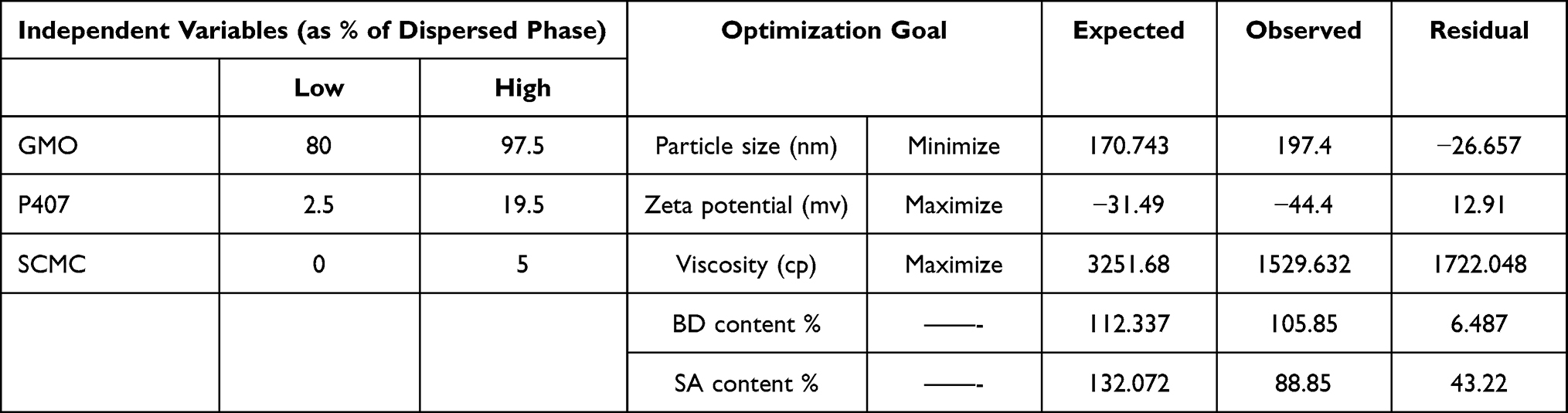 |
Table 1 Independent and Dependent Variables for Preparing BD/SA Cubosomal Dispersion |
DESIGN EXPERT® software suggested thirteen runs of 0.064% BD and 2% SA loaded cubosomes with different dispersed phase compositions accounting for 5% of the cubosomal dispersion dispersed in 95% of the aqueous phase. The dispersed phase composition with different GMO/P407/SCMC concentrations is presented in Table 2.
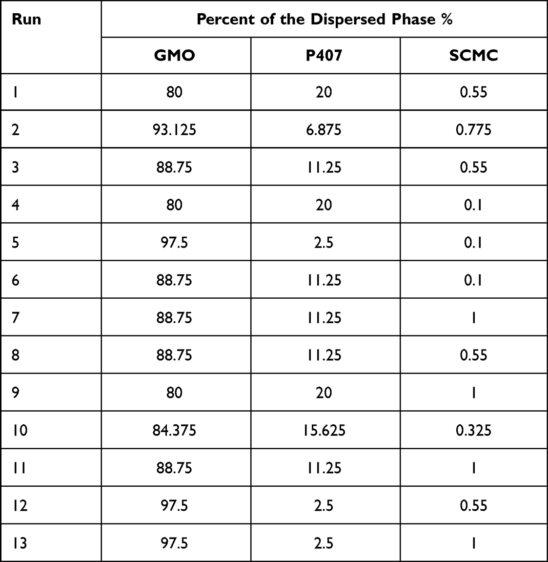 |
Table 2 Different Compositions of 0.064% BD 2% SA Loaded Cubosomes Dispersed Phase |
Preparation of BD and SA Liquid Cubosomal Dispersion
Cubosomes are commonly prepared via the top-down approach,23 this technique involves mixing the cubosomes forming lipid with a stabilizer; forming bulk viscous cubic aggregates which are then dispersed in an aqueous media using high energy input to form homogenous cubosomal dispersion.15,24
In this study, GMO and P407 were completely melted on a hot plate at a temperature of 80°C, then the BD and SA were dispersed in the oily phase, parallelly, the aqueous phase was maintained at a temperature of 70°C.25 The molten mixture was then added dropwise to the aqueous phase while maintaining mechanical stirring of 1500 rpm until the dispersion reaches room temperature, then SCMC was sprinkled over the surface while stirring the mixture at 200 rpm for 15 minutes to obtain a viscous dispersion which was stored in amber bottles for the future investigations.15,25
In-vitro Characterization of BD SA Cubosomal Dispersion
Particle Size, Polydispersity Index (PDI), Zeta Potential
The particle size distribution, polydispersity index (PDI), and zeta potential for the thirteen suggested runs were determined using the Zetasizer Nano series (Nano ZS, Malvern, UK). Each run was diluted 1:20 with deionized water and measured at a temperature of 25±0.5 °C in triplicate and an average reading was calculated.
Evaluation of Viscosity and Rheological Properties of the Prepared Cubosome Dispersions
Single point viscosity was determined to all 13 runs using the Brookfield viscometer (Brook field DV-III Ultra, USA) with spindle SC4-18 at rotation speed 2.00 rpm and at a room temperature of 26±2°C.
The runs’ rheological and flow properties were then evaluated by the same device under the same conditions, where the measurements were taken over a range of speed settings 0.2 to 1.6 rpm with 10 seconds between 2 successive speeds, and the results were only recorded when the torque was within the acceptable range (10–100%).
The rheological data obtained from the viscometer was the shear rate, shear stress, % torque and viscosity at different shear values; were incorporated into the power-law model to evaluate the cubosome dispersions’ rheological behaviour as follows:

where τ is the shear stress, K is the consistency index (sec.), γ is the rate of shear, n is the flow index at which a value of n = 0–1 indicates that the system is shear thinning, approaches 1 in Newtonian systems, and exceeds 1 for the dilatant systems.26
The obtained data were also fitted into 3 different equations namely Bingham, Casson, Carreau to analyse and verify whether the dispersions’ non-Newtonian behaviour followed a linear plastic system, a non-linear plastic system or a pseudo-plastic shear-thinning system respectively through comparing the equations regression coefficients (r2) of each dispersion.27
The equations were calculated as follows:

at which τ° is the yield value.


Drug Content
The BD drug content in the prepared cubosomal dispersions was assayed by 1260 Infinity II LC HPLC (Agilent Technology, USA) using a quasar C18 column, a mobile phase of water: acetonitrile in a ratio of 35:65, a flow rate of 1.0 mL/min, an injection volume of 10µL, at a wavelength of 254nm and a temperature of 25±0.5 °C.29 0.5mL of each run was diluted with 9.5mL of methanol HPLC grade and was assayed for BD content in triplicate at a retention time from 5–6 minutes.
The SA content in the cubosomes was assayed using a UV-visible double beam spectrophotometer (Shimadzu 1900i). 0.5mL of each run was diluted with 9.5 mL of methanol and then measured at a wavelength of 301nm in triplicate at a temperature of 25±0.5°C.30
Statistical Optimization
The desirability factor of an optimized formula (OF) was generated by Design Expert® software at which the preferred formulation is the one likely to offer the smallest particle size, the lowest zeta-potential and the highest viscosity shown in Table 1. The aim is to produce a formulation with enhanced skin permeability and in prolonged contact with the scalp through adjusting the formulation’s viscosity and flow properties.
OF Characterization
The particle size distribution, polydispersity index (PDI), zeta potential, single-point viscosity, rheological behaviour, BD and SA drug content for the OF have been evaluated with the same procedures and conditions used for the evaluation of the previous thirteen runs.
Physical Properties of the OF
The OF was visually inspected for its physical characteristics (eg, colour and homogeneity). The pH of the optimized formula was then measured by a pH meter after preparing 10% of the dispersion’s aqueous solution.
High-Resolution Transmission Electron Microscope (HRTEM)
The morphological shape of the BD SA loaded cubosomes in the OF was determined using HRTEM (JEM-2100, Japan). At first, the OF was diluted 1:20 with distilled water and then it was stained with 1% sodium phosphotungstate solution and placed on a carbon-coated copper grid, the excess fluid was drained by an absorbent filter paper and the sample was left to dry at room temperature for 30 mins before being examined by the HRTEM.31
In-vitro Release of BD/SA from the OF
The in-vitro release of BD and SA from the prepared cubosomes dispersed in 1%SCMC was determined using a dialysis method via a Spectra/Por® dialysis membrane with a 12,000–14,000 molecular weight cut-off.25 Prior to the experiment, the dialysis membrane was washed using deionized water and was then soaked in a phosphate buffer (pH7.4) for 2 hours.
The dialysis membrane was then injected with 2mL of the OF and was immersed in 100 mL of the dissolution medium of 1% sodium lauryl sulphate in a phosphate buffer (pH 7.4) maintained at 100 rpm at a temperature pf 37 ± 0.5 °C.32 5 mL aliquots were taken at time intervals 5, 10, 15, 30, 45, 60, 90, 120, 150, 180, 240 and 300 minutes and were replaced with 5mL of the dissolution medium.25 The released BD and SA were assayed by HPLC and UV spectrophotometer respectively using the previously discussed procedure.
In-vivo Evaluation of BD SA Cubosomal Dispersion in an Animal Model
The experimental design of the animal model and treatment of animals was reviewed and approved by the research ethics committee in the Faculty of Pharmacy, Cairo University, protocol number PI 3000. All study procedures strictly complied with the “Guide for the Care and Use of Laboratory Animals” published by the US National Institute of Health (NIH Publication No. 85-23, revised 2011).33
Experimental Design
Fifty male balb/c mice aged 7–10 weeks, weighing 29–35 grams were used in the experiment. The mice have been accommodated for 7 days at a room temperature of 28±5 °C, air humidity of 50±10% in a 12-hour light/dark cycle environment, and they were provided with food and water ad libitum. On the 8th day, the dorsal backs of all mice were shaved using an electric clipper and a hair removal cream.
The fifty mice were randomly divided into 5 equal groups in separate cages, briefly on day 1 and for 7 consecutive days, all groups except the healthy control group have received a daily topical dose of 62.5 mg of the 5% imiquimod (IMQ) Aldara® cream on their shaved dorsal backs as well as a dose of 3.25 mg on their right ear pinna.34,35 The prepared BD SA cubosomal dispersion and the BD SA commercial lotion were applied one hour after the application of the 5% IMQ cream. The 5 groups were divided as follows:
Group 1: Healthy negative control mice; that only received Vaseline on their dorsal back and inside their right ear pinna.
Group 2: Positive control mice; treated with 5% topical IMQ only on their dorsal back and inside their right ear pinna.
Group 3: Positive control mice; treated with 5% topical IMQ on their dorsal back and inside their right ear pinna and then received void cubosomes in 1% SCMC.
Group 4: Treated with 5% topical IMQ on their dorsal back and inside their right ear pinna and then received the BD SA commercial lotion.
Group 5: Treated with 5% topical IMQ on their dorsal back and inside their right ear pinna and then received the prepared BD SA cubosomal dispersion.
Pharmacological Evaluation
Ear Thickness
The ear thickness of the right ear pinna treated with 5% IMQ cream was measured using Vernier calliper (530–119 Mitutoyo, Japan) as a reflective estimate of the degree of skin inflammation.34,36
Psoriasis Area and Severity Index (PASI) Score
PASI scoring of the mice was conducted by the visual inspection of the mice dorsal back and right ear pinna, this scoring system is used as an objective tool to examine the severity of IMQ induced inflammation and psoriasis, based on the developed clinical Psoriasis Area and Severity Index.37 The PASI score measures the severity of erythema, scaling and thickness, each is given a score from 0–4 at which 0-none, 1- mild, 2- moderate. 3-marked, 4-severely marked, the cumulative score 0–12 is then calculated and is considered as an indicative parameter to the severity of inflammation.38
Spleen to Bodyweight Ratio
Spleen weight was assessed as an indication of the extent of immune stimulation.39 Each mouse was weighed just before its sacrifice, the spleen was then immediately harvested and weighted using the same digital balance. The spleen to body weight ratio was calculated as follows: Spleen weight in grams/ Bodyweight in grams.
Histopathological Examination
At the end of the experiment, tissue samples from both the dorsal back skin and the right ear pinna were harvested and fixed in 10% neutral buffered formalin for 72 hrs. Samples were processed in serial grades of ethanol, cleared in xylene, then infiltrated and embedded into paraffin wax embedding media. Five μn thick tissue sections were cut by rotary microtome and mounted on glass slides. Tissue sections were stained by Hematoxylin and Eosin (H&E) as a standard staining method for microscopic examination.40
6 random non-overlapping fields per tissue section of each sample were then scanned and analyzed for average epidermal thickness in micrometers for each group.
Results and Discussion
Particle Size, Polydispersity Index (PDI), Zeta Potential
The mean particle size, PDI and zeta potential for the 13 runs are recorded in Table 3, the cubosomes’ particle size ranged from 142.25±2.757nm to 511± 5.697nm which seems promising since nanoparticles more than 600nm might fail to effectively deliver the encapsulated drug through the skin and the drug might even be inclined to remain on the stratum corneum.41 Nanoparticles with a diameter of 300nm or less, even possess a higher skin penetration power and are more likely to deliver their contents into deeper skin layers, likewise, particles that range from 10nm to 210nm are even further privileged to penetrate the skin through the trans-follicular route.41
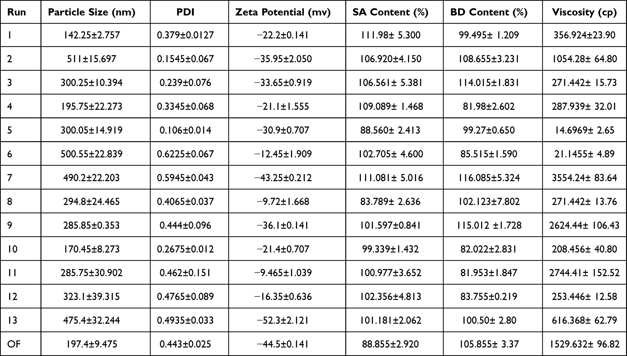 |
Table 3 Particle Size, PDI, Zeta Potential, BD/SA Content for the 13 Different Cubosomal Dispersions and OF |
It can be observed from Figure 1A and B that different GMO/P407 concentrations had a significant effect on the mean particle size among the different runs (p-value =0.0281). It has also been observed that decreasing GMO concentration and increasing P407 concentration from 2.5% till around 11% yielded smaller particle size, however at a higher concentration of P407, the mean particle size started to increase again, this could be attributed to the potential P407 impact on the nano-dispersion steric stability.23
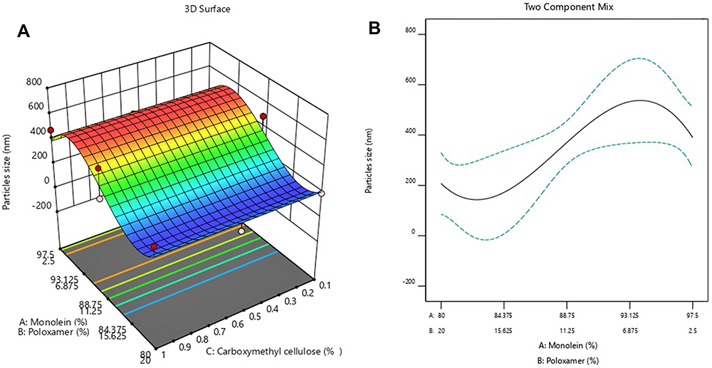 |
Figure 1 (A) 3D representation of the effect of different GMO/P407/SCMC concentrations on cubosomes average particle size. (B) Two components mix of the effect of different GMO/P407 concentrations on cubosomes average particle size. |
The Polydispersity index (PDI) also known as heterogeneity index; describes the uniformity of the particles size distribution, at which a PDI value greater than 0.7 acts as an indication for the broad range of size distribution.41,42 The PDI values ranged from 0.106 ± 0.014 to 0.6225 ± 0.067, which indicates that the particles sizes distribution within the same run is of a narrower range.
On the other hand, the zeta potential range has been investigated to determine the cubosome surface charge, and it can be observed that all the 13 cubosomes dispersions are anionic43 and the zeta potential was ranging from 9.72 ± 1.668 mv to −52.3 ± 2.121 mv. This might be attributed to the presence of the fatty acid GMO as well as adding P407 which resulted in more negative charge values due to the interaction between P407 hydroxyl ions with the aqueous medium.44 Negatively charged particles are advantageous in permeating the skin via channels created by the repulsive forces between negatively charged skin lipids and particles.45 However, it is observed that different GMO/P407 concentrations had a non-significant impact on the zeta-potential (p-value=0.5980).
Zeta potential reflects the stability of nano-dispersions at which values lower than −30mv or higher than +30mv indicate a good electrostatic stabilization. At higher zeta potential values; particle aggregation is less likely to be prominent due to electric repulsion, unlike at lower zeta potential values where coagulation and flocculation tend to be more pronounced with a negative impact on the dispersion’s stability.41,46
Evaluation of Viscosity and Rheological Properties of the BD SA Cubosomal Dispersions
The single point viscosity measured for each run recorded in Table 3 shows highly variable readings that ranged from 14.69 ± 2.65 cp and up to 3554.24 ± 83.64 cp. Different SCMC concentrations had a significant difference in the cubosomal runs viscosity (p-value=0.0054), irrespective of the change in the concentration of GMO or P407 as illustrated in Figure 2. SCMC is an anionic water-soluble polysaccharide that has been extensively used to maintain the intrinsic viscosity of drug nanocarrier because of its distinguished surface properties, mechanical strength capability and viscosity adjusting power; due to its high solubility in water.20,47
 |
Figure 2 3D representation of the effect of different GMO/P407/SCMC concentrations on single-point viscosity. |
Upon measuring the viscosity at different shear rates, it was clear that all flow indexes (n values) were less than 1 as recorded in Table 4, which could be interpreted as a shear-thinning behaviour and is strongly related to the spreadability of the dosage form and its ability to form a smooth layer on the skin.48
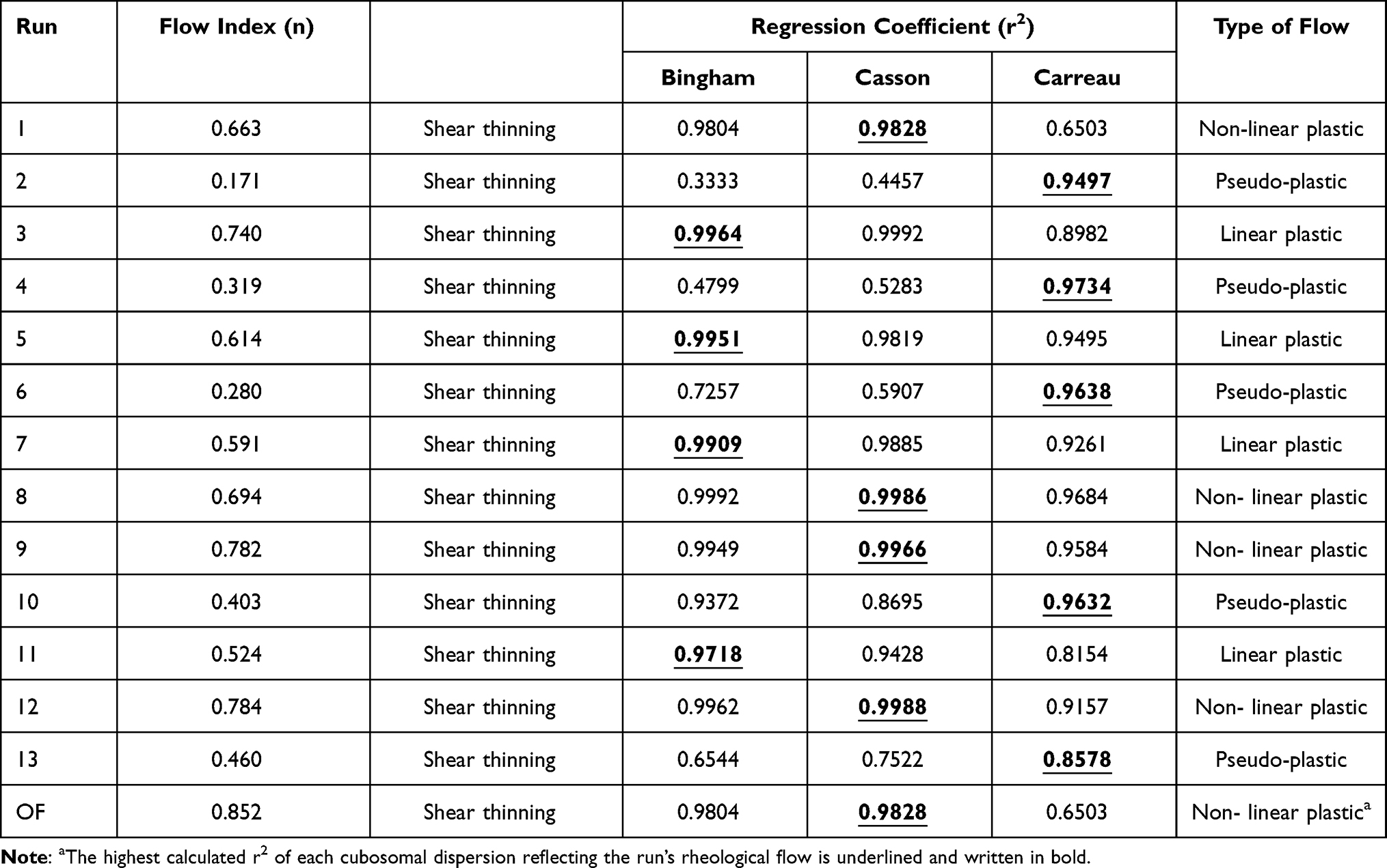 |
Table 4 Rheological Behaviour for the 13 Different Cubosomal Dispersions and OF |
However, the type of non-Newtonian behaviour varied from one formulation to the other, since runs 3, 5, 7 and 11 recorded the highest r2 when incorporated into the Bingham equation, thus, they follow a linear plastic behaviour which means the dispersion will not flow until critical yield stress is exceeded,27 while runs 1, 8, 9, 12 exhibited a non-linear plastic behaviour, thus they exhibit shear-thinning characteristics and is reduced to a Newtonian fluid at a very high wall shear stress.49
Runs 2, 4, 6, 10 and 13 followed a pseudoplastic behaviour which means they tend to illustrate both Newtonian characteristics at the low shear rate and power-law properties at high shear rates.50 At a high shear rate where n<1 apparent viscosity declines to show shear-thinning features but when viscosity increments n>1 the fluid tends to behave as a shear-thickening system, thus, those 5 runs have only exhibited shear-thinning behaviour.25,50
Drug Content
As observed in Table 3; the BD and SA content varied among the different runs with BD content ranging from 81.953±1.847% to 115.012 ±1.728% and SA content ranging from 83.789 ± 2.636% to 111.081 ± 5.016%. All runs except for runs 4, 7, 8, 9 are within the accepted USP drug ranges which varies from 85–115%.51,52
The different GMO/P407/SCMC had no significant impact on both the BD content (p-value =0.3054) nor the SA content (p-value = 0.1189). The high drug content and payloads is a result of high drug encapsulating power and high internal surface area of the cubosomes.15 The higher SA drug content could be possibly related to the potential nucleation of SA when it was initially melted with GMO.53
Statistical Optimization
The composition of the OF’s dispersed phase as generated by Design Expert® software was 84.32% GMO, 16.67% P407 and 0.99% SCMC, the software also predicted that the OFs mean particle size to be 170.743 nm, a zeta potential of −31.49, BD content of 112.337%, SA content of 132.072% and a single point viscosity of 3251.68 as shown in Table 1.
OF Characterization
However, as recorded in Table 3, there has been a disparity between the predicted and actual results since the OF’s actual average particle size of 197.4 ± 9.475, PDI of 0.443 ± 0.025. And zeta potential of −44.4±0.141 which collectively indicates that the OF has achieved the lowest particle size compared to all other runs with narrow particle size distribution and with expected stability achieved by the electrostatic forces.41,42 The BD and SA content was 105.85±2.290% and 88.855±2.920% respectively which is within the USP accepted drug range 85%-115%.51,52
The OF’s single point viscosity recorded at 2 rpm was 1529.632 with a non-linear plastic shear-thinning behaviour at different shear rates as recorded in Table 4. The OF high viscosity could enable the cubosomal dispersion to last in longer contact with the scalp with good spread-ability when compared to the commercial alcohol-based lotions.
Physical Properties of the Optimized Formula
The OF was homogenous milky-white in colour with a thickened viscous consistency with a pH of 7.8. The OF remained unchanged with no visual phase separation even after standing for 3 months in an amber glass bottle.
High-Resolution Transmission Electron Microscope (HRTEM)
The microphotographs obtained by the HRTEM confirmed that the OF was composed of cubic edged nanoparticles that fall in the nano-range. Interestingly, the particle size of some cubosomes was as small as 38.31 nm as shown in Figure 3 suggesting that not all produced cubosomes are of the same size; the presence of smaller cubosomes might further suggest enhanced skin penetration power and better eligibility for trans-follicular drug transport.41
 |
Figure 3 Micrographs by HRTEM shows cubic shaped nanoparticles with edged dimensions with different particle size within the nano-range. |
In-vitro Release of BD/SA from the OF
As illustrated in Figure 4, the release of both the BD and SA have started as soon as the dispersion was immersed in the dissolution medium with around 8.34% of BD and 11.33% of SA released after the first 5 minutes. The percentage release of both drugs was exponential, with peak release of BD at around 3 hours and of the SA at around 2 hours.
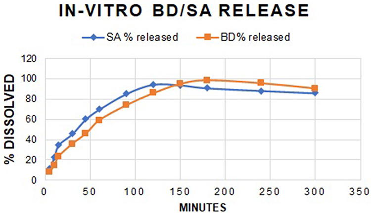 |
Figure 4 In-vitro release of BD/SA from the OF. |
The rapid release of drugs from cubosomal dispersion has been previously attributed to the small particle size of the loaded cubosomes with the high surface area making burst release inevitable.54
Pharmacological Evaluation
Ear Thickness
Change in ear thickness is considered a reflective parameter of the intensity of IMQ- induced psoriasis and skin inflammation in multiple studies.36,55,56 An increase in ear thickness has been observed in all groups receiving 5% IMQ on their right ear pinna compared to the control group as illustrated in Figure 5A and B. The change in ear thickness has been also accompanied by prominent erythema and scaling that progressed over the course of the experiment as demonstrated in Figure 6. The highest changes in ear thickness, redness and scaling have been observed in group 2 receiving 5%IMQ only and group 3 receiving 5%IMQ and treated with void cubosomes in 1%SCMC.
 |
Figure 5 (A) The change in ear thickness over the 7 days of the experimental model. (B) The change in ear thickness in all groups on day 7. |
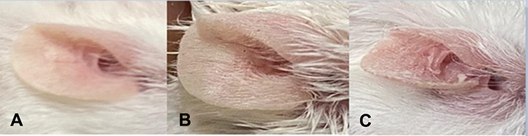 |
Figure 6 Changes in the right ear pinna receiving IMQ on days (A) 1, (B) 3 and (C) 7. |
On the other hand; group 5 receiving 5%IMQ and treated with the prepared BD/SA cubosomal dispersion in 1%SCMC has recorded the least change in ear thickness with an average of 11 x10−2mm, compared to group 4 receiving 5%IMQ and treated with the commercial BD/SA lotion with a change 17x10−2mm, indicating the higher anti-psoriatic power of the loaded cubosomes compared to the commercial products. However, both groups 4 and 5 were significantly different from diseased groups in a one-way ANOVA statistical test followed by Tukey post hoc test (p<0.05).
PASI Score
For decades; the PASI scoring system is a method for quantifying the extent of the disease, and evaluating psoriasis improvement during the course of treatment.57 The quantitative assessment of three hallmarks of psoriatic lesions, including; erythema (redness), infiltration (thickness), and desquamation (scaling) are crucial to evaluate the progression of the disease,37 as shown in Figures 7 and 8 the disease progression has varied from one group to another in terms of those three parameters.
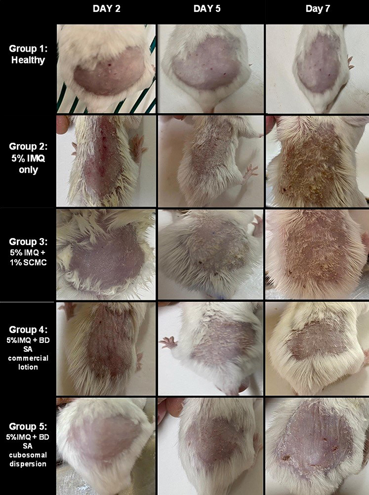 |
Figure 7 Psoriasis progression in all groups on days 2, 5 and 7. |
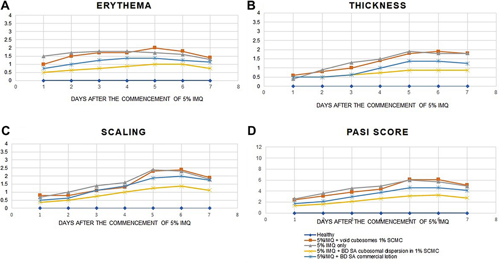 |
Figure 8 Graphical representation of the progression of (A) erythema, (B) thickness, (C) scaling, (D) cumulative PASI score in all groups over the 7 days of the experimental model. |
Group 2 and group 3 receiving 5%IMQ only and 5% IMQ then treated with void cubosomes in 1%SCMC respectively, showed the worst disease prognosis with the most thickened reddish skin with the most intense scaly lesions. On the other hand, groups 4 and 5 treated with the commercial lotion and the tested cubosomal formula respectively; have shown a better prognosis with a lower cumulative PASI score and were significantly different from the diseased group in a Kruskal–Wallis statistical test (p<0.05). Meanwhile, the tested BDSA cubosomes was significantly different from the BDSA commercial product in terms of PASI score using Kruskal–Wallis statistical test (p<0.05).
Spleen to Bodyweight Ratio
In multiple studies, Spleen size and weight have been considered a direct indicator of the degree of immune stimulation and proliferation of immune cells in 5%IMQ psoriasis induced models.39,58 As shown in Figures 9 and 10, the spleen harvested from the groups treated with the IMQ only and the IMQ with 1%SCMC were the most enlarged ones and with the highest spleen to bodyweight ratio.
 |
Figure 9 Representative photograph of harvested spleen in all groups on the 7th day of the experimental model. (A) Healthy group, (B) group receiving 5% IMQ only, (C) group receiving 5% IMQ + void cubosomes in 1%SCMC, (D) group receiving 5% IMQ + BD SA cubosomes in 1%SCMC and (E) group receiving 5% IMQ + commercial BD SA lotion. |
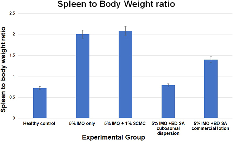 |
Figure 10 Spleen to body weight ratio among all groups on the 7th day of the experimental model. |
On the other hand, the groups treated with the tested BDSA cubosomes and the commercial product were significantly different in a one-way ANOVA statistical test following Tukey post hoc test (p<0.05) from the diseased group, however, there was no significant difference between the tested cubosomes and the commercially available product (p=1.00).
Histopathological Examination
Results of Microscopic examinations of the harvested skin of dorsal back samples are as follows:
Group 1: healthy control; demonstrating normal morphological features of skin layers with apparent intact thin epidermal layer (black arrow), intact dermal layer with well-organized collagen fibers and hair follicles (black star) without abnormal inflammatory cells infiltrates as well as intact subcutaneous tissue as shown in Figure 11A and B.
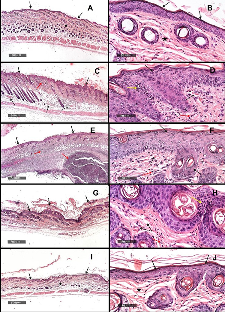 |
Figure 11 (A) Dorsal skin micrograph of healthy mice under X500um magnification, (B) dorsal skin micrograph of healthy mice under X50um magnification, (C) dorsal skin micrograph of mice receiving 5%IMQ only under X500um magnification, (D) dorsal skin micrograph of mice receiving 5%IMQ only under X50um magnification, (E) dorsal skin micrograph of mice receiving 5%IMQ and void cubosomes in 1%SCMC under X500um magnification, (F) dorsal skin micrograph of mice receiving 5%IMQ and void cubosomes in 1%SCMC under X50um magnification, (G) dorsal skin micrograph of mice receiving 5%IMQ and commercial BDSA lotion under X500um magnification, (H) dorsal skin micrograph of mice receiving 5%IMQ and commercial BDSA lotion under X50um magnification, (I) dorsal skin micrograph of mice receiving 5%IMQ and tested cubosomal BDSA dispersion in 1%SCMC under X500um magnification, (J) dorsal skin micrograph of mice receiving 5%IMQ and tested cubosomal BDSA dispersion in 1%SCMC under X50um magnification. |
Group 2: treated with 5% IMQ only, showed a significant increase of epidermal thickness (Acanthosis) throughout all tissue sections (black arrow) with mild clubbing of rete ridges. However, less extensive records of inflammatory cell infiltrate were observed all over the samples with more tendencies for focal subepidermal aggregates of inflammatory cells (red arrow). Moreover, moderate records of Munro abscesses (yellow arrow) with moderate congested and dilated subepidermal and subcutaneous blood vessels (BVs) (dashed arrow) as observed in Figure 11C and D.
Group 3: treated with 5% IMQ + 1% SCMC, showed significant acanthosis throughout all skin samples tissue sections (black arrow) with mild clubbing of rete ridges, accompanied with severe diffuse dermal and subcutaneous mixed inflammatory cells infiltrates (red arrow) with focal encapsulated subcutaneous aggregates of inflammatory cells. Moderate congested and dilated subepidermal BVs (dashed arrow) as presented in Figure 11E and F.
Group 4: treated with 5% IMQ + BD SA commercial lotion; showed moderate protective efficacy of skin samples with persistence higher records of epidermal thickening with moderate hyperkeratosis (black arrow) accompanied with mild records of Munro abscesses (yellow arrow). However, mild dermal or subcutaneous inflammatory cell infiltrates records were observed with the persistence of moderate congested subepidermal BVs (dashed arrow) as shown in Figure 11I and J.
Group 5: treated with 5% IMQ + BD SA prepared cubosomal dispersion; demonstrated obvious higher protective efficacy among treatment groups with almost intact well-organized histological features of different skin layers including epidermal (black arrow), dermal (star) and subcutaneous layers, with minor sporadic focal subcutaneous aggregates of inflammatory cells and mild congested BVs as shown in Figure 11G and H.
As generally observed in Figure 11, the skin micrographs show an increase in epidermal thickness as a result of keratinocytes hyperproliferation and reduced turnover time,59 thus, the epidermal thickness mirrors the progression of the disease. As shown in Figure 12, the epidermal thickness examined under the microscope was the highest in the diseased groups, however, both groups treated with the tested BDSA cubosomes and commercial BDSA lotion have shown a significant difference from the diseased groups in a one way ANOVA statistical test followed by Tukey post hoc test (p<0.05), Interestingly, there was a significant difference between the BDSA cubosomal dispersion and the commercial BDSA product indicating the higher antiproliferative action of the tested formula.
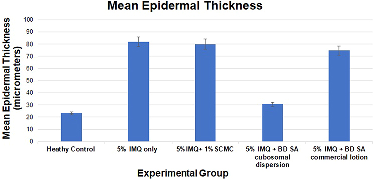 |
Figure 12 Average epidermal thickness in all groups at the 7th day of the experiment. |
Conclusion
The optimized cubosomal nanoparticle dispersion loaded with BD and SA was characterized by a small particle size of 197.4 ± 9.475 and PDI of 0.443 ± 0.025, which make it a very good candidate for topical skin penetration that will ultimately enhance the bioavailability.
The BD SA loaded cubosomes was stabilized by electrostatic forces as indicated by a zeta potential of −44.5 ±0.141, in addition to its high viscosity enabling the formulation to last longer in contact with the diseased area besides its shear-thinning rheological behaviour that made it spreadable to a thin smooth film. The cubosomes nanoparticle has also achieved a high BD and SA encapsulating power and served as an excellent drug transport carrier.
The application of BD SA loaded cubosomal nanoparticle dispersion in a 5% IMQ induced psoriasis animal model has ameliorated skin inflammation reflected in a lower change in ear thickness and immune-mediated splenomegaly as well as resulting in higher histopathological protective efficacy with intact skin layers with minor aggregates of inflammatory cells and mildly congested blood vessels when compared to the commercially available BD SA lotion.
Disclosure
The authors report no conflicts of interest in this work.
References
1. Rendon A, Schäkel K. Psoriasis pathogenesis and treatment. Int J Mol Sci. 2019;20(6):1475. doi:10.3390/ijms20061475
2. Papp K, Berth‐Jones J, Kragballe K, Wozel G, De La Brassinne M. Scalp psoriasis: a review of current topical treatment options. J Eur Acad Dermatol Venereol. 2007;21:151–160.
3. Chan CS, Van Voorhees AS, Lebwohl MG, et al. Treatment of severe scalp psoriasis: from the Medical Board of the National Psoriasis Foundation. J Am Acad Dermatol. 2009;60(6):962–971. doi:10.1016/j.jaad.2008.11.890
4. El-Komy MH, Mashaly H, Sayed KS, et al. Clinical and epidemiologic features of psoriasis patients in an Egyptian medical center. JAAD Int. 2021;1:81–90.
5. EBSCOhost. 128564902 | Scalp psoriasis: management and treatment. 2021.
6. van de Kerkhof PCM, de Hoop D, de Korte J, Kuipers MV. Scalp psoriasis, clinical presentations and therapeutic management. Dermatology. 1998;197(4):326–334. doi:10.1159/000018026
7. Armstrong AW, Read C. Pathophysiology, clinical presentation, and treatment of psoriasis: a review. JAMA. 2020;323(19):1945–1960. doi:10.1001/jama.2020.4006
8. Le Roux E, Frow H. Diagnosis and management of mild to moderate psoriasis. Prescriber. 2021;31(7–8):9–17. doi:10.1002/psb.1855
9. Dhar S, Seth J, Parikh D. Systemic side-effects of topical corticosteroids. Indian J Dermatol. 2014;59(5):460–464. doi:10.4103/0019-5154.139874
10. Coondoo A, Phiske M, Verma S, Lahiri K. Side-effects of topical steroids: a long overdue revisit. Indian Dermatol Online J. 2014;5(4):416–425. doi:10.4103/2229-5178.142483
11. Del Rosso JQ. Ceramide- and keratolytic-containing body cleanser and cream application in patients with psoriasis: outcomes from a consumer usage study. J Clin Aesthetic Dermatol. 2019;12(7):18–21.
12. Torsekar R, Gautam MM. Topical therapies in psoriasis. Indian Dermatol Online J. 2017;8(4):235. doi:10.4103/2229-5178.209622
13. Kravvas G, Gholam K. Use of topical therapies for pediatric psoriasis: a systematic review. Pediatr Dermatol. 2018;35(3):296–302. doi:10.1111/pde.13422
14. van de Kerkhof PCM, Vissers WH. The topical treatment of psoriasis. Skin Pharmacol Physiol. 2003;16(2):69–83. doi:10.1159/000069029
15. Gaballa SA, El Garhy OH, Abdelkader H. Cubosomes: composition, preparation, and drug delivery applications. J Adv Biomed Pharm Sci. 2020;3(1):1–9.
16. Patel D, Patel B, Thakkar H. Lipid based nanocarriers: promising drug delivery system for topical application. Eur J Lipid Sci Technol. 2021;123(5):2000264. doi:10.1002/ejlt.202000264
17. Garg G, Saraf S, Saraf S. Cubosomes: an overview. Biol Pharm Bull. 2007;30(2):350–353. doi:10.1248/bpb.30.350
18. Dhadwal A, Sharma DR, Pandit V, Ashawat MS, Kumar P. Cubosomes: a novel carrier for transdermal drug delivery. J Drug Deliv Ther. 2020;10(1):123–130. doi:10.22270/jddt.v10i1.3814
19. Hameed A, Fatima GR, Malik K, Muqadas A, Fazal-ur-Rehman M. Scope of nanotechnology in cosmetics: dermatology and skin care products. J Med Chem Sci. 2019;2(1):9–16.
20. Rahman MS, Hasan MS, Nitai AS, et al. Recent developments of carboxymethyl cellulose. Polymers. 2021;13(8):1345. doi:10.3390/polym13081345
21. SpringerLink. Viscosity properties of sodium carboxymethylcellulose solutions. 2021.
22. Nafee NA, Ismail FA, Boraie NA, Mortada LM. Mucoadhesive delivery systems. I. evaluation of mucoadhesive polymers for buccal tablet formulation. Drug Dev Ind Pharm. 2004;30(9):985–993. doi:10.1081/DDC-200037245
23. Esposito E, Eblovi N, Rasi S, et al. Lipid-based supramolecular systems for topical application: a preformulatory study. AAPS PharmSci. 2003;5(4):62–76. doi:10.1208/ps050430
24. SpringerLink. Cubosomes: structure, preparation and use as an antigen delivery system. 2021.
25. Morsi NM, Abdelbary GA, Ahmed MA. Silver sulfadiazine based cubosome hydrogels for topical treatment of burns: development and in vitro/in vivo characterization. Eur J Pharm Biopharm. 2014;86(2):178–189. doi:10.1016/j.ejpb.2013.04.018
26. Gad HA, El-Nabarawi MA, Abd El-Hady SS. Formulation and evaluation of PLA and PLGA in situ implants containing secnidazole and/or doxycycline for treatment of periodontitis. AAPS PharmSciTech. 2008;9(3):878. doi:10.1208/s12249-008-9126-9
27. Barnes HA, Hutton JF, Walters K. An Introduction to Rheology. Elsevier; 1989.
28. Khan M, Sardar H, Gulzar MM, Alshomrani AS. On multiple solutions of non-Newtonian Carreau fluid flow over an inclined shrinking sheet. Results Phys. 2018;8:926–932. doi:10.1016/j.rinp.2018.01.021
29. PerkinElmer. HPLC analysis of betamethasone dipropionate using a Quasar C18 column in accordance with the United States pharmacopeia. Available from: https://www.perkinelmer.com/libraries/ABR_HPLC-Analysis-of-BetamethasoneDipropionateUsing-a-QuasarC18Column.
30. Jani DH, Patel SA. Spectrophotometric determination of salicylic acid from single as well as combined dosage form. J Pharmaceutical Res. 2021;7:938–947.
31. Farag MM, Abd El Malak NS, Yehia SA, Ahmed MA. Sonocomplexation as an effective tool to enhance the antitumorigenic effect of metformin: preparation, in vitro characterization, molecular dynamic simulation & MiaPaCa-2 cell line hypoxia evaluation. J Drug Deliv Sci Technol. 2020;59:101968. doi:10.1016/j.jddst.2020.101968
32. Simon A, de Almeida Borges VR, Cabral LM, de Sousa VP. Development and validation of a discriminative dissolution test for betamethasone sodium phosphate and betamethasone dipropionate intramuscular injectable suspension. AAPS PharmSciTech. 2013;14(1):425–434. doi:10.1208/s12249-012-9920-2
33. Albus U. Guide for the care and use of laboratory animals (8th edn). Lab Anim. 2012;46(3):267–268. doi:10.1258/la.2012.150312
34. Agrawal YO, Mahajan UB, Mahajan HS, Ojha S. Methotrexate-loaded nanostructured lipid carrier gel alleviates imiquimod-induced psoriasis by moderating inflammation: formulation, optimization, characterization, in-vitro and in-vivo studies. Int J Nanomedicine. 2020;15:4763–4778. doi:10.2147/IJN.S247007
35. Satake K, Amano T, Okamoto T. Low systemic exposure and calcemic effect of calcipotriol/betamethasone ointment in rats with imiquimod-induced psoriasis-like dermatitis. Eur J Pharmacol. 2018;826:31–38. doi:10.1016/j.ejphar.2018.02.032
36. Mori H, Arita K, Yamaguchi T, Hirai M, Kurebayashi Y. Effects of topical application of betamethasone on imiquimod-induced psoriasis-like skin inflammation in mice. Kobe J Med Sci. 2016;62(4):E79–E88.
37. Harari M, Shani J, Hristakieva E, Stanimirovic A, Seidl W, Burdo A. Clinical evaluation of a more rapid and sensitive Psoriasis Assessment Severity Score (PASS), and its comparison with the classic method of Psoriasis Area and Severity Index (PASI), before and after climatotherapy at the dead‐sea. Int J Dermatol. 2022;39:913–918.
38. Avasatthi V, Pawar H, Dora CP, Bansod P, Gill MS, Suresh S. A novel nanogel formulation of methotrexate for topical treatment of psoriasis: optimization, in vitro and in vivo evaluation. Pharm Dev Technol. 2016;21(5):554–562. doi:10.3109/10837450.2015.1026605
39. Meng S, Sun L, Wang L, et al. Loading of water-insoluble celastrol into niosome hydrogels for improved topical permeation and anti-psoriasis activity. Colloids Surf B Biointerfaces. 2019;182:110352. doi:10.1016/j.colsurfb.2019.110352
40. Culling CFA. Handbook of Histopathological and Histochemical Techniques: Including Museum Techniques. Butterworth-Heinemann; 2013.
41. Danaei M, Dehghankhold M, Ataei S, et al. Impact of particle size and polydispersity index on the clinical applications of lipidic nanocarrier systems. Pharmaceutics. 2018;10(2):57. doi:10.3390/Pharmaceutics10020057
42. Bera B. Nanoporous silicon prepared by vapour phase strain etch and sacrificial technique. Int J Comput Appl. 2021;975:8887.
43. Clogston JD, Patri AK. Zeta potential measurement. In: McNeil SE, editor. Characterization of Nanoparticles Intended for Drug Delivery. Methods in Molecular Biology. Humana Press; 2011:63–70.
44. Rizwan SB, Hanley T, Boyd BJ, Rades T, Hook S. Liquid crystalline systems of phytantriol and glyceryl monooleate containing a hydrophilic protein: characterisation, swelling and release kinetics. J Pharm Sci. 2009;98(11):4191–4204. doi:10.1002/jps.21724
45. ScienceDirect. Potential use of nanoparticles for transcutaneous vaccine delivery: effect of particle size and charge. 2021.
46. Thatipamula RP, Palem CR, Gannu R, Mudragada S, Yamsani MR. Formulation and in vitro characterization of domperidone loaded solid lipid nanoparticles and nanostructured lipid carriers. DARU J Fac Pharm Tehran Univ Med Sci. 2011;19(1):23–32.
47. Oun AA, Rhim JW. Preparation and characterization of sodium carboxymethyl cellulose/cotton linter cellulose nanofibril composite films. Carbohydr Polym. 2015;127:101–109. doi:10.1016/j.carbpol.2015.03.073
48. SpringerLink. Rheological investigation of body cream and body lotion in actual application conditions. 2021.
49. Bird RB, Dai GC, Yarusso BJ. The rheology and flow of viscoplastic materials. Rev Chem Eng. 1983;1(1):1–70. doi:10.1515/revce-1983-0102
50. Waqas M, Khan MI, Hayat T, Alsaedi A. Numerical simulation for magneto Carreau nanofluid model with thermal radiation: a revised model. Comput Methods Appl Mech Eng. 2017;324:640–653. doi:10.1016/j.cma.2017.06.012
51. Al-sakini S, Maraie N. Optimization and in vitro evaluation of the release of class ii drug from its nanocubosomal dispersion. Int J Appl Pharm. 2019;11:86–90. doi:10.22159/ijap.2019v11i2.30582
52. Sayed S, Abdel-Moteleb M, Amin MM, Khowessah OM. Cubogel as potential platform for glaucoma management. Drug Deliv. 2021;28(1):293–305. doi:10.1080/10717544.2021.1872740
53. Mealey D, Croker DM, Rasmuson ÅC. Crystal nucleation of salicylic acid in organic solvents. Cryst Eng Comm. 2015;17(21):3961–3973. doi:10.1039/C4CE01428F
54. Bhosale R, Osmani R, Harkare B, Ghodake P. Cubosomes: the inimitable nanoparticulate drug carriers. Sch Acad J Pharm. 2013;2:481–486.
55. El Malki K, Karbach SH, Huppert J, et al. An alternative pathway of imiquimod-induced psoriasis-like skin inflammation in the absence of interleukin-17 receptor A signaling. J Invest Dermatol. 2013;133(2):441–451. doi:10.1038/jid.2012.318
56. Na Takuathung M, Wongnoppavich A, Panthong A, et al. Antipsoriatic effects of wannachawee recipe on imiquimod-induced psoriasis-like dermatitis in BALB/c mice. ECAM. 2018;2018:7931031. doi:10.1155/2018/7931031
57. Fredriksson T, Pettersson U. Severe psoriasis – oral therapy with a new retinoid. Dermatology. 1978;157(4):238–244. doi:10.1159/000250839
58. Liu F, Wang S, Liu B, Wang Y, Tan W. (R)-salbutamol improves imiquimod-induced psoriasis-like skin dermatitis by regulating the Th17/Tregs balance and glycerophospholipid metabolism. Cells. 2022;9:511. doi:10.3390/cells9020511
59. Ogawa E, Sato Y, Minagawa A, Okuyama R. Pathogenesis of psoriasis and development of treatment. J Dermatol. 2018;45(3):264–272. doi:10.1111/1346-8138.14139
 © 2022 The Author(s). This work is published and licensed by Dove Medical Press Limited. The
full terms of this license are available at https://www.dovepress.com/terms.php
and incorporate the Creative Commons Attribution
- Non Commercial (unported, v3.0) License.
By accessing the work you hereby accept the Terms. Non-commercial uses of the work are permitted
without any further permission from Dove Medical Press Limited, provided the work is properly
attributed. For permission for commercial use of this work, please see paragraphs 4.2 and 5 of our Terms.
© 2022 The Author(s). This work is published and licensed by Dove Medical Press Limited. The
full terms of this license are available at https://www.dovepress.com/terms.php
and incorporate the Creative Commons Attribution
- Non Commercial (unported, v3.0) License.
By accessing the work you hereby accept the Terms. Non-commercial uses of the work are permitted
without any further permission from Dove Medical Press Limited, provided the work is properly
attributed. For permission for commercial use of this work, please see paragraphs 4.2 and 5 of our Terms.