Back to Journals » International Journal of General Medicine » Volume 18
Correlation Between Serum Procalcitonin and Disease Activity Score in Patients with Ankylosing Spondylitis That Have Hip Involvement
Received 10 February 2025
Accepted for publication 16 May 2025
Published 17 December 2025 Volume 2025:18 Pages 7605—7614
DOI https://doi.org/10.2147/IJGM.S516658
Checked for plagiarism Yes
Review by Single anonymous peer review
Peer reviewer comments 2
Editor who approved publication: Dr Daniela Opriș-Belinski
XiaoXian Liang, BaoGui Deng
Department of Orthopedics, The First Affiliated Hospital of Guangzhou University of Chinese Medicine, Guangzhou, Guangdong, 510405, People’s Republic of China
Correspondence: BaoGui Deng, Department of Orthopedics, The First Affiliated Hospital of Guangzhou University of Chinese Medicine, No. 16, Airport Road, Baiyun District, Guangzhou, Guangdong, 510405, People’s Republic of China, Email [email protected]
Background: To study the correlation analysis between serum procalcitonin (PCT) and Ankylosing Spondylitis Disease Activity Score (ASDAS) in ankylosing spondylitis (AS) patients with hip involvement.
Methods: This prospective study collected 100 r-axSpA patients and 52 healthy controls. The BASRI-hip score was used to categorize r-axSpA patients into groups with no hip involvement (BASRI-hip=0, 1), mild involvement (BASRI-hip=2), and moderate-to-severe involvement (BASRI-hip=3, 4). Clinical characteristics of the groups were compared. The correlation between PCT, ASDAS, and BASRI-hip was assessed using the Spearman correlation analysis. ROC (Receiver Operating Characteristic) curves were produced to determine the area under the curve (AUC) of PCT and ASDAS in predicting the occurrence of hip involvement and the degree of hip involvement. Factors influencing hip involvement in r-axSpA were analyzed by multifactorial logistic regression.
Results: PCT levels were higher in r-axSpA patients than in healthy controls (p < 0.001). PCT and ASDAS were higher in the group with moderate-to-severe r-axSpA hip involvement than in the group without involvement and the group with mild involvement, while mild involvement was higher than that in the group without involvement (p < 0.05). Spearman correlation showed a positive correlation between PCT, ASDAS, and the degree of hip involvement. The area under the ROC curve (AUC) of PCT, ASDAS were 0.723 and 0.754 in predicting hip involvement, respectively; and the AUC of the severity of hip involvement were 0.733 and 0.718, respectively. PCT (OR = 1.11, 95% CI 1.04– 1.18, p < 0.01) and ASDAS (OR = 6.19, 95% CI 1.52– 235.12, p = 0.011) were the risk factors for hip involvement.
Conclusion: PCT and ASDAS show positive correlation with hip involvement in r-axSpA, and have a certain predictive value to react to hip involvement in r-axSpA.
Keywords: serum procalcitonin, disease activity score, ankylosing spondylitis, hip involvement, correlation analysis
Introduction
Ankylosing spondylitis (r-axSpA) is a chronic inflammatory disease that affects the central axis joints, with the spine, sacroiliac and hip joints mainly involved, and some patients can lead to lateral and posterior convex deformity in the late stage, with limited hip joint activities and even non-functional rigidity. Axial spondyloarthritis (axSpA) has a global prevalence ranging from 0.1% to 1.4%, with radiographic axSpA (r-axSpA) historically showing higher rates in men (male-to-female ratio ~2:1) and non-radiographic axSpA exhibiting a near-equal gender distribution. In China, the overall prevalence of spondyloarthritis (SpA) is approximately 1%, reflecting both axSpA and related subtypes. Regional variations exist, with lower prevalence observed in some Asian populations.1–3 The hip joint is important for weight bearing, balance and limb movement in the human body, and it is one of the commonly affected joints in r-axSpA.4,5 Hip involvement in r-axSpA patients results in functional impairment and disability.6 r-axSpA patients with hip involvement are diagnosed based on clinical symptoms, joint dysfunction, and X-ray or MRI findings.7 While the BASRI hip grading method is reliable, it does not accurately characterize the sensitivity of severe hip involvement.8,9 Accurately defining hip involvement in r-axSpA and describing the progression of hip involvement are clinically important to improve our knowledge of hip involvement in r-axSpA and to improve the clinical prognosis of these patients. Screening potential factors associated with the onset, progression, and severity of hip involvement in these patients can help to implement optimal treatment and management programs to prevent or delay hip deterioration to end-stage.
Procalcitonin (PCT) is often recognized as an acute phase biomarker of the inflammatory response.10,11 The diagnostic value of PCT as a sensitive inflammatory marker in infectious diseases has been widely recognized.12 PCT can be induced by a variety of stimuli including bacterial endotoxins, proinflammatory cytokines (such as TNF-α, IL-1 and IL-6) and triggering events such as trauma or cardiogenic shock. Although severe general bacterial, parasitic, or fungal infections with systemic manifestations are associated with increased serum PCT levels, localized bacterial infections, severe viral infections, or inflammatory reactions of noninfectious origin cause small to moderate increases in PCT levels. The diagnostic capacity of PCT is very important to distinguish systemic inflammations of infectious origin from etiologies other than infection. The role of PCT in r-axSpA remains limited.13 In real-world clinical decision-making, emerging biomarkers (eg, fecal calprotectin or novel stool proteins like chymotrypsin C and gelsolin) demonstrate advantages over CRP and ESR by offering higher specificity for localized intestinal inflammation, reduced susceptibility to systemic confounders, and better correlation with mucosal healing in conditions like inflammatory bowel disease (IBD). While CRP and ESR remain widely used due to low cost and rapid availability, their nonspecific elevation in non-intestinal inflammation (eg, infections, autoimmune disorders) often limits diagnostic precision, particularly in distinguishing IBD activity from extraintestinal inflammation. The value of newer biomarkers lies in their ability to complement existing markers through non-invasive monitoring of treatment response and relapse prediction. For instance, fecal biomarkers show superior sensitivity to early mucosal inflammation compared to CRP/ESR, enabling earlier therapeutic adjustments without repeated endoscopy. However, CRP retains utility in acute-phase systemic inflammation assessment (eg, sepsis or postoperative monitoring). Integrating both approaches—combining systemic markers (CRP/ESR) with tissue-specific biomarkers—may optimize clinical decision-making by balancing specificity, cost-effectiveness, and patient acceptability.
Studies have shown that in the absence of bacterial infection, elevated levels of PCT are observed in patients with certain inflammatory diseases, such as Kawasaki disease14 and gouty arthritis.15 In addition, previous studies have shown that PCT is associated with disease activity in a variety of diseases.15–17 However, the role and significance of PCT in non-infectious inflammatory diseases, such as r-axSpA, especially its association with disease activity and joint involvement, needs to be further explored. The aim of this study was to investigate the correlation between PCT levels, ASDAS, and hip involvement in patients with r-axSpA, with a view to providing new perspectives on the clinical diagnosis and disease monitoring.
Materials and Methods
Subjects of Study
This prospective study included 100 patients with r-axSpA from January 2020 to January 2024 in the First Affiliated Hospital of Guangzhou University of Chinese Medicine.
Inclusion criteria: (1) Patients who met the Diagnostic criteria for ankylosing spondylitis;18 (2) Patients with complete clinical data; (3) Patients with age ≥18 years; (4) Patients with disease duration ≥ 1 year.
Exclusion criteria: (1) patients with rheumatoid arthritis and systemic lupus erythematosus; (2) patients with abnormal spinal function due to other etiologies; (3) patients with hepatic, renal, and cardiac insufficiency; (4) patients with severe medical diseases; (5) patients with cerebrovascular diseases; (6) Patients with systemic infections caused by bacteria, fungi and viruses; (7) patients who had been treated with corticosteroids in the last 3 months; (8) patients with communication disorders. During the same period, 52 age- and gender-matched healthy subjects who came to our hospital for physical examination were included in the control group. The study was approved by the Ethics Committee of The First Affiliated Hospital of Guangzhou University of Chinese Medicine, and all patients gave informed consent and signed a protocol.
Clinical Data
The time of the day when the patient presented with clinical symptoms related to r-axSpA and was admitted to the hospital with a diagnosis of r-axSpA was used as the baseline period. Data on age, duration of disease, previous extra-articular manifestations (eg, uveitis, Achilles tendonitis, peripheral arthritis), smoking history, gender, BMI, and medication use were collected from all patients at enrollment. The BASDAI (Bath Ankylosing Spondylitis Disease Activity Index) and (BASFI Bath Ankylosing Spondylitis Functional Index) were also collected at baseline in the form of a completed scale, and the Ankylosing Spondylitis Disease Activity Score (ASDAS) was calculated on the basis of the collected data.
ASDAS
On the same day, patients underwent clinical and laboratory evaluations. Disease activity was assessed by ASDAS in each AS patient 24 h after admission to the hospital.19 ASDAS < 1.3, 1.3 ≤ ASDAS < 2.1, 2.1 ≤ ASDAS < 3.5, and ASDAS≥3.5 correspond to disease remission, low disease activity, high disease activity, and very high disease activity, respectively. ASDAS was calculated based on C-reactive protein (CRP) level: ASDAS-CRP = 0.12 × Back Pain + 0.06 × Duration of Morning Stiffness + 0.11 × Patient Global + 0.07 × Peripheral Pain/Swelling + 0.58 × Ln (CRP + 1).
Diagnosis of Hip Involvement
Hip involvement was evaluated using the BASRI-hip scale, ranging from 0 to 4. 0 signifies a normal condition (absence of injury, no hip changes as seen in radiographs); 1 indicates a dubious condition (possibly localized narrowing of the joint space), 2 denotes a mild condition (obvious hip lesion with a circumferential narrowing exceeding 2 mm); 3 represents a moderate condition (obvious hip lesion with a periarticular joint space constriction of ≤ 2 mm, or bone-to-bone alignment of ≤ 2 cm); 4 indicates a severe condition (bone distortion or bone-to-bone alignment under 2 cm or the necessity for complete hip replacement). Hip involvement in imaging was identified by a BASRI hip score of 2 or higher. Two skilled doctors assessed BASRI hip engagement using pre-surgery pelvic X-rays. In cases where the two doctors’ scores differed, the ultimate score was assigned by a different seasoned senior surgeon.20
Measurement of Serum PCT Levels and Blood Biochemical Levels
Fasting venous blood (5 mL) was collected from patients and centrifuged at 3000 rpm for 5 min. Serum PCT levels (normal range < 0.05 ng/mL) were measured using ECLIA (Roche, Cobas 6000) within 24 h of sample collection. Laboratory parameters such as ESR, CRP and HLA-B27 were measured by blood biochemical detection.
Statistical Analysis
All statistical analyses were performed by SPSS software 24.0. Continuous information was presented as mean ± standard deviation, continuous data not following a normal distribution as a median (Q1-Q3), and categorical factors as frequencies and percentages. The Student’s t-test or Mann–Whitney U-test facilitated the comparison of continuous variable differences, whereas the chi-square test or Fisher’s exact test was employed for categorical variables. Association between variables was detected by Spearman correlation analysis. BASDAI and ESR were analyzed as confounding variables using binary logistic regression models to analyze the correlates of r-axSpA hip involvement. The diagnostic value of PCT and ASDAS was investigated using ROC. P < 0.05 was considered a significant difference, and the data were visualized by Prism 8.0.
Results
Demographic Characteristics and General Clinical Information
The demographic characteristics and laboratory parameters between r-axSpA patients and healthy controls are shown in Table 1. The differences between age, gender, BMI, smoking history, alcohol history, and family history of r-axSpA between the two groups were not statistically significant (P > 0.05). WBC, CRP, ESR were higher in patients with r-axSpA than in healthy controls (P < 0.05). Leukocytes are often elevated in patients with infectious diseases, whereas our baseline results showed insignificant changes in serum leukocytes in patients with r-axSpA.
 |
Table 1 Demographic Characteristics and General Clinical Data of AS Patients and Healthy Patients |
Clinical Data of r-axSpA Patients with Hip Involvement
Based on the BASRI-hip scores of r-axSpA patients, the patients were divided into three subgroups, namely, no involvement group, mild involvement group, and moderate-to-severe involvement group (Table 2). The differences between the sex ratio, alcohol history, smoking history, family history of r-axSpA, disease duration, leukocytes, ESR, BASDAI, HLA-B27, proportion of different drug treatments, and extra-articular manifestations of the patients in the three groups were not statistically significant (P > 0.05). Differences between age, BMI, CRP, BASFI, and ASDAS were statistically significant (P < 0.05).
 |
Table 2 Clinical Characteristics Between the Three AS Subgroups |
PCT Expression Levels and ASDAS
R-axSpA patients had significantly higher serum PCT levels (0.08 [0.06–0.12] ng/mL) than healthy controls (0.03 [0.02–0.05] ng/mL), suggesting that PCT may not be the only marker of infection, but can also be an indicator of inflammation in patients with aseptic r-axSpA. The highest serum PCT levels were found in the hip moderate-to-severe involvement group [0.1 (0.06–0.12) ng/mL], followed by the mild involvement group [0.08 (0.06–0.11) ng/mL], and the lowest in the no involvement group [0.07 (0.05–0.1) ng/mL] (p < 0.05). Patients in the moderate-to-severe involvement group had highest ASDAS [3.19 (2.1–4.42)], followed by the mild involvement group [2.6 (1.82–3.95)] and no involvement group [2.44 (1.41–3.02)] (p < 0.05) (Figure 1).
 |
Figure 1 PCT expression levels and ASDAS between groups. *P < 0.05, **P < 0.01, ***P < 0.001. |
Correlation Between Serum PCT Levels, ASDAS, and Hip Involvement in Patients with r-axSpA
Spearman correlation analysis showed that serum PCT and ASDAS were positively correlated with the degree of hip involvement (rs = 0.490, rs = 0.488). Serum PCT levels showed a correlation with ASDAS in r-axSpA (rs = 0.423) (Figure 2).
 |
Figure 2 Spearman’s analysis of the correlation between serum PCT levels, ASDAS, and Hip joint involvement. |
Diagnostic Value of Serum PCT Level and ASDAS in Hip Involvement in Patients with r-axSpA
The AUC value of serum PCT in predicting hip involvement was 0.723, with a specificity of 60.00% and a sensitivity of 77.33%, and the AUC value of ASDAS was 0.754, with a specificity of 92.00% and a sensitivity of 54.67%. Further, we analyzed the diagnostic value of both serum PCT and ASDAS in the severity of hip joint involvement, in which the AUC of serum PCT was 0.733, with a specificity of 71.05% and a sensitivity of 64.86%, and the AUC of disease activity score was 0.718, with a specificity of 71.05% and a sensitivity of 70.27% (Figures 3 and 4, Tables 3 and 4).
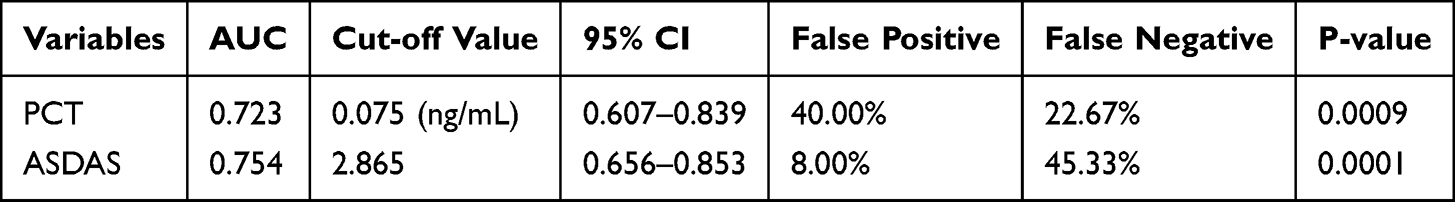 |
Table 3 ROC Analysis of PCT (0.05–0.5 ng/mL), ASDAS Scores in Differentiating Whether Hip Involvement Occurs in AS Patients |
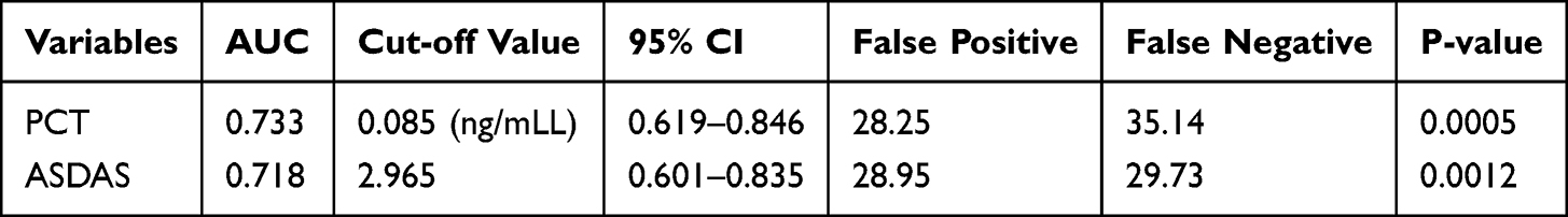 |
Table 4 ROC Analysis of PCT and ASDAS Scores in Differentiating the Severity of Hip Involvement in AS Patients |
 |
Figure 3 ROC curves of PCT and ASDAS in distinguishing Hip involvement in AS patients. |
 |
Figure 4 ROC curves of PCT and ASDAS in differentiating the severity of Hip involvement in AS patients. |
Multifactorial Analysis of Factors Affecting r-axSpA Hip Involvement
Next we performed a multifactorial logistic regression analysis, assigning a value of 0 to no involvement and a value of 1 to hip involvement. Factors with a significant difference of P < 0.05 in the above results were included in the multifactorial binary logistic regression analysis: age (P = 0.0063), BMI (Body Mass Index) (P = 0.0017), BASRI (Bath Ankylosing Spondylitis Radiology Index) (P < 0.0001), CRP (P = 0.0001), PCT (P < 0.0001), and ADDAS (P < 0.0001). Since BASDAI and ESR were reported to be statistically significant in r-axSpA hip involvement, these two metrics were also included in the multifactorial regression analysis. After adjusting for confounders, CRP, PCT, ASDAS, and BASFI scores were independently associated with hip involvement (Table 5).
 |
Table 5 Binary Logistic Regression Analysis of Hip Involvement in AS Patients |
Discussion
R-axSpA mainly involves the spine and sacroiliac joints, with the common symptoms of lumbosacral stiffness and pain.21 Hip joint involvement is also a common manifestation and one of the most significant causes of disability among AS patients.22 Hip joint involvement in r-axSpA indicates a gradual aggravation of the disease, which in severe cases can lead to joint deformity and disability.23,24 Early diagnosis of r-axSpA hip involvement is important for early intervention and reduction of disability. In patients with r-axSpA, elevated PCT levels may reflect an increased inflammatory state, which is closely related to disease activity and joint involvement. Among patients with r-axSpA, those with combined hip involvement have significantly higher levels of CRP than those without combined hip involvement in terms of laboratory markers, suggesting that hip involvement may be associated with higher levels of inflammatory response.22,25 Although the study did not directly mention changes in PCT levels, CRP and PCT are usually elevated together during inflammation.26 These findings imply that PCT levels may be associated with hip involvement in patients with r-axSpA, and that higher PCT levels may indicate a more active inflammatory process. Our findings validated the conjecture, as serum PCT was significantly elevated in r-axSpA patients compared with healthy controls. Furthermore, in a subgroup of r-axSpA patients with different degrees of hip involvement, we found that the higher the degree of hip involvement, the higher the level of PCT.
ASDAS has been performed using the BASDAI27,28 BASDAI is a combination of six indicators based on patient self-report, whereas ASDAS is a composite scoring system that combines patient self-report and laboratory markers of inflammation (CRP, ESR). BASDAI focuses on the patient’s subjective experience, whereas ASDAS provides a more comprehensive assessment by integrating CRP or ESR.29 In our study, ASDAS increased with increasing hip involvement in all three subgroups. Similar to our results, previous studies have found that patients with severe r-axSpA have higher ASDAS.6,30 We performed Spearman correlation analyses and found that PCT levels and ASDAS were positively correlated with the degree of r-axSpA hip involvement, respectively. In addition, there was also a correlation between PCT levels and ASDAS. PCT has been reported to be positively correlated with ASDAS in patients with r-axSpA, but no significant difference can be observed.16,31 Our data showed a smaller correlation coefficient between PCT levels and ASDAS, and the reason for statistical significance may be that our study had more cases. Finally, PCT level and ASDAS had diagnostic value in hip involvement and disease severity. These results suggest that the monitoring of serum PCT levels in patients with r-axSpA not only reflects inflammation, but may also predict fluctuations in disease activity, which is potentially valuable for the early recognition of disease progression.
Finally, our study has some limitations yet. Although some confounders were adjusted in logistic regression, others like concomitant infections, metabolic disorders, or medication effects (NSAIDs, biologics, DMARDs) were not deeply explored. These could influence both PCT and ASDAS. While PCT is shown to correlate with disease severity, its clinical applicability remains uncertain. First, this is a single-center study with a small sample size. Second, we excluded patients with co-infections. Future studies involving more patients with active r-axSpA and co-infections in patients with active r-axSpA will help to further evaluate the clinical relevance of PCT. Then, surgery or trauma may increase PCT levels, which should be measured repeatedly. And we did not follow the response to treatment and the dynamic changes in PCT levels and ASDAS after treatment. Although our study initially revealed the potential value of serum PCT in the assessment of r-axSpA hip involvement, further in-depth studies are needed in this area. Future studies should focus on the dynamic changes of PCT in different disease stages and treatment responses, as well as its comparison with other inflammatory markers, to determine the optimal use of PCT in the clinical management of r-axSpA. In addition, exploring the exact mechanism of PCT in r-axSpA, including its specific role in the inflammatory response, will help us to gain a deeper understanding of the pathophysiologic process of r-axSpA and provide new ideas for disease prevention and treatment.
Data Sharing Statement
The datasets used and/or analyzed during the present study are available from the corresponding author on reasonable request.
Ethics Approval
The present study was approved by the Ethics Committee of The First Affiliated Hospital of Guangzhou University of Chinese Medicine and written informed consent was provided by all patients prior to the study start. All procedures were performed in accordance with the ethical standards of the Institutional Review Board and The Declaration of Helsinki, and its later amendments or comparable ethical standards.
Funding
There is no funding to report.
Disclosure
The authors have no conflicts of interest to declare.
References
1. Braun J, Sieper J. Ankylosing spondylitis. Lancet. 2007;369(9570):1379–1390. doi:10.1016/S0140-6736(07)60635-7
2. Ward MM, Deodhar A, Gensler LS, et al. 2019 Update of the American College of Rheumatology/Spondylitis Association of America/Spondyloarthritis Research and Treatment Network recommendations for the treatment of ankylosing spondylitis and nonradiographic axial spondyloarthritis. Arthritis Rheumatol. 2019;71(10):1599–1613. doi:10.1002/art.41042
3. Ranganathan V, Gracey E, Brown MA, Inman RD, Haroon N. Pathogenesis of ankylosing spondylitis - recent advances and future directions. Nat Rev Rheumatol. 2017;13(6):359–367. doi:10.1038/nrrheum.2017.56
4. Wink F, Arends S, Maas F, et al. High prevalence of Hip involvement and decrease in inflammatory ultrasound lesions during tumour necrosis factor-alpha blocking therapy in ankylosing spondylitis. Rheumatology. 2019;58(6):1040–1046. doi:10.1007/s00296-012-2510-5
5. Chen S, Deng L. Risk factors for radiological hip involvement in patients with ankylosing spondylitis. Rev Assoc Med Bras. 2021;67(9):1293–1298. doi:10.1590/1806-9282.20210585
6. Yilmaz O, Tutoglu A, Garip Y, Ozcan E, Bodur H. Health-related quality of life in Turkish patients with ankylosing spondylitis: impact of peripheral involvement on quality of life in terms of disease activity, functional status, severity of pain, and social and emotional functioning. Rheumatol Int. 2013;33(5):1159–1163.
7. Ugalde PF, Gomariz EM, Estevez EC; por el Grupo Ie. [Indication for anti-TNF-alpha treatment in patients with ankylosing spondylitis in Spain]. Reumatol Clin. 2007;3(6):251–256. Estonian. doi:10.1016/S1699-258X(07)73699-9
8. Singh G, Lawrence A, Agarwal V, Misra R, Aggarwal A. Higher prevalence of extra-articular manifestations in ankylosing spondylitis with peripheral arthritis. J Clin Rheumatol. 2008;14(5):264–266. doi:10.1097/RHU.0b013e31817b8789
9. Vander Cruyssen B, Vastesaeger N, Collantes-Estevez E. Hip disease in ankylosing spondylitis. Curr Opin Rheumatol. 2013;25(4):448–454. doi:10.1097/BOR.0b013e3283620e04
10. Maruna P, Nedelnikova K, Gurlich R. Physiology and genetics of procalcitonin. Physiol Res. 2000;49(Suppl 1):S57–61.
11. Wacker C, Prkno A, Brunkhorst FM, Schlattmann P. Procalcitonin as a diagnostic marker for sepsis: a systematic review and meta-analysis. Lancet Infect Dis. 2013;13(5):426–435. doi:10.1016/S1473-3099(12)70323-7
12. Lee H. Procalcitonin as a biomarker of infectious diseases. Korean J Intern Med. 2013;28(3):285–291. doi:10.3904/kjim.2013.28.3.285
13. Ozmen M, Oktay E, Tarhan EF, Aslan O, Oflazoglu U, Koseoglu MH. Serum procalcitonin levels in patients with ankylosing spondylitis. Int J Rheum Dis. 2016;19(5):500–505. doi:10.1111/1756-185X.12386
14. Okada Y, Minakami H, Tomomasa T, et al. Serum procalcitonin concentration in patients with Kawasaki disease. J Infect. 2004;48(2):199–205. doi:10.1016/j.jinf.2003.08.002
15. Liu W, Sigdel KR, Wang Y, et al. High level serum procalcitonin associated gouty arthritis susceptibility: from a Southern Chinese Han population. PLoS One. 2015;10(7):e0132855. doi:10.1371/journal.pone.0132855
16. Liu Y, Shi J, Wang B, et al. Combining calcitonin and procalcitonin and rheumatoid arthritis-related biomarkers improve diagnostic outcomes in early rheumatoid arthritis. Dis Markers. 2021;2021:6331994. doi:10.1155/2021/6331994
17. Bruera S, Ventura MJ, Agarwal SK, Krause KJ, Lopez-Olivo MA. The utility of erythrocyte sedimentation rate, C-reactive protein, and procalcitonin in detecting infections in patients with systemic lupus erythematosus: a systematic review. Lupus. 2022;31(10):1163–1174. doi:10.1177/09612033221106157
18. van der Linden S, Valkenburg HA, Cats A. Evaluation of diagnostic criteria for ankylosing spondylitis. A proposal for modification of the New York criteria. Arthritis Rheum. 1984;27(4):361–368. doi:10.1002/art.1780270401
19. Fernandez-Espartero C, de Miguel E, Loza E, et al. Validity of the ankylosing spondylitis disease activity score (ASDAS) in patients with early spondyloarthritis from the Esperanza programme. Ann Rheum Dis. 2014;73(7):1350–1355. doi:10.1136/annrheumdis-2012-202976
20. Nhan DT, Caplan L. Patient-reported outcomes in axial spondyloarthritis. Rheum Dis Clin North Am. 2016;42(2):285–299. doi:10.1016/j.rdc.2016.01.011
21. MacKay K, Brophy S, Mack C, Doran M, Calin A. The development and validation of a radiographic grading system for the Hip in ankylosing spondylitis: the bath ankylosing spondylitis radiology hip index. J Rheumatol. 2000;27(12):2866–2872.
22. Zhao J, Zheng W, Zhang C, Li J, Liu D, Xu W. Radiographic Hip involvement in ankylosing spondylitis: factors associated with severe hip diseases. J Rheumatol. 2015;42(1):106–110. doi:10.3899/jrheum.140428
23. Katakam A, Bedair HS, Melnic CM. Do all rigid and unbalanced spines present the same risk of dislocation after total hip arthroplasty? A comparison study between patients with ankylosing spondylitis and history of spinal fusion. J Arthroplasty. 2020;35(12):3594–3600. doi:10.1016/j.arth.2020.06.048
24. Xu Y, Jiang W, Zhang H. Association between C-reactive protein gene variant and treatment efficacy of etanercept in ankylosing spondylitis patients receiving Hip arthroplasty. J Clin Lab Anal. 2020;34(8):e23343. doi:10.1002/jcla.23343
25. Chen HA, Chen CH, Liao HT, et al. Factors associated with radiographic spinal involvement and hip involvement in ankylosing spondylitis. Semin Arthritis Rheum. 2011;40(6):552–558. doi:10.1016/j.semarthrit.2010.07.008
26. Liu QH, Song MY, Yang BX, Xia RX. Clinical significance of measuring reticulated platelets in infectious diseases. Medicine. 2017;96(52):e9424. doi:10.1097/MD.0000000000009424
27. Garrett S, Jenkinson T, Kennedy LG, Whitelock H, Gaisford P, Calin A. A new approach to defining disease status in ankylosing spondylitis: the bath ankylosing spondylitis disease activity index. J Rheumatol. 1994;21(12):2286–2291.
28. da Costa IP, Bortoluzzo AB, Goncalves CR, et al. [Evaluation of performance of BASDAI (Bath Ankylosing Spondylitis Disease Activity Index) in a Brazilian cohort of 1,492 patients with spondyloarthritis: data from the Brazilian Registry of Spondyloarthritides (RBE)]. Rev Bras Reumatol. 2015;55(1):48–54. doi:10.1016/j.rbr.2014.05.005
29. Nam B, Koo BS, Lee TH, et al. Low BASDAI score alone is not a good predictor of anti-tumor necrosis factor treatment efficacy in ankylosing spondylitis: a retrospective cohort study. BMC Musculoskelet Disord. 2021;22(1):140. doi:10.1186/s12891-020-03941-8
30. Hu Y, Jiang WZ, Pan CL, Wang T. Active ankylosing spondylitis increases blood loss during total hip arthroplasty for a stiff hip joint. BMC Musculoskelet Disord. 2020;21(1):243. doi:10.1186/s12891-020-03278-2
31. Moldovan F. Role of serum biomarkers in differentiating periprosthetic joint infections from aseptic failures after total hip arthroplasties. J Clin Med. 2024;13(19):5716. doi:10.3390/jcm13195716
 © 2025 The Author(s). This work is published and licensed by Dove Medical Press Limited. The
full terms of this license are available at https://www.dovepress.com/terms
and incorporate the Creative Commons Attribution
- Non Commercial (unported, 4.0) License.
By accessing the work you hereby accept the Terms. Non-commercial uses of the work are permitted
without any further permission from Dove Medical Press Limited, provided the work is properly
attributed. For permission for commercial use of this work, please see paragraphs 4.2 and 5 of our Terms.
© 2025 The Author(s). This work is published and licensed by Dove Medical Press Limited. The
full terms of this license are available at https://www.dovepress.com/terms
and incorporate the Creative Commons Attribution
- Non Commercial (unported, 4.0) License.
By accessing the work you hereby accept the Terms. Non-commercial uses of the work are permitted
without any further permission from Dove Medical Press Limited, provided the work is properly
attributed. For permission for commercial use of this work, please see paragraphs 4.2 and 5 of our Terms.