Back to Journals » Clinical Ophthalmology » Volume 17
Corneal Outcomes Following Cataract Surgery Using Ophthalmic Viscosurgical Devices Composed of Chondroitin Sulfate-Hyaluronic Acid: A Systematic Review and Meta-Analysis
Authors Hsiao CW, Cheng H, Ghafouri R, Ferko NC, Ayres BD ![]()
Received 25 May 2023
Accepted for publication 11 July 2023
Published 24 July 2023 Volume 2023:17 Pages 2083—2096
DOI https://doi.org/10.2147/OPTH.S419863
Checked for plagiarism Yes
Review by Single anonymous peer review
Peer reviewer comments 2
Editor who approved publication: Dr Scott Fraser
Chia-Wen Hsiao,1 Hang Cheng,1 Rana Ghafouri,2 Nicole C Ferko,2 Brandon D Ayres3
1Alcon, Fort Worth, TX, USA; 2Eversana, Burlington, ON, Canada; 3Thomas Jefferson University Hospital, Philadelphia, PA, USA
Correspondence: Nicole C Ferko, Eversana Life Science Services, 3228 S Service Road, Burlington, ON, Canada, Tel +1 (905) 978-3111, Email [email protected]
Background: Ophthalmic viscosurgical devices (OVDs) are commonly used during cataract surgery to protect the corneal endothelium. A systematic literature review and meta-analysis were conducted to assess the clinical evidence of OVDs composed of chondroitin sulfate-hyaluronic acid (CS-HA) versus other OVDs in maintaining endothelial cell density (ECD) and corneal thickness (CT).
Methods: MEDLINE and EMBASE databases were searched from 2000 to 2020. Randomized controlled trials (RCTs, N ≥ 20 per group) comparing an OVD containing CS-HA (ie, VISCOAT®, DuoVisc® or DisCoVisc®) to any other OVD were included. The identified comparators were limited to the OVDs found in the literature, which included those composed of HA-only or hydroxypropyl methylcellulose (HPMC). Outcomes of focus included changes in ECD (baseline to 3 months) and CT (baseline to 24 hours). Meta-analyses were performed using R software, to assess mean differences (MD) in ECD and CT change between CS-HA OVDs and HA-only or HPMC OVDs.
Results: A total of 966 abstracts were screened, and data were extracted from 12 RCTs. Meta-analyses using a random-effects model revealed significantly lower percent (%) decrease in ECD for CS-HA OVDs compared to both HA-only (MD: − 4.10%; 95% CI: − 5.81 to − 2.40; p < 0.0001; 9 studies) and HPMC (MD: − 6.47%; 95% CI: − 10.41 to − 2.52; p = 0.001; 2 studies) products. Similarly, % CT increase was significantly lower with CS-HA than with HA-only OVDs (MD: − 3.22%; 95% CI: − 6.24% to − 0.20%; p = 0.04; 4 studies). However, there were no significant differences when comparing % CT change between CS-HA and HPMC OVDs (MD: 2.65%; 95% CI: − 0.43% to 0.95%; p = 0.4; 2 studies).
Conclusion: CS-HA OVDs lead to less postoperative loss of endothelial cells and may better protect corneal endothelium during cataract surgery, relative to other OVDs. Future randomized studies may be needed to solidify these findings.
Keywords: cataract, ophthalmic viscosurgical device, endothelial cell density, corneal thickness, endothelial protection
Introduction
The cornea, the transparent front layer of the eye, serves to protect inner contents and provide focusing and refractive power.1 The corneal endothelial cell layer functions to maintain the cornea in a dehydrated state, which is necessary for corneal clarity and transparency.2,3 A critical minimum cell density between 300 and 500 cells/mm2 is required to maintain proper functioning of the endothelium; a reduction below this range leads to severe corneal edema and bullous keratopathy, which may result in visual dysfunction and pain due to corneal decompensation.4 Corneal endothelial cells deteriorate gradually as a normal part of aging, but trauma to the cornea may result in considerable cell loss.2,3 Manipulation of the anterior segment of the eye during intraocular surgical procedures can cause damage to endothelial cells. As one of the most commonly performed anterior segment procedures, cataract surgery is a notable source of such damage.2 Endothelial cell loss (ECL) after uncomplicated cataract surgery has been reported to range from 4% to 25% of surgical cases.5–7 Risk factors for endothelial cell damage during cataract surgery include highly dense cataracts, short axial length, shallow anterior chamber, advanced age, long phacoemulsification time, and large infusion volume. The rate of ECL after cataract surgery is even greater in patients especially prone to cell loss, such as those with Fuchs corneal dystrophy and diabetes.4,5,8,9 Since corneal endothelial cells have limited regenerative capacity,3,5 protection is critical during intraocular surgery to minimize cell loss and avoid the negative and costly consequences associated with decompensation.
The major contributor to postoperative ECL is mechanical trauma, such as contact with surgical instruments, lens fragments, or “biophysical” adhesive effects between the intraocular lens (IOL) and the corneal endothelium.2 Ophthalmic viscosurgical devices (OVDs) are substances that are injected into the eye during cataract surgery, which serve to protect the corneal endothelium by reducing surgical trauma.10–12 OVDs are well established for use during cataract surgery in order to coat surgical instruments to minimize the risk of damage to intraocular tissue, create space and maintain stability in the anterior chamber, and protect the endothelium against fluid turbulence, free radicals, air bubbles, and lens fragmentation.10,11,13–15
A variety of OVDs exist on the market, each with different levels of viscosity, elasticity and cohesion based on their different rheologically active polymeric substances, concentrations, and chain lengths.10,16 An effective OVD should have the ability to coat the corneal endothelium and create space in the anterior chamber,16 thus viscosity and cohesion are considered the most important physical characteristics and are used for OVD classification. Dispersive OVDs have low viscosity and high retention and are mainly used to coat intraocular structures and instruments to protect the eye, while cohesive OVDs have high viscosity and low retention, and are mainly used to create and maintain space as well as manipulate tissues.16,17 Other classes of OVDs include viscoadaptives which change in rheologic behavior under varying conditions of turbulence in the eye, as well as viscous dispersives, which demonstrate properties of both higher and lower viscosity dispersives to adapt to surgical needs.16 The soft-shell technique sequentially uses dispersive and cohesive OVDs to maximize their respective properties.17
Current OVDs used in the surgical setting are composed of one or more of hyaluronic acid (HA), chondroitin sulfate (CS), and hydroxypropyl methylcellulose (HPMC). Hyaluronate is a natural biological wetting substance found in the vitreous of the eye, and its high viscosity and high molecular weight provide desirable characteristics for creating space and stability.16,18,19 CS is a biopolymer that can naturally be found in extracellular matrices of the human cartilage and cornea; it reduces molecular weight and provides lower viscosity when mixed with HA, which serves to promote adhesion to the endothelial tissue for protection.16,20,21 HPMC is a semi-synthetic derivative of cellulose that has very low viscosity, making it highly dispersive.22 Unlike HA and CS, HPMC is not naturally found in humans and thus cannot be fully metabolized. In OVDs, a combination of CS and HA creates a triple negative charge, promoting molecular attraction to corneal endothelial tissue and allowing the OVD to both disperse and stick to the endothelium, which may improve coating and protection of endothelial cells.21 There is evidence that residual deposits of HPMC after cataract extraction can lead to inflammation.18,23 In contrast, CS has been shown to reduce inflammation in the body and has been explored as therapy for a variety of inflammatory disorders such as osteoarthritis, psoriasis, and inflammatory bowel disease.24
Previous meta-analyses evaluating clinical outcomes of OVDs have not compared OVDs based on their composition. Further, these meta-analyses are outdated12 or do not evaluate OVDs based on important outcomes such as ECL.11 The aim of the current study was to conduct a systematic review and meta-analysis of randomized-controlled trials (RCTs) to assess outcomes of OVDs by categorizing OVDs based on their constituents and comparing their ability to protect the corneal endothelium (ie, CS-HA, HA alone, and HPMC), by assessing changes to endothelial cell density (ECD) and corneal thickness (CT).
Materials and Methods
Search Strategy
This systematic review and meta-analysis followed the Preferred Reporting Items for Systematic Reviews and Meta-Analyses (PRISMA) guidelines. A systematic search of MEDLINE and EMBASE was conducted for relevant systematic reviews, randomized controlled trials (RCTs), and observational studies (prospective or retrospective cohort and case–control studies) using a search strategy developed by a medical information specialist that involved controlled vocabulary and keywords related to our research question (eg, “ophthalmic viscosurgical device”, “viscoelastic substance”, “cataract extraction”, “eye surgery”) (Appendix 1). The original search strategy was limited to English language articles published on or after January 1, 1996. The search was performed on November 20, 2020.
Study Selection
Studies were selected for inclusion based on pre-defined PICOS criteria (ie, population, intervention, comparator, outcomes, and study design). Studies were considered for inclusion in the meta-analysis if they were RCTs evaluating effects of CS-HA OVDs compared to other OVD types in patients undergoing cataract surgery. These criteria were used to screen the titles and abstracts of publications to determine whether they were eligible for inclusion. Studies deemed eligible upon title and abstract screening were screened in full-text. Additional exclusion criteria for the meta-analysis were studies that were outdated (ie, published before January 1, 2000), studies with small sample size (ie, N < 20 per treatment group), and studies not evaluating ECD and CT. Publications were reviewed in duplicate at each stage, and discrepancies were resolved by consensus or by adjudication by a third reviewer.
Data Extraction and Study Outcomes
Baseline characteristics and outcomes from the included studies were extracted using a standardized extraction form developed in Microsoft Excel. Key study characteristics were extracted, including preoperative, postoperative and change measurements for clinical outcomes. Where not reported, the percentage change was calculated by dividing the difference between postoperative and preoperative values by the preoperative value and multiplying by one hundred (Appendix 2, Equation 1). For values where the standard deviation (SD) was not reported, SDs were estimated based on the reported variances in other studies, using the Cochrane Handbook guidelines for imputing standard deviations.25 Variances for percentage changes were calculated using a formula derived from the Delta method (Appendix 2, Equation 2). Data were extracted by one reviewer and then examined for accuracy and completeness by a second reviewer.
The outcomes of interest were changes in ECD from baseline (preoperative) to 3 months postoperative and changes in CT from preoperative values to 1 day postoperative. For studies reporting different timepoints, the latest timepoint for ECD and the earliest timepoint for CT were extracted. For ECD, 3 months was selected as the timepoint of interest, as postoperative cell loss tends to occur in the first three months,26 and it is the most commonly reported timepoint for ECL. The timepoint of 1 day was selected for CT, as corneal swelling at 24 hours has been correlated to long-term ECL,27 and this was the most commonly reported timepoint for CT. Percentage change was selected for the meta-analysis as a relative change accounts for any variance in baseline values and offers more clinically relevant interpretation of results.
Risk of Bias Assessment
The quality of studies included in the meta-analyses was assessed using the Cochrane Risk of Bias (RoB) tool for RCTs. The following components were assessed to be present, absent, or unclear (not specified) in each RCT: randomization, concealment of treatment allocation, blinding, group variance, and outcomes not reported (Table 1). A rating of high risk, unclear risk, or low risk was assigned across the categories. The quality of included studies was assessed by a single reviewer with a second reviewer consulted for questions or uncertainties.
 |
Table 1 Quality Appraisal of Included Studies |
Data Synthesis and Statistical Methods
Pairwise meta-analyses were performed for each head-to-head comparison. A random effects model was the primary methodology used for presentation of the meta-analysis results, and forest plots were created. The weighted mean difference (MD) for continuous outcomes and corresponding 95% confidence interval (CI) were calculated.
I2 values were calculated to describe the percentage of variance attributable to heterogeneity between studies. The following ranges were used to interpret I2 values regarding the degree of heterogeneity present between the synthesized studies for each comparison: 0–40% represented minimal heterogeneity, 30–60% represented moderate heterogeneity, 50–90% represented substantial heterogeneity, and 75–90% represented considerable heterogeneity.25 If I2 value was from 75 to 90%, but the CIs for the effect measures overlapped between studies, then the heterogeneity was classified as substantial. If the I2 was from 75 to 90% and the CIs for the effect measures did not overlap between studies, then the heterogeneity was classified as considerable.
Sub-group analyses were performed for both ECD and CT if there were at least two studies in each group. The subgroup analyses included (1) CS-HA versus HA OVDs, (2) CS-HA versus HPMC OVDs, (3) dispersive versus cohesive OVDs (determined by manufacturer labelling), and (4) soft-shell versus other OVDs. For the latter subgroup analysis, any study that included the soft-shell technique (ie, use of a dispersive OVD to coat the endothelium followed by a cohesive OVD to flatten the anterior lens capsule) was included.
Sensitivity analyses were performed to assess alternative methods (ie, fixed effects model) and study quality (ie, exclusion of lower-quality studies, defined as any RCT with high risk for any domain of the RoB tool).
All analyses were performed using R Statistical Software version 3.6.1.
Results
Study Selection and Characteristics
A total of 1278 studies were identified from the systematic review. After removing duplicates, 966 studies published between January 1996 and November 2020 were identified for screening (Figure 1). Of these, 845 were excluded for various reasons (eg, non-cataract surgery, non-RCT, non-human, not English), and 121 were screened at the full-text stage. In full-text screening, 106 were excluded for date of publication (before January 1, 2000), not reporting any outcomes of interest (ie, ECD or CT), or having N < 20 per comparison group. In total, 14 RCTs were identified that fulfilled the inclusion criteria. An additional two studies were excluded for having multiple comparators due to inappropriate data for comparative purposes. Finally, 12 unique studies were included for meta-analysis, with 11 reporting on ECD and 6 reporting CT outcomes. Of these, 10 compared CS-HA OVDs to HA OVDs.28–30,32–37,39 and 2 compared CS-HA OVDs to OVDs composed of HPMC.31,38
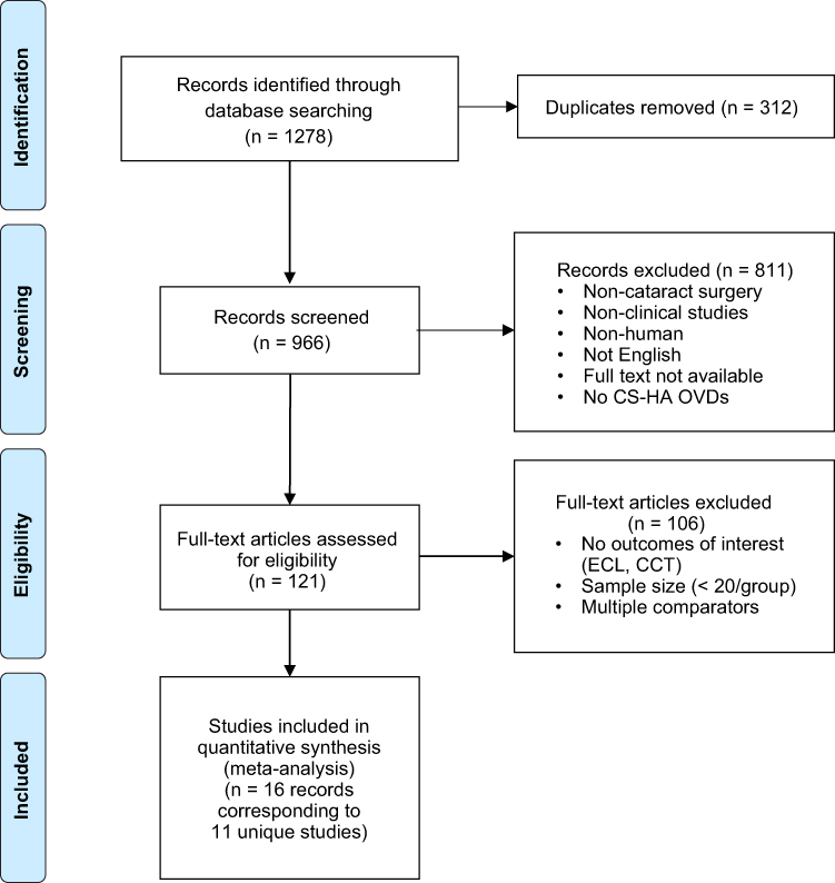 |
Figure 1 Prisma diagram of included studies. |
Percent change was reported in 2 studies for ECD;31,35 for the remaining 10 studies, percent change and variance were calculated for ECD and CT according to reported baseline and follow-up outcome results. Study characteristics are summarized in Table 2. In total, the combined sample size of patients totaled 1349 (ranging 20–163 per group) for ECD and 556 (ranging 20–111 per group) for CT, with CS-HA OVDs used in 691 and 287 patients, respectively. Statistically significant differences between OVDs were reported in 5 studies evaluating ECD30,33,35,37,38 and 2 studies reporting CT changes.34,37
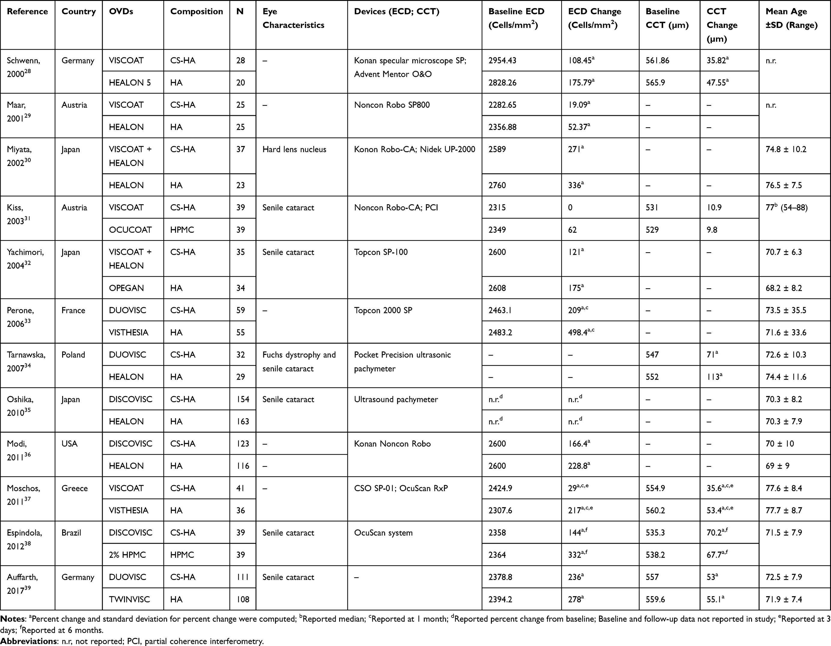 |
Table 2 Characteristics of RCTs Evaluating OVDs in Cataract Surgery Patients |
Quality Assessment
The quality of the included studies was assessed using the National Institute for Health and Care Excellence (NICE) risk of bias tool and the risk of bias algorithm outlined by the Cochrane guidelines. Based on seven prespecified domains (randomization, allocation concealment, baseline group differences, blinding of participants and personnel, blinding of outcome assessment, selective outcome reporting), publications were scored as having low, unclear, or high risk of bias. The results of the quality assessment are summarized in Figure 2. Blinding of outcome assessors was specified in 9 of the 12 studies evaluated,28,29,31–33,35,36,38,39 and blinding of participants was noted in 5 studies.31–33,36,38 Due to the differences in physical properties of OVDs, blinding of care providers was not possible, and adequate concealment of treatment allocation was specified in 5 studies (Table 1).28,35,36,38,39 One study evaluating CT was performed in a patient population with a high-risk of corneal endothelial dysfunction due to Fuchs dystrophy.34
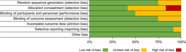 |
Figure 2 Risk of bias assessment for studies meeting inclusion criteria. |
Analyses
Endothelial Cell Density
The results of the meta-analysis for ECD are shown in Figure 3. OVDs composed of CS-HA were associated with significantly lower endothelial cell loss (ECL) from baseline to 3 months postoperatively when compared to all other OVD types (MD: −4.54%; 95% CI: −6.30% to −2.77%; p < 0.0001; 11 studies; Figure 3A).28–33,35–39 When compared to HA-only OVDs, CS-HA OVDs had significantly lower ECL (MD: −4.10%; 95% CI: −5.81 to −2.40; p < 0.0001; 9 studies; Figure 3B).28–30,32,33,35–37,39 Similarly, ECL was significantly lower with CS-HA OVDs compared to HPMC OVDs (MD: −6.47%; 95% CI: −10.41 to −2.52; p = 0.001; 2 studies; Figure 3C).31,38
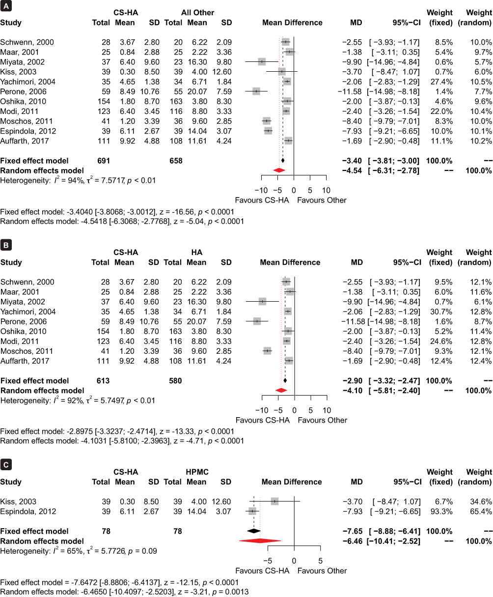 |
Figure 3 Forest plot of meta-analysis results for percent change in ECD from baseline to 3 months postoperative with CS-HA OVDs versus other OVDs. (A) Comparison between CS-HA and all other OVDs. (B) Comparison between CS-HA and HA. (C) Comparison between CS-HA and HPMC. |
Corneal Thickness
Figure 4 summarizes the meta-analysis results for CT change. The meta-analysis revealed that when compared against other OVDs, those containing CS-HA demonstrated significantly less increase in CT from baseline to 1 day postoperatively with the fixed effect model (MD: −1.47%; 95% CI: −1.89% to −1.05%; p < 0.0001; 6 studies) but did not reach statistical significance at the 5% level with the random effects model (MD: −2.02%; 95% CI: −4.24% to 0.18%; p = 0.07; 6 studies; Figure 4A).28,31,34,37–39 When compared to HA-only OVDs, CS-HA OVDs had statistically significantly less CT change with the random effects model (MD: −3.22%; 95% CI: −6.24% to −0.20%; p = 0.04; 4 studies; Figure 4B).28,34,37,39 However, meta-analysis revealed no significant difference between CS-HA OVDs and those composed of HPMC (MD: 2.65%; 95% CI: −0.43% to 0.95%; p = 0.4; 2 studies; Figure 4C).31,38
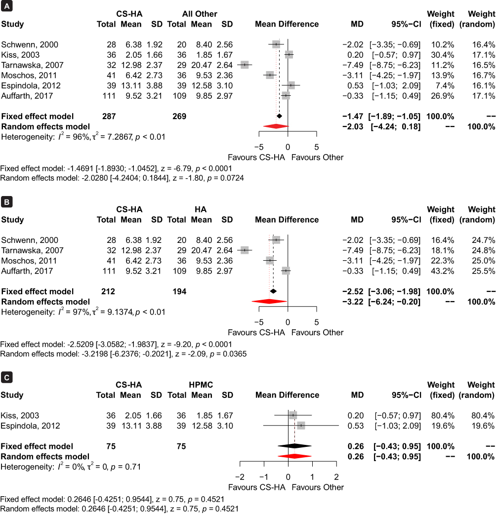 |
Figure 4 Forest plot of meta-analysis results for percent CT change from baseline to 1 day postoperative with CS-HA OVDs versus other OVDs. (A) Comparison between CS-HA and all other OVDs. (B) Comparison between CS-HA and HA. (C) Comparison between CS-HA and HPMC. |
Other Subgroup analyses
In additional subgroup analyses of ECD and CT changes evaluated in specific OVD types, such as soft-shell and dispersive OVDs, results remained in favor of soft-shell technique and CS-HA OVDs. Compared with other OVDs without CS-HA, meta-analysis revealed that the soft-shell technique with CS-HA had significantly less ECL at 3 months (MD: −2.78%; 95% CI: −4.70% to −0.86%; p < 0.005; 3 studies; Figure 5A).30,32,39 When evaluating CT, Soft-shell OVDs (Duovisc) had significantly less CT increase 1 day after surgery with the fixed effect model (MD: −2.44%; 95% CI: −3.12% to −1.75%; p < 0.0001), but this change was not statistically significant with the random effects model (MD: −3.89%; CI: −10.90% to 3.12%; p = 0.28; 2 studies; Figure 5B).34,39
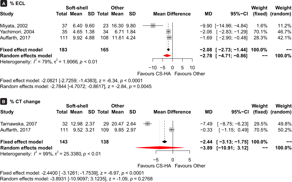 |
Figure 5 Forest plot of meta-analysis results for Soft-shell versus other OVDs. (A) Comparison between Soft-shell and other OVDs for percent ECL. (B) Comparison between Soft-shell and other OVDs for percent CT change. |
Similarly, dispersive CS-HA OVDs demonstrated significantly less reduction in ECD than comparator OVDs (MD: −4.05%; 95% CI: −7.78% to −0.31%; p = 0.03; 4 studies; Figure 6A).28,29,31,37 Meta-analysis for CT change revealed that dispersive OVDs composed of CS-HA had significantly less of an increase in CT when using the fixed effect model (MD: −1.06%; 95% CI: −1.63% to −0.48%; p = 0.0003), but this difference was not statistically significant with the random effect model (MD: −1.61%; 95% CI: −3.77% to 0.55%; p = 0.1; 3 studies; Figure 6B).28,31,37
 |
Figure 6 Forest plot of meta-analysis results for VISCOAT versus other OVDs. (A) Comparison between VISCOAT and other OVDs for percent ECL. (B) Comparison between VISCOAT and other OVDs for percent CT change. |
Discussion
This systematic review and meta-analysis assessed the effect of various OVDs in maintaining ECD and CT in patients undergoing cataract surgery. The results of this meta-analysis demonstrate that OVDs composed of CS-HA generally result in better outcomes for the corneal endothelium compared to other OVD types. Specifically, CS-HA OVDs resulted in less postoperative loss of corneal endothelial cells at 3 months compared to OVDs composed of HA-only or HPMC. Additionally, the analysis showed that OVDs composed of CS-HA caused less increase in CT at 1-day postoperatively compared to HA-only OVDs. Further, subgroup analyses revealed that the soft-shell technique using CS-HA similarly led to significantly less cell loss at 3 months following cataract surgery. These findings suggest that the protective effect of CS-HA OVDs on ECD may be related to its impact on CT. However, it is important to note that the relationship between ECD and CT is complex and multifactorial, and other factors such as age, underlying medical conditions, and surgical techniques may also affect these outcomes.4,5,8,9
Only two previous meta-analyses assessing outcomes of OVDs have been published to date. These previous studies have not considered OVDs based on their constituents, which is a notable element that plays a big role in the functioning of OVDs. The current study also differed from the previous literature by exploring different comparison arms and/or outcomes of interest. One earlier meta-analysis by Van den Bruel et al assessed ECL across 21 studies, comparing OVDs based on Arshinoff classifications (eg, viscoadaptives, viscous cohesives, low viscosity dispersives, etc.).12 The study, published in 2011, found that that viscoadaptives lead to lower cell loss compared with very low viscosity dispersives and super-viscous cohesives.12 Conversely, our subgroup analysis showed that dispersive OVDs led to less endothelial cell density loss when compared to other OVDs. This earlier meta-analysis only captured RCTs from 1991 to 2007; as such, the results may be outdated due to relatively lower sample sizes. The discrepant results may be partially due to the inclusion of older studies which may use outdated technologies and surgical methods, with newer studies not being captured, as well as methodological study differences. Notably, our meta-analysis compared relative changes in ECD and CT by evaluating percentage changes from preoperative to postoperative values, thus accounting for any baseline differences between patients, while Van den Bruel et al conducted comparisons using absolute differences between preoperative and postoperative values.12
Our current meta-analysis did not assess intraocular pressure (IOP), as the results of a more recent meta-analysis by Malvankar-Mehta et al compared OVDs based on changes in IOP and best corrected visual acuity (BCVA) across 28 RCTs. However, the outcomes of ECD and CT were not assessed and remained an area to be explored.11 Malvankar-Mehta et al conducted single-arm meta-analyses of specific OVDs (eg, Healon, Viscoat, OcuCoat, etc.), and the results showed that there is a significant increase in post-operative IOP up to 5 hours postoperative, irrespective of OVD used. Malvankar-Mehta et al noted that there is a significant increase in postoperative IOP at 1-day follow-up with Healon, Viscoat, and Soft-shell, nonsignificant increase with Healon GV, Healon5, 2% HPMC, and OcuCoat, and a nonsignificant decrease with Viscoat + ProVisc.11 Notably, Viscoat is a CS-HA OVD and is combined with ProVisc for the soft-shell technique, and interestingly, this specific soft-shell combination led to a decrease in IOP, while increased IOP was identified with other soft-shell combinations. One limitation of the study by Malvankar-Mehta et al was the combination of comparative and single-arm studies analyzed, as the relative effect of treatment cannot be estimated with the lack of comparator in single-arm studies, leading to lower-quality results.
The primary outcome of the current meta-analysis showed that OVDs made of a combination of CS and HA may lead to lower rates of corneal endothelial cell loss after cataract surgery, compared to those composed of HA alone or HPMC. The ingredients used to produce OVDs differ in rheologic properties, thus affecting important characteristics of OVDs, such as viscosity, cohesion, elasticity and pseudoplasticity. Considering the differing and unique compositions of OVDs, it is important for clinicians to take these into account when selecting a product to use. Selection of the most appropriate OVD is particularly critical in challenging cases where patients have comorbidities such as pseudoexfoliation syndrome, Fuchs endothelial dystrophy, diabetes, or intraoperative floppy iris syndrome,4,5,8,9,18,40 or where patients show characteristic risk factors for cell loss such as hard cataracts or flat anterior chambers.7,41 In these cases, preservation of the endothelial cells is of the utmost importance, as these patients are especially vulnerable to corneal endothelial decompensation and bullous keratopathy. In fact, corneal edema that develops after cataract surgery is the second leading cause of visually significant corneal endothelial dysfunction.4 This irreversible corneal edema leads to blurred or loss of vision and may cause severe pain, with the only possible treatment being corneal transplantation.4,42 Corneal transplants are costly, ranging from $6000 to $17,000 in the US, and it has been reported that only 1 in 70 needs for corneal transplantation worldwide are met due to shortage of corneal graft tissue.43–47 Thus, the use of an effective OVD to minimize postoperative cell loss is paramount. Notably, one RCT included in this meta-analysis compared a CS-HA OVD to a HA-only OVD in a patient population with Fuchs endothelial dystrophy.34 In that particular study, compared to HA OVD, the group treated with CS-HA OVD using the soft-shell technique had statistically significantly lower mean CT measured at 1 day, 1 week, 1 month, and 6 months postoperatively, indicating that the soft-shell method with CS-HA OVDs may provide better protection than HA OVDs for the compromised corneal endothelium in patients with Fuchs dystrophy.34
While cohesive OVDs can be used to enlarge pupils and maximize space in the anterior capsule, the literature shows that dispersive OVDs are the preferred choice for prolonged protection of the corneal endothelium.12,15,29 Due to their dispersive nature, attributed to the addition of chondroitin sulfate, CS-HA OVDs are able to successfully coat corneal tissue as well as surgical instruments, providing an adhesive and protective effect.16,18,21
This quantitative systematic review focused only on randomized trials, with low risk of bias generally present across studies, filling a gap in the literature regarding differentiation of OVDs on endothelial cell protection. However, it is important to note that variability seen in the results may be attributed to factors including surgeon experience, cataract severity, choice of phacoemulsifier, and surgical technique. A key limitation of this meta-analysis was the differences and inconsistency in the reporting of outcomes for the studies included (ie, absolute versus relative change, difference values versus preoperative and postoperative values), requiring computation of certain missing variables. Furthermore, high heterogeneity between the studies was present in several of the analyses. This is not unexpected due to the wide variation in patient populations such as subjects with Fuchs endothelial dystrophy or hard lens nucleus in addition to cataract, and variation in surgical methods and study methodologies such as measurement tools or devices used for ECD and CT. Some of these challenges cannot be completely addressed given the limited quantity of overall randomized data; however, we conducted subgroup analyses to help control for some of these differences. Finally, there were limited data on HPMC as a comparator, as only two included RCT evaluated HPMC. As more randomized studies become available, more robust analyses may be achievable. There is important evolution in OVD technology, and remaining abreast of how different constituents, formulations, and combinations help to optimize endothelial cell protection, while minimizing intraocular pressure changes, will be central to inform surgical decision-making in cataract and other anterior ophthalmic surgeries.
Conclusion
In summary, CS-HA OVDs provide effective corneal protection and minimize the loss of valuable corneal endothelial cells following cataract surgery, relative to other OVDs. When compared to HA-only OVDs, those composed of CS-HA resulted in statistically significantly less reduction in ECD, and significantly lower or similar increase in CT. The selection of the most suitable OVD is crucial for optimal postoperative outcomes in patients, especially in complex cases, where CS-HA OVDs should be strongly considered.
Data Sharing Statement
The data supporting this meta-analysis are from previously reported studies and datasets, which have been cited. The processed data are available from the corresponding author upon request.
Acknowledgments
Results from this meta-analysis were presented at the European Society of Cataract and Refractive Surgeons on September 18, 2022.
Funding
This study was sponsored by Alcon Vision, LLC.
Disclosure
Chia-Wen Hsiao and Hang Cheng are employees of Alcon. Rana Ghafouri and Nicole C. Ferko are employees of EVERSANA, a consultancy for various pharmaceutical, medical device, and biotechnology companies. Brandon D. Ayres is a consultant to Alcon. The authors report no other conflicts of interest in this work.
References
1. Meek KM, Knupp C. Corneal structure and transparency. Prog Retin Eye Res. 2015;49:1–16. doi:10.1016/j.preteyeres.2015.07.001
2. Tuft SJ, Coster DJ. The corneal endothelium. Eye. 1990;4(Pt 3):389–424. doi:10.1038/eye.1990.53
3. Ventura AC, Wälti R, Böhnke M. Corneal thickness and endothelial density before and after cataract surgery. Br J Ophthalmol. 2001;85(1):18–20. doi:10.1136/bjo.85.1.18
4. Feizi S. Corneal endothelial cell dysfunction: etiologies and management. Ther Adv Ophthalmol. 2018;10:2515841418815802. doi:10.1177/2515841418815802
5. Ho JW, Afshari NA. Advances in cataract surgery: preserving the corneal endothelium. Curr Opin Ophthalmol. 2015;26(1):22–27. doi:10.1097/icu.0000000000000121
6. Mencucci R, Ponchietti C, Virgili G, Giansanti F, Menchini U. Corneal endothelial damage after cataract surgery: microincision versus standard technique. J Cataract Refract Surg. 2006;32(8):1351–1354. doi:10.1016/j.jcrs.2006.02.070
7. O’Brien PD, Fitzpatrick P, Kilmartin DJ, Beatty S. Risk factors for endothelial cell loss after phacoemulsification surgery by a junior resident. J Cataract Refract Surg. 2004;30(4):839–843. doi:10.1016/s0886-3350(03)00648-5
8. Aoki T, Kitazawa K, Inatomi T, et al. Risk Factors for Corneal Endothelial Cell Loss in Patients with Pseudoexfoliation Syndrome. Sci Rep. 2020;10(1):7260. doi:10.1038/s41598-020-64126-w
9. Bamdad S, Bolkheir A, Sedaghat MR, Motamed M. Changes in corneal thickness and corneal endothelial cell density after phacoemulsification cataract surgery: a double-blind randomized trial. Electron Physician. 2018;10(4):6616–6623. doi:10.19082/6616
10. Arshinoff SA, Hofman I. Prospective, randomized trial comparing Micro Visc Plus and Healon GV in routine phacoemulsification. J Cataract Refract Surg. 1998;24(6):814–820. doi:10.1016/s0886-3350(98)80137-5
11. Malvankar-Mehta MS, Fu A, Subramanian Y, Hutnik C. Impact of Ophthalmic Viscosurgical Devices in Cataract Surgery. J Ophthalmol. 2020;2020:7801093. doi:10.1155/2020/7801093
12. Van den Bruel A, Gailly J, Devriese S, Welton NJ, Shortt AJ, Vrijens F. The protective effect of ophthalmic viscoelastic devices on endothelial cell loss during cataract surgery: a meta-analysis using mixed treatment comparisons. Br J Ophthalmol. 2011;95(1):5–10. doi:10.1136/bjo.2009.158360
13. Arshinoff SA, Albiani DA, Taylor-Laporte J. Intraocular pressure after bilateral cataract surgery using Healon, Healon5, and Healon GV. J Cataract Refract Surg. 2002;28(4):617–625. doi:10.1016/s0886-3350(01)01262-7
14. Davis EA, Lindstrom RL. Corneal thickness and visual acuity after phacoemulsification with 3 viscoelastic materials. J Cataract Refract Surg. 2000;26(10):1505–1509. doi:10.1016/s0886-3350(00)00436-3
15. Holzer MP, Tetz MR, Auffarth GU, Welt R, Völcker HE. Effect of Healon5 and 4 other viscoelastic substances on intraocular pressure and endothelium after cataract surgery. J Cataract Refract Surg. 2001;27(2):213–218. doi:10.1016/s0886-3350(00)00568-x
16. Arshinoff SA, Jafari M. New classification of ophthalmic viscosurgical devices--2005. J Cataract Refract Surg. 2005;31(11):2167–2171. doi:10.1016/j.jcrs.2005.08.056
17. Arshinoff SA. Dispersive-cohesive viscoelastic soft shell technique. J Cataract Refract Surg. 1999;25(2):167–173. doi:10.1016/s0886-3350(99)80121-7
18. Borkenstein AF, Borkenstein EM, Malyugin B. Ophthalmic Viscosurgical Devices (OVDs) in Challenging Cases: a Review. Ophthalmol Ther. 2021;10(4):831–843. doi:10.1007/s40123-021-00403-9
19. Tram NK, Swindle-Reilly KE. Rheological Properties and Age-Related Changes of the Human Vitreous Humor. Front Bioeng Biotechnol. 2018;6:199. doi:10.3389/fbioe.2018.00199
20. Wang X, Majumdar S, Ma G, et al. Chondroitin Sulfate-Based Biocompatible Crosslinker Restores Corneal Mechanics and Collagen Alignment. Invest Ophthalmol Vis Sci. 2017;58(10):3887–3895. doi:10.1167/iovs.16-21292
21. Alcon. DuoVisc. Fort Worth, Texas; 2014.
22. Long Y, Zhao X, Liu S, et al. Collagen-Hydroxypropyl Methylcellulose Membranes for Corneal Regeneration. ACS Omega. 2018;3(1):1269–1275. doi:10.1021/acsomega.7b01511
23. Behera UC, Sivakumar RR, Prajna L, Agrawal N, Rajan RP. Epiretinal deposits post cataract extraction. Retin Cases Brief Rep. 2013;7(4):359–361. doi:10.1097/ICB.0b013e3182964f91
24. Vallières M, du Souich P. Modulation of inflammation by chondroitin sulfate. Osteoarthritis Cartilage. 2010;18 Suppl 1:S1–6. doi:10.1016/j.joca.2010.02.017
25. Higgins TJ, Chandler J, Cumpston M, Li T, Page MJ, Welch VA. Cochrane Handbook for Systematic Reviews of Interventions version 6.2. Available from: www.training.cochrane.org/handbook.
26. Gupta PK, Berdahl JP, Chan CC, et al. The corneal endothelium: clinical review of endothelial cell health and function. J Cataract Refract Surg. 2021;47(9):1218–1226. doi:10.1097/j.jcrs.0000000000000650
27. Lundberg B, Jonsson M, Behndig A. Postoperative corneal swelling correlates strongly to corneal endothelial cell loss after phacoemulsification cataract surgery. Am J Ophthalmol. 2005;139(6):1035–1041. doi:10.1016/j.ajo.2004.12.080
28. Schwenn O, Dick HB, Krummenauer F, Christmann S, Vogel A, Pfeiffer N. Healon5 versus Viscoat during cataract surgery: intraocular pressure, laser flare and corneal changes. Graefes Arch Clin Exp Ophthalmol. 2000;238(10):861–867. doi:10.1007/s004170000192
29. Maár N, Graebe A, Schild G, Stur M, Amon M. Influence of viscoelastic substances used in cataract surgery on corneal metabolism and endothelial morphology: comparison of Healon and Viscoat. J Cataract Refract Surg. 2001;27(11):1756–1761. doi:10.1016/s0886-3350(01)00985-3
30. Miyata K, Nagamoto T, Maruoka S, Tanabe T, Nakahara M, Amano S. Efficacy and safety of the soft-shell technique in cases with a hard lens nucleus. J Cataract Refract Surg. 2002;28(9):1546–1550. doi:10.1016/s0886-3350(02)01323-8
31. Kiss B, Findl O, Menapace R, et al. Corneal endothelial cell protection with a dispersive viscoelastic material and an irrigating solution during phacoemulsification: low-cost versus expensive combination. J Cataract Refract Surg. 2003;29(4):733–740. doi:10.1016/s0886-3350(02)01745-5
32. Yachimori R, Matsuura T, Hayashi K, Hayashi H. Increased intraocular pressure and corneal endothelial cell loss following phacoemulsification surgery. Ophthalmic Surg Lasers Imaging. 2004;35(6):453–459.
33. Perone JM, Popovici A, Ouled-Moussa R, Herasymyuk O, Reynders S. Safety and efficacy of two ocular anesthetic methods for phacoemulsification: topical anesthesia and viscoanesthesia (VisThesia). Eur J Ophthalmol. 2007;17(2):171–177. doi:10.1177/112067210701700204
34. Tarnawska D, Wylegała E. Effectiveness of the soft-shell technique in patients with Fuchs’ endothelial dystrophy. J Cataract Refract Surg. 2007;33(11):1907–1912. doi:10.1016/j.jcrs.2007.06.049
35. Oshika T, Bissen-Miyajima H, Fujita Y, et al. Prospective randomized comparison of DisCoVisc and Healon5 in phacoemulsification and intraocular lens implantation. Eye. 2010;24(8):1376–1381. doi:10.1038/eye.2010.47
36. Modi SS, Davison JA, Walters T. Safety, efficacy, and intraoperative characteristics of DisCoVisc and Healon ophthalmic viscosurgical devices for cataract surgery. Clin Ophthalmol. 2011;5:1381–1389. doi:10.2147/opth.S22243
37. Moschos MM, Chatziralli IP, Sergentanis TN. Viscoat versus Visthesia during phacoemulsification cataract surgery: corneal and foveal changes. BMC Ophthalmol. 2011;11:9. doi:10.1186/1471-2415-11-9
38. Espíndola RF, Castro EF, Santhiago MR, Kara-Junior N. A clinical comparison between DisCoVisc and 2% hydroxypropylmethylcellulose in phacoemulsification: a fellow eye study. Clinics. 2012;67(9):1059–1062. doi:10.6061/clinics/2012(09)13
39. Auffarth GU, Auerbach FN, Rabsilber T, et al. Comparison of the performance and safety of 2 ophthalmic viscosurgical devices in cataract surgery. J Cataract Refract Surg. 2017;43(1):87–94. doi:10.1016/j.jcrs.2016.10.025
40. Arshinoff SA, Norman R. Tri-soft shell technique. J Cataract Refract Surg. 2013;39(8):1196–1203. doi:10.1016/j.jcrs.2013.06.011
41. Hwang HB, Lyu B, Yim HB, Lee NY. Endothelial Cell Loss after Phacoemulsification according to Different Anterior Chamber Depths. J Ophthalmol. 2015;2015:210716. doi:10.1155/2015/210716
42. Pricopie S, Istrate S, Voinea L, Leasu C, Paun V, Radu C. Pseudophakic bullous keratopathy. Rom J Ophthalmol. 2017;61(2):90–94. doi:10.22336/rjo.2017.17
43. Price MO, Mehta JS, Jurkunas UV, Price FW. Corneal endothelial dysfunction: evolving understanding and treatment options. Prog Retin Eye Res. 2021;82:100904. doi:10.1016/j.preteyeres.2020.100904
44. Lewin Group I. Cost-Benefit Analysis of Corneal Transplant: final Report. Prepared for Eye Bank Association of America; 2013. Available from: http://www.lewin.com/content/dam/Lewin/Resources/Site_Sections/Publications/EBAARpt.pdf.
45. Salmon HA, Chalk D, Stein K, Frost NA. Cost effectiveness of collagen crosslinking for progressive keratoconus in the UK NHS. Eye. 2015;29(11):1504–1511. doi:10.1038/eye.2015.151
46. Yong KL, Nguyen HV, Cajucom-Uy HY, et al. Cost Minimization Analysis of Precut Cornea Grafts in Descemet Stripping Automated Endothelial Keratoplasty. Medicine. 2016;95(8):e2887. doi:10.1097/md.0000000000002887
47. Gain P, Jullienne R, He Z, et al. Global Survey of Corneal Transplantation and Eye Banking. JAMA Ophthalmol. 2016;134(2):167–173. doi:10.1001/jamaophthalmol.2015.4776
 © 2023 The Author(s). This work is published and licensed by Dove Medical Press Limited. The
full terms of this license are available at https://www.dovepress.com/terms
and incorporate the Creative Commons Attribution
- Non Commercial (unported, 3.0) License.
By accessing the work you hereby accept the Terms. Non-commercial uses of the work are permitted
without any further permission from Dove Medical Press Limited, provided the work is properly
attributed. For permission for commercial use of this work, please see paragraphs 4.2 and 5 of our Terms.
© 2023 The Author(s). This work is published and licensed by Dove Medical Press Limited. The
full terms of this license are available at https://www.dovepress.com/terms
and incorporate the Creative Commons Attribution
- Non Commercial (unported, 3.0) License.
By accessing the work you hereby accept the Terms. Non-commercial uses of the work are permitted
without any further permission from Dove Medical Press Limited, provided the work is properly
attributed. For permission for commercial use of this work, please see paragraphs 4.2 and 5 of our Terms.