Back to Journals » International Journal of Nanomedicine » Volume 16
Comparison of Three Different Aqueous Microenvironments for Enhancing Oral Bioavailability of Sildenafil: Solid Self-Nanoemulsifying Drug Delivery System, Amorphous Microspheres and Crystalline Microspheres
Authors Kim JS ![]() , Din FU
, Din FU ![]() , Lee SM, Kim DS, Woo MR, Cheon S, Ji SH, Kim JO, Youn YS
, Lee SM, Kim DS, Woo MR, Cheon S, Ji SH, Kim JO, Youn YS ![]() , Oh KT
, Oh KT ![]() , Lim SJ, Jin SG
, Lim SJ, Jin SG ![]() , Choi HG
, Choi HG
Received 10 June 2021
Accepted for publication 9 August 2021
Published 24 August 2021 Volume 2021:16 Pages 5797—5810
DOI https://doi.org/10.2147/IJN.S324206
Checked for plagiarism Yes
Review by Single anonymous peer review
Peer reviewer comments 4
Editor who approved publication: Dr Yan Shen
Jung Suk Kim,1,* Fakhar ud Din,2,* Sang Min Lee,1,* Dong Shik Kim,1 Mi Ran Woo,1 Seunghyun Cheon,1 Sang Hun Ji,1 Jong Oh Kim,3 Yu Seok Youn,4 Kyung Taek Oh,5 Soo-Jeong Lim,6 Sung Giu Jin,7 Han-Gon Choi1
1College of Pharmacy, Hanyang University, Ansan, South Korea; 2Department of Pharmacy, Quaid-I-Azam University, Islamabad, Pakistan; 3College of Pharmacy, Yeungnam University, Gyongsan, South Korea; 4School of Pharmacy, Sungkyunkwan University, Suwon, South Korea; 5College of Pharmacy, Chung-Ang University, Seoul, South Korea; 6Department of Bioscience and biotechnology, Sejong University, Seoul, South Korea; 7Department of Pharmaceutical Engineering, Dankook University, Cheonan, South Korea
*These authors contributed equally to this work
Correspondence: Sung Giu Jin; Han-Gon Choi Tel +82 41-550-3558
; +82 31-400-5802
Fax +82 41-559-7945
; +82 31-400-5958
Email [email protected]; [email protected]
Background: The purpose of this study was to screen various drug delivery systems for improving the aqueous solubility and oral bioavailability of sildenafil. Three representative techniques, solid self-nanoemulsifying drug delivery systems (SNEDDS), amorphous microspheres and crystalline microspheres, were compared.
Methods: Both microspheres systems contained sildenafil:Labrasol:PVP at a weight ratio of 1:1:6. The amorphous microspheres were manufactured using ethanol, while crystalline microspheres were generated using distilled water. Liquid SNEDDS was composed of sildenafil:Labrasol:Transcutol HP:Captex 300 in the ratio of 1:70:15:15 (w:w:w:w). The solidification process in SNEDDS was performed using HDK N20 Pharma as a solid carrier.
Results: The amorphous microspheres appeared spherical with significantly decreased particle size compared to the drug powder. The crystalline microspheres exhibited a rough surface with no major particle-size difference compared with sildenafil powder, indicating that the hydrophilic excipients adhered to the sildenafil crystal. Solid SNEDDS presented a smooth surface, assuming that the oily liquid was adsorbed to the porous solid carrier. According to the physicochemical evaluation, the crystalline state maintained in crystalline microspheres, whereas the crystal state changed to amorphous state in other formulations. Amorphous microspheres, crystalline microspheres and solid SNEDDS produced about 79, 55, 82-fold increased solubility, compared to drug powder. Moreover, the prepared formulations provided a higher dissolution rate (%) and plasma concentration than did the drug powder (performance order; solid SNEDDS ≥ amorphous microspheres ≥ crystalline microspheres > drug powder). Among the formulations, solid SNEDDS demonstrated the highest improvement in oral bioavailability (AUC; 1508.78 ± 343.95 h·ng/mL).
Conclusion: Therefore, solid SNEDDS could be recommended as an oral dosage form for enhancing the oral bioavailability of sildenafil.
Keywords: sildenafil, amorphous microspheres, crystalline microspheres, solid self-nanoemulsifying drug delivery system, aqueous microenvironment, oral bioavailability
Introduction
Sildenafil reversibly inhibits the phosphodiesterase-5 enzyme and is clinically used for the treatment of erectile dysfunction.1 Moreover, sildenafil induces pulmonary vasodilation and promotes gas exchange in the lung; and hence, it has been prescribed to patients with pulmonary arterial hypertension.2,3 Sildenafil possesses inadequate aqueous solubility. It is reported that drugs with low aqueous solubility are precipitated when in contact with gastrointestinal fluid. The precipitated drugs hardly permeate intestinal lumen, resulting in poor oral bioavailability.4,5 The citrate form has been the most common technique to enhance the solubility of sildenafil.6–8 However, salt forms lack lipophilic properties, reducing permeability through the gastrointestinal membrane, thereby decreasing oral bioavailability.9 Therefore, in the present study, various drug delivery techniques other than salt formation were studied for improving the aqueous solubility and oral bioavailability of sildenafil.
Developing microspheres is one of the representative solubilising technologies.10,11 In the microspheres system, poorly water-soluble drugs are disseminated in hydrophilic excipients giving spherical shape with micrometre size.12 Microspheres are produced by utilising various methods such as spray-drying and hot melt extrusion.13,14 The former method is prepared by dissolving/suspending drugs and ingredients in proper solvent.15 Depending on the type of solvent, microspheres system can be classified into amorphous and crystalline system. In amorphous systems, drugs and carriers are completely dissolved in organic solvents and spray-dried. As a result, the physicochemical properties of the drug were changed, thereby the oral bioavailability increased.16 In contrast, crystalline systems are generated using distilled water as a solvent. The use of distilled water allows free from the residual organic solvent issue. Furthermore, improved oral bioavailability of the drug was achieved without alteration of physicochemical properties.17,18
Self-nanoemulsifying drug delivery systems (SNEDDS) are composed of oil, surfactant and co-surfactant.19,20 When water penetrates drug-loaded SNEDDS, oil-in-water (o/w) nanoemulsions are naturally created. These nano-sized emulsions have a large surface area in contact with water, which increases the solubility of the drug. Likewise, o/w nanoemulsion is formed in the gastrointestinal (GI) tract, when drug-loaded SNEDDS is mixed with GI fluid. Furthermore, oil exerts permeation enhancement, resulting in increased absorption in the GI tract.21–23 To improve convenience and stability, porous inorganic carriers are usually added and spray dried for producing solid SNEDDS.24 For these reasons, solid SNEDDS has been widely studied for improving oral bioavailability of poorly water-soluble drugs, these days.
Recently, amorphous microspheres, crystalline microspheres and solid SNEDDS are three representative drug delivery systems for improving the oral bioavailability of poorly water-soluble drugs. Therefore, in order to increase the aqueous solubility and oral bioavailability of sildenafil the most, those three representative techniques must be compared. Additionally, this is the first study that has compared microspheres and solid SNEDDS in order to improve the oral bioavailability of sildenafil, till date. Spray-dryer was used to fabricate each drug delivery system. Spray-drying was chosen due to its widespread use in pharmaceutical industry and laboratory-scale experiments.25–27 Compared to other techniques, such as supercritical fluid-based approach or microfluidics, spray-dryer has several advantages including continuous mode of operation, reproducibility and low production cost.28–30 Amorphous microspheres, crystalline microspheres and solid SNEDDS were compared according to aqueous solubility, dissolution profile and physicochemical properties. The schematic design of this study is illustrated in Figure 1. Each physicochemical property was expected to induce different solubilising mechanisms and aqueous microenvironments.31 These differences resulted in performance order in aqueous solubility, according to the previous studies.32 Moreover, pharmacokinetic studies in rats were conducted to compare the oral bioavailability of sildenafil among amorphous microspheres, crystalline microspheres, solid SNEDDS and drug powder. Finally, a drug delivery system exhibiting the highest oral bioavailability of sildenafil was selected.
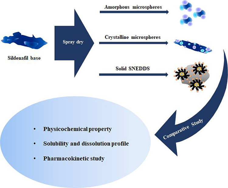 |
Figure 1 Schematic design of this study. |
Materials and Methods
Materials
Sildenafil (base) was supplied by Huons Pharm. Co. (Ansan, South Korea). Polyvinylpyrrolidone (PVP, K30), Solutol HS15 and Cremophor EL were purchased from BASF (Ludwigshafen, Germany). Dextran, polyvinyl alcohol (PVA), hydroxypropyl methylcellulose 2910 (HPMC), pyrogenic silica (HDK N20 Pharma), hydroxypropyl cellulose low viscosity (HPC-L), carboxymethyl cellulose sodium (Na-CMC), poloxamer 188, PEG 4000 and hydroxypropyl beta-cyclodextrin (HP-β-CD) were kindly offered from Hanmi Pharm. Co. (Suwon, South Korea). Soybean oil, sorbitan monooleate (Span 80), olive oil, polysorbate (Tween 80) and corn oil were provided by Daejung Chem. Co. (Siheugn, South Korea). Peceol, Capryol 90 (propylene glycol monocaprylate), Plurol diisosterarique, Labrafil M2125CS, Transcutol HP, Lauroglycol FCC and Labrasol were acquired from Gattefosse (Saint-Priest Cedex, France). Medium-chain triglycerides (Captex 300) were purchased from Abitec (Columbus, OH, USA). Mineral oil and linseed oil were obtained from Samchun Chemical Co. (Pyeongtaek, South Korea). Gelatin and sesame oil were purchased from Wako Chemicals Co. (Tokyo, Japan). All other chemicals and solvents were of reagent grade; they were used with or without further purification.
Animals
Male Sprague-Dawley (SD) rats weighing 300 ± 20 g were supplied by Orient Bio (Sungnam, South Korea). All healthy rats were stored in animal cages at 25–27 °C and 55 ± 5% relative humidity during the whole process. The SD rats were fasted for 12 h; however, they were allowed free access to water before drug administration and pharmacokinetic handling. The entire animal care and processes for the animal studies were conducted according to the NIH Policy. Additionally, the protocols of pharmacokinetic studies were accepted by the Institutional Animal Care and Use Committee (IACUC) at Hanyang University.
Aqueous Solubility of Sildenafil
Approximately 100 mg of sildenafil (to reach a supersaturated state) was added to 1 mL of 1% (w/v) hydrophilic polymer aqueous solution (PVP, Na-CMC, gelatin, PVA, HP-β-CD, HPC-L HPMC and dextran), 5% (w/v) surfactant aqueous solution (Tween 80, Span 80, Cremophor EL, Labrafil M2125CS, Solutol HS15, Plurol diisosterarique Capryol 90, Lauroglycol FCC, poloxamer 188, PEG 4000, Transcutol HP and Labrasol), and pure oil (mineral oil, soybean oil, corn oil, olive oil, linseed oil and Captex 300) to perform the solubility test. After 5 min of vortex-mixing, the samples were placed in a shaking water bath (Daihan Scientific, Wonju, South Korea) for 5 days (25 °C and shaking speed of 100 rpm) to generate a supersaturated state. Three days later, the samples were centrifuged (10,000 × g for 15 min) using a 5430R centrifuge (Eppendorf, Hamburg, Germany). Next, the supernatant was filtered (0.45 μm, nylon) and diluted with the mobile phase for analysing sildenafil by utilising a high-performance liquid chromatography (HPLC) system (Agilent 1260 Infinity, Agilent Technologies, Santa Clara, CA, USA). The HPLC system was arrayed with the Agilent Chem Station software, G1311C 1260 Quat Pump and G1314B 1260 VWD VL detector. The mobile phase, column and gradient methods used were consistent with those used in previous studies.20
Development of Sildenafil-Loaded Amorphous Microspheres and Crystalline Microspheres
According to the aqueous solubility tests, suitable polymer and surfactant were chosen. Sildenafil-loaded amorphous microspheres were manufactured by mixing numerous ratios of polymer, surfactant and sildenafil in ethanol. The experimental plan of formulations is shown in Table S1. The resultant clear solution was introduced into a Büchi mini spray dryer and continuously pushed to the pneumatic nozzle (0.7 mm diameter) by employing a peristaltic pump at a flow rate of 5.4 mL/min. The inlet temperature was set at 90 °C and the outlet temperature was maintained at 60–70 °C. The aspirator was fixed at 100% and the pressure of the spraying air was 4 kg/cm2. To optimise the ratio of polymer, surfactant and sildenafil, solubility and dissolution tests were performed. After selecting the configuration of amorphous microspheres, the solvent was changed from ethanol to distilled water to manufacture the crystalline microspheres. Then, the resultant suspension was spray dried while continuously stirring using a magnetic bar (500 rpm). The inlet temperature was maintained at 140 °C and the outlet temperature was set at 80–90 °C. The pneumatic nozzle, pump rate, aspirator value and spraying air pressure were consistent with the preparation conditions of the amorphous microspheres.
Fabrication of Sildenafil-Loaded Solid SNEDDS
The composition of liquid SNEDDS was selected on the basis of an aqueous solubility test. Afterwards, numerous ratios of oil, surfactant and co-surfactant were combined to create liquid SNEDDS formulations. The formulations containing various mixtures of oil, surfactant and co-surfactant were plotted in ternary-phase diagram (Figure S1). Furthermore, their dispersion in water was visually evaluated. Additionally, the nanoemulsion droplet size was assessed only for the formulations providing excellent nanoemulsifying ability (achieving the visual standard of the preceding experiment). The nanoemulsion droplet size was analysed via a Zetasizer Nano ZS (Malvern Instruments, Worcestershire, UK). Among the formulations, the liquid SNEDDS with the smallest emulsion droplet size was selected. Next, sildenafil was incorporated into the chosen liquid SNEDDS and highly porous inorganic materials with large specific surface areas (500 mg) were added for solidification. The suspension flowed through a pneumatic nozzle of 0.7 mm diameter in a Büchi mini spray dryer (B-290; Flawil, Switzerland) at a flow rate of 5.0 mL/min. The inlet temperature was set at 130 °C and the outlet temperature was maintained at 80 °C. The aspirator was set at 90% and the pressure of the spraying air was 4 kg/cm2. During the entire spray-drying process, the suspension was continuously stirred (500 rpm) using a magnetic bar to maintain a stable suspension state.
Physicochemical Properties of Various Sildenafil-Loaded Drug Delivery Sytems
Surface Morphological Features
The shape and surface of the samples were examined by employing an S-4800 scanning electron microscope (Hitachi; Tokyo, Japan). Double-sided adhesive tape was utilized to fix the samples. To render the sample electrically conductive, a platinum coating (4 min at 15 mA) was applied by utilising the EmiTeck Sputter Coater (K575 K) at a speed of 6 nm/min under vacuum (0.8 Pa).
Particle-Size Distribution
Particle-size analysis was carried out using Mastersizer 3000 (Malvern, Worcestershire, UK). The instrument was optimised under the following conditions: air pressure, 1 bar; feed rate, 50%; gab, 1.
Thermal Analysis
The thermal analysis was conducted via a differential scanning calorimetry (DSC Q200; TA Instruments, New Castle, DE, USA). Each sample (approximately 5 mg) was placed in standard aluminium pans and dry nitrogen was employed as the effluent gas. All samples were scanned at a temperature ramp speed of 10 °C/min and heat flow from 0 to 300 °C.
Solid-State Characterisation
X-ray powder scattering measurements were performed by means of an X-ray diffractometer (D/MAX-2500, Rigaku, Japan) at room temperature. Monochromatic Cu Kα radiation (λ = 1.54178 Å) was used at 100 mA and 40 kV. An angular increment of 0.02° per second was selected over a range of 2θ angles from 3° to 50°.
Dissolution Test
Dissolution profiles of sildenafil-loaded amorphous microspheres, crystalline microspheres, solid SNEDDS and the drug powder (equivalent to 10 mg of sildenafil) were investigated using Vision Classic 6 (Hanson Research Co.; Los Angeles, CA, USA). USP apparatus II (paddle method) was utilised. The dissolution medium was 900 mL of distilled water and the temperature was maintained at 37 ± 0.5 °C. The speed of the paddle was set to 50 rpm. At specific time intervals (0, 5, 10, 15, 30 and 45 min), an aliquot (3 mL) of the sample was collected, filtered through a 0.45 µm nylon syringe filter and assayed for the content of sildenafil by employing HPLC, as described above.
Pharmacokinetic Study
Twenty-four rats were non-selectively divided into four groups (each group consisted of six rats). After the anaesthesia process, the femoral artery of the rats was inserted with a surgical tube and fixed on a surgical board. Before surgery, heparin (50 IU/mL) was prepared to coat the inside of the polyethylene tube. The sildenafil-loaded amorphous microspheres, crystalline microspheres, solid SNEDDS and the drug powder were placed in a #9 gelatin capsule and administered orally at a dose of 20 mg/kg. Plasma samples (300 µL) were collected at predetermined time points via the cannulated tube. After centrifuging the sample at 20,000 g for 10 min, the supernatant (100 µL) was separated. The protein precipitation method was applied while using revaprazan as an internal standard. Finally, the amount of sildenafil was analysed through HPLC.
Statistical Analysis
Statistically significant difference was confirmed by the Student’s t-test (for a pair of groups) and one-way ANOVA followed by Tukey’s post hoc test (for more than two groups). For the statistical calculation, SPSS Version 26 (IBM, Armonk, NY, USA) was utilised. A p-value of <0.05 was considered significant.33
Results and Discussion
Development of Sildenafil-Loaded Microspheres
Among the investigated ingredients, PVP and Labrasol were selected as a proper polymer and surfactant, respectively, due to their high solubility as shown in Figure S2-A and B. Afterwards, various amorphous microspheres were prepared (Table S1). To determine the optimal weight ratio of the drug, PVP and Labrasol, aqueous solubility and dissolution test were carried out as shown in Figure S3-A and B. As a result, the optimised amorphous microspheres were composed of PVP/ Labrasol/drug at the weight ratio of 6/1/1, showing the highest solubility and dissolution rate (%). The same configuration selected in the amorphous microspheres was utilised in the crystalline microspheres; however, this drug delivery system was produced using distilled water instead of an organic solvent. The final compositions of amorphous and crystalline microspheres are presented in Table 1.
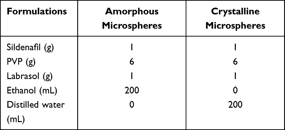 |
Table 1 Composition of Sildenafil-Loaded Amorphous and Crystalline Microspheres |
Fabrication of Sildenafil-Loaded Solid SNEDDS
In this study, ingredients that maximise drug solubility were selected as components of SNEDDS, according to the previously published studies.20,31 The drug showed the highest solubility in Captex 300 among the oil stocks (Figure S4). Furthermore, Labrasol and Transcutol significantly increased the solubility of the drug, compared to others. Therefore, Captex 300, Labrasol and Transcutol were selected. The appropriate ratio of Captex 300, Labrasol and Transcutol was determined by the nanoemulsion-inducing ability and nanoemulsion droplet size, established in the previous methods.34,35 In the absence of sildenafil, the nanoemulsifying capability of liquid SNEDDS in water was evaluated along with visual standards as shown in Figure 2A. Among the formulations with superior self-nanoemulsifying ability, emulsion droplet size of each drug delivery system was investigated (Figure 2B and C). As a result, the liquid SNEDDS composed of Labrasol, Transcutol HP and Captex 300 at a weight ratio of 70/15/15% was chosen, generating the smallest droplet size (150.3 ± 2.7 nm). The small-sized nanoemulsion has better solubilising capability than large-sized nanoemulsion, due to the increased surface area in contact with water. Additionally, nanoemulsion with fine droplet size possesses wide range of drug loading capacity and excellent biocompatibility. For this reason, SNEDDS with small droplet is usually preferred.36,37 Subsequently, sildenafil was loaded into the liquid SNEDDS. A spray-drying technique was utilised for solidifying liquid SNEDDS. It is crucial to convert liquid form into solid form, since liquid SNEDDS has several drawbacks. Generally, liquid SNEDDS is encapsulated in soft gelatin capsules and these are susceptible to humidity. In addition, incompatibility issues between gelatin shell and liquid SNEDDS might occur in the soft capsules. For those reasons, there are many studies solidifying liquid SNEDDS using spray dryer.38,39 In this study, HDK N20 Pharma was chosen as a solid carrier, due to its porous surface and high BET value (175–225 m2/g).40,41 The sildenafil-loaded liquid SNEDDS was dispersed in water and HDK N20 Pharma was suspended. Consequently, the suspension was spray dried to manufacture sildenafil-loaded solid SNEDDS. To fabricate free-flowing powder, 500 mg of HDK N20 Pharma was required in order to absorb 1000 mg of liquid SNEDDS.
 |
Figure 2 Composition of liquid SNEDDS: (A) Ternary phase diagram; (B) Effect of Captex 300 concentration on the emulsion droplet size; (C) Effect of Transcutol HP concentration on the emulsion droplet size. Each value represents the mean ± S.D. *Indicates p < 0.05 when compared with 80/20, 75/25 and 70/30. #Indicates p < 0.05 when compared with 75/10/15, 65/20/15 and 60/25/15. |
Surface Morphological Features and Particle-Size Distribution
The surface morphological features were confirmed through a scanning electron microscope (SEM). Sildenafil exhibited irregular and oblong shape, assuming a crystalline solid form (Figure 3A). Amorphous microspheres appeared as small spherical particles clustered together (Figure 3B). Compared to the drug powder, decreased particle size was observed in amorphous microspheres. Nonetheless, the SEM image of the crystalline microspheres indicated that the crystalline drug adhered to hydrophilic excipients, such as PVP and Labrasol (Figure 3C). Unlike the amorphous microspheres, the crystalline microspheres presented no significant particle size reduction, compared to the drug powder. However, the external region of the drug consisted of hydrophilic ingredients, which led to the production of an uneven surface in crystalline microspheres. Furthermore, irregular spherules with smooth surfaces were scattered in the solid SNEDDS (Figure 3D). HDK N20 Pharma is well reported to exhibit a highly porous surface and rough appearance.42,43 During the solidification process, oily liquid covered the porous surface of HDK N20 Pharma, resulting in the creation of a smooth surface. The particle-size distribution results are presented in Figure 4. The portions of particles with diameters below D50 are 50%. In general, the value of D50 indicates the median diameter. The drug powder gave an average size of 26.6 ± 0.9 µm. In amorphous microspheres, a significant particle size reduction occurred (9.3 ± 1.0 µm), compared to sildenafil powder (p < 0.05). On the other hand, the crystalline microspheres exhibited a particle size of 27.8 ± 1.5 µm, similar to the particle size for the drug powder. The tendency of the particle-size analysis was consistent with the SEM results. The drug and ingredients were completely dissolved in ethanol and spray dried, leading to a decrease in particle size in the amorphous microspheres. In the preparation of crystalline microspheres, PVP and Labrasol were dissolved; however, hydrophobic sildenafil was undissolved in distilled water and the aqueous suspension was spray dried. Thus, the dissolved spray-dried hydrophilic excipients were attached to the undissolved spray-dried sildenafil, resulting in no significant change in particle size compared to the drug powder. In solid SNEDDS, the drug was completely dissolved in liquid SNEDDS before the solidifying process. Hence, the particle size of solid SNEDDS (10.4 ± 0.4 µm) was similar to that of amorphous microspheres; however, it was considerably reduced compared to pure drug powder.
 |
Figure 3 Scanning electron micrographs: (A) Sildenafil powder (× 3000); (B) Amorphous microspheres (× 3000); (C) Crystalline microspheres (× 2000); and (D) Solid SNEDDS (× 3000). |
 |
Figure 4 Particle-size distribution of sildenafil and prepared formulations. |
Thermal Analysis and Solid-State Characterisation
The outcomes of differential scanning calorimetry are depicted in Figure 5A. At 189 °C, a distinguishing peak was produced, showing crystalline characteristics; this result was consistent with the melting point of sildenafil. The correlative peak appeared in two types of physical mixtures. The results for the physical mixtures demonstrated that the drug maintained its crystallinity and there was no significant physicochemical interaction between the drug and excipients. The specific peak disappeared in the amorphous microspheres and solid SNEDDS. In contrast to the amorphous microspheres, a small endothermic peak was observed in crystalline microspheres, corresponding to the melting point of sildenafil. The results of X-ray diffraction are depicted in Figure 5B. Several representative peaks are shown over a range of diffraction angles in sildenafil, indicating crystalline characteristics. Both physical mixtures exhibited similar peak patterns, similar to that of the drug powder. Conserving the designated peak pattern indicated that the drug presented good compatibility with the ingredients. The unique pattern was not observed in amorphous microspheres or solid SNEDDS. However, a peak pattern was detected for the crystalline microspheres, analogous to the sildenafil powder. Based on the thermal analysis and solid-state characterisation, the crystal form of sildenafil changed to an amorphous form in amorphous microspheres and solid SNEDDS, while it was preserved in the crystalline microspheres. As the drug was completely dissolved in ethanol or liquid SNEDDS, the physicochemical characteristics were altered in the amorphous microspheres and solid SNEDDS. Nevertheless, sildenafil was not dissolved but was suspended in distilled water for preserving the crystalline characteristics in the crystalline microspheres.
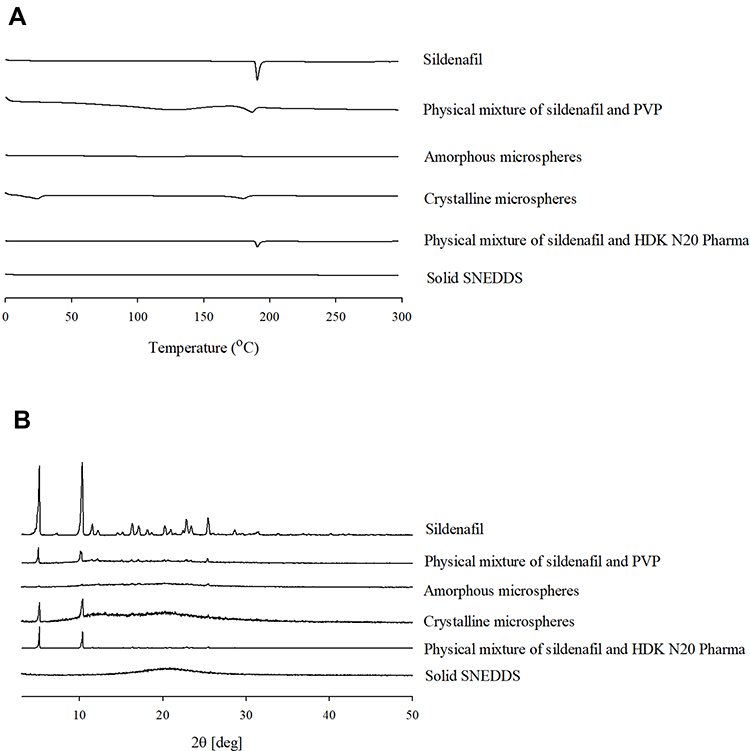 |
Figure 5 Differential scanning calorimetry thermograms (A) and X-ray diffractometer results (B). |
Aqueous Solubility and Dissolution Profile of Prepared Sildenafil-Loaded Formulations
Aqueous solubility results are presented in Figure 6. All three formulations showed significantly increased solubility compared to the drug powder (p < 0.05). Compared to the drug powder, amorphous microspheres, crystalline microspheres and solid SNEDDS enhanced sildenafil solubility by 79, 55 and 82 times, respectively. Additionally, amorphous microspheres and solid SNEDDS demonstrated highly improved solubility compared with crystalline microspheres (p < 0.05). Solid SNEDDS showed the highest aqueous solubility among the formulations, although there were no significant differences between solid SNEDDS and amorphous microspheres. The dissolution profile reflected the trend in aqueous solubility results (Figure 7). All of the manufactured drug delivery systems produced an enhanced dissolution rate (%) compared to the drug powder at every sampling time (p < 0.05). Solid SNEDDS presented superior dissolution rate (%) to other formulations. Both solid SNEDDS and amorphous microspheres gave a significantly improved dissolution rate (%) at 45 min, compared with crystalline microspheres (drug powder; 8.4 ± 1.3%, amorphous microspheres; 74.3 ± 0.9%, crystalline microspheres; 45.9 ± 1.3%, solid SNEDDS; 76.6 ± 6.0%) (p < 0.05). Those results can be explained by the fact that three different aqueous microenvironments in each formulation resulted increased solubility and dissolution. In crystalline microspheres, the creation of hydrophilic microenvironment on the surface of a hydrophobic drug was a crucial factor for enhancing the aqueous solubility. When the water contacted with the surface of the formulation, PVP and Labrasol rapidly leached out, forming aqueous microenvironment on the drug surface. In this microenvironment, which was not observed in a simple blend of drug and hydrophilic excipients, the drug was instantaneously super-saturated, and the solubility highly increased. In amorphous microspheres, particle size reduced and crystal form changed to amorphous form. The reduced particle size resulted increased surface area of amorphous microspheres in contact with water. Consequently, the increased surface area promoted the drug to immediately hydrate in water, preventing aggregation and leading improved aqueous solubility. In addition, it is well reported that amorphous state possesses higher free energy than crystal state. This excess free energy in amorphous microspheres provided an energy advantage when the drug dissolved in water. Furthermore, the polymer (PVP) in amorphous microspheres prevented the drug from recrystallizing and the surfactant (Labrasol) lowered the surface tension between water and the drug, producing drug-loaded polymer-based micelles. These mechanisms enhanced the aqueous solubility of sildenafil in amorphous microspheres. In solid SNEDDS, crystalline form of the drug changed to amorphous form and particle size was considerably reduced. Those results are consistent with the amorphous microspheres. On the other hand, different property was observed in solid SNEDDS, compared to amorphous microspheres. When solid SNEDDS contacted water, o/w nanoemulsions were spontaneously formed.44 In this system, the drug was encapsulated in the oil phase surrounded by hydrophilic surfactants, resulting the hydrophobic drug super-saturated in water. Moreover, “nano-scale” nanoemulsions might possess larger surface area than “micro-scale” microspheres, facilitating the hydration of the drug. For those reasons, solid SNEDDS could exhibit relatively higher aqueous solubility and dissolution rate (%) than amorphous microspheres. Overall, sildenafil dissolved better in water when it was converted to its amorphous form than when it remained crystalline. In the amorphous state of the drug, it is reasonable to assume that the solid SNEDDS is more appropriate system than amorphous microspheres in increasing aqueous solubility, though there was no significant difference between the two systems. When the three sildenafil-loaded systems were stored under room temperature (25 ± 1 °C) for 6 months, significant differences were not observed in terms of content (> 97%), solubility and dissolution profile (data not shown).
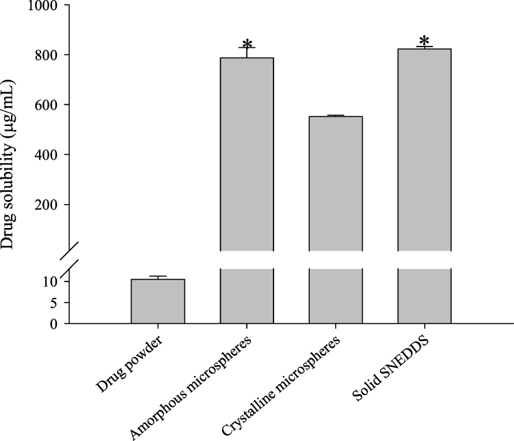 |
Figure 6 Comparison of amorphous microspheres, crystalline microspheres, solid SNEDDS and drug powder in aqueous solubility. *Indicates p < 0.05 when compared with crystalline microsphere and drug powder. |
 |
Figure 7 Dissolution profile of amorphous microspheres, crystalline microspheres, solid SNEDDS and drug powder. |
Pharmacokinetic Study
The plasma concentration–time profiles of sildenafil are shown in Figure 8. Compared with drug powder, three formulations including amorphous microspheres, crystalline microspheres and solid SNEDDS produced significantly increased plasma concentrations at 0.08, 0.25, 0.5, 1.0 and 1.5 h (p < 0.05). Amorphous microspheres and solid SNEDDS generated significantly increased plasma concentrations compared to the crystalline microspheres at 0.08, 0.25, 0.5 and 1.0 h (p < 0.05). In addition, solid SNEDDS produced relatively higher plasma concentrations than amorphous microspheres up to 2 h. The conforming pharmacokinetic parameters in rats are exhibited Table 2. All the prepared formulations showed significantly enhanced AUC values when compared to the drug powder (p < 0.05). The values were ranked in the following order: solid SNEDDS (1508.78 ± 343.95 h⋅ng/mL) ≥ amorphous microspheres (1339.90 ± 416.52 h⋅ng/mL) ≥ crystalline microspheres (1042.90 ± 119.90 h⋅ng/mL) > drug powder (733.57 ± 184.15 h⋅ng/mL). In addition, amorphous microspheres and solid SNEDDS produced significantly improved Cmax values compared with crystalline microspheres and the drug powder (p < 0.05); the performance order was as follows: solid SNEDDS ≥ amorphous microspheres > crystalline microspheres > drug powder. Among the formulations, solid SNEDDS produced the highest increase in oral bioavailability, according to the AUC and Cmax values. However, there was no major difference in other pharmacokinetic parameters, such as Tmax, t1/2 and Kel, among the drug powder and formulations. The trend in the pharmacokinetics was consistent with the solubility and dissolution profiles. It can be inferred that the oral bioavailability of sildenafil was superior when the drug was in an amorphous form than when exposed to the hydrophilic microenvironment while retaining its crystalline form in the gastrointestinal tract. Comparing the two amorphous-form-inducing systems, solid SNEDDS had a relatively higher AUC and Cmax values than amorphous microspheres, even if they were not significantly different. The results indicated that relatively higher aqueous solubility and dissolution rate (%) of solid SNEDDS resulted in relatively higher oral bioavailability than amorphous microspheres. Furthermore, it is assumed that the oil phase in solid SNEDDS played as a permeation enhancer in the gastrointestinal tract, leading to slightly improved drug absorption.
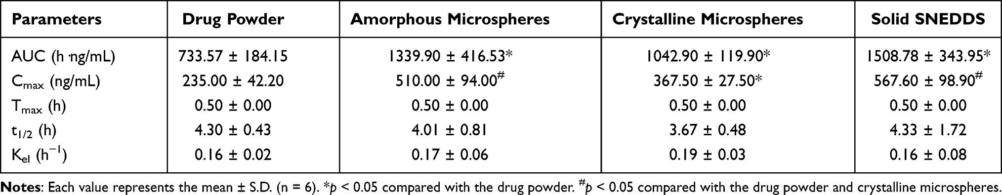 |
Table 2 Pharmacokinetic Parameters |
 |
Figure 8 Plasma concentration–time profiles of sildenafil after oral administration of amorphous microspheres, crystalline microspheres, solid SNEDDS, and drug powder. Each value represents the mean ± S.D. (n = 6). |
Conclusion
The effects of three different aqueous microenvironments on the oral bioavailability of sildenafil were evaluated in this study. Amorphous microspheres, crystalline microspheres and solid SNEDDS considerably improved the solubility, dissolution rate (%) and oral bioavailability of sildenafil compared to the drug powder. The superiority of the drug delivery systems was in the following order: solid SNEDDS ≥ amorphous microspheres > crystalline microspheres. Solid SNEDDS exhibited relatively higher aqueous solubility, dissolution rate (%) and oral bioavailability than amorphous microspheres, although there were no significant differences between the two systems. As a result, the solid SNEDDS, which converts the crystalline form of the drug to its amorphous form via an o/w nanoemulsion, could be recommended for enhancing the oral bioavailability of sildenafil in this research. In addition, further experiments should be performed, including dissolution-equivalent study on various pHs, stability study and pharmacokinetic investigation in larger animal models, prior to carry out clinical research.
Acknowledgments
This work was supported by the National Research Foundation of South Korea (NRF) grant funded by the South Korea government (MEST) (No. 2018R1A2B2004668 & 2020R1C1C1009663). The authors would like to gratefully acknowledge the Bio-Medical Engineering Core Facility Center at Dankook University for providing critical reagents and equipment.
Disclosure
Prof. Dr. Sung Giu Jin reports grants from the National Research Foundation of South Korea, outside the submitted work. The authors report no other potential conflicts of interest for this work.
References
1. Jannini EA, Droupy S. Needs and expectations of patients with erectile dysfunction: an update on pharmacological innovations in phosphodiesterase type 5 inhibition with focus on sildenafil. Sex Med. 2019;7:1–10.
2. Ghofrani HA, Wiedemann R, Rose F, et al. Sildenafil for treatment of lung fibrosis and pulmonary hypertension: a randomised controlled trial. The Lancet. 2002;360:895–900.
3. Hao Y, Zhu Y, Mao Y, et al. Efficacy and safety of Sildenafil treatment in pulmonary hypertension caused by chronic obstructive pulmonary disease: a meta-analysis. Life Sci. 2020;257:118001.
4. Paranjpe M, Finke JH, Richter C, et al. Physicochemical characterization of sildenafil-loaded solid lipid nanoparticle dispersions (SLN) for pulmonary application. Int J Pharm. 2014;476:41–49.
5. Zhao L, Mustapha O, Shafique S, et al. Electrospun gelatin nanocontainers for enhanced biopharmaceutical performance of piroxicam: in vivo and in vitro investigations. Int J Nanomedicine. 2020;15:8819.
6. Doghri Y, Chetaneau F, Rhimi M, et al. Sildenafil citrate long-term treatment effects on cardiovascular reactivity in a SHR experimental model of metabolic syndrome. PLoS One. 2019;14:e0223914.
7. Sawatdee S, Atipairin A, Yoon SA, Srichana T, Changsan N. Enhanced dissolution of sildenafil citrate as dry foam tablets. Pharm Dev Technol. 2019;24:1–11.
8. Renshall LJ, Cottrell EC, Cowley E, et al. Antenatal sildenafil citrate treatment increases offspring blood pressure in the placental-specific Igf2 knockout mouse model of FGR. Am J Physiol Heart Circ Physiol. 2020;318:H252–263.
9. Sarveiya V, Templeton JF, Benson HAE. Ion-pairs of ibuprofen: increased membrane diffusion. J Pharm Pharmacol. 2004;56:717–724.
10. Choi JS, Cho NH, Kim DH, Park JS. Comparison of paclitaxel solid dispersion and polymeric micelles for improved oral bioavailability and in vitro anti-cancer effects. Mater Sci Eng C. 2019;100:247–259.
11. Chen S, Guo R, Xie C, Liang Q, Xiao X. Biomimetic mineralization of nanocrystalline hydroxyapatites on aminated modified polylactic acid microspheres to develop a novel drug delivery system for alendronate. Mater Sci Eng C. 2020;110:110655.
12. Wei MY, Lei XP, Fu JJ, et al. The use of amphiphilic copolymer in the solid dispersion formulation of nimodipine to inhibit drug crystallization in the release media: combining nano-drug delivery system with solid preparations. Mater Sci Eng C. 2020;111:110836.
13. Kim KS, Kim JC, Jin SG, et al. Development of novel prasugrel base microsphere-loaded tablet with enhanced stability: physicochemical characterization and in vivo evaluation in beagle dogs. Colloids Surf B Biointerfaces. 2016;146:754–761.
14. Kim JS, Park JH, Jeong SC, et al. Novel revaprazan-loaded gelatin microsphere with enhanced drug solubility and oral bioavailability. J Microencapsul. 2018;35:421–427.
15. Xiong X, Zhang M, Hou Q, et al. Solid dispersions of telaprevir with improved solubility prepared by comilling: formulation, physicochemical characterization, and cytotoxicity evaluation. Mater Sci Eng C. 2019;105:110012.
16. Mustapha O, Kim KS, Shafique S, et al. Comparison of three different types of cilostazol-loaded solid dispersion: physicochemical characterization and pharmacokinetics in rats. Colloids Surf B Biointerfaces. 2017;154:89–95.
17. Giri BR, Kim JS, Park JH, et al. Improved bioavailability and high photostability of methotrexate by spray-dried surface-attached solid dispersion with an aqueous medium. Pharmaceutics. 2021;13:13010111.
18. Park JH, Cho JH, Kim DS, et al. Revaprazan-loaded surface-modified solid dispersion: physicochemical characterization and in vivo evaluation. Pharm Dev Technol. 2019;24:788–793.
19. Bannow J, Yorulmaz Y, Lobmann K, Mullertz A, Rades T. Improving the drug load and in vitro performance of supersaturated self-nanoemulsifying drug delivery systems (super-SNEDDS) using polymeric precipitation inhibitors. Int J Pharm. 2020;575:118960.
20. Kim JS, Din FU, Lee SM, et al. Comparative study between high-pressure homogenisation and Shirasu porous glass membrane technique in sildenafil base-loaded solid SNEDDS: effects on physicochemical properties and in vivo characteristics. Int J Pharm. 2021;592:120039.
21. Michaelsen MH, Siqueira Jorgensen SD, Abdi IM, Wasan KM, Rades T, Mullertz A. Fenofibrate oral absorption from SNEDDS and super-SNEDDS is not significantly affected by lipase inhibition in rats. Eur J Pharm Biopharm. 2019;142:258–264.
22. Patil SC, Tagalpallewar AA, Kokare CR. Natural anti-proliferative agent loaded self-microemulsifying nanoparticles for potential therapy in oral squamous carcinoma. J Pharm Investig. 2019;49:527–541.
23. Kazi M, Alhajri A, Alshehri SM, et al. Enhancing oral bioavailability of apigenin using a bioactive self-nanoemulsifying drug delivery system (Bio-SNEDDS): in vitro, in vivo and stability evaluations. Pharmaceutics. 2020;12:12080749.
24. Ye J, Wu H, Huang C, et al. Comparisons of in vitro Fick’s first law, lipolysis and in vivo rat models for oral absorption on BCS II drugs in SNEDDS. Int J Nanomedicine. 2019;14:5623–5636.
25. Davis M, Walker G. Recent strategies in spray drying for the enhanced bioavailability of poorly water-soluble drugs. J Control Release. 2018;269:110–127.
26. Lee YC, McNevin M, Ikeda C, et al. Combination of colloidal silicon dioxide with spray-dried solid dispersion to facilitate discharge from an agitated dryer. AAPS PharmSciTech. 2019;20:182.
27. Weers JG, Miller DP, Tarara TE. Spray-dried PulmoSphere™ formulations for inhalation comprising crystalline drug particles. AAPS PharmSciTech. 2019;20:103.
28. Kankala RK, Lin XF, Song HF, et al. Supercritical fluid-assisted decoration of nanoparticles on porous microcontainers for codelivery of therapeutics and inhalation therapy of diabetes. ACS Biomater Sci Eng. 2018;4:4225–4235.
29. Kankala RK, Zhao J, Liu CG, et al. Highly porous microcarriers for minimally invasive in situ skeletal muscle cell delivery. Small. 2019;15:1901397.
30. Poozesh S, Bilgili E. Scale-up of pharmaceutical spray drying using scale-up rules: a review. Int J Pharm. 2019;562:271–292.
31. Kim DW, Kwon MS, Yousaf AM, et al. Comparison of a solid SMEDDS and solid dispersion for enhanced stability and bioavailability of clopidogrel napadisilate. Carbohydr Polym. 2014;114:365–374.
32. Park JH, Kim DS, Mustapha O, et al. Comparison of a revaprazan-loaded solid dispersion, solid SNEDDS and inclusion compound: physicochemical characterisation and pharmacokinetics. Colloids Surf B Biointerfaces. 2018;162:420–426.
33. Ramasamy T, Ruttala HB, Sundaramoorthy P, et al. Multimodal selenium nanoshell-capped Au@ mSiO2 nanoplatform for NIR-responsive chemo-photothermal therapy against metastatic breast cancer. NPG Asia Mater. 2018;10:197–216.
34. Singh D, Tiwary AK, Bedi N. Canagliflozin loaded SMEDDS: formulation optimization for improved solubility, permeability and pharmacokinetic performance. J Pharm Investig. 2019;49:67–85.
35. Assi RA, Abdulbaqi IM, Ming TS, et al. Liquid and solid self-emulsifying drug delivery systems (SEDDs) as carriers for the oral delivery of azithromycin: optimization, in vitro characterization and stability assessment. Pharmaceutics. 2020;12:12111052.
36. Wang J, Yang H, Li Q, et al. Novel nanomicelles based on rebaudioside A: a potential nanoplatform for oral delivery of honokiol with enhanced oral bioavailability and antitumor activity. Int J Pharm. 2020;590:119899.
37. Kim JS, Choi YJ, Woo MR, et al. New potential application of hydroxypropyl-β-cyclodextrin in solid self-nanoemulsifying drug delivery system and solid dispersion. Carbohydr Polym. 2021;271:118433.
38. Bezerra-Souza A, Fernandez-Garcia R, Rodrigues GF, et al. Repurposing butenafine as an oral nanomedicine for visceral leishmaniasis. Pharmaceutics. 2019;11:353.
39. Smith L, Serrano DR, Mauger M, et al. Orally bioavailable and effective buparvaquone lipid-based nanomedicines for visceral leishmaniasis. Mol Pharm. 2018;15:2570–2583.
40. Beringhs AO, Minatovicz BC, Zhang GGZ, et al. Impact of porous excipients on the manufacturability and product performance of solid self-emulsifying drug delivery systems. AAPS Pharmscitech. 2018;19:3298–3310.
41. Choi MJ, Kim JS, Yu H, et al. Comparison of the physicochemical properties, aqueous solubility, and oral bioavailability of rivaroxaban-loaded high-pressure homogenised and Shirasu porous glass membrane emulsified solid self-nanoemulsifying drug delivery systems. J Mol Liq. 2021;117057. doi:10.1016/j.molliq.2021.117057
42. Mustapha O, Kim KS, Shafique S, et al. Development of novel cilostazol-loaded solid SNEDDS using a SPG membrane emulsification technique: physicochemical characterization and in vivo evaluation. Colloids Surf B Biointerfaces. 2017;150:216–222.
43. Kim DS, Kim JS, Lim SJ, et al. Comparison of 1-palmitoyl-2-linoleoyl-3-acetyl-rac-glycerol-loaded self-emulsifying granule and solid self-nanoemulsifying drug delivery system: powder property, dissolution and oral bioavailability. Pharmaceutics. 2019;11:11080415.
44. Kim DS, Cho JH, Park JH, et al. Self-microemulsifying drug delivery system (SMEDDS) for improved oral delivery and photostability of methotrexate. Int J Nanomedicine. 2019;14:4949–4960.
 © 2021 The Author(s). This work is published and licensed by Dove Medical Press Limited. The
full terms of this license are available at https://www.dovepress.com/terms
and incorporate the Creative Commons Attribution
- Non Commercial (unported, 3.0) License.
By accessing the work you hereby accept the Terms. Non-commercial uses of the work are permitted
without any further permission from Dove Medical Press Limited, provided the work is properly
attributed. For permission for commercial use of this work, please see paragraphs 4.2 and 5 of our Terms.
© 2021 The Author(s). This work is published and licensed by Dove Medical Press Limited. The
full terms of this license are available at https://www.dovepress.com/terms
and incorporate the Creative Commons Attribution
- Non Commercial (unported, 3.0) License.
By accessing the work you hereby accept the Terms. Non-commercial uses of the work are permitted
without any further permission from Dove Medical Press Limited, provided the work is properly
attributed. For permission for commercial use of this work, please see paragraphs 4.2 and 5 of our Terms.