
Back to Journals » Cancer Management and Research » Volume 18

Clonal Seborrheic Keratosis with Adjacent Bowen’s Disease in a Non-Sun-Exposed Patient: Collision or Transformation?
Authors Wang Z, Mao S, Xiao H, Zhao Q
Received 24 February 2026
Accepted for publication 16 June 2026
Published 15 July 2026 Volume 2026:18 602855
DOI https://doi.org/10.2147/CMAR.S602855
Checked for plagiarism Yes
Review by Single anonymous peer review
Peer reviewer comments 2
Editor who approved publication: Dr Javier-David Benitez-Fuentes
Zhi Wang,1 Sen Mao,2 Haibo Xiao,2 Qian Zhao2
1Department of Hepatobiliary Surgery, Hunan University of Medicine General Hospital, HuaiHua, Hunan, People’s Republic of China; 2Department of Clinical Pathology, Hunan University of Medicine General Hospital, HuaiHua, Hunan, People’s Republic of China
Correspondence: Qian Zhao, Department of Clinical Pathology, Hunan University of Medicine General Hospital, HuaiHua, Hunan, People’s Republic of China, Email [email protected]
Abstract: We report a case of clonal seborrheic keratosis (CPSK) adjacent to Bowen’s disease (BD) in a 29‑year‑old male with no history of chronic sun exposure. The lesion presented as a brown, flat papule on the right side of the back. Histopathologically, the CPSK component featured well‑demarcated intraepidermal nests of basaloid keratinocytes, consistent with the Borst‑Jadassohn phenomenon. Adjacent areas showed full‑thickness epidermal atypia, nuclear pleomorphism, and numerous mitotic figures, including atypical forms, characteristic of BD. No clear transitional zone was identified between the two components. These findings align most closely with a collision tumor-coincidental coexistence of two independent neoplasms-rather than true malignant transformation of CPSK into BD. This case underscores the need for careful histologic evaluation to distinguish collision from transformation, particularly in the absence of chronic ultraviolet exposure.
Keywords: clonal seborrheic keratosis, Bowen’s disease, collision tumor, malignant transformation
Introduction
Clonal seborrheic keratosis is a subtype of seborrheic keratosis, with clinical manifestations similar to those of ordinary seborrheic keratosis. Histopathologically, it is consistent with Borst-Jadassohn intraepidermal epithelioma. Whether SK can undergo malignant transformation into BD remains controversial: some studies support true transformation based on transitional zones or molecular data, while others consider it a collision tumor interpretation. Distinguishing between these possibilities is clinically relevant. Here we report a case of CPSK with adjacent BD in a patient without chronic sun exposure. No clear transitional zone was identified. We present the histopathological and immunohistochemical findings and argue that this case favors a collision tumor rather than malignant transformation.
Case Report
A 29-year-old male patient developed a painless, slightly pruritic brown macule on the right side of his back five years prior, with no identifiable precipitating factors. Over time, the lesion progressed into a 0.8 cm × 0.5 cm oval, dark brown plaque exhibiting a rough surface with fine white scales but no ulceration, bleeding, or tenderness. Systemic physical examination showed no abnormalities. Dermatologic assessment confirmed a well-demarcated, moderately firm plaque without surrounding inflammatory changes. There was no similar medical history in his family.
Histopathological examination revealed a biphasic lesion. The epidermis showed hyperkeratosis, verrucous hyperplasia with pseudokeratotic cysts, and acanthosis. Within the spinous layer, well-demarcated small uniform basaloid cells arranged in whorls or nests formed intraepidermal epithelial nests with sharp borders from surrounding cells, consistent with CPSK; small foci of necrosis were observed within some nests, and the cells had abundant clear vacuolated cytoplasm with small normochromatic nuclei and no significant atypia. In adjacent areas, however, the epidermis showed full-thickness architectural disarray. The squamous epithelial cells exhibited marked atypia, with variation in nuclear size, shape, and chromatin density. Numerous mitotic figures, including atypical mitoses, were present within the epidermis, diagnostic of Bowen’s disease. Importantly, no clear transitional zone was identified between the two components. The basement membrane remained intact, and a moderate lymphoplasmacytic infiltrate was present at the dermoepidermal junction (Figure 1).
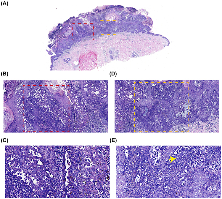 |
Figure 1 CPSK adjacent to BD with an abrupt interface and no transitional zone. (A) Low-power view showing the overall lesion architecture. The CPSK component is outlined by red dashed boxes (left portion of the image), and the BD component is outlined by yellow dashed boxes (right portion). The two components meet at an abrupt interface. Normal skin is present at both lateral margins. (B) Higher magnification of the CPSK area (red dashed box in A). The image shows hyperkeratosis, acanthosis, and multiple well-demarcated intraepidermal nest-like structures (Borst-Jadassohn phenomenon). Focal necrosis, acantholysis, and scattered pseudohorn cysts are present. (C) Higher magnification of the nested area (red dashed box in B). The cells within the nests are basaloid and uniform, with small monotonous nuclei and fine chromatin. Cytologic atypia is absent or minimal. Mitotic figures are rare or absent. (D) Higher magnification of the BD area (yellow dashed box in A). The tumor cells are disorganized with loss of polarity. The basement membrane remains intact, with no dermal infiltration. (E) Higher magnification of the area indicated by the yellow dashed box in D. The tumor cell nuclei are enlarged and irregular, with a nuclear-to-cytoplasmic ratio exceeding 1:2. Yellow arrows indicate mitotic figures. |
Immunohistochemically, the Bowen’s disease component showed diffuse positivity for p53 and p16, with a Ki-67 proliferation index of 70–80% in hotspot areas. The CPSK component exhibited scattered p53 positivity and low Ki-67 expression. Melanocytic markers (S100, Melan A, HMB-45) were negative throughout the lesion, excluding melanoma. CK7 and CEA were negative, ruling out extramammary Paget’s disease. Ber-EP4, Bcl-2, CD10, CK20, and AR were also negative, which, together with the absence of peripheral palisading, retraction clefts, and myxoid stroma, supported the exclusion of Basal cell carcinoma (BCC) (Figure 2).
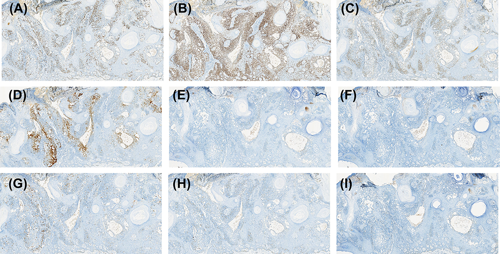 |
Figure 2 Immunohistochemical staining. (A) Ki-67 immunostaining. Diffuse nuclear immunoreactivity was identified in the Bowen’s disease compartment, with a proliferation index ranging from 70% to 80%, while only sparse nuclear immunopositivity was detectable in the CPSK component. (B) p63 immunostaining. Marked nuclear expression was diffusely distributed throughout the full thickness of the epidermis in both the Bowen’s disease and CPSK components. (C) p53 immunostaining. Widespread nuclear labeling was observed in the Bowen’s disease lesions, whereas only scattered positive nuclear signals were present in the CPSK component. (D) p16 immunostaining. Intense nuclear immunopositivity was confined exclusively to the basal epidermal layer in both pathological components. (E) CK7 immunostaining. No specific immunopositive signals were detected in either tissue component. (F) CEA immunostaining. No detectable immunoreactive signals were observed in either lesion component. (G) S100 immunostaining. Focal immunopositivity was observed in scattered cells. (H) Melan A immunostaining. Isolated focal positive signals were detected in occasional cells. (I) BCL-2 immunostaining. Negative immunostaining was confirmed in the tested samples. |
Discussion
CPSK is a subtype of seborrheic keratosis (SK),1 characterized by unique histological features compared to other subtypes. Within the stratum spinosum of a background acanthotic epidermis, CPSKs are distinguished by the presence of distinct nests of cytologically benign or cytologically atypical keratinocytes. Yoshimi et al found that these basal-like cell nests express both K14 and K17, indicating their differentiation into basal-like undifferentiated cells.2 Immunohistochemical analysis of lesions with clonal growth patterns showed increased expression of antigens associated with keratinocyte growth and differentiation, including EGFR, p53, and p63, compared to adjacent normal keratinocytes. An elevated rate of keratinocyte proliferation within the cell nests was found by evaluating Ki-67 expression.3
Given the proliferative features of CPSK described above, the relationship between this subtype and malignant tumors has been of particular interest. The debate centers on whether their coexistence reflects collision tumors or true malignant transformation.4–6 Takayama et al described a case of clonal Bowen’s disease with sebaceous differentiation arising from CPSK. The presence of intraepidermal nested structures in all lesional areas confirmed that the tumor cells within the intraepidermal nests of SK had malignantly transformed into BD with sebaceous differentiation, rather than being the consequence of a coincidental collision.7 In contrast, other authors have interpreted such coexistence as collision tumors.6 Boyd and Rapini, in an analysis of 69 cutaneous collision tumors, noted that basal cell carcinoma and seborrheic keratosis was among the most common combinations, and concluded that most such associations likely represent the juxtaposition of common lesions by coincidence.8
In our case, several features argue against transformation. First, no clear transitional zone was identified despite step sectioning. The interface between CPSK and BD showed an abrupt change rather than gradual cytologic progression. Second, the Borst-Jadassohn nests within the CPSK component lacked significant atypia or mitotic activity. Third, the immunohistochemical profile (p53, Ki-67) showed diffuse positivity in BD but only scattered positivity in CPSK, with no intermediate pattern at the junction. Soini et al demonstrated that p53 accumulation and Ki-67 elevation can occur in benign skin lesions including seborrheic keratoses, and such findings are not specific for malignancy.9 The observed pattern is more consistent with two independent lesions than with clonal evolution. Additionally, our patient had no history of chronic sun exposure, was young and immunocompetent. We therefore classify this case as a collision tumor, in which CPSK and BD coexist independently.
Several differential diagnoses were considered and excluded. Acantholytic squamous cell carcinoma in situ (aSCCIS) was initially considered due to the presence of acantholysis in this case. However, the acantholytic changes were confined to the CPSK component, where the cells showed no significant atypia or mitotic figures. Tagami et al described acantholysis as a rare variant of irritated seborrheic keratosis.10 Chen et al found acantholysis in 29 of 524 SK specimens (5.5%), predominantly in the irritated type, and noted that these changes were not associated with malignancy.11 In aSCCIS, acantholysis is typically accompanied by pronounced cytologic atypia and full-thickness epidermal disorganization. The absence of atypia in the acantholytic areas of CPSK, together with the preserved nested architecture, does not support a diagnosis of aSCCIS. Negative staining for CK7 excluded pagetoid Bowen’s disease and extramammary Paget’s disease (EMPD). Pagetoid Bowen’s disease expresses EMA, CK5/6, and p63 but is negative for CEA, CK8, and S-100. EMPD is usually CK7 positive. The CK7-negative result in this case effectively ruled out EMPD, whereas the adjacent component exhibited full-thickness atypia with atypical mitotic figures, consistent with classic Bowen’s disease. Pagetoid melanoma in situ can mimic pagetoid morphology but can be differentiated by positive staining for S-100 and HMB45. Our case demonstrated no melanocytic markers, and the lesion was entirely intraepidermal without nesting at the dermoepidermal junction. BCC may show nested growth, but BCC typically exhibits peripheral palisading, retraction clefts, and a myxoid stroma. None of these features were present in this case. Additionally, immunohistochemical staining for Ber-EP4, a sensitive marker of BCC, yielded negative results.
Finally, clinical management of CPSK deserves consideration regardless of its pathogenesis. Goldsmith et al reported that CPSK shows a higher overall recurrence rate than non-CPSK at sites of incomplete excision or adjacent locations, and incompletely excised CPSKs with cytological atypia recur more frequently than those without atypia; such lesions carry a substantial risk of progressing to at least squamous cell carcinoma in situ upon recurrence.12 Therefore, when histopathological examination reveals a clonal pattern in seborrheic keratoses, pathologists should document its presence, note any cytological atypia, and evaluate the lesion margins. Incompletely excised CPSKs warrant close clinical follow-up, particularly when cytological atypia is present. In our case, the lesion was completely excised, and no recurrence has been observed to date.
In summary, we report a case of CPSK with adjacent BD in a young patient without chronic sun exposure. The absence of a transitional zone, the abrupt histologic interface, and the differential immunohistochemical patterns support a collision tumor rather than true malignant transformation. Clinically, CPSK often lacks the typical clinical and dermoscopic features of conventional seborrheic keratosis, so histopathological examination is essential when such lesions present atypically. Several limitations must be acknowledged. A microscopic transitional zone might have been missed despite step sectioning. Molecular or clonality studies were not performed to confirm or exclude a common origin. As a single case report, the findings cannot be generalized. Nevertheless, this case underscores the diagnostic challenge of distinguishing collision from transformation and emphasizes the need for careful histologic evaluation with appropriate immunohistochemistry.
Abbreviations
aSCCIS, Acantholytic squamous cell carcinoma in situ; BCC, Basal cell carcinoma; BD, Bowen’s disease; CEA, Carcinoembryonic antigen; CPSK, Seborrheic keratosis with clonal pattern; EMPD, Extramammary Paget’s disease; SK, Seborrheic keratosis.
Data Sharing Statement
All relevant data generated and analyzed during the current study are fully included in this published case report. No additional datasets are available for sharing beyond the content presented in the manuscript and its associated figures, and all data and materials are available in Hunan University of Medicine General Hospital’s Pathology Department.
Ethics Approval and Consent to Participate
Institutional approval was required for the publication of this case report. This study was approved by the Ethics Committee of Hunan University of Medicine General Hospital. The patient provided written informed consent for publication of the case report and any accompanying images.
Author Contributions
All authors made a significant contribution to the work reported, whether that is in the conception, study design, execution, acquisition of data, analysis and interpretation, or in all these areas; took part in drafting, revising or critically reviewing the article; gave final approval of the version to be published; have agreed on the journal to which the article has been submitted; and agree to be accountable for all aspects of the work.
Funding
This work was supported by a research grant from Hunan University of Medicine General Hospital.
Disclosure
The authors declare that they have no competing interests.
References
1. Roh NK, Hahn HJ, Lee YW, Choe YB, Ahn KJ. Clinical and histopathological investigation of seborrheic keratosis. Ann Dermatol. 2016;28(2):152–5. doi:10.5021/ad.2016.28.2.152
2. Yoshimi N, Imai Y, Kakuno A, Tsubura A, Yamanishi K, Kurokawa I. Epithelial keratin and filaggrin expression in seborrheic keratosis: evaluation based on histopathological classification. Int J Dermatol. 2014;53(6):707–713. doi:10.1111/j.1365-4632.2012.05828.x
3. Lora V, Chouvet B, Kanitakis J. The “intraepidermal epithelioma” revisited: immunohistochemical study of the Borst-Jadassohn phenomenon. Am J Dermatopathol. 2011;33(5):492–497. doi:10.1097/DAD.0b013e3181fe6f90
4. Cascajo CD, Reichel M, Sánchez JL. Malignant neoplasms associated with seborrheic keratoses. An analysis of 54 cases. Am J Dermatopathol. 1996;18(3):278–282. doi:10.1097/00000372-199606000-00007
5. Vun Y, De’Ambrosis B, Spelman L, et al. Seborrhoeic keratosis and malignancy: collision tumour or malignant transformation? Australas J Dermatol. 2006;47(2):106–108. doi:10.1111/j.1440-0960.2006.00242.x
6. Meng X, Zhang Y, Ren X, et al. A case of generalized Bowen’s Disease accompanied by generalized seborrheic keratosis with literature review. Case Rep Dermatol. 2025;17(1):505–513. doi:10.1159/000548328
7. Takayama E, Takai T, Fujimoto M, Yoshioka A. Bowen disease with sebaceous differentiation arising from clonal seborrheic keratosis. J Dermatol. 2024;51(10):1350–1354. doi:10.1111/1346-8138.17180
8. Boyd AS, Rapini RP. Cutaneous collision tumors. An analysis of 69 cases and review of the literature. Am J Dermatopathol. 1994;16(3):253–257. doi:10.1097/00000372-199406000-00005
9. Soini Y, Pääkkö P, Vähäkangas K, et al. Aberrant accumulation of p53 associates with Ki67 and mitotic count in benign skin lesions. Br J Dermatol. 1994;131(4):514–520. doi:10.1111/j.1365-2133.1994.tb08552.x
10. Tagami H, Urano-Suehisa S, Hattori H. Seborrheic keratosis: an acantholytic variant. J Cutan Pathol. 1978;5(3):125–132. doi:10.1111/j.1600-0560.1978.tb00951.x
11. Chen M, Toto PD, Takemiya M, et al. Acantholytic variant of seborrheic keratosis. J Cutan Pathol. 1990;17(1):27–31. doi:10.1111/j.1600-0560.1990.tb01674.x
12. Goldsmith JF, Montaser Kouhsari L, Tahan SR. Clonal-pattern seborrheic keratosis: risk of recurrence and progression to carcinoma. Am J Surg Pathol. 2022;46(12):1642–1649. doi:10.1097/PAS.0000000000001973
 © 2026 The Author(s). This work is published and licensed by Dove Medical Press Limited. The
full terms of this license are available at https://www.dovepress.com/terms
and incorporate the Creative Commons Attribution
- Non Commercial (unported, 4.0) License.
By accessing the work you hereby accept the Terms. Non-commercial uses of the work are permitted
without any further permission from Dove Medical Press Limited, provided the work is properly
attributed. For permission for commercial use of this work, please see paragraphs 4.2 and 5 of our Terms.
© 2026 The Author(s). This work is published and licensed by Dove Medical Press Limited. The
full terms of this license are available at https://www.dovepress.com/terms
and incorporate the Creative Commons Attribution
- Non Commercial (unported, 4.0) License.
By accessing the work you hereby accept the Terms. Non-commercial uses of the work are permitted
without any further permission from Dove Medical Press Limited, provided the work is properly
attributed. For permission for commercial use of this work, please see paragraphs 4.2 and 5 of our Terms.