
Back to Journals » Journal of Inflammation Research » Volume 18
Clinical Value of Lymphocyte Count in Risk Prediction of Papillary Thyroid Carcinoma: A Case-Control Study in a Chinese Population
Authors Qiu XP, Xie RY, Chen Y, Tu M, Wang T
Received 13 March 2025
Accepted for publication 1 September 2025
Published 19 September 2025 Volume 2025:18 Pages 12971—12988
DOI https://doi.org/10.2147/JIR.S528176
Checked for plagiarism Yes
Review by Single anonymous peer review
Peer reviewer comments 4
Editor who approved publication: Dr Felix Marsh-Wakefield
Xiu-Ping Qiu, Ru-Ying Xie, Yang Chen, Mei Tu, Ting Wang
Department of Endocrinology, Longyan First Affiliated Hospital of Fujian Medical University, Longyan, Fujian, 364000, People’s Republic of China
Correspondence: Ting Wang, Department of Endocrinology, Longyan First Affiliated Hospital of Fujian Medical University, Longyan, 364000, Fujian, People’s Republic of China, Tel +86 13956044997, Tel +86 13956044997, Email [email protected] Mei Tu, Department of Endocrinology, Longyan First Affiliated Hospital of Fujian Medical University, Longyan, 364000, Fujian, People’s Republic of China, Tel +86 13859596628, Email [email protected]
Objective: This study aimed to evaluate the association between preoperative peripheral blood lymphocyte count and the risk of papillary thyroid carcinoma (PTC) in a Chinese population using a case-control design.
Methods: A retrospective analysis was conducted on 1832 patients who underwent thyroid surgery, including 1007 diagnosed with benign thyroid nodules and 825 with PTC. Clinical parameters, including lymphocyte count, nodule volume, thyroid function, and thyroid autoantibody levels, were assessed. Multivariate regression and stratified analyses were conducted to examine the relationship between lymphocyte count and PTC risk.
Results: The mean lymphocyte count was significantly higher in patients with PTC compared to those with benign thyroid nodules (1.20 ± 0.63 vs 1.87 ± 0.53, p < 0.001). Multivariate regression analysis indicated a dose-response relationship, with patients in the highest lymphocyte count group exhibiting an odds ratio of 15.02 (95% CI: 7.52– 30.03, p < 0.0001). Receiver operating curve analysis demonstrated good diagnostic performance (AUC = 0.8068). Stratified analyses revealed variations in risk patterns across subgroups stratified by age, sex, and nodule volume.
Conclusion: Lymphocyte count may serve as a potential predictor of PTC risk. This finding may provide new insights into early clinical risk stratification and personalized screening strategies in the management of PTC.
Keywords: biomarker, immune microenvironment, inflammatory markers, papillary thyroid carcinoma, peripheral blood lymphocyte count, risk assessment, thyroid nodules
Introduction
Thyroid nodules represent a prevalent endocrine disorder globally, with an increasing incidence observed in recent years. According to the latest epidemiological data published in 2023, the global detection rate of thyroid nodules has shown a significant upward trend. Epidemiological studies estimate that the detection rate of thyroid nodules in the general population ranges from 19% to 68%, with a significantly higher prevalence among women compared to men.1 In China, the widespread application of high-resolution ultrasonography has contributed to a detection rate exceeding 40%.2,3 Although most thyroid nodules are benign, the incidence of malignant nodules, predominantly thyroid cancer, has risen substantially. According to global cancer statistics, the annual incidence of thyroid cancer is approximately 6.7 cases per 100,000 individuals.4 In China, thyroid cancer ranks as the fourth most frequently diagnosed malignancy among women.4 Given these trends, distinguishing between benign and malignant thyroid nodules remains a key aspect of clinical practice, facilitating the development of optimized diagnostic and therapeutic strategies. Distinguishing benign and malignant thyroid nodules is a major challenge in clinical practice. Existing diagnostic approaches for thyroid nodules have significant limitations: (1) Ultrasound is highly operator-dependent, with poor interobserver consistency and variabilities in sensitivity and specificity; (2) Fine-needle aspiration is invasive, prone to false negatives and sampling bias, and are often associated with low patient compliance; and (3) Histological confirmation requires surgical intervention, which increases both patient risk and medical costs. In contrast, serum inflammatory markers offer a non-invasive, reproducible, and cost-effective detection methods, demonstrating significant advantages in resource-limited areas and holding promising for improving the accuracy and affordability of thyroid nodule screening strategies.
Lymphocytes, a crucial subset of immune cells in peripheral blood, primarily comprise of T cells, B cells, and natural killer (NK) cells. These cells play essential roles in immune surveillance and the regulation of inflammatory responses.5 Lymphocyte count serves as a key indicator of immune status, and deviations from normal levels have been closely linked to the development and adverse prognosis of various diseases. For example, lymphopenia has been associated with poorer clinical outcomes in several malignancies, including lung, gastric, and breast cancer.6 Additionally, reduced lymphocyte counts have been correlated with disease severity in chronic inflammatory conditions, such as rheumatoid arthritis, as well as infectious diseases, such as sepsis. Potential mechanisms underlying these associations include immune evasion within the tumor microenvironment and immune cell exhaustion induced by sustained inflammatory responses.7
Currently, there has been limited research specifically examining the role of lymphocyte count in thyroid nodules. However, inflammation-related markers, such as the neutrophil-to-lymphocyte ratio (NLR) and platelet-to-lymphocyte ratio (PLR), have been investigated for their potential diagnostic and prognostic value in thyroid nodules.8–11 There are significant inconsistencies between existing studies on NLR and PLR in thyroid cancer, which may result from the following key factors: (1) sample size limitations; (2) heterogeneity of the study population; (3) degree of standardization of test methods; and (4) differences in adjustment for clinical covariates.10 Single lymphocyte count has fewer confounding factors than composite inflammatory markers, potentially providing a more stable and reliable method of thyroid cancer risk assessment.
The core hypothesis of this study is that peripheral blood lymphocyte count can be used as a potential predictive marker of thyroid malignancy. This hypothesis is based on the following scientific arguments: (1) inflammation plays a critical regulatory role in tumorigenesis; (2) changes in the number of lymphocytes, an important component of the immune system, may reflect the immunological characteristics of the tumor microenvironment; and (3) individual lymphocyte counts have fewer confounding factors than composite inflammatory markers, potentially providing a more stable tumor risk assessment. Through a case-control study design, we aimed to test the potential value of lymphocyte counts in the diagnosis of thyroid malignancies. By specifically examining lymphocyte count as a single inflammatory marker, this study seeks to contribute novel insights and empirical evidence for the diagnostic evaluation of thyroid nodules. The findings may support the development of a simple and cost-effective auxiliary diagnostic tool, enhancing risk assessment strategies in clinical practice.
A key strength of this study is its focus on lymphocyte count as an independent indicator, assessed within the framework of a case-control design to optimize analytical efficiency and specificity. This approach aims to elucidate the potential relationship between lymphocyte count and thyroid malignancies, providing a foundation for future large-scale studies.
Methods
Study Population
A retrospective case-control study was conducted, including patients diagnosed with thyroid nodules who underwent thyroid surgery at Longyan First Hospital between January 2021 and December 2023. A total of 1832 patients were enrolled, with 1007 classified as having benign thyroid nodules and 825 diagnosed with papillary thyroid carcinoma (PTC). PTC diagnoses were established based on the 2017 World Health Organization (WHO) Classification of Tumors of the Thyroid and were confirmed by two independent pathologists: (1) Grouping process: Predefined Pathological Classification Criteria Using Standardized Pathological Classification System for Thyroid Nodules Blinded Assessment by Two Independent Pathologists; (2) Grouping Time Points: Pathological assessment was performed immediately prior to initial enrollment, and a second independent pathological review was performed using preoperative biopsy specimens; (3) Discordance handling: Establish a third senior pathologist as an arbitrator, use consensus meetings to resolve differences, and if no agreement can be reached, exclude the case and document inconsistencies and resolutions. Clinical data were extracted from the electronic medical record system of the hospital using a standardized process to ensure accuracy and completeness.
Inclusion Criteria (Adapted from Prior Studies):11,12
The inclusion criteria were strictly defined as: (1) age 18–75 years old; (2) no history of other malignant tumors; (3) no serious immune system diseases; (4) no inflammatory diseases before surgery; (5) no long-term use of drugs that affect immune function.
Exclusion criteria included: (1) concomitant vital organ dysfunction; (2) incomplete clinical data; and (3) recent infection or inflammation. Exclusion criteria (based on prior studies):9 1) Presence of acute inflammation or infection. 2) Diagnosis of autoimmune diseases. 3) History of other malignancies. 4) Long-term use of immunomodulatory drugs. 5) Presence of endocrine system diseases. 6) Incomplete clinical data.
Data Processing and Statistical Transformation: Continuous variables with non-normal distribution, including thyroid nodule volume, cholesterol, thyroid function markers (FT3, FT4, TSH), thyroid antibodies (TPOAB, TGAB, TRAB), and leukocyte subsets (neutrophils, monocytes, white blood cells), were logarithmically transformed using SPSS. The transformation with, 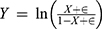 , ε = 0.001 for variables ranging from 0 to 1 generated negative values during the normalization process. Due to the presence of values between 0 and 1 in thyroid volume, TRAB, and monocyte measurements, logarithmic transformation resulted in negative values. This mathematical characteristic requires careful interpretation and may necessitate alternative normalization strategies.
Variables
Exposure Variable
Peripheral blood lymphocyte count was measured within 24 hours of hospital admission using an automated hematology analyzer (Sysmex XN-9000). The results were reported in units of 109/L and classified according to clinical guidelines.
Outcome Variable
PTC was confirmed through pathological examination and independently assessed by two experienced pathologists, who were blinded to clinical data. The outcome was recorded as a binary variable (0 = benign, 1 = malignant).
Covariates
Demographic Characteristics
Age (continuous) and sex (categorical).
Clinical Indicators
Nodule volume, neutrophil count, and monocyte count (all continuous).
Thyroid Autoantibodies
Thyroid peroxidase antibody (TPOAb) and thyroglobulin antibody (TGAb) (both continuous).
Covariates were selected based on clinical relevance and previously identified confounding factors. Missing data (> 5%) were addressed using multiple imputation methods, and robustness was evaluated through sensitivity analyses, including complete-case analysis and assessments using imputed datasets.
Ethical Statement
Ethical approval for this study was obtained from the Ethics Committee of the Longyan First Affiliated Hospital of Fujian Medical University (Approval Number: LYREC2024-K043-01), and all procedures adhered to the principles of the Declaration of Helsinki. As a retrospective observational study using anonymized data, the requirement for informed consent was waived in accordance with institutional ethics guidelines. Patient privacy was rigorously safeguarded, with all personally identifiable information de-identified to maintain confidentiality. The data were used exclusively for academic purposes.
Statistical Analysis
Descriptive Statistics
Continuous variables: Presented as mean ± standard deviation for normally distributed data or as median (minimum, maximum) for non-normally distributed data.
Categorical variables: Expressed as frequency or percentage.
Statistical tests: Categorical variables: Analyzed using the chi-square test. Normally distributed continuous variables: Compared using the independent samples t-test. Non-normally distributed continuous variables: Assessed using the Mann–Whitney U-test.
Association Analysis
Step 1: Regression Models
Model 1: No covariate adjustment.
Model 2: Adjusted for demographic characteristics, including age and sex.
Model 3: Adjusted for additional covariates, including nodule volume, neutrophil count, monocyte count, TPOAb, FT3, FT4, TRAb, and TGAb.
Purpose: The association between lymphocyte count and thyroid malignancy was evaluated under different adjustment strategies.
Step 2: Nonlinearity Testing
A Generalized Additive Model (GAM) was applied with smooth curve fitting using penalized splines.
If nonlinearity was detected, the inflection point was determined using a recursive algorithm, and a piecewise linear model was constructed.
Standard linear regression and piecewise models were compared using log-likelihood ratio tests.
Step 3: Subgroup Analysis
Stratified linear regression or GAM was applied based on clinical cut-points or tertiles of continuous covariates.
Interaction tests were performed, and effect modification was assessed using likelihood ratio tests.
Sensitivity Analysis
Lymphocyte count was converted into categorical variables, and trend p-values were calculated.
The results of the continuous variable analysis were validated, and potential nonlinear relationships were explored.
Statistical Software
All analyses were conducted using R software (version 4.2.0). A two-sided p-value of < 0.05 was considered statistically significant.
Results
Characteristics of Study Participants
A total of 1832 patients were included in the study, with 1007 classified in the benign thyroid nodule group and 825 in the PTC group. Significant differences were identified in multiple clinical parameters between the two groups.
Age: The mean age was slightly higher in the PTC group compared to the benign nodule group (47.36 ± 12.12 years vs 44.28 ± 10.95 years, p < 0.001).
Thyroid Nodule Volume: Significantly larger nodule volumes were observed in the benign thyroid nodule group (0.59 ± 2.34 vs −1.23 ± 1.97, p < 0.001).
Endocrine and Inflammatory Marker Analysis
Substantial differences were observed in endocrine-related parameters:
Thyroid function and autoantibodies: Statistically significant inter-group variations were identified in free triiodothyronine (FT3), free thyroxine (FT4), TPOAb, TGAb, and thyroid-stimulating hormone receptor antibodies (TRAb) (p < 0.001).
Lymphocyte Count: The lymphocyte count was significantly higher in the PTC group compared to the benign nodule group (1.87 ± 0.53 vs 1.20 ± 0.63, p < 0.001), indicating a potential relationship between lymphocyte dynamics and thyroid malignancy.
Inflammatory Cellular Markers
Significant inter-group differences were also identified in inflammatory cellular markers:
Neutrophil Count: Significantly higher in the PTC group (1.76 ± 0.59 vs 1.43 ± 0.81, p < 0.001).
Monocyte Count: Increased in the PTC group (−1.07 ± 0.44 vs.-1.26 ± 0.63, p < 0.001).
Leukocyte Count: Elevated in the PTC group (2.06 ± 0.46 vs 1.69 ± 0.48, p < 0.001).
Sex Distribution
Although significant differences were identified in clinical parameters, sex distribution did not differ significantly between the two groups (p = 0.216). Females constituted the majority in both groups, representing 80.9% of the benign nodule group and 83.2% of the PTC group (Table 1).
 |
Table 1 Baseline Characteristics of Patients with Benign and Malignant Thyroid Nodules |
Univariate Analysis Results
Lymphocyte Count and Disease Risk Correlation
Univariate analysis identified a significant dose-response relationship between lymphocyte count and disease risk. Compared to the low-value group, a significantly increased risk was observed in the medium-value group (OR = 11.93, 95% CI: 6.61–21.51, p < 0.0001), while an even greater risk was noted in the high-value group (OR = 41.75, 95% CI: 23.34–74.67, p < 0.0001). These findings indicate that lymphocyte count serves as a key predictive biomarker for thyroid malignancy.
Age and Clinical Characteristics
The mean age of the group was 45.66 ± 11.59 years. Age-stratified analysis demonstrated that older adult patients had a significantly higher risk of thyroid malignancy (OR = 1.88, 95% CI: 1.50–2.37, p < 0.0001), confirming age as an independent risk factor for disease progression.
Thyroid Nodule Volume
Thyroid nodule volume was strongly positively correlated with disease risk. A significantly elevated risk was observed in the high-volume group (OR = 7.10, 95% CI: 5.51–9.16, p < 0.0001), indicating that nodule volume serves as a potential predictive indicator for malignancy.
Endocrine and Inflammatory Markers
Several endocrine and inflammatory markers were significantly associated with disease risk:
FT3: A significantly increased risk was observed in the high-value group (OR = 1.89, 95% CI: 1.50–2.39, p < 0.0001).
FT4: A lower risk was identified in the high-value group (OR = 0.52, 95% CI: 0.41–0.65, p < 0.0001).
TRAb: An elevated risk was noted in the high-value group (OR = 4.88, 95% CI: 3.68–6.48, p < 0.0001).
Among inflammatory cell markers, neutrophils, monocytes, and leukocytes exhibited negative correlations with disease risk. However, an increased risk was maintained in the high-value monocyte group, indicating a complex relationship between inflammatory markers and thyroid malignancy.
Sex Differences
Sex differences were not statistically significant in this study (OR = 0.86, 95% CI: 0.68–1.09, p = 0.216). Females constituted the majority in both the benign and malignant groups (Table 2).
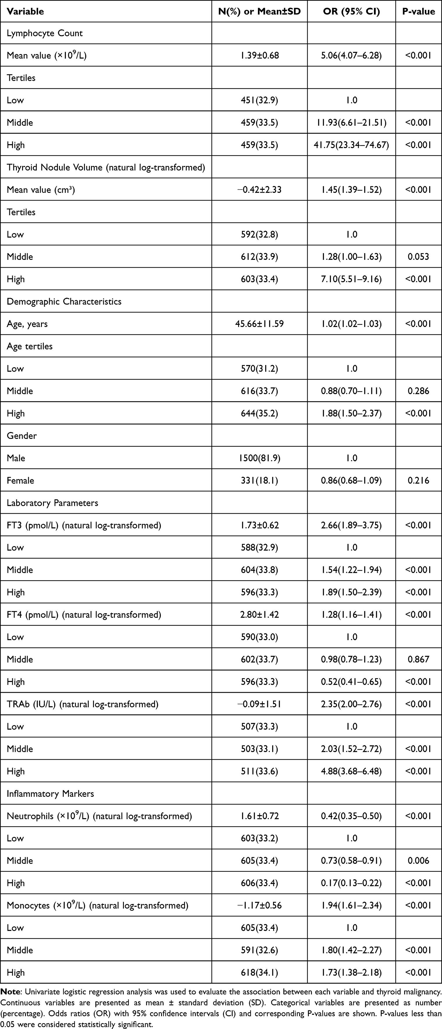 |
Table 2 Univariate Analysis of Risk Factors for Thyroid Malignancy |
Multidimensional Analysis of the Relationship Between Lymphocyte Count and the Risk of PTC
This study highlights the complex relationship between lymphocyte count and the risk of PTC through a systematic stratified analysis.
Nodule volume and malignancy risk: Thyroid nodule volume significantly influenced the risk of malignant transformation. Compared to the low-volume group (OR = 2.92, 95% CI: 2.06–4.14), the medium-volume group (OR = 4.11, 95% CI: 2.86–5.89) and high-volume group (OR = 9.47, 95% CI: 5.67–15.81) exhibited a significant dose-dependent increase in malignancy risk (p < 0.0001). Notably, within different nodule volume strata, the interaction with lymphocyte count was more pronounced. In the high-volume group, the malignancy risk in the high-lymphocyte subgroup reached 47.21 times (95% CI: 16.20–137.54). The p-value for interaction was 0.0005, demonstrating a highly significant modifying effect. Thus, thyroid nodule volume significantly modifies the relationship between absolute lymphocyte count and TEAM risk. The effect of lymphocyte count on TEAM risk is much stronger in patients with larger nodular volume.
Sex-specific risk: Both males (OR = 5.43, 95% CI: 4.25–6.95) and females (OR = 4.17, 95% CI: 2.57–6.77) demonstrated significant malignancy risks, with a slightly higher risk observed in males. Within the male subgroup, the malignancy risk in the high-lymphocyte group reached 48.52 times (95% CI: 25.04–94.01), indicating that sex may influence tumor transformation by modulating the inflammatory microenvironment. Stratified by sex, the association between absolute lymphocyte count and TEAM remained statistically significant in both male (OR = 5.43, 95% CI: 4.25–6.95, p < 0.0001) and female (OR = 4.17, 95% CI: 2.57–6.77, p < 0.0001) participants. The p-value for interaction was 0.3480, indicating no significant interaction effect. Thus, sex does not significantly modify the association between absolute lymphocyte count and TEAM risk.
Age gradient effect: Stratified age analysis identified a significant risk gradient, with the highest malignancy risk observed in the older adult group (OR = 7.25, 95% CI: 4.80–10.93). This increased risk may be attributed to age-related changes in immune function and tumorigenesis mechanisms. In the older adult group, the malignancy risk in the high-lymphocyte subgroup reached 58.08 times (95% CI: 22.45–150.20). Interaction analysis after adjustment for age showed that the positive association between absolute lymphocyte count and TEAM remained significant within each TEAM tertile: Low tertile: OR = 4.6 (95% CI: 3.2–6.7, p < 0.0001); Middle tertile: OR = 3.8 (95% CI: 2.6–5.6, p < 0.0001); High tertile: OR = 7.2 (95% CI: 4.8–10.9, p < 0.0001). The interaction p-value was 0.0713, indicating a trend toward a differential (non-uniform) effect across subgroups. However, this did not reach conventional statistical significance (p < 0.05).
Endocrine indicators and malignant transformation: Stratification by endocrine indicators revealed more complex risk patterns:
FT3 Group: The malignancy risk in the high-lymphocyte subgroup was as high as 88.45 times (95% CI: 21.06–371.49).
FT4 Group: The malignancy risk in the high-lymphocyte subgroup was 33.57 times (95% CI: 14.61–77.17) (Table 3).
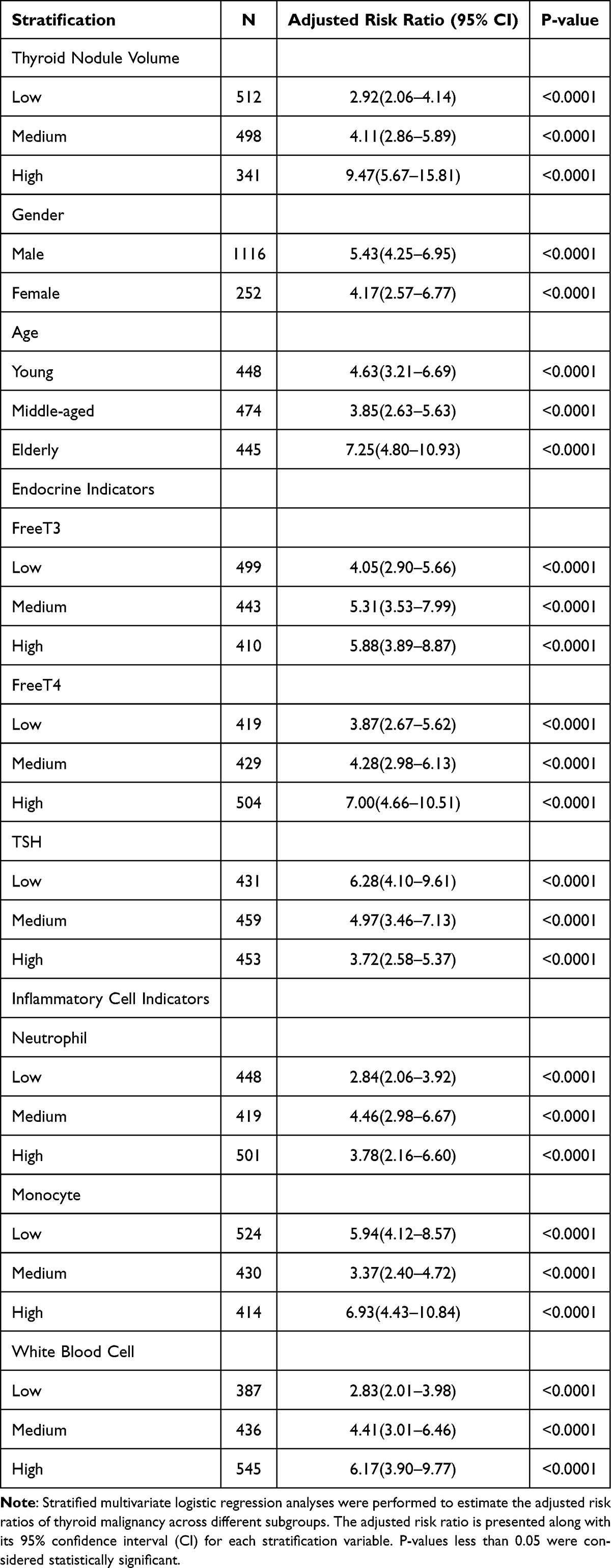 |
Table 3 Stratified Analysis of Risk Factors for Thyroid Malignancy |
Overall Risk Association
Lymphocyte count was identified as an independent risk factor for the development of PTC (Adjusted Risk Ratio: 2.77, 95% CI: 2.05–3.73, p < 0.0001).
Significance: The relationship remained statistically significant after adjustment for confounding factors, including demographic characteristics and clinical parameters.
Stratified Risk Analysis
Moderate lymphocyte levels were associated with a significantly increased cancer risk (Risk Ratio: 6.63, 95% CI: 3.33–13.22, p < 0.0001).
Elevated lymphocyte levels demonstrated a dose-dependent association with a higher cancer risk (Risk Ratio: 15.02, 95% CI: 7.52–30.03, p < 0.0001) (Table 4).
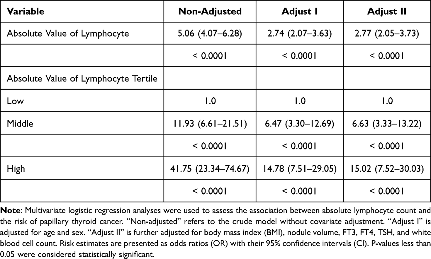 |
Table 4 Multivariate Regression Analysis of Lymphocyte Absolute Count and Papillary Thyroid Cancer Risk |
Smooth Curve Fitting
A nonlinear relationship between lymphocyte count and the risk of papillary thyroid carcinoma was identified using a GAM. The reported estimated effective degrees of freedom (eg, 4.45 for the smooth term) reflects the complexity of the fitted relationship: higher values allow greater flexibility, while lower values produce smoother fits. The smooth curve demonstrated a complex, nonmonotonic association between lymphocyte count and cancer risk, indicating the involvement of intricate immune-mediated mechanisms in thyroid carcinogenesis. In our data, the GAM demonstrated a lower AIC and BIC, as well as a higher proportion of deviance explained (30.3% for the GAM model), compared to the linear model, supporting the superiority of the nonlinear specification. The model was adjusted for multiple covariates, yielding an adjusted R-squared of 0.317, indicating substantial explanatory power. As shown in the smoothed plot (Figure 1), the relationship exhibited an “inverted U-shaped” (∩-shaped) trend. Specifically, TEAM (risk indicator for PTC) rose with increasing lymphocyte count at low to moderate levels, reaching a plateau and subsequently showing a slight decline at higher lymphocyte counts. This suggests that at lower levels, increasing lymphocytes may be associated with rising risk, but after reaching an optimal mid-range, further increases in lymphocytes may actually correspond to reduced risk or a regulatory effect (Figure 1, Tables 5–8).
 |
Table 5 Generalized Additive Model Analysis of the Nonlinear Relationship Between Lymphocyte Count and Papillary Thyroid Carcinoma Risk |
 |
Table 6 Chi-Square Tests for Linear Terms |
 |
Table 7 Approximate Significance of Smooth Terms |
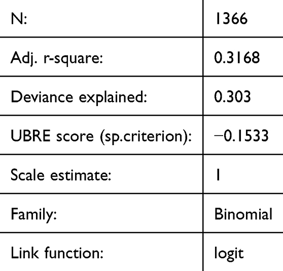 |
Table 8 Model Statistics |
 |
Figure 1 Smooth curve fitting analysis of lymphocyte count and risk of PTC. This figure illustrates the relationship between lymphocyte count (x-axis, absolute number, range 0–5) and the clinical outcome (y-axis, TEAM – Transforming Effect Value) using a smoothing spline fitting model. The red solid line represents the fitted trend, while the blue dashed lines indicate the 95% CIs. The blue and red curves depict risk trends for different groups, with shaded regions representing 95% CIs. |
Receiver Operating Curve Analysis for Continuous Predictor
ROC analysis for lymphocyte count yielded an area under the curve (AUC) of 0.8068 (95% CI: 0.7840–0.8297), indicating good diagnostic performance. The optimal classification threshold was identified as 1.2050, achieving a sensitivity of 92.51% and a specificity of 61.71%. The model demonstrated an overall accuracy of 70.42%. The optimal threshold identified by ROC analysis was 1.205, and this value was used to define the subgroups (≤1.205 vs >1.205).
The Positive Likelihood Ratio was calculated as 2.4160, while the Negative Likelihood Ratio was 0.1214, resulting in a Diagnostic Odds Ratio of 19.8962. The Positive Predictive Value was 48.77%, while the Negative Predictive Value was 95.43%, highlighting the effectiveness of the model in predicting the condition based on lymphocyte counts.(Figure 2).
 |
Figure 2 ROC Curve. This ROC curve demonstrates the diagnostic performance of lymphocyte count in differentiating between malignant and benign thyroid nodules. The blue curve represents the ROC curve, with sensitivity (true positive rate) on the vertical axis and 1 - specificity (false positive rate) on the horizontal axis. The green diagonal line denotes the reference for random guessing (area under the curve, AUC = 0.5). The closer the curve is to the top - left corner, the higher the diagnostic accuracy. The AUC reflects the overall diagnostic performance. |
Discussion
Summary of Findings
This study examined the relationship between absolute peripheral blood lymphocyte count (exposure variable) and papillary thyroid carcinoma (PTC, outcome variable) using a retrospective case-control design with a large sample size (1832 cases). The findings systematically characterized the complex relationship between lymphocyte count and thyroid malignancy risk, emphasizing the potential role of lymphocyte count and related markers in the pathogenesis and progression of thyroid cancer.
The results indicated that lymphocyte count was significantly elevated in patients with thyroid cancer and was closely associated with nodule volume, thyroid function parameters, and thyroid autoantibody levels. Stratified analysis further demonstrated that individuals in the high lymphocyte count group (Tertile High) exhibited a significantly increased risk of thyroid cancer (OR = 41.75, 95% CI: 23.34–74.67, p < 0.0001). These findings indicate that lymphocyte count may play a key role in the development and progression of thyroid malignancy.
The results indicate that lymphocyte count may serve as a potential biomarker for disease risk assessment. Multivariate regression analysis revealed a significant dose-response relationship between lymphocyte count and PTC risk. Following multiple adjustments, the risk ratio for the high lymphocyte group reached 15.02 (95% CI: 7.52–30.03, p < 0.0001). ROC curve analysis demonstrated an AUC of 0.8068 (95% CI: 0.7840–0.8297) for lymphocyte count, indicating good diagnostic performance.
Relationship Between Lymphocytes and the Immune Microenvironment of Thyroid Cancer
Anti-Tumor Immune Function
Lymphocytes constitute a key component of the tumor immune microenvironment and play a fundamental role in immune surveillance.13 CD8+ T cells and NK cells contribute to tumor suppression through direct cytotoxic activity or cytokine secretion, while CD4+ T cells facilitate anti-tumor immunity by modulating immune responses.14 In this study, the higher lymphocyte count observed in patients with thyroid cancer indicates an enhanced immune response to tumor cells.
Dual Role of Inflammation in Tumorigenesis
Chronic inflammation is a key contributor to tumorigenesis, with lymphocytes playing a key role in inflammatory responses.15,16 In this study, nodule volume was significantly larger in the non-cancer group compared to the cancer group (0.59 ± 2.34 vs −1.23 ± 1.97, p < 0.001). Additionally, patients in the high nodule volume group (Tertile High) exhibited a significantly increased risk of thyroid cancer (OR = 9.47, 95% CI: 5.67–15.81, p < 0.0001). These findings indicate that lymphocytes contribute to thyroid cancer development by mediating chronic inflammatory responses.
Immune Evasion and Tumor Progression
Although lymphocytes play a key role in anti-tumor immunity, their function can be suppressed by tumor cells through immune evasion mechanisms.17 For instance, tumor cells can express immune checkpoint molecules such as PD-L1, which inhibit T-cell activity and facilitate tumor progression.18 In this study, TRAb levels were significantly higher in the thyroid cancer group compared to the non-cancer group (0.35 ± 2.31 vs −0.32 ± 0.73, p < 0.001), indicating a potential role of immune evasion in thyroid cancer progression.
As key components of immune surveillance, lymphocytes are actively involved in tumor immune evasion processes.19 Changes in lymphocyte count and function may directly impact tumor development and progression.19 Within the thyroid tumor microenvironment, lymphocyte dysfunction may contribute to immune evasion and sustained tumor growth.17
Relationship Between Lymphocytes, Thyroid Function, and Autoantibodies
Changes in Thyroid Function Indicators
FT3 and FT4 levels were significantly higher in the thyroid cancer group compared to the non-cancer group (FT3: 1.86 ± 0.88 vs 1.62 ± 0.25, p < 0.001; FT4: 3.03 ± 2.08 vs 2.63 ± 0.38, p < 0.001), whereas TSH levels did not differ significantly between the two groups (p = 0.699). These findings indicate that compensatory changes in thyroid function may occur in patients with thyroid cancer.20
Elevated Levels of Autoantibodies
TPOAb and TGAb levels were significantly higher in the thyroid cancer group compared to the non-cancer group (TPOAb: 13.87 ± 85.98 vs 2.34 ± 8.46, p < 0.001; TGAb: 10.78 ± 75.13 vs 2.47 ± 12.76, p < 0.001). Additionally, elevated TPOAb and TGAb levels were significantly associated with an increased risk of thyroid cancer (TPOAb: OR = 3.29, 95% CI: 2.24–4.83, p < 0.0001; TGAb: OR = 3.91, 95% CI: 2.59–5.89, p < 0.0001). These findings indicate that autoimmune responses contribute to the pathogenesis of thyroid cancer.21,22
Clinical Implications of Lymphocyte Count
Potential Biomarker for Thyroid Cancer
ROC curve analysis demonstrated that lymphocyte count had an AUC of 0.8068 (95% CI: 0.7840–0.8297), with a sensitivity of 92.51% and a specificity of 61.71% at an optimal threshold of 1.2050. These findings indicate that lymphocyte count possesses high diagnostic value and serves as a potential biomarker for thyroid cancer.
Guiding Individualized Treatment
Stratified analysis demonstrated that patients in the high lymphocyte count group had a significantly increased risk of thyroid cancer (OR = 41.75, p < 0.0001). These findings indicate that lymphocyte count may be used in clinical practice for risk stratification and the development of individualized treatment strategies.23 For instance, patients with higher lymphocyte counts benefit from enhanced immune monitoring or early intervention.
This study is the first systematic analysis of the clinical significance of lymphocyte count in PTC patients, an important indicator rarely focused on in previous research, providing a new perspective for PTC diagnosis and prognosis assessment. This approach enables rapid risk prediction through routine blood tests, providing substantial translational potential compared to existing studies that rely on complex immunological testing or expensive imaging modalities. By integrating multidimensional stratified analysis, this study provides a new preliminary basis for clinical decision making.
The evaluation of lymphocyte count facilitates preliminary risk assessment for malignant transformation of thyroid nodules, thereby guiding subsequent diagnostic and therapeutic decisions, such as the necessity for further biopsy or imaging studies. Future research should aim to validate the external generalizability of these findings, refine multi-marker risk prediction models, and investigate the regulatory role of lymphocytes in thyroid tumorigenesis to generate more robust scientific evidence for clinical applications.
Existing research further supports our findings. Dov et al’s machine learning study confirms the potential of whole slide images in thyroid cytopathology screening, providing technical support for the clinical application of lymphocyte count.24 The Delphi expert consensus study of Marletta et al emphasized the importance of integrating new technologies and biomarkers in thyroid cytopathology.25 Eccher et al and Gern et al studies explored donor thyroid nodule management and intraoperative frozen section techniques, respectively, providing additional clinical context for lymphocyte count as a risk prediction indicator. These studies collectively demonstrate that lymphocyte count is not only a potential biomarker but may also become an important tool for personalized thyroid disease management.26,27
Absolute single lymphocyte counts have significant methodological advantages over traditional immunological ratios such as NLR and PLR. Absolute values can more directly reflect actual changes in immune cell numbers, avoiding complex statistical biases that ratio calculations can introduce. Ratio indicators are susceptible to nonlinearity in denominator and numerator cell counts, which may mask or distort true immunological changes. In contrast, single lymphocyte counts provide a more stable and direct way to assess immune status, accurately capturing small changes in immune cell numbers. In addition, the absolute value method reduces the potential mathematical error caused by calculating the ratio, providing a more reliable and transparent biomarker for tumor immunology research. Our results support this view and suggest that single lymphocyte counts have unique and important clinical value in the immunological assessment of thyroid cancer.28
Strengths and Limitations
This study possesses several notable strengths. First, a rigorous and innovative study design was used, incorporating a large sample size (1832 cases) within a retrospective case-control framework. Strict inclusion and exclusion criteria were applied to enhance the representativeness of the study population and ensure data reliability. Second, the use of advanced statistical methods, including multivariate linear regression, GAM, and smooth curve fitting, facilitated a comprehensive exploration of the complex nonlinear relationship between lymphocyte count and PTC risk. Unlike traditional studies that rely on a single statistical approach, this multidimensional analytical framework substantially improved the depth and breadth of the study.
Additionally, potential confounders such as age, sex, and endocrine markers, were rigorously controlled through multivariate adjustments, enhancing internal validity. ROC curve analysis demonstrated the strong diagnostic performance of lymphocyte count (AUC = 0.8068), providing a reliable risk prediction tool for clinical application. Finally, this study is the first to systematically examine the differential risk patterns of lymphocyte count across subgroups, providing a new immunological perspective on thyroid tumorigenesis and contributing to its originality and academic significance.
However, several limitations should be acknowledged. First, as a single-center retrospective case-control study, the generalizability and applicability of the findings are restricted. Although 1832 patients were included, all participants were recruited from Longyan First Hospital, which may introduce selection bias. Therefore, validation through multicenter, large-scale prospective studies is necessary. Second, the study population primarily consisted of individuals of Chinese Han ethnicity, limiting the direct applicability of the findings to other ethnic and regional populations. Moreover, the inclusion and exclusion criteria excluded patients with acute inflammation, autoimmune diseases, other malignancies, long-term use of immunomodulatory drugs, and endocrine disorders. As a result, the findings may not be generalizable to these specific populations. In addition, the large standard deviations observed suggest considerable individual variation, meaning that the detected differences may have limited practical implications for specific cases. To better assess the real-world utility of these results, future research could benefit from larger and more diverse samples or stratified analyses based on key covariates. Third, as an observational study, this research establishes an association between lymphocyte count and PTC risk but does not infer causality. Although adjustments were made for measurable confounders, the presence of unrecognized potential confounders cannot be ruled out, which impacts the accuracy of the results. Finally, the study focused solely on lymphocyte count without investigating the underlying biological mechanisms, highlighting an important direction for future research.
Conclusion
This study is the first to systematically reveal a critical link between lymphocyte count and risk of papillary thyroid cancer. Our study not only provides new biomarkers for clinical risk stratification but also opens up new avenues for personalized thyroid disease management. Future studies could further validate the findings of this study and explore the potential predictive value of lymphocyte counts in different thyroid diseases. It can be used as one of the tools to improve the diagnostic efficacy in combination with existing criteria such as thyroid ultrasound.
Abbreviations
PTC, Papillary thyroid carcinoma; NK, Natural killer; NLR, Neutrophil-to-lymphocyte ratio; PLR, Platelet-to-lymphocyte ratio; WHO, World Health Organization; TPOAb, Thyroid peroxidase antibody; TGAb, Thyroglobulin antibody; GAM, Generalized Additive Model; TRAb, Thyroid-stimulating hormone receptor antibodies; NPV, Negative Predictive Value; PPV, Positive Predictive Value.
Data Sharing Statement
All data generated or analysed during this study are included in this article. Research data can be obtained from the first author upon request (Xiu-Ping Qiu).
Ethics Approval and Consent to Participate
The study was conducted in accordance with the Declaration of Helsinki (as was revised in 2013). The study was approved by Ethics Committee of the Longyan First Affiliated Hospital of Fujian Medical University (Approval Number: LYREC 2024-K043-01). As a retrospective observational study using anonymized data, the requirement for informed consent was waived in accordance with institutional ethics guidelines. Patient privacy was rigorously safeguarded, with all personally identifiable information de-identified to maintain confidentiality. The data were used exclusively for academic purposes.
Acknowledgments
We would like to express our sincere gratitude to the staff and medical team at Longyan First Affiliated Hospital of Fujian Medical University for their invaluable support in data collection and patient management. We also thank the Ethics Committee of Longyan First Affiliated Hospital for their guidance and approval of this study. Special thanks to the laboratory team for their assistance in conducting hematological analyses and ensuring the accuracy of the data.
We are deeply grateful to our mentors and colleagues for their constructive feedback and insightful discussions throughout the research process. Additionally, we acknowledge the financial and logistical support provided by Longyan First Affiliated Hospital of Fujian Medical University, which made this study possible.
Finally, we extend our heartfelt appreciation to the patients and their families for their participation and trust, without which this research would not have been possible.
Funding
Funding Support for the Longyan Medical Science and Technology Team for Oncology and Metabolic Diseases.
Disclosure
The authors declare that they have no competing interests.
References
1. Grani G, Sponziello M, Filetti S, Durante C. Thyroid nodules: diagnosis and management. Nat Rev Endocrinol. 2024;20(12):715–728. doi:10.1038/s41574-024-01025-4
2. Haugen BR, Alexander EK, Bible KC, et al. 2015 American Thyroid Association Management Guidelines for adult patients with thyroid nodules and differentiated thyroid cancer: the American Thyroid Association Guidelines Task Force on thyroid nodules and differentiated thyroid cancer. Thyroid. 2016;26(1):1–133. doi:10.1089/thy.2015.0020
3. Uppal N, Collins R, James B. Thyroid nodules: global, economic, and personal burdens. Front Endocrinol. 2023;14:1113977. doi:10.3389/fendo.2023.1113977
4. Bray F, Ferlay J, Soerjomataram I, Siegel RL, Torre LA, Jemal A. Global cancer statistics 2018: GLOBOCAN estimates of incidence and mortality worldwide for 36 cancers in 185 countries [published correction appears in CA Cancer J Clin. 2020 Jul;70(4):313. doi: 10.3322/caac.21609]. CA Cancer J Clin. 2018;68(6):394–424. doi:10.3322/caac.21492
5. Bozan MB, Yazar FM, Kale İT, Yüzbaşıoğlu MF, Boran ÖF, Azak Bozan A. Delta neutrophil index and neutrophil-to-lymphocyte ratio in the differentiation of thyroid malignancy and nodular goiter. World J Surg. 2021;45(2):507–514. doi:10.1007/s00268-020-05822-6
6. Cui SS, Shen Y, Yang RQ. Predictive value of lymphocyte-to-neutrophil ratio and platelet-to-neutrophil ratio on PD-L1 expression in lung cancer. Clin Respir J. 2024;18(8):e13821. doi:10.1111/crj.13821
7. Gasparyan AY, Ayvazyan L, Mukanova U, Yessirkepov M, Kitas GD. The platelet-to-lymphocyte ratio as an inflammatory marker in rheumatic diseases. Ann Lab Med. 2019;39(4):345–357. doi:10.3343/alm.2019.39.4.345
8. Yildiz S, Eker E, Ozturk M, Alay M. A comparison of haemogram parameters of patients with thyroid papillary cancer and nodular goiter in Van, Turkey. J Pak Med Assoc. 2019;69(11):1642–1646. doi:10.5455/JPMA.301839
9. Deniz MS. A novel proportional index to differentiate between demographically and clinically matched cases with papillary thyroid carcinoma or non-cancerous nodule: PLR-to-PDW ratio. Am J Transl Res. 2023;15(4):2820–2827.
10. Manatakis DK, Tseleni-Balafouta S, Balalis D, et al. Association of baseline neutrophil-to-lymphocyte ratio with clinicopathological characteristics of papillary thyroid carcinoma. Int J Endocrinol. 2017;2017:8471235. doi:10.1155/2017/8471235
11. Kim SM, Kim EH, Kim BH, et al. Association of the preoperative neutrophil-to-ymphocyte count ratio and platelet-to-lymphocyte count ratio with clinicopathological Characteristics in patients with papillary thyroid cancer. Endocrinol Metab. 2015;30(4):494–501. doi:10.3803/EnM.2015.30.4.494
12. Manatakis DK, Tseleni-Balafouta S, Tzelves L, et al. Diagnostic accuracy of preoperative neutrophil-to-lymphocyte and platelet-to-lymphocyte ratios in detecting occult papillary thyroid microcarcinomas in benign multinodular goitres. J Thyroid Res. 2018;2018:3470429. doi:10.1155/2018/3470429
13. Tsai CH, Chuang YM, Li X, et al. Immunoediting instructs tumor metabolic reprogramming to support immune evasion. Cell Metab. 2023;35(1):118–133.e7. doi:10.1016/j.cmet.2022.12.003
14. Fridman WH, Pagès F, Sautès-Fridman C, Galon J. The immune contexture in human tumours: impact on clinical outcome. Nat Rev Cancer. 2012;12(4):298–306. doi:10.1038/nrc3245
15. Singh N, Baby D, Rajguru JP, Patil PB, Thakkannavar SS, Pujari VB. Inflammation and cancer. Ann Afr Med. 2019;18(3):121–126. doi:10.4103/aam.aam_56_18
16. Kennel KB, Bozlar M, De Valk AF, Greten FR. Cancer-associated fibroblasts in inflammation and antitumor immunity. Clin Cancer Res. 2023;29(6):1009–1016. doi:10.1158/1078-0432.CCR-22-1031
17. Pardoll DM. The blockade of immune checkpoints in cancer immunotherapy. Nat Rev Cancer. 2012;12(4):252–264. doi:10.1038/nrc3239
18. Chen DS, Mellman I. Oncology meets immunology: the cancer-immunity cycle. Immunity. 2013;39(1):1–10. doi:10.1016/j.immuni.2013.07.012
19. Paijens ST, Vledder A, de Bruyn M, Nijman HW. Tumor-infiltrating lymphocytes in the immunotherapy era. Cell Mol Immunol. 2021;18(4):842–859. doi:10.1038/s41423-020-00565-9
20. Fiore E, Rago T, Provenzale MA, et al. Lower levels of TSH are associated with a lower risk of papillary thyroid cancer in patients with thyroid nodular disease: thyroid autonomy may play a protective role. Endocr Relat Cancer. 2009;16(4):1251–1260. doi:10.1677/ERC-09-0036
21. He M, Zhang T, Huang Q, Tang X, Wu B, Liu Y. Pathological correlation between eosinophils and thyroid nodules based on medical image testing. SLAS Technol. 2024;29(5):100189. doi:10.1016/j.slast.2024.100189
22. Cinar I, Sengul I. Coexistence of Hashimoto’s thyroiditis and papillary thyroid carcinoma revisited in thyroidology, an experience from an endemic region: fad or future? Rev Assoc Med Bras. 2024;70(4):e20231380. doi:10.1590/1806-9282.20231380
23. Pagès F, Mlecnik B, Marliot F, et al. International validation of the consensus Immunoscore for the classification of colon cancer: a prognostic and accuracy study. Lancet. 2018;391(10135):2128–2139. doi:10.1016/S0140-6736(18)30789-X
24. Dov D, Kovalsky SZ, Feng Q, et al. Use of machine learning-based software for the screening of thyroid cytopathology whole slide images. Arch Pathol Lab Med. 2022;146(7):872–878. doi:10.5858/arpa.2020-0712-OA
25. Marletta S, Salatiello M, Pantanowitz L, et al. Delphi expert consensus for whole slide imaging in thyroid cytopathology. Cytopathology. 2023;34(6):581–589. doi:10.1111/cyt.13279
26. Eccher A, Girolami I, D’Errico A, et al. Management of thyroid nodules in deceased donors with comparison between fine needle aspiration and intraoperative frozen section in the setting of transplantation. Prog Transplant. 2019;29(4):316–320. doi:10.1177/1526924819873898
27. Gern J, Arbogast M, Alakus H, et al. Intraoperative thyroid frozen section: indications, results and consequences. Gland Surg. 2024;13(5):630–639. doi:10.21037/gs-23-105
28. Lee KH, Seok EY, Kim EY, et al. Different prognostic values of individual hematologic parameters in papillary thyroid cancer due to age-related changes in immunity. Ann Surg Treat Res. 2019;96(2):70–77. doi:10.4174/astr.2019.96.2.70
 © 2025 The Author(s). This work is published and licensed by Dove Medical Press Limited. The
full terms of this license are available at https://www.dovepress.com/terms
and incorporate the Creative Commons Attribution
- Non Commercial (unported, 4.0) License.
By accessing the work you hereby accept the Terms. Non-commercial uses of the work are permitted
without any further permission from Dove Medical Press Limited, provided the work is properly
attributed. For permission for commercial use of this work, please see paragraphs 4.2 and 5 of our Terms.
© 2025 The Author(s). This work is published and licensed by Dove Medical Press Limited. The
full terms of this license are available at https://www.dovepress.com/terms
and incorporate the Creative Commons Attribution
- Non Commercial (unported, 4.0) License.
By accessing the work you hereby accept the Terms. Non-commercial uses of the work are permitted
without any further permission from Dove Medical Press Limited, provided the work is properly
attributed. For permission for commercial use of this work, please see paragraphs 4.2 and 5 of our Terms.