
Back to Journals » International Journal of Nanomedicine » Volume 21
Clinical Applications and Future Prospects of Metallic Nanoparticles in Diagnosis and Therapy
Authors Galarza-Arévalo GE  , González MP , Romero MP
Received 16 September 2025
Accepted for publication 11 February 2026
Published 16 April 2026 Volume 2026:21 567931
DOI https://doi.org/10.2147/IJN.S567931
Checked for plagiarism Yes
Review by Single anonymous peer review
Peer reviewer comments 4
Editor who approved publication: Professor Jie Huang
Gabriela E Galarza-Arévalo, Myriam P González, Maria P Romero
Escuela Politécnica Nacional, Quito, 170525, Ecuador
Correspondence: Maria P Romero, Email [email protected]
Abstract: Metallic nanoparticles (MNPs) have emerged as versatile platforms for addressing unmet clinical needs in diagnosis, therapy, and theranostics. This review synthesizes recent advances in the clinical application of iron-, gold-, hafnium-, gadolinium-, silver-, copper-, titanium-, and zinc-based nanoparticles across oncology, infection control, biomedical coatings, and diagnostic imaging. In oncology, superparamagnetic iron oxide nanoparticles (SPIONs) have demonstrated a median overall survival of 13.4 months in patients with recurrent glioblastoma and achieved intratumoral temperatures of 42– 48.5 °C during magnetic hyperthermia. Gold–silica nano shells generated tumor-free ablation zones in up to 87.5% of prostate lesions, with oncologic success reported in 73% of treated patients. CYT-6091, a ~27 nm PEGylated gold nanoparticle conjugated with TNF-α, enabled systemic delivery of this cytokine without inducing severe hypotension, showing a substantially improved safety profile compared with free TNF-α. In antimicrobial applications, Nano Silver Fluoride reduced dentin caries progression by more than 30% compared with controls, while copper nanoparticle-reinforced universal adhesives preserved mechanical performance and retention rates over 48 months. Collectively, these findings highlight the capacity of MNPs to integrate diagnostic and therapeutic functions, including MRI/CT contrast enhancement, magnetic hyperthermia, photothermal and photodynamic therapies, and gene delivery. Despite significant progress, challenges related to heterogeneous biodistribution, long-term toxicity, and regulatory approval remain, emphasizing the need for the development of safer and more efficient metallic nanomedicines aligned with emerging clinical demands in personalized medicine.
Keywords: metallic nanoparticles, theranostics, cancer nanomedicine, magnetic hyperthermia, antimicrobial nanomaterials, personalized medicine
Introduction
Metallic nanoparticles have emerged as transformative tools in modern medicine due to their unique physicochemical properties and nanoscale precision, enabling a wide range of diagnostic and therapeutic applications. Typically ranging from 1 to 100 nanometers in size (Figure 1), MNPs exhibit high surface-to-volume ratios, tunable optical, magnetic and electronic properties, and the ability to interact with biological systems at the molecular and cellular levels.1 These features make them particularly attractive for overcoming key limitations of conventional medical approaches, such as the nephrotoxicity of traditional contrast agents, the poor selectivity of chemotherapy and radiotherapy, and the resulting systemic toxicities that compromise treatment efficacy.1
 |
Figure 1 Clinical imaging of metallic nanoparticle-based therapies in oncology: (a) Recurrent glioblastoma treated with intratumoral instillation of magnetic nanoparticles: pre-treatment MRI (ai,aii), post-instillation CT showing nanoparticle deposits (aiii, aiv) with isothermal treatment lines (40–50 °C), and 3D reconstructions of fused MRI/CT illustrating tumor (brown), nanoparticle fluid (blue), and thermometry catheter (green) (av, avi) Three-dimensional reconstruction obtained from fused magnetic resonance imaging and computed tomography data, showing the tumor (brown), the magnetic fluid (blue), and the thermometry catheter (green).2 (b) Overall survival after diagnosis of first tumor recurrence/progression (OS-2) in 59 glioblastoma patients treated with combined thermo-/radiotherapy.2 (c) Integrated treatment evaluation with metallic nanoparticles showing (ci) the reconstructed distribution of implanted magnetic nanoparticles derived from CT-based volumetric maps and (cii) the experimentally validated temperature distribution obtained through intratumoral thermometry during interstitial magnetic nanoparticle hyperthermia.3 (d) CT scans of a patient with recurrent prostate cancer before injection (di), immediately after intraprostatic nanoparticle instillation (dii), and six weeks post-treatment (diii), demonstrating persistent nanoparticle depots enabling serial thermotherapy without reinjection.4 (e) CT scans from a recurrent glioblastoma patient before (left) and after combined intracavitary thermotherapy with iron oxide nanoparticles and radiotherapy (right), showing pronounced peritumoral edema around the nanoparticle deposits.5 (f) MRI of multiple brain metastases on T1-weighted gadolinium-enhanced imaging, representative of patient inclusion criteria in the NANO-RAD trial evaluating gadolinium-based nanoparticles.6 Reproduced from Maier-Hauff et al (2011), Wust et al (2006), Johannsen et al (2005), Grauer et al (2019), and Verry et al (2019) under a Creative Commons Attribution-NonCommercial 4.0 License. |
From a materials perspective, metallic nanomaterials used in medicine can be broadly classified into several major categories based on the composition and functional properties: noble metal nanoparticles (eg., gold and silver), magnetic metal oxide nanoparticles (eg., iron oxide), transition metal and metal oxide nanoparticles (eg., copper, zinc oxide, titanium dioxide), and emerging hybrid or doped metallic nanostructures. Each class exhibits distinct physicochemical characteristics that dictate its biomedical utility.1
Gold nanoparticles (AuNPs) are among the most extensively studied MNPs due to their excellent biocompatibility, chemical stability, and ease of surface functionalization. Their strong surface plasmon resonance enables efficient light absorption and heat generation, making them ideal for diagnostic imaging, biosensing, and photothermal therapy. For instance, AuNPs played a pivotal role in the development of rapid and highly sensitive diagnostic sensors during the COVID-19 pandemic, significantly improving viral detection and contributing to public health management.7 AuNP-based platforms have also been integrated into silicone-covered self-expandable metal stents and gold–silica nano shells systems for localized hyperthermia therapy, where near-infrared (NIR) laser irradiation induces controlled tissue ablation in esophageal models.8
Silver nanoparticles (AgNPs) are primarily valued for their potent antimicrobial activity. Their ability to release silver ions and disrupt microbial membranes makes them highly effective against Gram-positive and Gram-negative bacteria, including antibiotic-resistant strains. Consequently, AgNPs are widely used in wound dressings, medical devices coating, and antimicrobial surfaces to prevent biofilm formation and healthcare-associated infections. Similarly, Copper nanoparticles (CuNPs) contribute to biofilm prevention and pathogen control, while iron oxide nanoparticles are used in diagnostic and therapeutic settings.9,10 Their ability to inhibit bacterial adhesion and biofilm formation, particularly in silver and copper nanoparticles, further underscores their potential in combating antibiotic-resistant infections in medical settings.
Magnetic iron oxide nanoparticles (IONPs), typically composed of magnetite (Fe3O4) or maghemite (γ-Fe2O3), possess superparamagnetic properties that enable their use as contrast agents in magnetic resonance imaging (MRI) and as therapeutic agents in magnetic hyperthermia.11 By generating localized heat under alternating magnetic fields, IONPs can selectively induce tumor cell death while also enhancing imaging sensitivity and spatial resolution.12
Other metallic and metal oxide nanoparticles, zinc oxide (ZnO), and titanium dioxide (TiO2), further expand the biomedical landscape of MNPs. ZnO and TiO2 nanoparticles act as photosensitizers capable of generating reactive oxygen species (ROS) under light activation, making them effective for photodynamic therapy to induce apoptosis in tumor cells with minimal damage to healthy tissues.13,14 Beyond individual applications, the integration of metallic nanoparticles into theranostic platforms nano systems capable of simultaneous diagnostic imaging and targeted therapy represents a major advancement in personalized medicine. By combining imaging agents and therapeutic payloads within a single nanoparticle, theranostics enable real-time monitoring of treatment response while enhancing therapeutic precision and reducing systemic toxicity.15
Despite these advances, significant unmet clinical needs continue to drive innovation in metallic nanoparticles. In oncology, conventional treatments still lack adequate specificity, cause substantial systemic toxicity, and remain insufficient for detecting and eradicating early micro metastatic disease. In antimicrobial therapy, the rapid spread of multidrug-resistant pathogens, the limited efficacy of antibiotics against biofilms, and the scarcity of new therapeutic molecules demand more effective alternatives. In diagnostic imaging, current modalities often suffer from limited sensitivity, narrow signal windows, and difficulties integrating structural, functional, and molecular information within a single platform. These gaps have catalyzed the development of metallic nanoparticles engineered to enhance therapeutic selectivity, overcome microbial resistance, and strengthen advanced imaging approaches with built-in theranostic capabilities.
This review therefore provides a comprehensive overview of the clinical applications of metallic nanoparticles in diagnosis and therapy, with a particular focus on imaging techniques such as MRI, computed tomography, and nuclear medicine, as well as therapeutic strategies including photothermal therapy, photodynamic therapy, and gene delivery. Finally, the review discusses current challenges and future perspectives of MNPs, emphasizing their potential to transform personalized healthcare and advance modern medicine.
 |
Table 1 Clinically Investigated Metallic-Based Nanoparticles in Cancer Therapy and Imaging |
Clinical Applications of Metallic Nanoparticles
Metallic nanoparticles have gained significant attention for their ability to address complex medical challenges, particularly in diagnostics and therapeutics (Table 1) presents various regulatory approvals of metallic nanoparticles for cancer therapy and imaging.23 Due to their nanoscale properties, MNPs offer innovative disease detection, antimicrobial activity, drug delivery, and tissue regeneration solutions. The most widely researched MNPs include superparamagnetic iron oxide nanoparticles (SPIONs), gold, silver, and copper nanoparticles (CuNPs), each with a range of clinical applications.15 Building on their remarkable versatility, metallic nanoparticles are used across various medical fields. The following sections will analyze their roles in different diagnostic and therapeutic strategies.
Cancer Therapy and Imaging
Iron-Based Nanoparticles
Iron-based nanoparticles, particularly superparamagnetic iron oxide nanoparticles and ultrasmall SPIONs (USPIOs, <50 nm), have emerged as versatile platforms in oncology. Their biomedical value derives from two complementary functions: (i) as therapeutic agents, they can generate localized heating under an alternating magnetic field, enabling magnetic hyperthermia and enhancement of radiotherapy; (ii) as diagnostic agents, they shorten T1 and T2 relaxation times, providing sensitive, prolonged contrast in magnetic resonance imaging for tumor detection, staging, and monitoring.
Clinical Trials in Hyperthermia Therapy
The most extensively investigated therapeutic formulation is NanoTherm®, an aqueous dispersion of aminosilane-coated SPIONs (Fe3O4 cores ~12–15 nm, 112–120 mg Fe/mL) developed by MagForce AG. In a Phase II trial, Maier-Hauff et al (2011)2 treated 59 patients with recurrent glioblastoma multiforme (GBM) through neuronavigational intratumoral installation of NanoTherm® followed by six semi-weekly hyperthermia sessions combined with reduced-dose radiotherapy (Figure 1a). The therapy was feasible and safe, yielding a median overall survival of 13.4 months after recurrence, exceeding historical controls, with manageable side effects such as seizures and edema (Figure 1b).
In a broader feasibility study, Wust et al (2006)3 evaluated 22 patients with recurrent, unresectable tumors of various origins (glioblastoma, prostate, cervical, ovarian, rectal, sarcoma). Nanoparticles were injected under CT or ultrasound guidance, and hyperthermia sessions achieved intratumoral temperatures of 42–44 °C with ablative peaks above 46 °C, demonstrating wide applicability with mostly mild, transient toxicities (Figure 1c).
The first prostate cancer application was reported by Johannsen et al (2005),4 who treated a 67-year-old patient with locally recurrent carcinoma using transperineal ultrasound-guided NanoTherm® injections into 24 prostate depots followed by six weekly hyperthermia sessions. Intraprostatic temperatures reached 48.5 °C, and nanoparticles remained stable in situ for at least six weeks, confirming feasibility and tolerability (Figure 1d).
More recently, Grauer et al (2019)5 introduced the “NanoPaste” technique in six GBM patients, coating the resection cavity walls with NanoTherm® prior to hyperthermia and, in four cases, re-irradiation. Although delayed peritumoral edema required corticosteroids and re-surgery in most patients, histological analysis revealed extensive necrosis, HSP70 upregulation, and infiltration of immune cells, indicating both local cytotoxicity and systemic immunomodulation. Notably, two patients achieved durable remissions beyond 23 months, highlighting the potential of SPION-based hyperthermia to synergize with radiotherapy and possibly immunotherapy (Figure 1e).
Taken together, these trials demonstrate that SPION-mediated hyperthermia is technically feasible, safe under controlled conditions, and capable of producing therapeutic heating across multiple tumor types. Glioblastoma studies suggest survival benefits when combined with radiotherapy, while prostate studies confirm nanoparticle retention and reproducibility. Importantly, the NanoPaste results underscore that SPION hyperthermia may not only ablate tumor cells but also stimulate immune responses. Remaining challenges include heterogeneous nanoparticle distribution, variable heating profiles, and treatment-related edema, all of which require further optimization in ongoing phase I/II studies.
Clinical Use of Iron Oxide Nanoparticles as MRI Contrast Agents
Beyond hyperthermia, iron oxide nanoparticles have been widely investigated as magnetic resonance imaging contrast agents in oncology. Their superparamagnetic properties shorten T1 and T2 relaxation times, producing sensitive and long-lasting tumor enhancement. Among the most studied agents is ferumoxtran-10 (Combidex®/Sinerem®), a dextran-coated USPIO of approximately 30 nm, which demonstrated prolonged tumor enhancement for 24 to 72 hours in gliomas and in lymph node staging for prostate and pelvic cancers, often revealing tumor regions undetected by conventional gadolinium chelates; although initially approved by the EMA in 2006, it was later withdrawn in 2007.16,17 Ferumoxytol (Feraheme®), a semisynthetic carbohydrate-coated USPIO of similar size, was originally approved by the FDA for iron deficiency anemia but has since been repurposed in multiple oncology trials; it functions as a blood pool agent and is avidly taken up by tumor-associated macrophages, proving useful in imaging lymphomas, sarcomas, and brain tumors, while also offering safety advantages for patients with renal impairment at risk for gadolinium-induced nephrogenic systemic fibrosis.16,17 In pediatric oncology, ferumoxytol-enhanced whole-body MRI has achieved diagnostic accuracy above 98% compared with18 F-FDG PET/CT while avoiding ionizing radiation19. Earlier formulations such as ferumoxides (Feridex®/Endorem®), dextran-coated SPIOs of 120–180 nm, were approved in 1996 for liver and spleen imaging and used to detect hepatic metastases, though they were discontinued in 2008.17 Similarly, ferucarbotran (Resovist®/Cliavist®), a carboxydextran-coated SPIO of ~60 nm, was approved in Europe and Asia in 2001 and demonstrated high sensitivity for detecting small hepatic lesions before being withdrawn in 2009. More recently, carboxydextran-coated iron oxide nanoparticles were approved by the EMA in 2011 for sentinel lymph node detection in breast and rectal cancers, providing a non-radioactive alternative for surgical staging.17 Other iron formulations, such as iron sucrose or gluconate colloids, are primarily approved for anemia rather than oncology and are not directly relevant to cancer therapy.16
Iron oxide nanoparticles offer significant advantages in oncology, including prolonged MRI contrast, improved detection of small metastatic lesions, and the ability to generate controlled therapeutic hyperthermia. However, their clinical use remains limited by heterogeneous intratumoral distribution, variable heating efficiency, treatment-related adverse effects, such as edema, and regulatory withdrawals of several formulations. Key challenges include optimizing biodistribution, standardizing dosing parameters, and generating stronger clinical evidence to support their routine diagnostic and therapeutic implementation in cancer care.
 |
Figure 2 Therapeutic mechanisms and tumor-targeting strategies of metallic nanoparticles in oncology: (a) Metallic nanoparticles induce apoptosis in cancer cells through multiple mechanisms, including ROS generation, transcriptional inhibition, cell cycle arrest, and immune modulation. (b) Nanoparticles enhance phototherapy efficacy by acting as photosensitizers that amplify the effects of light, heat, and radiation, while modulating immune responses.23 (c) Schematic representation illustrating how structural abnormalities in tumor vasculature promote nanoparticle accumulation through the enhanced permeability and retention (EPR) effect, in contrast to normal vasculature. (ci) Tumor vessels exhibit a disorganized and tortuous architecture, characterized by widened endothelial junctions, pericyte loss, and an irregular basement membrane, all of which increase vascular permeability. (cii) Passive extravasation and accumulation of nanoparticles within the tumor microenvironment, driven by elevated vascular permeability, irregular blood flow, and the presence of tumor cells, macrophages, fibroblasts, and a disordered extracellular matrix; a process that defines the EPR effect and supports the use of nanoparticles as carriers for antineoplastic, antiangiogenic, or pH- and temperature-responsive therapies.24 Reproduced form Mundekkad et al (2022), and Andleeb et al (2021) under a Creative Commons Attribution-NonCommercial 4.0 License. |
God-Based Nanoparticles in Clinical Trials in Hyperthermia Therapy
Gold nanoparticles have been extensively studied for their role in cancer treatment. AuNPs, known for their biocompatibility and ease of functionalization, are widely used in various therapeutic modalities, such as photothermal therapy (PTT) and photodynamic therapy (PDT), as well as for targeted drug delivery (Figure 2a and b).25 In photothermal therapy, AuNPs absorb light and convert it into heat. Due to their surface plasmon resonance, gold nanoparticles are particularly effective at absorbing near-infrared light, which can penetrate deeply into tissues.23 This absorbed light is converted into heat, leading to localized hyperthermia that selectively destroys cancer cells without damaging surrounding healthy tissue. AuNPs can be functionalized with ligands that target specific cancer cell receptors, ensuring that the nanoparticles accumulate at the tumor site prior to laser irradiation. This method has shown high efficacy in treating solid tumors, such as breast and prostate cancers.26
In this context, AuNPs, particularly gold–silica nanoshells (GSNs), have been investigated as photothermal agents for focal cancer ablation due to their ability to strongly absorb near-infrared light and convert it into localized heat. In a first-in-human pilot study, Rastinehad et al (2019)18 evaluated 16 men with low to intermediate risk localized prostate cancer treated with intravenous GSN infusion followed by MR/US-fusion guided laser excitation of the prostate lesion. The treatment was feasible in 94% of patients and showed tumor-free ablation zones in 62.5% of lesions at 3 months and 87.5% at 12 months, with no grade ≥3 toxicities and preservation of urinary and sexual function. In a larger multi-institutional trial including 46 men (44 completed treatment), the same protocol demonstrated oncologic success in 73% of patients at 12 months, as confirmed by negative targeted and systematic biopsies and significant PSA reductions. Both trials underscore that GSN-mediated photothermal ablation is safe, feasible, and achieves promising short-term tumor control in prostate cancer while minimizing side effects associated with whole-gland therapies.18
Beyond focal ablation, gold nanoparticles have also been engineered as systemic nanomedicines. In a Phase I dose-escalation trial, Libutti et al (2010)19 investigated CYT-6091 (Aurimune®), a PEGylated 27 nm colloidal gold nanoparticle conjugated with recombinant human TNF-α (rhTNF), in 29 patients with advanced solid tumors, including melanoma, colorectal, pancreatic, breast, and sarcoma. Patients received two intravenous infusions (50–600 μg/m2 rhTNF equivalent) 14 days apart. The treatment was well tolerated up to the highest dose, with fever as the main manageable toxicity; unlike native rhTNF, no dose-limiting hypotension or severe hepatotoxicity occurred. Importantly, fever is the main manageable toxicity; unlike native rhTNF, no dose-limiting hypotension is observed. Electron microscopy confirmed gold nanoparticle accumulation in tumor biopsies 24 h post-infusion, and preliminary efficacy included one partial response (ocular melanoma) and four cases of stable disease. These findings demonstrate that efficacy included gold nanoparticles can safely deliver potent cytokines systemically at otherwise toxic doses, with selective tumor targeting.19
Gold nanoparticles used in clinical hyperthermia trials have demonstrated strong therapeutic potential due to their efficient near-infrared absorption, precise heat generation, and targeted delivery capabilities. Studies with gold–silica nano shells show good tolerability and promising short-term tumor control in prostate cancer. However, their performance remains limited by heterogeneous nanoparticle distribution, variable thermal profiles, and insufficient long-term efficacy data. Advancements are needed in surface functionalization, laser dosimetry standardization, and larger clinical trials to support their therapeutic integration.
Hafnium-Based Nanoparticles
Hafnium oxide nanoparticles (NBTXR3, Hensify®) are the first inorganic nanomedicine to obtain European CE marking as a radio enhancer in oncology. These crystalline nanoparticles (~50 nm) are administered via a single intratumoral injection and remain inert until activated by ionizing radiation, where they amplify local dose deposition selectively in tumor tissue.20 In a pivotal phase II/III trial including 180 patients with locally advanced soft tissue sarcoma (STS) of the extremities or trunk wall, NBTXR3 plus preoperative radiotherapy significantly improved pathological complete response rates compared with radiotherapy alone, leading to its clinical approval in Europe.20 Additional evidence from a phase I/II trial in hepatocellular carcinoma (HCC) and liver metastases (NCT02721056) demonstrated the safety and feasibility of NBTXR3 combined with stereotactic body radiotherapy (SBRT). In this study, patients received a single intralesional injection of NBTXR3 followed by SBRT (45–50 Gy in 3–5 fractions). Among 7 evaluable HCC patients, 3 achieved complete responses and 4 partial responses, while among 5 patients with liver metastases, 2 achieved partial responses and 1 stable disease. Importantly, no dose-limiting toxicities were observed up to the 33% dose level, with only mild to moderate procedure-related adverse events. Collectively, these clinical trials show that NBTXR3 is well tolerated, integrates seamlessly into existing radiotherapy workflows, and enhances local tumor control in sarcoma, HCC, and liver metastases, establishing hafnium oxide nanoparticles as a promising new class of radio enhancers in cancer therapy.27
Gadolinium-Based Nanoparticles
Gadolinium-based nanoparticles represent a new class of theranostic agents that combine the magnetic resonance imaging contrast properties of gadolinium with the ability to enhance the effects of ionizing radiation. The best characterized formulation is AGuIX®, an ultrasmall nanoparticle (~3 nm) with a polysiloxane core covalently grafted with gadolinium chelates (Gd-DOTA).21 Its dual functionality allows both sensitive MRI visualization and localized radio sensitization when activated by X-rays.21
The first clinical evidence comes from a phase I trial in 12 patients with locally advanced cervical cancer, where intravenous was administered alongside chemoradiation and brachytherapy. MRI confirmed nanoparticle accumulation in tumors, and treatment was well tolerated with no dose-limiting toxicities. Remarkably, all patients achieved complete remission of the primary tumor, with only one distant recurrence reported, highlighting the potential of AGuIX® as a safe and effective theranostic nanomedicine, chemoradiation and brachytherapy.21
A second clinical investigation, the NANO-RAD phase I trial, enrolled patients with multiple brain metastases from solid tumors. In this study, 15–18 patients received intravenous AGuIX® followed by whole-brain radiotherapy (WBRT, 30 Gy in 10 fractions). The trial demonstrated good tolerability across dose levels, and MRI imaging confirmed preferential tumor uptake of nanoparticles, supporting their role as both imaging enhancers and radiosensitizers.6
More recently, the NanoBrainMets phase II trial evaluated 23 patients with 129 brain metastases. In this randomized, double-blinded design, patients received stereotactic radiosurgery with or without AGuIX®. Advanced MRI mapping confirmed measurable tumor uptake (0.012–0.17 mg/mL), with larger tumors accumulating higher concentrations. Uptake patterns varied between patients, reflecting differences in tumor vasculature and suggesting a need for patient-specific optimization.22 Figure 1f shows MRI of multiple brain metastases in patients with the NANO-RAD trial evaluating gadolinium-based nanoparticles.
Gadolinium-based nanoparticles, such as AGuIX®, represent a promising theranostic platform that combines highly sensitive MRI contrast with localized radio sensitization. Clinical studies have demonstrated good tolerability, selective tumor accumulation, and encouraging therapeutic responses in cervical cancer and brain metastases. However, their effectiveness is limited by tumor uptake variability and by dependence on vascular permeability. Further optimization of delivery strategies and larger clinical trials are needed to validate long-term benefits and support their integration into precision radiotherapy.
Cancer Therapy and Imaging
Iron-Based Nanoparticles
Iron-based nanoparticles, particularly superparamagnetic iron oxide nanoparticles and ultrasmall SPIONs (USPIOs, <50 nm), have emerged as versatile platforms in oncology. Their biomedical value derives from two complementary functions: (i) as therapeutic agents, they can generate localized heating under an alternating magnetic field, enabling magnetic hyperthermia and enhancement of radiotherapy; (ii) as diagnostic agents, they shorten T1 and T2 relaxation times, providing sensitive, prolonged contrast in magnetic resonance imaging for tumor detection, staging, and monitoring.
Clinical Trials in Hyperthermia Therapy
The most extensively investigated therapeutic formulation is NanoTherm®, an aqueous dispersion of aminosilane-coated SPIONs (Fe3O4 cores ~12–15 nm, 112–120 mg Fe/mL) developed by MagForce AG. In a phase II trial, Maier-Hauff et al (2011)2 treated 59 patients with recurrent glioblastoma multiforme (GBM) through neuronavigational intratumoral instillation of NanoTherm® followed by six semi-weekly hyperthermia sessions combined with reduced-dose radiotherapy (Figure 1a). The therapy was feasible and safe, yielding a median overall survival of 13.4 months after recurrence, exceeding historical controls, with manageable side effects such as seizures and edema (Figure 1b).
In a broader feasibility study, Wust et al (2006)3 evaluated 22 patients with recurrent, unresectable tumors of various origins (glioblastoma, prostate, cervical, ovarian, rectal, sarcoma). Nanoparticles were injected under CT or ultrasound guidance, and hyperthermia sessions achieved intratumoral temperatures of 42–44 °C with ablative peaks above 46 °C, demonstrating wide applicability with mostly mild, transient toxicities (Figure 1c).
The first prostate cancer application was reported by Johannsen et al (2005),4 who treated a 67-year-old patient with locally recurrent carcinoma using transperineal ultrasound-guided NanoTherm® injections into 24 prostate depots followed by six weekly hyperthermia sessions. Intraprostatic temperatures reached 48.5 °C, and nanoparticles remained stable in situ for at least six weeks, confirming feasibility and tolerability (Figure 1d).
More recently, Grauer et al (2019)5 introduced the “NanoPaste” technique in six GBM patients, coating the resection cavity walls with NanoTherm® prior to hyperthermia and, in four cases, re-irradiation. Although delayed peritumoral edema required corticosteroids and re-surgery in most patients, histological analysis revealed extensive necrosis, HSP70 upregulation, and infiltration of immune cells, indicating both local cytotoxicity and systemic immunomodulation. Notably, two patients achieved durable remissions beyond 23 months, highlighting the potential of SPION-based hyperthermia to synergize with radiotherapy and possibly immunotherapy (Figure 1e).
Taken together, these trials demonstrate that SPION-mediated hyperthermia is technically feasible, safe under controlled conditions, and capable of producing therapeutic heating across multiple tumor types. Glioblastoma studies suggest survival benefits when combined with radiotherapy, while prostate studies confirm nanoparticle retention and reproducibility. Importantly, the NanoPaste results underscore that SPION hyperthermia may not only ablate tumor cells but also stimulate immune responses. Remaining challenges include heterogeneous nanoparticle distribution, variable heating profiles, and treatment-related edema, all of which require further optimization in ongoing phase I/II studies.
Clinical Use of Iron Oxide Nanoparticles as MRI Contrast Agents
Beyond hyperthermia, iron oxide nanoparticles have been widely investigated as magnetic resonance imaging contrast agents in oncology. Their superparamagnetic properties shorten T1 and T2 relaxation times, producing sensitive and long-lasting tumor enhancement. Among the most studied agents is ferumoxtran-10 (Combidex®/Sinerem®), a dextran-coated USPIO of approximately 30 nm, which demonstrated prolonged tumor enhancement for 24 to 72 hours in gliomas and in lymph node staging for prostate and pelvic cancers, often revealing tumor regions undetected by conventional gadolinium chelates; although initially approved by the EMA in 2006, it was later withdrawn in 2007.16,16,17 Similarly, ferucarbotran (Resovist®/Cliavist®), a carboxydextran-coated SPIO of ~60 nm, was approved in Europe and Asia in 2001 and demonstrated high sensitivity for detecting small hepatic lesions before being withdrawn in 2009. More recently, carboxydextran-coated iron oxide nanoparticles were approved in 2011 by the EMA for sentinel lymph node detection in breast and rectal cancers, providing a non-radioactive alternative for surgical staging.17 Other iron formulations, such as iron sucrose or gluconate colloids, are primarily approved for anemia rather than oncology and are not directly relevant to cancer therapy.16
Collectively, these clinical experiences demonstrate that iron oxide nanoparticles are not only effective and safe contrast agents but also extend MRI capabilities in cancer patients, from tumor delineation and metastasis detection to nodal mapping, underscoring their potential as theranostic platforms that integrate diagnosis and therapy.
Gold-Based Nanoparticles
Gold nanoparticles have been extensively studied for their role in cancer treatment (Figure 2). AuNPs, known for their biocompatibility and ease of functionalization, are widely used in various therapeutic modalities, such as photothermal therapy (PTT) and photodynamic therapy (PDT), as well as for targeted drug delivery (Figure 2a and b).18 In photothermal therapy, AuNPs absorb light and convert it into heat. Due to their surface plasmon resonance, gold nanoparticles are particularly effective at absorbing near-infrared light, which can penetrate deeply into tissues.23 This absorbed light is converted into heat, leading to localized hyperthermia that selectively destroys cancer cells without damaging surrounding healthy tissue. AuNPs can be functionalized with ligands that target specific cancer cell receptors, ensuring that the nanoparticles accumulate in the tumor site before laser irradiation. This method has shown high efficacy in treating solid tumors, such as breast and prostate cancers.26
In this context, AuNPs, particularly gold–silica nanoshells (GSNs), have been investigated as photothermal agents for focal cancer ablation due to their ability to strongly absorb near-infrared light and convert it into localized heat. In a first-in-human pilot study, Rastinehad et al (2019)18 evaluated 16 men with low to intermediate risk localized prostate cancer treated with intravenous GSN infusion followed by MR/US-fusion guided laser excitation of the prostate lesion. The treatment was feasible in 94% of patients and showed tumor-free ablation zones in 62.5% of lesions at 3 months and 87.5% at 12 months, with no grade ≥3 toxicities and preservation of urinary and sexual function. In a larger multi-institutional trial including 46 men (44 completed treatment), the same protocol demonstrated oncologic success in 73% of patients at 12 months, as confirmed by negative targeted and systematic biopsies and significant PSA reductions. Both trials underscore that GSN-mediated photothermal ablation is safe, feasible, and achieves promising short-term tumor control in prostate cancer while minimizing side effects associated with whole-gland therapies.18
Beyond focal ablation, gold nanoparticles have also been engineered as systemic nanomedicines. In a phase I dose-escalation trial, Libutti et al (2010)19 investigated CYT-6091 (Aurimune®), a PEGylated 27 nm colloidal gold nanoparticle conjugated with recombinant human TNF-α (rhTNF), in 29 patients with advanced solid tumors, including melanoma, colorectal, pancreatic, breast, and sarcoma. Patients received two intravenous infusions (50–600 μg/m2 rhTNF equivalent) 14 days apart. The treatment was well tolerated up to the highest dose, with fever as the main manageable toxicity; unlike native rhTNF, no dose-limiting hypotension or severe hepatotoxicity occurred. Importantly, fever is the main manageable toxicity; unlike native rhTNF, no dose-limiting hypotension is observed. Electron microscopy confirmed gold nanoparticle accumulation in tumor biopsies 24 h post-infusion, and preliminary efficacy included one partial response (ocular melanoma) and four cases of stable disease. These findings demonstrate that efficacy included gold nanoparticles can safely deliver potent cytokines systemically at otherwise toxic doses, with selective tumor targeting.19
Collectively, clinical evidence indicates that AuNPs can act as both focal photothermal agents and systemic drug carriers (CYT-6091). In both contexts, they exhibit acceptable safety profiles and early signals of efficacy, positioning them as promising candidates for further clinical development.18,19
Hafnium-Based Nanoparticles
Hafnium oxide nanoparticles (NBTXR3, Hensify®) are the first inorganic nanomedicine to obtain European CE marking as a radio enhancer in oncology. These crystalline nanoparticles (~50 nm) are administered via a single intratumoral injection and remain inert until activated by ionizing radiation, where they amplify local dose deposition selectively in tumor tissue.20 In a pivotal phase II/III trial including 180 patients with locally advanced soft tissue sarcoma (STS) of the extremities or trunk wall, NBTXR3 plus preoperative radiotherapy significantly improved pathological complete response rates compared with radiotherapy alone, leading to its clinical approval in Europe.20 Additional evidence from a phase I/II trial in hepatocellular carcinoma (HCC) and liver metastases (NCT02721056) demonstrated the safety and feasibility of NBTXR3 combined with stereotactic body radiotherapy (SBRT). In this study, patients received a single intralesional injection of NBTXR3 followed by SBRT (45–50 Gy in 3–5 fractions). Among 7 evaluable HCC patients, 3 achieved complete responses and 4 partial responses, while among 5 patients with liver metastases, 2 achieved partial responses and 1 stable disease. Importantly, no dose-limiting toxicities were observed up to the 33% dose level, with only mild to moderate procedure-related adverse events. Collectively, these clinical trials show that NBTXR3 is well tolerated, integrates seamlessly into existing radiotherapy workflows, and enhances local tumor control in sarcoma, HCC, and liver metastases, establishing hafnium oxide nanoparticles as a promising new class of radio enhancers in cancer therapy.27
Gadolinium-Based Nanoparticles
Gadolinium-based nanoparticles represent a new class of theranostic agents that combine the magnetic resonance imaging contrast properties of gadolinium with the ability to enhance the effects of ionizing radiation. The best characterized formulation is AGuIX®, an ultrasmall nanoparticle (~3 nm) with a polysiloxane core covalently grafted with gadolinium chelates (Gd-DOTA). Its dual functionality allows both sensitive MRI visualization and localized radio sensitization when activated by X-rays.21
The first clinical evidence comes from a phase I trial in 12 patients with locally advanced cervical cancer, where intravenous was administered alongside chemoradiation and brachytherapy. MRI confirmed nanoparticle accumulation in tumors, and treatment was well tolerated with no dose-limiting toxicities. Remarkably, all patients achieved complete remission of the primary tumor, with only one distant recurrence reported, highlighting the potential of AGuIX® as a safe and effective theranostic nanomedicine, chemoradiation and brachytherapy.21
A second clinical investigation, the NANO-RAD phase I trial, enrolled patients with multiple brain metastases from solid tumors. In this study, 15–18 patients received intravenous AGuIX® followed by whole-brain radiotherapy (WBRT, 30 Gy in 10 fractions). The trial demonstrated good tolerability across dose levels, and MRI imaging confirmed preferential tumor uptake of nanoparticles, supporting their role as both imaging enhancers and radiosensitizers.6
More recently, the NanoBrainMets phase II trial evaluated 23 patients with 129 brain metastases. In this randomized, double-blinded design, patients received stereotactic radiosurgery with or without AGuIX®. Advanced MRI mapping confirmed measurable tumor uptake (0.012–0.17 mg/mL), with larger tumors accumulating higher concentrations. Uptake patterns varied between patients, reflecting differences in tumor vasculature and suggesting a need for patient-specific optimization.22 Figure 1f shows MRI of multiple brain metastases in patients with NANO-RAD trial evaluating gadolinium-based nanoparticles.
Taken together, these three studies establish that AGuIX® nanoparticles are safe, accumulate in tumors in a quantifiable manner, and significantly improve both tumor visualization and radiotherapy efficacy. While the cervical cancer trial demonstrated strong clinical efficacy with complete local control, brain metastasis studies provided robust evidence of biodistribution and imaging-guided theragnostic potential. Collectively, nanoparticles are a promising first-in-class gadolinium-based nanoparticle with applications in multiple solid tumors, bridging diagnostic imaging and therapeutic enhancement.
Based on the clinical cases described, it is evident that although metallic nanoparticles have shown encouraging outcomes in magnetic hyperthermia, photothermal ablation, and radiosensitization, much of the existing clinical evidence is derived from studies with very small sample sizes. Studies such as Johannsen et al (2005), which involved a single prostate cancer patient, and early evaluations of NanoTherm® in glioblastoma (with only six patients) clearly reflect this limitation. Comparable constraints are observed in initial trials of gold–silica nanoshells (16 participants), the systemic nanomedicine CYT-6091 (29 patients), and preliminary investigations of NBTXR3 and AGuIX® (7–23 patients). These small cohorts reduce statistical power, yield imprecise efficacy estimates, and limit the generalizability of the findings to broader oncology populations. Additional factors, including non-randomized designs, single-arm methodologies, and short follow-up periods, further hinder comparisons with standard therapies and limit the assessment of long-term treatment durability. Altogether, the available evidence indicates therapeutic promise but highlights the need for larger, controlled, and methodologically rigorous clinical trials to strengthen the translational potential of these nanoparticle-based platforms in oncology.
Antimicrobial Applications
Metallic nanoparticles have attracted significant attention for their potent antimicrobial properties, making them effective agents for combating a wide range of pathogens, including bacteria, fungi, and viruses. Due to their small size and large surface area, MNPs can interact with microbial cells in ways that conventional antibiotics cannot, providing new solutions to the growing challenge of antimicrobial resistance.28
Silver Nanoparticles
Silver nanoparticles are the most extensively studied metallic nanoparticles for antimicrobial applications. Their ability to disrupt bacterial cell membranes and interfere with essential cellular processes makes them highly effective against Gram-positive and Gram-negative bacteria (Figure 3). The antimicrobial action of AgNPs is primarily attributed to the release of silver ions (Ag+), which penetrate microbial cells, generate reactive oxygen species, and induce oxidative stress. This oxidative damage destroys bacterial proteins, DNA, and cell membranes, ultimately resulting in cell death.29
 |
Figure 3 Antibacterial activity and morphological effects of silver nanoparticles on Escherichia coli: (a) Inhibition of E. coli colony growth at different silver concentrations using AgNO3 (ai) and spherical nanoparticles (aii). Complete inhibition was observed at ≥12.5 μg of AgNO3 and at 6 μg of spherical nanoparticles. Growth suppression follows a first-order exponential decay. (b) EFTEM images showing ultrastructural changes in E. coli: (bi) untreated cells; (bii) cells exposed to AgNO3 with partially damaged membranes (arrows); (biii) cells treated with triangular nanoparticles showing surface deposits; (biv) cells treated with spherical nanoparticles; (bv) magnified membrane damage in cells exposed to triangular nanoparticles.30 Images adapted from Pal, S. et al (2007) under a Creative Commons Attribution-NonCommercial 4.0 License. |
AgNPs are particularly effective against antibiotic-resistant bacteria, such as methicillin-resistant Staphylococcus aureus (MRSA) and Escherichia coli strains 31. This makes them valuable in developing coatings for medical devices, wound dressings, and disinfectants to reduce hospital-acquired infections. Studies have shown that silver nanoparticle-infused wound dressings prevent bacterial colonization and accelerate wound healing by promoting granulation tissue formation and reducing inflammation.31
The study by Ammar et al (2022) evaluates the antibacterial effect and impact on caries activity of nanosilver fluoride (NSF) and silver diamine fluoride (SDF) in primary teeth through a randomized controlled clinical trial. The study involved 50 children (ages 4–6) with active dentin caries, randomly assigned to either the NSF or SDF group. Microbiological samples were collected at baseline and 1 month later to assess changes in Streptococcus mutans and Lactobacilli counts, as well as lesion activity. Results showed a significant reduction in bacterial counts and active caries lesions in both groups, with NSF demonstrating a greater reduction in S. mutans (21.3%) than SDF (10.5%), though no significant difference was observed in lactobacilli reduction. Both treatments led to caries inactivation in approximately two-thirds of lesions, with no significant difference between groups. The study concludes that NSF is as effective as SDF in short-term antibacterial efficacy and caries arrest while avoiding the tooth discoloration associated with SDF, suggesting that NSF may be a promising alternative for minimally invasive caries management. However, further long-term studies are required to confirm clinical effectiveness.32
Copper Nanoparticles
In addition to AgNPs, copper nanoparticles (CuNPs) have demonstrated strong antimicrobial activity. CuNPs can generate ROS, like AgNPs, which disrupt microbial cell membranes and cause oxidative damage to intracellular components. Copper ions released by CuNPs are toxic to bacteria and fungi, making them highly effective at preventing microbial biofilm growth on medical devices and hospital surfaces. Biofilms are particularly difficult to treat with traditional antibiotics because of their resistance to drug penetration, but CuNPs offer a promising solution by preventing biofilm formation and eradicating established biofilm.33
A 48-month double-blind randomized clinical trial evaluated the clinical performance of a copper-containing universal adhesive in non-carious cervical lesions (NCCLs). The study included 36 patients, in whom 216 Class V restorations were placed and randomly assigned to four groups: ERcu (etch-and-rinse with 0.1% CuNPs), ERct (etch-and-rinse without CuNPs), SEcu (self-etch with 0.1% CuNPs), and SEct (self-etch without CuNPs). Restorations were assessed at 6, 12, 18, 36, and 48 months using FDI and USPHS criteria. At 48 months, retention rates were 74.1% (ERcu), 81.5% (ERct), and 64.8% (SEcu and SEct), with statistically significant differences between SEct vs. ERct and SEcu vs. ERct (p < 0.05), but no significant differences in other parameters. The findings suggest that adding copper nanoparticles to universal adhesives does not negatively impact long-term clinical performance, supporting their potential use in restorative dentistry.34
Other Metallic Systems Used as Antimicrobial
The use of iron oxide nanoparticles as antimicrobial agents is also being explored. While IONPs lack significant antimicrobial activity, they can be functionalized with antimicrobial agents or antibiotics to enhance their effectiveness. For example, IONPs coated with antibiotics can be magnetically directed to infection sites, delivering high drug concentrations and improving therapeutic outcomes while reducing the likelihood of off-target side effects.35
Gold nanoparticles have also been investigated for their antimicrobial properties, particularly when functionalized with antimicrobial peptides or other biomolecules. Although AuNPs alone have limited direct antimicrobial action, their ability to carry and release antimicrobial agents at specific infection sites makes them valuable in developing targeted antimicrobial therapies.36
Additionally, AuNPs have shown potential in combination therapies, where they are used alongside conventional antibiotics to enhance treatment effectiveness and reduce the risk of resistance development.37 In healthcare settings, metallic nanoparticle coatings are increasingly used to prevent the colonization of pathogens on medical devices, such as catheters, implants, and surgical instruments. By incorporating AgNPs or CuNPs into the surface of these devices, hospitals can reduce the incidence of device-related infections, which are a major source of morbidity and mortality in clinical environments. Nanoparticle-coated surfaces release ions that inhibit microbial growth providing long-lasting protection without additional disinfection.38 Metallic nanoparticles are also being integrated into disinfectants and sanitizers to enhance their antimicrobial efficacy. AgNP-based disinfectants are particularly effective against multidrug-resistant bacteria and are used in hospital settings to reduce the spread of infectious diseases. These disinfectants are less likely to contribute to the development of resistance compared to traditional chemical disinfectants, making them a valuable tool in infection control.39
While MNPs offer promising antimicrobial benefits, there remain challenges regarding their safety and potential toxicity. Long-term exposure to silver or copper nanoparticles may lead to the accumulation of metal ions in tissues, resulting in cytotoxicity or oxidative stress in human cells. Therefore, further research is needed to optimize the dosage, formulation, and delivery methods of MNPs to ensure their safe use in medical and industrial applications.40
Wound Healing and Tissue Regeneration
Metallic nanoparticles (MNPs) have emerged as promising agents for promoting wound healing and tissue regeneration due to their unique physicochemical properties, including antimicrobial activity, biocompatibility, and nanoscale interactions with biological tissues. These properties enable MNPs to accelerate healing by preventing infection, promoting cell proliferation, and enhancing tissue regeneration.28
Silver nanoparticles are widely recognized for their application in wound healing. Their potent antimicrobial properties help reduce the risk of infections, a critical factor in healing chronic wounds, burns, and ulcers. AgNPs are commonly incorporated into wound dressings, releasing silver ions (Ag+) and inhibiting bacterial and fungal growth at the wound site. This helps to create a sterile environment conducive to healing.31 Furthermore, AgNPs have been shown to stimulate the migration of keratinocytes and fibroblasts, two cell types essential for wound closure and tissue regeneration.41
Studies have demonstrated that AgNP-infused dressings promote faster healing than conventional wound care products. This is especially significant in the treatment of burn wounds and diabetic ulcers, were infections often complicate recovery. AgNPs’ ability to maintain a moist environment, reduce inflammation, and prevent biofilm formation further enhances their effectiveness in wound management.42
In addition to silver, copper nanoparticles have shown potential in accelerating tissue regeneration. Copper plays a vital role in angiogenesis, the process by which new blood vessels form, which is crucial for delivering nutrients and oxygen to healing tissues.33 CuNPs have been found to promote angiogenesis by stimulating the production of vascular endothelial growth factor (VEGF), a key signaling molecule in blood vessel formation.43 This makes CuNPs particularly beneficial in treating difficult-to-heal wounds, such as chronic ulcers, where inadequate blood supply impairs healing.44
CuNPs also exhibit antimicrobial properties, making them dual-functional agents in wound healing. By preventing infections while promoting tissue repair, CuNPs offer a comprehensive solution for wound management. Their ability to stimulate collagen production and enhance the structural integrity of newly formed tissues further contributes to their regenerative potential.45
Gold nanoparticles have also been investigated for their role in tissue regeneration. Although they lack the direct antimicrobial activity of AgNPs and CuNPs, AuNPs are highly biocompatible and can be functionalized with growth factors or peptides to promote tissue repair.36 AuNPs have been shown to enhance wound healing by promoting fibroblast proliferation and collagen deposition, which are essential for tissue reconstruction. Additionally, AuNPs can be combined with other nanoparticles or therapies to enhance their regenerative effects.37
Zinc oxide nanoparticles (ZnONPs) are another class of metallic nanoparticles being explored for their wound-healing potential. ZnO NPs possess inherent antibacterial properties and can modulate immune responses and promote epithelialization, which is crucial for skin regeneration. Zinc is a vital trace element in wound healing, and ZnONPs help regulate enzyme activity and protein synthesis, accelerating the repair of damaged tissues.46 ZnONPs are also known for their anti-inflammatory properties, which help reduce swelling and redness around the wound, allowing for faster recovery.
The development of nanocomposite dressings that combine different types of nanoparticles, such as AgNPs, CuNPs, TiO2NPs and ZnO NPs, is a growing area of research. These dressings leverage the unique properties of each nanoparticle to provide a more effective wound-healing environment. Combining antimicrobial, anti-inflammatory, and regenerative functions, these nanocomposites offer a multifaceted approach to wound care.46
Despite the promising potential of MNPs for wound healing and tissue regeneration, there remain challenges related to their toxicity and long-term safety. Prolonged exposure to nanoparticles, particularly silver and copper, can lead to cytotoxic effects, which may delay healing or cause adverse reactions in some patients. Therefore, optimizing nanoparticles’ dosage and release profile in wound dressings is critical to minimizing these risks while maximizing therapeutic benefits.40
Medical Device Coatings
One of the most promising applications of metallic nanoparticles (MNPs) is their use in medical device coatings to prevent microbial colonization and biofilm formation, which are leading causes of device-related infections. These infections are of particular concern in hospital settings, where the use of catheters, implants, and other medical devices can lead to severe complications if they become contaminated with bacteria, fungi, or other pathogens. The integration of MNPs into the surfaces of these devices provides a potent antimicrobial solution that can significantly reduce infection risk and improve patient outcomes.31
Due to their potent and broad-spectrum antimicrobial activity, silver nanoparticles (AgNPs) are the most widely used MNPs in medical device coatings. AgNPs release silver ions (Ag⁺), which disrupt bacterial cell membranes, inhibit metabolic processes, and ultimately lead to cell death. This bactericidal action makes AgNPs highly effective in preventing bacterial colonization on medical devices such as catheters, orthopedic implants, and surgical instruments.29
AgNP-coated devices have shown significant potential in reducing hospital-acquired infections (HAIs), particularly those caused by multidrug-resistant (MDR) pathogens such as methicillin-resistant Staphylococcus aureus (MRSA) and Pseudomonas aeruginosa.41 These pathogens pose a serious challenge to conventional antibiotics, but using AgNPs in medical device coatings provides an alternative strategy to combat these infections. Studies have demonstrated that silver-coated catheters and wound dressings exhibit reduced bacterial colonization and infection rates, making them critical in managing infections in high-risk patients.42
In addition to their antimicrobial properties, AgNPs help prevent biofilm formation, a significant challenge in medical device-related infections. Bacterial biofilms form when bacteria adhere to surfaces and produce a protective extracellular matrix, making them highly resistant to antibiotics and immune responses. AgNPs can penetrate biofilms, disrupt their structure, and kill the bacteria within, thus preventing biofilm-associated infections. In a study, the therapeutic effectiveness of silver nanoparticle-based dressing (Ag coat) was evaluated for the healing of skin blisters caused by limb fractures. The study included 31 patients with lower extremity fractures, who were randomly assigned to receive either an Ag coat dressing or a Gaz Vaseline dressing. The healing process, pain levels (measured via the Visual Analogue Scale - VAS), wound size reduction, dressing changes, and cost-effectiveness were assessed over 14 days. Results showed that the Ag coat dressing significantly accelerated wound healing, reduced pain levels, and lowered hospitalization time compared to the Vaseline dressing.47
Additionally, it required fewer applications and proved more cost-effective. These findings suggest that silver-coated dressings offer a superior alternative for managing fracture blisters, promoting faster recovery, reduced inflammation, and improved patient comfort. However, further studies with larger sample sizes and extended follow-up periods are needed to confirm long-term clinical efficacy.47 Copper nanoparticles also possess notable antimicrobial properties and are increasingly being explored for use in medical device coatings. Like silver, copper ions (Cu2⁺) disrupt bacterial membranes and generate reactive oxygen species, damaging microbial DNA, proteins, and lipids. CuNPs are particularly effective against Gram-negative bacteria, such as Escherichia coli and Klebsiella pneumonia, which are common causes of device-related infections. Moreover, copper is known for its antifungal and antiviral properties, making CuNP-coated devices a versatile option for preventing a wide range of infections.44
The use of copper in medical device coatings extends beyond preventing infections. CuNPs have been shown to promote angiogenesis and tissue regeneration, making them beneficial for implant coatings that need to integrate with surrounding tissues, such as bone implants and vascular stents. By supporting tissue regeneration and preventing infections simultaneously, CuNPs offer a dual functionality that is highly advantageous in medical applications.43
Titanium dioxide nanoparticles (TiO2NPs) are another class of nanoparticles used in medical device coatings, particularly for orthopedic and dental implants. TiO2 NPs are known for their photocatalytic properties, which, when exposed to light, generate ROS that kill bacteria.45 This makes them effective in sterilizing surfaces and preventing infections without continuous antimicrobial release. Moreover, TiO2 coatings improve the biocompatibility of medical devices by enhancing osseointegration, the process by which an implant integrates with surrounding bone tissue. TiO2-coated implants have demonstrated superior stability and longevity, which is critical in orthopedic and dental procedures.48
Iron oxide nanoparticles are also being explored in medical coatings, particularly magnetically active devices. Fe3O4 NPs exhibit superparamagnetic properties, which can be harnessed for both antimicrobial applications and magnetic hyperthermia, a therapeutic process in which magnetic nanoparticles are heated to kill bacteria or cancer cells. In medical devices, Fe3O4 coatings provide antimicrobial benefits while enabling theragnostic applications for infection prevention and targeted treatment.49
While integrating MNPs into medical device coatings presents numerous advantages, there are also challenges related to their long-term safety and biocompatibility. For instance, prolonged exposure to silver or copper nanoparticles can cause cytotoxic effects in human tissues, particularly at high concentrations. Therefore, developing controlled-release coatings that gradually release antimicrobial nanoparticles over time is crucial to minimizing potential toxicity while maintaining efficacy.40
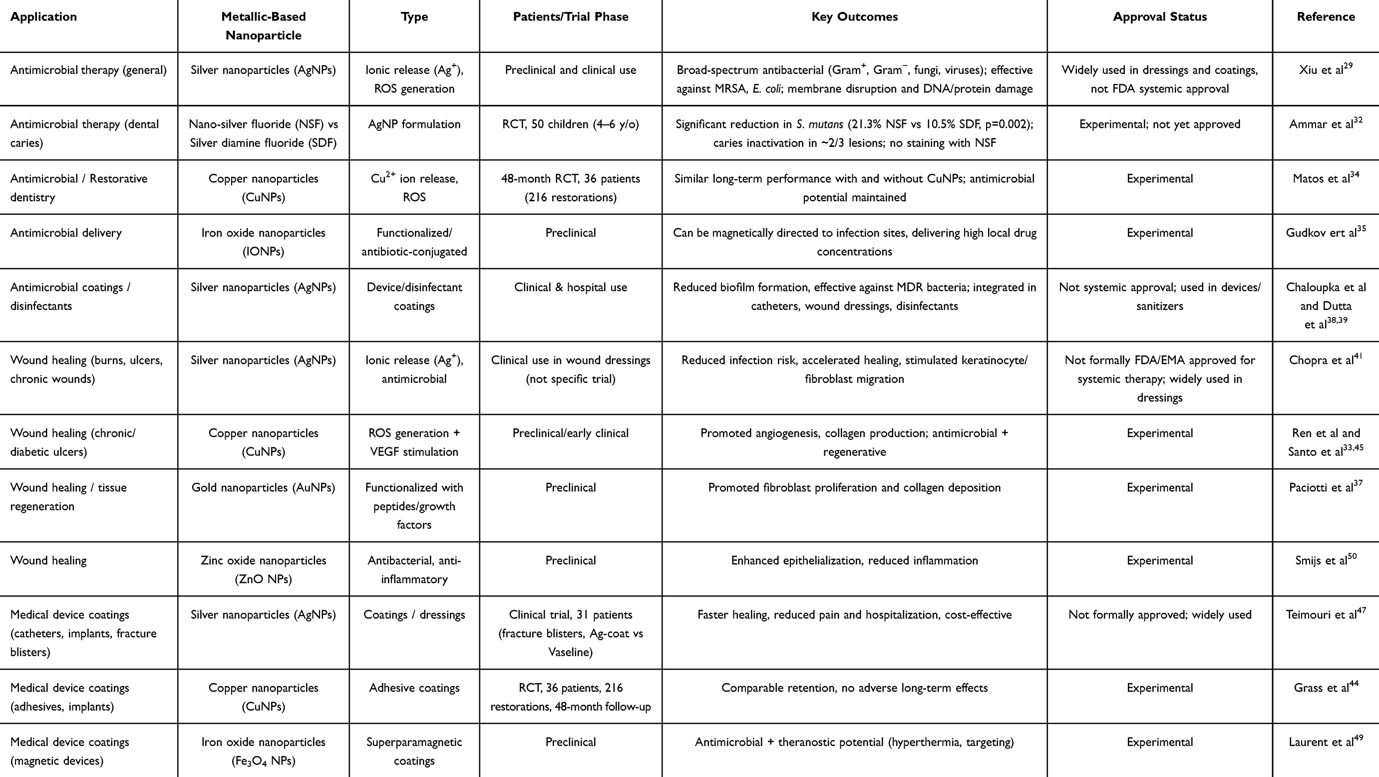 |
Table 2 Clinical and Preclinical Applications of Metallic-Based Nanoparticles in Antimicrobial Therapy, Wound Healing, and Medical Device Coatings |
Moreover, the regulatory hurdles for approving nanoparticle-based medical devices remain significant (Table 2) Regulatory bodies require extensive data on these materials’ safety, efficacy, and environmental impact before they can be widely used in clinical settings Research is ongoing to optimize nanoparticle coatings to ensure their safety for long-term use while maintaining their antimicrobial effectiveness.51
Perspective of Metallic Nanoparticles in Advanced Medical Applications
The evolution of metallic nanoparticles (MNPs) has enabled the development of increasingly sophisticated biomedical technologies, driven by their tunable physicochemical characteristics, high surface-area-to-volume ratio, and capacity to interact with biological systems at the molecular scale (Table 3).52 While earlier sections of this review highlight the clinical progress of MNPs in oncology, antimicrobial therapy, wound care, and device coatings, emerging research is now focused on advanced platforms designed to address complex and unmet medical needs through multifunctional, programmable nanosystems.
 |
Table 3 Clinical and Preclinical Perspectives of Metallic Nanoparticles in Advanced Medical Applications |
A major future trend involves the development of hybrid metallic nanoplatforms that integrate multiple components (such as imaging reporters, therapeutic agents, and targeting ligands) within a single construct These hybrid systems enable multimodal imaging, controlled drug release, and externally triggered therapeutic activation, representing a major step toward fully integrated theranostic technologies.35 By combining diagnosis and treatment in one nanoscale entity, such platforms aim to improve treatment precision, reduce systemic toxicity, and enable real-time monitoring of therapeutic outcomes.53
Advanced applications are also emerging in cardiovascular medicine, where gold-based nanoparticles and other MNPs are being investigated to enhance imaging modalities such as MRI and CT for early detection of atherosclerotic plaques and myocardial injury. These nanoparticles also offer opportunities for targeted drug delivery to diseased vascular sites, potentially reducing the risk of thrombotic events, heart attacks, and stroke. Their ability to accumulate at pathological vascular regions makes them attractive candidates for future cardiovascular theranostics.61
Metallic nanoparticles are likewise being integrated into gene therapy strategies. Gold nanoparticles and iron oxide nanoparticles can be functionalized with DNA, RNA, siRNA, or gene-editing constructs such as CRISPR components, enabling efficient nucleic acid delivery and protection against enzymatic degradation.62 Additionally, magnetic-field guidance allows iron oxide nanoparticles to be directed to specific organs or tissues, improving targeting efficiency and reducing off-target genetic modulation.
Despite these promising advances, several cross-cutting challenges must be addressed before MNPs can be fully translated into routine clinical use. Toxicity remains one of the most important concerns: certain nanoparticles (particularly silver and copper) can induce oxidative stress, membrane disruption, and mitochondrial dysfunction at high doses or after prolonged exposure.51 Biodistribution and clearance also pose significant challenges, as many nanoparticles tend to accumulate in reticuloendothelial organs such as the liver and spleen, raising questions regarding long-term biocompatibility and potential chronic toxicity. Regulatory agencies have highlighted the need for rigorous characterization, standardized manufacturing practices, comprehensive toxicological profiles, and environmental impact assessments before approving new nanomedicine products.50
Looking forward, metallic nanoparticles are expected to play a central role in the expansion of personalized medicine. Their ability to integrate diagnostic and therapeutic functionalities makes them ideal platforms for individualized treatment strategies guided by genomic, proteomic, or molecular imaging data.63 By enabling tailored drug delivery, early detection of disease biomarkers, and localized therapy with minimal systemic exposure, MNP-based systems could significantly enhance therapeutic precision and patient outcomes. Finally, the continued development of theranostic nanoparticles (capable of simultaneously identifying pathological processes and delivering targeted therapy) represents a transformative direction in next-generation medical care, offering earlier interventions, improved efficacy, and reduced toxicity.54
Applications in Therapy
Applying metallic nanoparticles (MNPs) in treatments has garnered significant attention due to their unique properties, enabling effective integration into various medical interventions (Table 3).
Gene Delivery
Gene therapy, which delivers genetic material into cells to treat diseases caused by genetic defects, has seen significant advances with the introduction of metallic nanoparticles. MNPs, particularly gold and iron oxide nanoparticles, have proven effective gene delivery carriers due to their ability to protect genetic material, enhance cellular uptake, and target specific cells or tissues.37
One of the primary advantages of using MNPs for gene delivery is their ability to complex with DNA, RNA, or small interfering RNA (siRNA), which can be delivered into cells to either repair defective genes or regulate gene expression (Figure 4). Gold nanoparticles are especially favored due to their biocompatibility and ease of surface modification. By attaching targeting ligands, such as antibodies or peptides, to the surface of AuNPs, these nanoparticles can be directed to specific cell types, improving the precision of therapy and reducing off-target effects.61
 |
Figure 4 Gene delivery and cell reprogramming strategies using magnetic nanoparticles: (a) Schematic overview of cell reprogramming and transplantation: patient-derived cells are expanded in chemically defined media, transfected with magnetic nanoparticles (MNPs) carrying therapeutic genes, differentiated into target cell populations, isolated by fluorescence-activated cell sorting (FACS), and subsequently transplanted back into the patient. (b) Comparison of gene delivery systems: (bi) conventional transfection using a cationic polymer (polyplex), which moves randomly in culture medium; and (bii) magnetofection system, where plasmid–polymer complexes are coated with MNPs (magnetoplex) and directed to the cell surface under magnetic force, enhancing gene transfer efficiency60. Image adapted from: Kami et al (2011)64 under a Creative Commons Attribution-NonCommercial 4.0 License. |
In addition to targeted delivery, MNPs protect genetic material during its journey through the bloodstream and into target cells. Nucleic acids are vulnerable to degradation by enzymes such as nucleases, but MNPs act as protective carriers, shielding the genetic material and ensuring it reaches the desired cells intact. Once inside the cell, the nanoparticles release their genetic payload in response to specific intracellular conditions, such as pH changes or enzyme activity, ensuring the therapy is activated only in the desired location.53
Due to their magnetic properties, iron oxide nanoparticles offer an additional advantage in gene therapy. These nanoparticles can be directed to specific tissues or organs using an external magnetic field, thereby improving the targeting efficiency of the gene-delivery process. This method, known as magnetofection, has been shown to enhance the uptake of genetic material in target cells while minimizing systemic distribution and reducing the risk of side effects.65
Another key feature of MNPs in gene delivery is their ability to enhance transfection efficiency, the process by which foreign genetic material is introduced into cells. MNPs facilitate endocytosis, improving the uptake of genetic material into the cell. By functionalizing the nanoparticles with cationic polymers such as polyethyleneimine (PEI), researchers have significantly improved the internalization of nanoparticles and their genetic cargo, increasing the overall effectiveness of gene therapy.66
In cancer treatment, gene therapy using MNPs is particularly promising. For example, AuNPs can be functionalized with siRNA molecules that silence genes responsible for tumor growth and proliferation. This approach has been studied extensively in oncology, where MNPs deliver therapeutic genes directly to tumor cells, reducing tumor growth while sparing healthy tissues.67
However, despite these advantages, challenges remain in the clinical application of MNPs for gene delivery. Issues such as toxicity, immune response, and long-term stability must be addressed to ensure these nanoparticles can be safely used in humans. Additionally, further research is required to improve the targeting specificity of MNPs, reduce potential off-target effects, and increase the efficiency of gene transfer across different cell types.51
Photodynamic Therapy (PDT)
Photodynamic therapy is a minimally invasive treatment that utilizes light to activate a photosensitizer in the presence of oxygen, producing reactive oxygen species that can destroy cancer cells, pathogens, or abnormal tissue. Metallic nanoparticles, especially gold nanoparticles, have emerged as powerful tools in PDT due to their unique optical properties, enhancing the precision and efficacy of this therapeutic approach.54
In PDT, a photosensitizing agent is first administered to the patient and allowed to accumulate in the target tissue, such as a tumor. Once localized in the abnormal cells, a specific wavelength of light is used to activate it. The interaction among light, the photosensitizer, and oxygen generates ROS, leading to cell death via oxidative damage. MNPs, particularly AuNPs, improve this process by increasing the efficiency of photosensitizer delivery and enhancing light absorption, leading to more effective treatment outcomes.54
Gold nanoparticles are especially suited for PDT because of their surface plasmon resonance (SPR), a phenomenon where electrons on the nanoparticle surface resonate in response to light, producing strong absorption and scattering. This property makes AuNPs excellent enhancers of the light-activated process in PDT. By conjugating photosensitizers such as porphyrins or phthalocyanines to AuNPs, researchers have created nanoplatforms that localize more efficiently in cancerous tissues and improve ROS generation upon light exposure.67
Furthermore, gold nanorods (a specific type of AuNP) are highly effective in PDT due to their ability to absorb light in the near-infrared region, which can penetrate deeper into tissues compared to visible light. This deeper penetration allows PDT to target tumors located below the skin’s surface, such as in breast or brain cancers. Gold nanorods functionalized with photosensitizers have shown promising results in preclinical studies, demonstrating the ability to selectively destroy tumor cells while minimizing damage to surrounding healthy tissue.68
In addition to AuNPs, other metallic nanoparticles, such as silver and iron oxide nanoparticles, are being investigated for their role in enhancing PDT. AgNPs, known for their antimicrobial properties, can be combined with photosensitizers to enhance ROS generation, making them useful for treating skin infections and promoting wound healing.41 IONPs offer the advantage of combining PDT with magnetic targeting, where an external magnetic field guides the nanoparticles to the desired location before light activation. This targeted approach improves the precision of PDT and reduces systemic exposure to the photosensitizer.49
Another benefit of using MNPs in PDT is the potential for multimodal therapies, such as combining PDT with other treatments, such as photothermal therapy or chemotherapy. For instance, gold nanoparticles can be used in dual-functional therapies, enhancing PDT’s efficacy while generating localized heat upon exposure to NIR light, thereby inducing hyperthermia to further destroy cancer cells. This combination of light-activated therapies increases the overall therapeutic outcome and has been shown to reduce tumor recurrence in animal models.54
Photothermal Therapy (PTT)
Photothermal therapy is a targeted therapeutic approach that utilizes the heat generated by nanoparticles upon exposure to light, typically in the near-infrared region, to destroy cancer cells selectively. Among various nanoparticles, metallic nanoparticles, especially gold nanoparticles, are extensively used due to their strong surface plasmon resonance, which enables efficient light absorption and conversion to heat.54
In PTT, gold nanoparticles are introduced into the body, where they accumulate in tumor tissues, often through the enhanced permeability and retention (EPR) effect, allowing them to accumulate passively in the tumor’s leaky vasculature. It is now recognized that the contribution of the EPR effect can vary across tumor types and patient-specific physiological conditions, potentially influencing the extent of nanoparticle accumulation in vivo. When these nanoparticles are exposed to NIR light, they absorb and convert it into heat, raising the local temperature in the tumor tissue. This localized hyperthermia (usually between 42–45°C) induces apoptosis or necrosis in cancer cells while minimizing damage to surrounding healthy tissues.69
Gold nanorods are particularly well-suited for PTT because they efficiently absorb NIR light. Unlike spherical gold nanoparticles, which resonate at visible light wavelengths, gold nanorods can be tuned to absorb NIR light by adjusting their aspect ratio. This makes them effective in treating deep-seated tumors, such as those in the liver, brain, or breast, where NIR light can penetrate more deeply than visible light. Gold nanorods conjugated with targeting ligands, such as antibodies or peptides, further enhance specificity by ensuring accumulation preferentially in cancer cells.54
Another advantage of PTT using AuNPs is its minimal invasion. The treatment requires only external NIR light application to the area where the nanoparticles have accumulated, making it a promising alternative to traditional cancer therapies such as chemotherapy or radiation, which often have severe side effects. Furthermore, AuNP-based PTT can be combined with other treatments, such as chemotherapy or immunotherapy, to achieve a synergistic effect, thereby improving overall therapeutic outcome.70
In addition to gold nanoparticles, silver nanoparticles and iron oxide nanoparticles are also being explored for PTT. AgNPs, due to their broad absorption spectrum, have been studied for their ability to generate heat under light exposure, though their use is often limited by concerns regarding cytotoxicity. On the other hand, iron oxide nanoparticles, known for their magnetic properties, offer the possibility of combining magnetic hyperthermia with PTT, where heat is generated by light and applying an external magnetic field.49
Recent advancements in PTT include the development of hybrid nanoparticles, combining properties of different metals or nanomaterials. For example, gold-silica nanoshells, consisting of a dielectric silica core coated with a thin layer of gold, have been shown to absorb NIR light and generate heat efficiently. These nanoparticles are particularly advantageous because their core-shell structure can be tailored to optimize optical properties and drug delivery capabilities, allowing for multifunctional therapeutic platforms.54
In clinical applications, PTT has demonstrated significant promise in treating various cancers, including breast, lung, and prostate. Early-phase clinical trials using gold nanoparticle-assisted PTT have reported positive outcomes, particularly in tumor reduction and patient safety. However, like other nanoparticle-based therapies, challenges such as long-term biocompatibility, biodistribution, and potential toxicity must be addressed before PTT can become a widely adopted cancer treatment.51
Future directions for PTT research include improving nanoparticle targeting specificity to minimize off-target effects, developing multifunctional nanoparticles for diagnostic imaging to enable real-time treatment monitoring, and optimizing nanoparticle design to reduce the risk of immune clearance and biodistribution issues. Advances in nanomedicine will likely continue to expand the scope of PTT in treating cancer and other conditions, such as infectious diseases and inflammatory disorders.70
Magnetotherapy: Antibacterial and Antitumoral
Magnetotherapy is an emerging therapeutic approach that utilizes the magnetic properties of nanoparticles, particularly iron oxide nanoparticles, for both antibacterial and antitumoral applications. The unique superparamagnetic behavior of IONPs makes them highly versatile for treatments that rely on magnetic fields to guide and activate therapeutic effects.49 These nanoparticles have shown great promise in magnetic hyperthermia and targeted antibacterial therapies, addressing key challenges in cancer treatment and microbial infections.
In cancer therapy, magnetic hyperthermia involves alternating magnetic fields to heat superparamagnetic nanoparticles introduced into tumor tissues. When exposed to an external magnetic field, these nanoparticles generate localized heat, raising the temperature in the tumor microenvironment to 42°C-45°C, inducing apoptosis or necrosis in cancer cells while leaving surrounding healthy tissues unaffected.71 This method is especially effective in treating tumors that are difficult to reach using traditional therapies, such as those in the brain or deep within the body.
IONPs can be functionalized with targeting ligands, such as antibodies or peptides, to enhance their accumulation in tumor tissues, thereby increasing the precision and effectiveness of the therapy. Combining magnetic hyperthermia with other treatments like chemotherapy or radiotherapy can significantly improve the overall therapeutic efficacy, as hyperthermia sensitizes cancer cells to these treatments, leading to more comprehensive tumor destruction.72
In addition to their antitumoral applications, IONPs play a key role in antibacterial magnetotherapy. Bacterial infections, particularly those involving biofilms or antibiotic-resistant strains, pose significant treatment challenges. Magnetic nanoparticles, functionalized with antibiotics or antimicrobial peptides, can be guided to infection sites using external magnetic fields. Once localized, they enhance the targeted delivery of antibacterial agents, leading to more effective treatment with reduced systemic side effects. The magnetic properties of IONPs enable precise control of nanoparticle accumulation and retention in infected tissue, particularly useful for treating chronic infections or those involving implantable medical devices where biofilms are prevalent.73
IONPs also exhibit inherent antibacterial properties. When exposed to a magnetic field, they generate reactive oxygen species, causing oxidative damage to bacterial cell walls and intracellular structures. This effect makes them particularly effective against many bacteria, including Gram-negative and Gram-positive strains. Moreover, IONPs can disrupt biofilms, communities of bacteria that adhere to surfaces and are notoriously resistant to antibiotics. The ability of IONPs to penetrate and destabilize biofilms represents a significant advancement in treating persistent infections, especially in clinical environments where multidrug-resistant bacteria are a concern.74
Magnetically assisted wound healing is another area where magnetotherapy has shown potential. Applying magnetic fields to areas treated with IONPs can accelerate tissue regeneration and enhance the antimicrobial efficacy of the nanoparticles. This approach is particularly valuable in treating chronic wounds, diabetic ulcers, and surgical site infections, where conventional treatments may be less effective.73
Recent research has focused on developing multifunctional magnetic nanoparticles, which can serve diagnostic and therapeutic purposes (termed theragnostic). These nanoparticles can be used in magnetic resonance imaging to diagnose and monitor disease progression while delivering therapeutic effects such as hyperthermia or targeted drug delivery. This dual capability is especially useful in personalized medicine, where treatments can be tailored to the patient’s specific needs and therapeutic progress can be tracked in real time.75
However, challenges remain in the widespread adoption of magnetotherapy. Ensuring the biocompatibility and long-term safety of IONPs is critical, as there is still limited understanding of their biodistribution and potential toxicity over extended periods. Additionally, the scalability and cost-effectiveness of producing high-quality IONPs for clinical use must be addressed to make magnetotherapy a viable option for mainstream medical treatments.76
Future research in magnetotherapy is expected to focus on optimizing the design and functionalization of IONPs to improve target efficiency, reduce potential cytotoxicity, and enhance therapeutic performance in cancer treatment and infection control. Integrating smart drug-delivery systems with magnetic guidance is promising for achieving precision medicine across various clinical settings.76
Disinfection
The use of metallic nanoparticles for disinfection has attracted significant attention due to their potent antimicrobial properties and ability to inactivate a wide range of pathogens, including bacteria, viruses, and fungi. Among the most studied nanoparticles in this field are silver nanoparticles, copper nanoparticles, and iron oxide nanoparticles, which have shown effectiveness in disinfecting medical devices, surfaces and water systems, as well as preventing the spread of infectious diseases in healthcare and community settings.55
Silver nanoparticles are widely regarded for their strong antimicrobial activity, primarily due to the release of silver ions (Ag⁺). These ions interact with microbial cell membranes, increasing permeability, disrupting enzyme function, and ultimately leading to cell death. AgNPs also induce the production of reactive oxygen species, causing oxidative stress and further damage to microbial cells.55 Due to these properties, AgNPs have been incorporated into various disinfectants, coatings, and medical devices to prevent infections, particularly in environments where antibiotic-resistant bacteria are a concern.
In addition to silver, copper nanoparticles are highly effective disinfectants due to their strong antimicrobial and antiviral properties. CuNPs disrupt microbial cell membranes by generating ROS, interfering with essential metabolic processes and causing cell death.77 These nanoparticles are increasingly used in coating high-touch surfaces, such as doorknobs, railings, and hospital furniture, to prevent pathogen transmission in public spaces and healthcare facilities. Copper surfaces have been shown to reduce the viability of various bacteria, including Escherichia coli and Staphylococcus aureus, within minutes of exposure, making them valuable tools in combating the spread of communicable diseases.78
Another significant application of metallic nanoparticles in disinfection is in water purification systems. Iron oxide nanoparticles, particularly magnetite (Fe3O4), have been utilized for their ability to adsorb contaminants and microbial pathogens from water.49 These nanoparticles can be used in filtration systems to remove harmful bacteria and viruses, providing a cost-effective solution for water disinfection in areas with limited access to clean drinking water. The magnetic properties of IONPs allow for easy removal from water systems after use, making them an environmentally friendly option for water treatment.79
Furthermore, hybrid nanoparticles that combine metals such as silver, copper, and iron are being developed to enhance disinfectant efficacy. These multifunctional nanoparticles exhibit synergistic effects, thereby improving their ability to kill a broader range of pathogens while reducing the concentration of nanoparticles required for disinfection. Such advancements are particularly relevant in controlling pandemics, where effective surface and water disinfection is paramount.80
Other Applications
Orthopedics
Metallic nanoparticles, mainly titanium (TiNPs) and silver nanoparticles, have become essential in orthopedics due to their unique properties that enhance the performance and longevity of implants and prosthetics. These nanoparticles offer significant advantages in promoting osseointegration, antimicrobial protection, and improving the overall biocompatibility of orthopedic devices.81
Recent advancements have also explored the use of magnesium nanoparticles (MgNPs) in orthopedic applications. Magnesium is biodegradable and promotes bone regeneration, making it suitable for temporary implants and scaffolds. These scaffolds are designed to provide mechanical support while gradually degrading as the bone heals, reducing the need for a second surgery to remove the implant. Moreover, iron oxide nanoparticles have shown potential in magnetic-assisted orthopedic therapies. These nanoparticles can be incorporated into bone grafts or scaffolds and manipulated with external magnetic fields to stimulate bone growth and enhance fracture healing. This approach, known as magnetic hyperthermia, also enables targeted delivery of therapeutic agents to the injury site, thereby improving the efficacy of bone regeneration treatments.82
Dental Applications
In the field of dentistry, metallic nanoparticles, especially silver, gold, and titanium nanoparticles, are revolutionizing dental materials and treatments due to their antimicrobial properties, biocompatibility, and ability to enhance the durability of dental restorations and implants.83
One of the primary applications of silver nanoparticles in dentistry is their integration into dental composites, adhesives, and sealants. AgNPs are renowned for their antibacterial properties, which are critical for preventing dental caries and biofilm formation that can lead to tooth decay and gum disease. Incorporating AgNPs into dental materials enables the creation of surfaces that continuously release silver ions (Ag⁺), inhibiting bacterial growth and maintaining oral hygiene over time.56 This is especially useful in high-risk patients prone to infections or those undergoing orthodontic treatments, where biofilm buildup around braces or retainers is common.
Beyond their role in restorative materials, silver nanoparticles have also been clinically evaluated as preventive and therapeutic agents against dental caries. The most advanced example is Nano Silver Fluoride (NSF), a formulation composed of silver nanoparticles (~3.2 ± 1.2 nm) stabilized with chitosan and combined with sodium fluoride. A randomized, double-blind clinical trial conducted in 60 schoolchildren (130 decayed primary teeth) demonstrated that NSF was highly effective in arresting dentine caries. After 7 days, 81% of treated lesions showed arrest versus 0% in controls; after 5 months, 72.7% versus 27.4%; and at 12 months, 66.7% versus 34.7%. The preventive fraction at 12 months was 50%, with a number needed to treat (NNT) of only three, highlighting the clinical efficiency of this approach. Importantly, NSF treatment did not produce the undesirable black staining characteristic of silver diamine fluoride, and no adverse effects were reported.84 These findings confirm that silver-based nanomaterials can be successfully translated into clinical dentistry, offering a low-cost, non-invasive, and effective solution for caries arrest in pediatric populations, particularly in underserved communities.
Gold nanoparticles (AuNPs) have also found applications in dentistry, particularly in diagnostic imaging and as part of advanced therapeutic approaches. AuNPs are biocompatible and can be functionalized to target specific dental tissues, aiding in precision diagnostics such as early detection of oral cancers or periodontal diseases.57 Their optical properties, particularly their ability to absorb and scatter light, make them excellent contrast agents for dental imaging techniques, providing clear and precise images of soft and hard tissues in the oral cavity.85 Furthermore, AuNPs have been investigated for photothermal therapy, where targeted heat generation can treat oral infections or even destroy tumorous tissues within the oral cavity.
Titanium nanoparticles are widely used in dental implants due to their high strength, corrosion resistance, and biocompatibility. Titanium-based implants are well-known for their ability to integrate with bone tissue, a process known as osseointegration, which is crucial for the long-term success of dental implants. TiNPs coatings on implant surfaces enhance this process by promoting bone cell adhesion and accelerating bone growth around the implant. This reduces healing time and improves the stability and durability of the implant, particularly in load-bearing areas like the jaw.86 Additionally, the antibacterial properties of TiNPs help prevent peri-implantitis, an infection that can compromise implant success.
Recent advancements have also led to the use of zinc oxide nanoparticles (ZnONPs) in dental cements and root canal sealers. ZnONPs exhibit antibacterial activity and help maintain a sterile environment in dental restorations, reducing the risk of secondary infections. These nanoparticles inhibit the growth of common oral pathogens and improve the mechanical properties of dental materials, such as strength and wear resistance.87
Application in Diagnosis
Applying metallic nanoparticles in medical diagnostics has revolutionized imaging techniques by enhancing sensitivity, specificity, and resolution. MNPs, particularly gold, silver, and iron oxide nanoparticles, are used as contrast agents in various imaging modalities due to their unique physical and chemical properties. Their ability to improve image contrast and their capacity for functionalization with targeting molecules make them indispensable tools in modern diagnostic techniques (Table 3).88
Magnetic Resonance Imaging (MRI)
Magnetic Resonance Imaging is one of the most powerful non-invasive diagnostic tools in modern medicine, and integrating metallic nanoparticles has significantly enhanced their capabilities. Iron oxide nanoparticles have emerged as highly effective contrast agents due to their superparamagnetic properties, significantly improving MRI scans’ contrast and resolution (Figure 5).73
 |
Figure 5 Iron oxide nanoparticles (IONPs) as T2-weighted MRI contrast agents for tumor targeting and diagnostic imaging: (a) Schematic illustration showing functionalized iron oxide nanoparticles (IONPs) circulating within the bloodstream and accumulating in the tumor microenvironment through passive and active targeting mechanisms. IONPs, represented as iron oxide cores (Fe3O4) coated with silica and targeting ligands, are internalized by tumor cells and macrophages, leading to local shortening of the transverse relaxation time (T2). This results in reduced signal intensity (darker contrast) in T2-weighted MRI images, as illustrated by the comparison between pre-contrast and IONP-enhanced scans, thereby enabling improved tumor visualization and diagnostic accuracy. (b) Diagnostic enhancement in imaging: cone-beam CT (CBCT) versus contrast-enhanced T1-weighted MR image (MRgRT) of a liver tumor. The MR image shows superior visualization of the gross tumor volume (GTV) in a patient with hepatocellular carcinoma (red arrows).73 Image adapted from: M Rahman et al (2023) and Mittaeur et al (2018) under a Creative Commons Attribution-NonCommercial 4.0 License. |
Iron oxide nanoparticles, typically composed of magnetite (Fe3O4) or maghemite (γ-Fe2O3), are widely used to enhance T2-weighted MRI by shortening the transverse relaxation time (T2), which results in darker signal intensities in areas where these nanoparticles accumulate (Figure 5a). This property is particularly useful for imaging tumors, inflammatory sites, and other pathological conditions where the localization of nanoparticles can help highlight abnormalities.89
 |
Figure 6 Illustration of the mechanism by which ligand-functionalized iron oxide nanoparticles selectively accumulate in tumors, enhancing magnetic resonance imaging contrast through their interaction with tumor cells and the vascular microenvironment. |
Functionalizing IONPs with targeting ligands, such as antibodies, peptides, or small molecules, enables selective accumulation in specific tissues or cell types (Figure 6). This allows for precision diagnostics by focusing the contrast enhancement on areas of interest, such as tumor tissues, inflammatory cells, or regions with abnormal blood flow. For example, IONPs conjugated with antibodies targeting cancer cell receptors can be explicitly directed to tumor sites, improving the detection of early-stage tumors and metastases that might otherwise be difficult to visualize with traditional MRI.58
Beyond their role as contrast agents, IONPs are also being explored in theranostic applications, which serve diagnostic and therapeutic purposes. In this approach, IONPs improve imaging precision and can also be used in magnetic hyperthermia. When exposed to an alternating magnetic field, IONPs generate localized heat that selectively destroys cancer cells while sparing surrounding healthy tissues. This dual function of IONPs makes them invaluable tools in personalized medicine, as they enable real-time monitoring of disease progression while simultaneously delivering treatment.90
Another area where metallic nanoparticles are advancing MRI technology is the development of multimodal imaging techniques. These combine MRI with other imaging modalities, such as positron emission tomography (PET) or computed tomography, to integrate anatomical, functional, and molecular information in a single scan. For instance, gold-coated iron oxide nanoparticles can be used as contrast agents in MRI and CT, providing complementary imaging data that enhances diagnostic accuracy.91
However, while the benefits of using metallic nanoparticles in MRI are clear, challenges remain regarding these nanoparticles’ biocompatibility, toxicity, and long-term safety. Ensuring IONPs are safely cleared from the body without causing toxicity or immune reactions is critical for their successful clinical application. Surface modifications, such as polyethylene glycol (PEG) coating, are commonly employed to improve the biocompatibility and circulation time of IONPs, reducing the risk of aggregation and promoting efficient excretion via the liver or kidneys.58
Computed Tomography (CT)
Computed Tomography has long been a cornerstone of medical imaging, and the use of metallic nanoparticles as contrast agents has brought significant advancements to the field. Among the various types of nanoparticles, gold nanoparticles are particularly noteworthy due to their high atomic number (Z = 79), which provides superior X-ray attenuation compared to conventional iodine-based contrast agents. This allows for enhanced contrast and better resolution in CT scans, especially in oncological applications.61
The high electron density of AuNPs results in stronger interaction with X-rays, leading to greater absorption and attenuation of the beam and improving the clarity and detail of CT images. This property is especially valuable in imaging tumors, vascular structures, and other anatomical abnormalities, where precise delineation is crucial for accurate diagnosis and treatment planning. Studies have demonstrated that gold nanoparticles can enhance the visualization of tumor margins, making them particularly useful in cancer diagnostics.92
Functionalized gold nanoparticles offer even greater potential, as they can be engineered to target specific tissues or cell types, thereby increasing contrast in areas of interest. For example, antibody-conjugated AuNPs have been designed to specifically target tumor cells, allowing for more precise molecular imaging. This approach enables early detection of small tumors or metastases that conventional contrast agents might miss, improving early-stage cancer diagnosis and monitoring of disease progression.59
In addition to improving diagnostic accuracy, gold nanoparticles have also shown potential in theranostic applications, which function as contrast agents and therapeutic enhancers. In radiotherapy, AuNPs can act as radiosensitizers, increasing radiation absorption in tumor tissues and enhancing the overall efficacy of the treatment. By concentrating the radiation dose within the tumor, gold nanoparticles help to minimize the damage to surrounding healthy tissues, leading to more targeted cancer treatments.93
Recent research has further explored using hybrid nanoparticles that combine gold with other metals or polymer coatings to enhance their stability, biocompatibility, and multifunctionality. For instance, gadolinium-coated gold nanoparticles offer the benefits of both CT and magnetic resonance imaging, providing dual-modality imaging that combines the high spatial resolution of CT with the soft tissue contrast of MRI.93 This hybrid approach allows clinicians to gather more comprehensive data from a single scan, improving both diagnostic precision and treatment planning.
Despite these advances, challenges are still associated with the clinical use of metallic nanoparticles in CT. One major concern is the biodistribution and clearance of nanoparticles from the body. AuNPs, if not properly cleared, can accumulate in organs such as the liver and spleen, leading to potential toxicity. To mitigate these risks, nanoparticles are often coated with biocompatible materials like polyethylene glycol, which helps prolong circulation time and improve kidney excretion.94 Ensuring the safe clearance of these nanoparticles is crucial to minimizing long-term health risks.
Moreover, while gold nanoparticles have shown great promise, their relatively high cost compared to traditional contrast agents may limit widespread use in routine clinical practice. However, ongoing research is focused on developing more cost-effective production methods and improving the scalability of nanoparticle synthesis to make these advanced contrast agents more accessible in medical settings.25
Nuclear Medicine
In the realm of nuclear medicine, the integration of metallic nanoparticles has significantly advanced diagnostic imaging and therapeutic applications. Nuclear medicine employs radioactive isotopes to diagnose and treat various diseases, with techniques like positron emission tomography (PET) and single-photon emission computed tomography (SPECT) being key modalities. Metallic nanoparticles, particularly those composed of gold and iron oxide, have enhanced the specificity and sensitivity of these imaging techniques, enabling better visualization of molecular processes and disease states (Table 3).95 Moreover, their diagnostic capabilities are intrinsically connected to emerging theranostic applications, where the same nanoparticles enable both high-sensitivity imaging and image-guided delivery of therapeutic radionuclides.
One of the significant advantages of using metallic nanoparticles in nuclear medicine is their ability to serve as multifunctional agents. In addition to their role in imaging, MNPs can be loaded with therapeutic agents or radionuclides, creating a platform for theranostic a combination of diagnostics and therapy on a single platform. This dual functionality allows for real-time monitoring of treatment efficacy while delivering a therapeutic dose directly to the targeted tissue, reducing the risk of off-target effects and improving the overall treatment outcome.96
In addition to improving imaging accuracy, metallic nanoparticles have expanded nuclear medicine’s capabilities by developing novel radiopharmaceuticals. When conjugated with radioisotopes such as Technetium-99m or Iodine-131, gold nanoparticles can act as highly efficient tracers for SPECT imaging, providing high-contrast images with minimal radiation exposure to the patient. The flexibility of nanoparticle functionalization also allows for targeted imaging of specific biomarkers in diseases such as cancer, cardiovascular conditions, and neurodegenerative disorders, improving the detection and monitoring of these diseases.97
Theranostic Applications in Nuclear Medicine
In nuclear medicine, the diagnostic use of metallic nanoparticles has laid the foundation for the development of theranostic platforms by enabling precise targeting, real time imaging, and quantitative assessment of nanoparticle biodistribution using PET and SPECT modalities. Building upon these diagnostic capabilities, theranostic applications of metallic nanoparticles have been extensively explored, particularly in oncology.
By integrating imaging agents and therapeutic payloads within a single nanoparticle platform, clinicians can simultaneously monitor tumor localization and treatment response while delivering radiation or chemotherapeutic agents directly to the tumor site. This dual functionality allows real-time feedback on therapeutic efficacy, minimizes systemic toxicity, and supports personalized treatment strategies tailored to individual disease profiles.60
Despite these advantages, significant challenges remain in the clinical translation of metallic nanoparticles for nuclear medicine applications. Key limitations include their biodistribution, long-term retention, and clearance, as prolonged accumulation in organs such as the liver and spleen raises concerns regarding chronic toxicity. Ongoing research focuses on improving nanoparticle biocompatibility and biodegradability, as well as developing surface modification strategies such as polyethylene glycol (PEG) coating to reduce immune recognition, optimize circulation time, and promote safer clearance.98
Furthermore, interactions with biological systems may indices unintended effects, including immune e activation or excessive reactive oxygen species (ROS) generation, potentially affecting healthy tissues. Regulatory challenges further complicate the clinical approval of nanotheranostic platforms, underscoring the need for comprehensive safety, dosimetry, and long-term toxicity evaluations.99
Conclusion
Metallic nanoparticles (MNPs) have evolved from experimental materials into clinically relevant platforms with demonstrated impact across multiple medical domains. Current clinical evidence supports their therapeutic and diagnostic potential. For example, in oncology, gold–silica nanoshells achieved tumor-free ablation zones in 87.5% of prostate cancer lesions at 12 months, while hafnium oxide nanoparticles (NBTXR3) significantly increased pathological complete response rates when combined with radiotherapy in soft-tissue sarcomas. Likewise, gadolinium-based nanoparticles (AGuIX®) produced complete remission of the primary tumor in all patients enrolled in the phase I trial for locally advanced cervical cancer.
Beyond oncology, silver, copper, titanium, and zinc-based systems have demonstrated robust antimicrobial performance, improved biofilm disruption, enhanced wound healing, and increased durability of dental and orthopedic biomaterials. Collectively, these clinical and preclinical findings reinforce the role of MNPs as versatile, multifunctional platforms capable of addressing unmet medical needs in cancer therapy, infection control, wound management, and advanced regenerative medicine.
Despite these advances, important limitations and challenges still hinder the broad translation of metallic nanomedicines into routine clinical practice. Many clinical studies rely on small cohorts, single-arm designs and short follow-up, limiting the robustness and generalizability of the reported benefits. At the biological level, issues related to biodistribution, long-term retention in reticuloendothelial organs, ion release and oxidative stress raise concerns about chronic toxicity and cumulative exposure, particularly for silver and copper systems. Tumor targeting remains highly heterogeneous, with variable contributions of enhanced permeability and retention effect and inconsistent nanoparticle accumulation across patients and tumor types. In addition, the lack of standardized protocols for nanoparticle synthesis, characterization, dosing and imaging readouts complicates cross-study comparison. These scientific and technical hurdles are compounded by complex regulatory pathways that require rigorous physicochemical characterization, comprehensive toxicological assessment, quality-by-design manufacturing and evaluation of environmental and occupational risks.
Looking ahead, the most promising opportunities for metallic nanoparticles lie in their rational integration as theranostic platforms for personalized medicine. Future directions include the design of multifunctional, stimuli-responsive nanoconstructs that combine imaging reporters, therapeutic payloads and active targeting ligands within a single system, enabling real-time image-guided therapy, adaptive dosing and longitudinal monitoring of treatment response. Improved control of surface chemistry, stealth coatings and active targeting strategies will be essential to optimize biodistribution, enhance tumor or infection-site accumulation and minimize off-target toxicity. Equally important will be the implementation of larger, well-controlled, multicenter clinical trials, supported by standardized characterization frameworks and harmonized regulatory guidance, to generate high-quality evidence on safety, efficacy and cost-effectiveness. If these challenges are addressed, metallic nanoparticles are poised to consolidate their role as core components of next-generation theranostic platforms, contributing significantly to safer, more precise and truly personalized diagnostic and therapeutic strategies.
Disclosure
The authors report no conflicts of interest in this work.
References
1. Aminzai MT, Yildirim M, Yabalak E. Metallic nanoparticles unveiled: synthesis, characterization, and their environmental, medicinal, and agricultural applications. Talanta. 2024;280:126790. doi:10.1016/j.talanta.2024.126790
2. Maier-Hauff K, Ulrich F, Nestler D, et al. Efficacy and safety of intratumoral thermotherapy using magnetic iron-oxide nanoparticles combined with external beam radiotherapy on patients with recurrent glioblastoma multiforme. J Neurooncol. 2010;103:24–32.
3. Wust P, Gneveckow U, Wust P, et al. Magnetic nanoparticles for interstitial thermotherapy - Feasibility, tolerance and achieved temperatures. Int J Hyperthermia. 2006;22(8):673–685. doi:10.1080/02656730601106037
4. Johannsen M, Gneveckow U, Eckelt L, et al. Clinical hyperthermia of prostate cancer using magnetic nanoparticles: presentation of a new interstitial technique. Int J Hyperthermia. 2005;21(7):637–647. doi:10.1080/02656730500158360
5. Grauer O, Jaber M, Hess K, et al. Combined intracavitary thermotherapy with iron oxide nanoparticles and radiotherapy as local treatment modality in recurrent glioblastoma patients. J Neurooncol. 2019;141(1):83–94. doi:10.1007/s11060-018-03005-x
6. Verry C, Sancey L, Dufort S, et al. Treatment of multiple brain metastases using gadolinium nanoparticles and radiotherapy: NANO-RAD, a phase I study protocol. BMJ Open. 2019;9(2):e023591. doi:10.1136/bmjopen-2018-023591
7. Yakoubi A, Dhafer CEB. Advanced Plasmonic Nanoparticle-Based Techniques for the Prevention, Detection, and Treatment of Current COVID-19. Plasmonics. 2023;18(1):311–347. doi:10.1007/s11468-022-01754-0
8. Tsauo J, Liu Y, Zhang X, et al. Local hyperthermia mediated by gold nanoparticle-integrated silicone-covered stent: feasibility and tissue response in a rat esophageal model. Eur Radiol Exp. 2024;8(1). doi:10.1186/s41747-024-00438-0.
9. Silva AKA, Espinosa A, Kolosnjaj-Tabi J, Wilhelm C, Gazeau F. Medical applications of iron oxide nanoparticles. In: Iron Oxides: From Nature to Applications. 2016:425–472.
10. Yang Y, Zheng X, Chen L, et al. Multifunctional Gold Nanoparticles in Cancer Diagnosis and Treatment. Int J Nanomed. 2022;17:2041–2067. doi:10.2147/IJN.S355142
11. Lange A, Grzenia A, Wierzbicki M, et al. Silver and Copper Nanoparticles Inhibit Biofilm Formation by Mastitis Pathogens. Animals. 2021;11(7):1884. doi:10.3390/ani11071884
12. Sharma S, Lamichhane N, Parul, Sen T, Roy I. Iron Oxide Nanoparticles Conjugated with Organic Optical Probes for In Vivo Diagnostic and Therapeutic Applications. Nanomedicine. 2021;16(11):943–962. doi:10.2217/nnm-2020-0442
13. Hlapisi N, Songca SP, Ajibade PA. Capped Plasmonic Gold and Silver Nanoparticles with Porphyrins for Potential Use as Anticancer Agents—A Review. Pharmaceutics. 2024;16(10):1268. doi:10.3390/pharmaceutics16101268
14. Sargazi S, Er S, Sacide Gelen S, et al. Application of titanium dioxide nanoparticles in photothermal and photodynamic therapy of cancer: an updated and comprehensive review. J Drug Deliv Sci Technol. 2022;75:103605. doi:10.1016/j.jddst.2022.103605
15. Anani T, Rahmati S, Sultana N, David AE. MRI-traceable theranostic nanoparticles for targeted cancer treatment. Theranostics. 2021;11(2):579. doi:10.7150/thno.48811
16. Neuwelt EA, Hamilton BE, Varallyay CG, et al. Ultrasmall superparamagnetic iron oxides (USPIOs): a future alternative magnetic resonance (MR) contrast agent for patients at risk for nephrogenic systemic fibrosis (NSF)? Kidney Int. 2009;75(5):465–474. doi:10.1038/ki.2008.496
17. Jin R, Lin B, Li D, Ai H. Superparamagnetic iron oxide nanoparticles for MR imaging and therapy: design considerations and clinical applications. Curr Opin Pharmacol. 2014;18:18–27. doi:10.1016/j.coph.2014.08.002
18. Rastinehad AR, Anastos H, Wajswol E, et al. Gold nanoshell-localized photothermal ablation of prostate tumors in a clinical pilot device study. Proc Natl Acad Sci U S A. 2019;116(37):18590–18596. doi:10.1073/pnas.1906929116
19. Libutti SK, Paciotti GF, Byrnes AA, et al. Phase I and pharmacokinetic studies of CYT-6091, a novel PEGylated colloidal gold-rhTNF nanomedicine. Clin Cancer Res. 2010;16(24):6139–6149. doi:10.1158/1078-0432.CCR-10-0978
20. Baere TD, Pracht M, Rolland Y, et al. Hafnium oxide nanoparticles activated by SBRT: a new interventional radiation therapy approach for the treatment of unresectable liver cancers. Ann Oncol. 2019;30:iv122. doi:10.1093/annonc/mdz157.005
21. Chargari C, Maury P, Texier M, et al. Theragnostic Gadolinium-Based Nanoparticles Safely Augment X-ray Radiation Effects in Patients with Cervical Cancer. ACS Nano. 2024;18(26):16516–16529. doi:10.1021/acsnano.3c12537
22. Bennett S, Verry C, Kaza E, et al. Quantifying gadolinium-based nanoparticle uptake distributions in brain metastases via magnetic resonance imaging. Sci Rep. 2024;14(1). doi:10.1038/s41598-024-62389-1.
23. Mundekkad D, Cho WC. Nanoparticles in Clinical Translation for Cancer Therapy. Int J Mol Sci. 2022;23(3):1685. doi:10.3390/ijms23031685
24. Souto EB, Blanco-Llamero C, Krambeck K, et al. Regulatory insights into nanomedicine and gene vaccine innovation: safety assessment, challenges, and regulatory perspectives. Acta Biomater. 2024;180:1–17. doi:10.1016/j.actbio.2024.04.010
25. Boisselier E, Astruc D. Gold nanoparticles in nanomedicine: preparations, imaging, diagnostics, therapies and toxicity. Chem Soc Rev. 2009;38(6):1759–1782. doi:10.1039/b806051g
26. Loo C, Lowery A, Halas N, West J, Drezek R. Immunotargeted nanoshells for integrated cancer imaging and therapy. Nano Lett. 2005;5(4):709–711. doi:10.1021/nl050127s
27. Chajon E, Pracht M, De Baere T, et al. A phase I/II trial of hafnium oxide nanoparticles activated by radiotherapy in hepatocellular carcinoma and liver metastasis. Ann Oncol. 2018;29:v92. doi:10.1093/annonc/mdy150.001
28. Rai M, Yadav A, Gade A. Silver nanoparticles as a new generation of antimicrobials. Biotechnol Adv. 2009;27(1):76–83. doi:10.1016/j.biotechadv.2008.09.002.
29. Xiu ZM, Zhang QB, Puppala HL, Colvin VL, Alvarez PJJ. Negligible particle-specific antibacterial activity of silver nanoparticles. Nano Lett. 2012;12(8):4271–4275. doi:10.1021/nl301934w
30. Andleeb A, Andleeb A, Asghar S, et al. A Systematic Review of Biosynthesized Metallic Nanoparticles as a Promising Anti-Cancer-Strategy. Cancers. 2021;13(11):2818. doi:10.3390/cancers13112818
31. Shrivastava S, Bera T, Roy A, et al. Characterization of enhanced antibacterial effects of novel silver nanoparticles. Nanotechnology. 2007;18:225103. doi:10.1088/0957-4484/18/22/225103
32. Ammar N, El-Tekeya MM, Essa S, et al. The antibacterial effect of nanosilver fluoride in relation to caries activity in primary teeth: a protocol for a randomized controlled clinical trial. Trials. 2022;23(1):558. doi:10.1186/s13063-022-06477-5
33. Ren G, Hu D, Cheng EWC, et al. Characterisation of copper oxide nanoparticles for antimicrobial applications. Int J Antimicrob Agents. 2009;33(6):587–590. doi:10.1016/j.ijantimicag.2008.12.004
34. Matos TP, Ñaupari-Villasante R, Kunz PVM, et al. 48-month clinical evaluation of a copper-containing universal adhesive in non-carious cervical lesions: a double-blind randomised clinical trial. Dent Mater. 2023;39(9):820–830. doi:10.1016/j.dental.2023.07.002
35. Gudkov SV, Burmistrov DE, Serov DA, et al. Do Iron Oxide Nanoparticles Have Significant Antibacterial Properties? Antibiotics. 2021;10(7):884. doi:10.3390/antibiotics10070884
36. Bhattacharya R, Mukherjee P. Biological properties of ‘naked’ metal nanoparticles. Adv Drug Delivery Rev. 2008;60(11):1289–1306. doi:10.1016/j.addr.2008.03.013.
37. Paciotti GF, Myer L, Weinreich D, et al. Colloidal gold: a novel nanoparticle vector for tumor directed drug delivery. Drug Delivery J Delivery Target Therap Agents. 2004;11(3):169–183. doi:10.1080/10717540490433895
38. Chaloupka K, Malam Y, Seifalian AM. Nanosilver as a new generation of nanoproduct in biomedical applications. Trends Biotechnol. 2010;28(11):580–588. doi:10.1016/j.tibtech.2010.07.006.
39. Dutta T, Barman A, Bhattacherjee S, Chakraborty J, Dutta T. Antimicrobial silver nanoparticles for water disinfection: a short review on recent advances. Nanotechnol Environ Eng. 2024;9(1):111–131. doi:10.1007/s41204-023-00354-5
40. Schröfel A, Kratošová G, Šafařík I, et al. Applications of biosynthesized metallic nanoparticles - A review. Acta Biomater. 2014;10(10):4023–4042. doi:10.1016/j.actbio.2014.05.022
41. Chopra I. The increasing use of silver-based products as antimicrobial agents: a useful development or a cause for concern? J Antimicrob Chemother. 2007;59(4):587–590. doi:10.1093/jac/dkm006.
42. Storm-Versloot MN, Vos CG, Ubbink DT, Vermeulen H. Topical Silver for Preventing Wound Infection (Review). Available from: http://www.thecochranelibrary.com.
43. Cioffi N, Torsi L, Ditaranto N, et al. Copper nanoparticle/polymer composites with antifungal and bacteriostatic properties. Chem Mater. 2005;17(21):5255–5262. doi:10.1021/cm0505244
44. Grass G, Rensing C, Solioz M. Metallic copper as an antimicrobial surface. Appl Environ Microbiol. 2011;77(5):1541–1547. doi:10.1128/AEM.02766-10.
45. Santo CE, Lam EW, Elowsky CG, et al. Bacterial killing by dry metallic copper surfaces. Appl Environ Microbiol. 2011;77(3):794–802. doi:10.1128/AEM.01599-10
46. Mirakhorli T, Ardebili ZO, Ladan-Moghadam A, Danaee E. Bulk and nanoparticles of zinc oxide exerted their beneficial effects by conferring modifications in transcription factors, histone deacetylase, carbon and nitrogen assimilation, antioxidant biomarkers, and secondary metabolism in soybean. PLoS One. 2021;16:e0256905. doi:10.1371/journal.pone.0256905
47. Teimouri M, Lalehzar S. Evaluation of the therapeutic effect of dressing containing Silver (Ag coat) in the process of healing skin blisters caused by limb fractures: a clinical trial study. BMC Surg. 2023;23(1):101. (). doi:10.1186/s12893-023-02012-8
48. Ziental D, Czarczynska-Goslinska B, Mlynarczyk DT, et al. Titanium dioxide nanoparticles: prospects and applications in medicine. Nanomaterials. 2020;10(2):387. doi:10.3390/nano10020387
49. Laurent S, Forge D, Port M, et al. Magnetic iron oxide nanoparticles: synthesis, stabilization, vectorization, physicochemical characterizations and biological applications. Chem Rev. 2008;108(6):2064–2110. doi:10.1021/cr068445e
50. Smijs TG, Pavel S. Titanium dioxide and zinc oxide nanoparticles in sunscreens: focus on their safety and effectiveness. Nanotechnol Sci Applications. 2011;4:95–112. doi:10.2147/nsa.s19419
51. Dobrovolskaia MA, Mcneil E. Immunological Properties of Engineered Nanomaterials. (2007. Available from: www.orthobiotech.com.
52. Al-Samydai A, Abu Hajleh MN, Al-Sahlawi F, et al. Advancements of metallic nanoparticles: a promising frontier in cancer treatment. Sci Prog. 2024;107(4):00368504241274967. doi:10.1177/00368504241274967
53. Daniel M-C, Astruc D. Gold Nanoparticles: assembly, Supramolecular Chemistry, Quantum-Size-Related Properties, and Applications toward Biology, Catalysis, and Nanotechnology. Chem Rev. 2004;104(1):293–346. doi:10.1021/cr030698
54. Huang X, El-Sayed MA. Gold nanoparticles: optical properties and implementations in cancer diagnosis and photothermal therapy. J Adv Res. 2010;1(1):13–28. doi:10.1016/j.jare.2010.02.002.
55. Deshmukh SP, Patil SM, Mullani SB, Delekar SD. Silver nanoparticles as an effective disinfectant: a review. Mater Sci Eng C. 2019;97:954–965. doi:10.1016/j.msec.2018.12.102
56. Bourgi R, Doumandji Z, Cuevas-Suárez CE, et al. Exploring the Role of Nanoparticles in Dental Materials: a Comprehensive Review. Coatings. 2025;15(1):33. doi:10.3390/coatings15010033
57. Tosan F, Rahnama N, Sakhaei D, Fathi AH, Yari A. Effects of doping metal nanoparticles in hydroxyapatite in Improving the physical and chemical properties of dental implants. Nanomed Res J. 2021;6:327–336.
58. Chung S, Revia RA, Zhang M. Iron oxide nanoparticles for immune cell labeling and cancer immunotherapy. Nanoscale Horiz. 2021;6(9):696–717. doi:10.1039/D1NH00179E
59. Tiwari PM, Vig K, Dennis VA, Singh SR. Functionalized gold nanoparticles and their biomedical applications. Nanomaterials. 2011;1(1):31–63. doi:10.3390/nano1010031
60. Drude N, Tienken L, Mottaghy FM. Theranostic and nanotheranostic probes in nuclear medicine. Methods. 2017;130:14–22. doi:10.1016/j.ymeth.2017.07.004
61. Hainfeld JF, Slatkin DN, Smilowitz HM. The use of gold nanoparticles to enhance radiotherapy in mice. Phys Med Biol. 2004;49(18):N309–N315. doi:10.1088/0031-9155/49/18/N03
62. Chatterjee DK, Fong LS, Zhang Y. Nanoparticles in photodynamic therapy: an emerging paradigm. Adv Drug Delivery Rev. 2008;60(15):1627–1637. doi:10.1016/j.addr.2008.08.003.
63. Puttasiddaiah R, Basavegowda N, Lakshmanagowda NK, et al. Emerging nanoparticle-based diagnostics and therapeutics for cancer: innovations and challenges. Pharmaceutics. 2025;17(1):70. doi:10.3390/pharmaceutics17010070
64. Pal S, Yu kyung T, Song J. Does the Antibacterial Activity of Silver Nanoparticles Depend on the Shape of the Nanoparticle? A Study of the Gram-Negative Bacterium Escherichia coli. Appl Environ Microbiol. 2007;73(6):1712–1720. doi:10.1128/AEM.02218-06
65. Zhang J, Zhang T, Gao J. Biocompatible iron oxide nanoparticles for targeted cancer gene therapy: a review. Nanomaterials. 2022;12(19):3323. doi:10.3390/nano12193323
66. Riley MK, Vermerris W. Recent advances in nanomaterials for gene delivery—a review. Nanomaterials. 2017;7(5):94. doi:10.3390/nano7050094
67. Shetty K, Yasaswi S, Dutt S, Yadav KS. Multifunctional nanocarriers for delivering siRNA and miRNA in glioblastoma therapy: advances in nanobiotechnology-based cancer therapy. 3 Biotech. 2022;12(11):301. doi:10.1007/s13205-022-03365-2
68. Bhana S, O’Connor R, Johnson J, et al. Photosensitizer-loaded gold nanorods for near infrared photodynamic and photothermal cancer therapy. J Colloid Interface Sci. 2016;469:8–16. doi:10.1016/j.jcis.2016.02.012
69. Riley RS, Day ES. Gold nanoparticle-mediated photothermal therapy: applications and opportunities for multimodal cancer treatment. Wiley Interdiscip Rev Nanomed Nanobiotechnol. 2017;9(4):e1449. doi:10.1002/wnan.1449
70. Li Y, Zhu X, Dong Y, et al. Nanomedicine-Enabled Mild Photothermal Therapy Strategies for Enhanced Antitumor Treatment. Adv Nanobiomed Res. 2024;4(3):2300094. doi:10.1002/anbr.202300094
71. Chiu-Lam A, Rinaldi C. Nanoscale thermal phenomena in the vicinity of magnetic nanoparticles in alternating magnetic fields. Adv Funct Mater. 2016;26(22):3933–3941. doi:10.1002/adfm.201505256
72. Sachdeva V, Monga A, Vashisht R, et al. Iron Oxide Nanoparticles: the precise strategy for targeted delivery of genes, oligonucleotides and peptides in cancer therapy. J Drug Deliv Sci Technol. 2022;74:103585. doi:10.1016/j.jddst.2022.103585
73. Rahman M. Magnetic resonance imaging and iron-oxide nanoparticles in the era of personalized medicine. Nanotheranostics. 2023;7(4):424. doi:10.7150/ntno.86467
74. Sahli C, Moya SE, Lomas JS, et al. Recent advances in nanotechnology for eradicating bacterial biofilm. Theranostics. 2022;12(5):2383. doi:10.7150/thno.67296
75. Yan K, Li P, Zhu H, et al. Recent advances in multifunctional magnetic nanoparticles and applications to biomedical diagnosis and treatment. RSC Adv. 2013;3(27):10598–10618. doi:10.1039/c3ra40348c
76. Mohsin A, Hussain MH, Mohsin MZ, et al. Recent advances of magnetic nanomaterials for bioimaging, drug delivery, and cell therapy. ACS Appl Nano Mater. 2022;5(8):10118–10136. doi:10.1021/acsanm.2c02014
77. Crisan MC, Teodora M, Lucian M. Copper nanoparticles: synthesis and characterization, physiology, toxicity and antimicrobial applications. Appl Sci. 2021;12(1):141. doi:10.3390/app12010141
78. Noyce JO, Michels H, Keevil CW. Potential use of copper surfaces to reduce survival of epidemic meticillin-resistant Staphylococcus aureus in the healthcare environment. J Hosp Infect. 2006;63(3):289–297. doi:10.1016/j.jhin.2005.12.008
79. Joseph TM, Al-Hazmi HE, Śniatała B, Esmaeili A, Habibzadeh S. Nanoparticles and nanofiltration for wastewater treatment: from polluted to fresh water. Environ Res. 2023;238:117114.
80. Shabatina TI, Vernaya OI, Melnikov MY. Hybrid nanosystems of antibiotics with metal nanoparticles—novel antibacterial agents. Molecules. 2023;28:1603.
81. Wang N, Fuh JYH, Dheen ST, Senthil Kumar A. Functions and applications of metallic and metallic oxide nanoparticles in orthopedic implants and scaffolds. J Biomed Mater Res B Appl Biomater. 2021;109(2):160–179. doi:10.1002/jbm.b.34688
82. Saberi A, Baltatu MS, Vizureanu P. Recent advances in magnesium–magnesium oxide nanoparticle composites for Biomedical Applications. Bioengineering. 2024;11(5):508. doi:10.3390/bioengineering11050508
83. Vasiliu S, Racovita S, Gugoasa IA, et al. The benefits of smart nanoparticles in dental applications. Int J Mol Sci. 2021;22:2585.
84. Dos Santos Jr VE, Filho AV, Ribeiro Targino AG, et al. A new “silver-bullet” to treat caries in children–nano silver fluoride: a randomised clinical trial. J Dent. 2014;42(8):945–951. doi:10.1016/j.jdent.2014.05.017
85. Alonazi RS, Alqubaysi HA, Al-Mutairi NH. Biomaterials in dentistry: advances in tissue engineering for dental restoration. Int J Health Sci. 2024;8:999–1015.
86. Bokobza L. On the use of nanoparticles in dental implants. Materials. 2024;17(13):3191. doi:10.3390/ma17133191
87. Sirelkhatim A, Mahmud S, Seeni A, et al. Review on zinc oxide nanoparticles: antibacterial activity and toxicity mechanism. Nanomicro Lett. 2015;7(3):219–242. doi:10.1007/s40820-015-0040-x
88. Niemirowicz K, Markiewicz KH, Wilczewska AZ, Car H. Magnetic nanoparticles as new diagnostic tools in medicine. Adv Med Sci. 2012;57(2):196–207. doi:10.2478/v10039-012-0031-9
89. Sohani Z, Aval HJ, Rabiee SM. Bioactive glass and iron oxide nanoparticle composite coatings for Ti-6Al-4V implants: microstructure, corrosion behavior, bioactivity and cellular response. Appl Surf Sci Adv. 2025;27:100734. doi:10.1016/j.apsadv.2025.100734
90. Ajinkya N, Yu X, Kaithal P, et al. Magnetic iron oxide nanoparticle (IONP) synthesis to applications: present and future. Materials. 2020;13(20):4644. doi:10.3390/ma13204644
91. Carril M, Fernández I, Rodríguez J, García I, Penadés S. Gold-Coated Iron Oxide Glyconanoparticles for MRI, CT, and US Multimodal Imaging. Part Part Syst Charact. 2014;31(1):81–87. doi:10.1002/ppsc.201300239
92. Pickford Scienti O. On the potential of multi-spectral x-ray and photoacoustic imaging to facilitate gold nanoparticle mediated dose-enhanced radiotherapy. (2021).
93. Zhou W, Gao X, Liu D, Chen X. Gold nanoparticles for in vitro diagnostics. Chem Rev. 2015;115(19):10575–10636. doi:10.1021/acs.chemrev.5b00100
94. Sani A, Cao C, Cui D. Toxicity of gold nanoparticles (AuNPs): a review. Biochem Biophys Rep. 2021;26:100991. doi:10.1016/j.bbrep.2021.100991
95. Schuemann J, Bagley AF, Berbeco R, et al. Roadmap for metal nanoparticles in radiation therapy: current status, translational challenges, and future directions. Phys Med Biol. 2020;65(21):21RM02. doi:10.1088/1361-6560/ab9159
96. Kumar R, Münstedt H. Silver ion release from antimicrobial polyamide/silver composites. Biomaterials. 2005;26(14):2081–2088. doi:10.1016/j.biomaterials.2004.05.030
97. Kairdolf BA, Qian X, Nie S. Bioconjugated nanoparticles for biosensing, in vivo imaging, and medical diagnostics. Anal Chem. 2017;89(2):1015–1031. doi:10.1021/acs.analchem.6b04873
98. Kleynhans J, Sathekge M, Ebenhan T. Obstacles and recommendations for clinical translation of nanoparticle system-based targeted alpha-particle therapy. Materials. 2021;14(17):4784. doi:10.3390/ma14174784
99. Kumar P, Singh R, Kush P. Biocompatibility, Toxicity Concerns, Environmental and Safety Considerations, and Legal Aspects of Functionalized Magnetic Nanoparticles. In: Functionalized Magnetic Nanoparticles for Theranostic Applications; 2024:533–558. doi:10.1002/9781394172917.ch17
 © 2026 The Author(s). This work is published and licensed by Dove Medical Press Limited. The
full terms of this license are available at https://www.dovepress.com/terms
and incorporate the Creative Commons Attribution
- Non Commercial (unported, 4.0) License.
By accessing the work you hereby accept the Terms. Non-commercial uses of the work are permitted
without any further permission from Dove Medical Press Limited, provided the work is properly
attributed. For permission for commercial use of this work, please see paragraphs 4.2 and 5 of our Terms.
© 2026 The Author(s). This work is published and licensed by Dove Medical Press Limited. The
full terms of this license are available at https://www.dovepress.com/terms
and incorporate the Creative Commons Attribution
- Non Commercial (unported, 4.0) License.
By accessing the work you hereby accept the Terms. Non-commercial uses of the work are permitted
without any further permission from Dove Medical Press Limited, provided the work is properly
attributed. For permission for commercial use of this work, please see paragraphs 4.2 and 5 of our Terms.