Back to Journals » International Medical Case Reports Journal » Volume 16
Chronic Uterine Inversion in 54 Year Old Woman: Case Report
Authors Asefa E, Abdulhay F, Dhugasa D
Received 31 May 2023
Accepted for publication 21 September 2023
Published 28 September 2023 Volume 2023:16 Pages 627—631
DOI https://doi.org/10.2147/IMCRJ.S411300
Checked for plagiarism Yes
Review by Single anonymous peer review
Peer reviewer comments 3
Editor who approved publication: Dr Scott Fraser
Eyob Asefa,1 Fedlu Abdulhay,2 Dereje Dhugasa3
1Department of Obstetrics and Gynecology, Nekemte Compressive Specialized Hospital, Nekemte, Ethiopia; 2Department of Obstetrics and Gynecology, Jima University, Jima, Ethiopia; 3Department of General Surgery, Nekemte Compressive Specialized Hospital, Nekemte, Ethiopia
Correspondence: Eyob Asefa, Nekemte Compressive Specialized Hospital, Nekemte, Ethiopia, Tel +251 917472260, Email [email protected]
Introduction: Uterine inversion is a disease characterized by the folding of the uterine fundus into the uterine cavity or beyond the cervix. It is a rare complication following parturition. Acute uterine inversion presents immediately following vaginal delivery. Prevalence of acute uterine inversion is 1 in 20,000– 50,000 cases. Chronic uterine inversion is a rare disease presentation in post-menopausal women. It is commonly associated with uterine pathology like leiomyoma, leiomyosarcoma, or endometrial polyps. It is very rare without associated factors. In the post-menopausal age group, the diagnosis is confirmed with high index of suspicion and physical examination. Typically, inverted uterine fundus is a leading point of protrusion but it could be the uterine cervix in uterine prolapse.
Case Presentation: A 54 year old woman came to our hospital with the complaint of a painless mass in her vagina of 3 years duration. Three years ago, she encountered a protrusion of mass through her vagina, which gradually grew in size over time. On physical examination, uterine fundus was the leading point of the mass and it protruded 7 cm below the hymenal ring. As a result, she was diagnosed with chronic uterine inversion and underwent an abdominal hysterectomy. She was discharged home improved. We report this case because of an unidentified factor eliciting the uterine inversion, late presentation of the disease and difficulty in surgical treatment.
Conclusion: Chronic uterine inversion is a rare disease presentation especially when there is no associated uterine pathology like leiomyoma. It is seen in a broad range of age groups, from reproductive to postmenopausal. A strong index of suspicion and physical examination are used to reach the diagnosis. Surgical technique should be anticipated to be difficult as it is a rare case, outside the experience of most surgeons.
Keywords: chronic uterine inversion, hysterectomy, uterine pathology
Introduction
Uterine inversion is the descent of the uterine fundus, inside out, through the cervix, either partially or fully.1,2 It is grossly classified as puerperal and non-puerperal, puerperal uterine inversion being the most common.3 Non-puerperal uterine inversion is classified as subacute (48 hour post-partum–4 weeks) and chronic inversion (greater than 4 weeks post-partum).2–4 Acute uterine inversion is one of the rarer obstetric complications.5 Prevalence of acute uterine inversion is reported in 1 in 20,000–50,000 cases.6 Estimated maternal death rate is about 15–20% if untreated and neurogenic and hemorrhage shocks are a common cause of death.2,6–8 Maternal death estimated to be higher in areas where there is limited access to an equipped health facility. If this fatal obstetric complication is not treated and the mother survives, it will progress to non-puerperal uterine inversion after 48 hours of acute inversion.5,7
Chronic uterine inversion is commonly associated with other uterine pathology, mainly submucous uterine leiomyoma.4,5,9,10 Other pathologies can be endometrial polyps, uterine neoplasms, and leiomyoma other than submucous.1–3,9 Increased intra-abdominal pressure and post-menopausal hormonal replacement are promoting factors.5 Chronic uterine inversion is one of the rarer disease presentations and even some gynecologists do not see a single case in their lifetime.1,5,7
Diagnosis mainly depends on clinical history, physical examination and imaging, mainly ultrasound.1,5 Once the diagnosis of chronic uterine inversion is established, and the preoperative assessment has been completed, the management strategy will be decided.5 Manual reposition and surgical treatment are two options.9 Chronic uterine inversion almost always requires surgical management, either uterine repositioning surgery or hysterectomy.6 There are two grossly classified surgical approaches for uterine repositioning surgery, especially in women in need of uterus preservation, either abdominal approach (Haultain’s method, Huntiginton’s method and hysterectomy) or vaginal approach (Kustner’s and Spinelli’s methods).6,8 The type of surgery to be performed is determined by the patient’s fertility need and any associated complication such as uterine necrosis.7 Hysterectomy is performed in cases where a woman does not need fertility and for associated complications that preclude uterine preservation, like uterine necrosis.2,3,7,9 We report this case because there was no identified cause eliciting the uterine inversion, the late presentation of the disease, which existed for 3 years, and the difficulty associated with surgical treatment.
Presentation of Case
The patient was a 54 year old, para 2 mother, who came from a rural area to the Nekemte Compressive Specialized Hospital with the complaint of a mass in her vagina of 3 years duration. Three years ago, she encountered a protrusion of mass through her vagina, which gradually grew in size over time. She had two babies, last delivery being 12 years ago. Both deliveries were at home as the health facility is too far from her residency area, being assisted by traditional birth attendant, and she claimed that it was smooth. Initially, 3 years back, there was bleeding and an offensive discharge from the mass but that stopped in the last year. She developed lacerations on the mass 6 months back. For this reason she divorced and isolated herself from all social life for this long period of time. Now, she was brought to hospital by her older sister and other relative specifically for reason of the mass lacerations. She had no other known chronic medical illness.
Pertinent PE Finding
Vital sign: BP = 100/60 mm hg, PR = 84 bpm, RR = 20 bpm, T° = 36.5C°, weight = 47 kg.
GUS: there is pinkish mass per vagina which is about 7 cm from hymenal ring.
Uterine fundus is a leading part of the mass.
There was abouta 5 cm sized longitudinal laceration on the right side of the mass.
There was no bleeding and discharge from the mass nor laceration area.
Laboratory
CBC: hemoglobin = 12 g/dl, PLT = 285 thousand, neutrophil = 74% percent.
Urine analysis: normal finding.
Blood group = A, RH = positive.
HIV test = negative.
HBSAg = negative.
Ultrasound: partially full bladder seen in abdomen, uterus not seen in abdomen. Otherwise no other pertinent finding.
Chest X-ray: normal Chest X-ray finding.
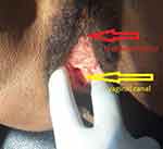
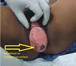
For this finding, she was diagnosed with chronic uterine inversion. Images were taken pre-operatively and post-operatively, the uterine fundus was the leading point of the mass which protruded completely through the vagina (Figure 1) and there was chronic laceration developed on the endometrial surface (Figure 2) and after the operation, normal vaginal canal was seen (Figure 3). After reviewing management options with her and her family, she was scheduled for surgery and an abdominal hysterectomy was performed the following day. It was abdominal surgery and it was easy to take the uterus into the abdomen. As tissue was redundant and distorted, anatomical reference and surgical landmarks are difficult to identify. Due to this, the surgery was difficult and there was an intraoperative transactional right ureteral injury and an end=to=end anastomosis was done. She was discharged on her 10th day of operation being improved. She was given a follow up appointment 2 weeks later, and when she was evaluated it was found that she was doing well.
 |
Figure 3 Vaginal canal seen post hysterectomy. Notes: Red colored arrow shows external urethral orifice and yellow colored arrow shows vaginal canal which was taken on 12th post hysterectomy. |
Discussion
Chronic uterine inversion is a rare disease presentation in the gynecology discipline. Ninety seven percent of persistent chronic uterine inversions are associated with other uterine pathology. Uterine leiomyoma is the most frequent cause, occurring in up to 80% of cases as fundal attachment and submucous type.1 Other causes are leiomyosarcoma, endometrial polyps, and endometrial carcinoma.5,9 For this reason, chronic uterine inversion could be seen in a broader age range, from reproductive to post-menopausal.11 In different case reports, it was reported from ages 28 to 70 years of age.5,9 In post-menopausal women, any uterine mass or lesion should be presented to pathology.5
In this case, there was no uterine mass causing uterine inversion, which is a rare presentation. In addition, she is post-menopausal. Therefore, in post-menopausal women with a vaginal mass, persistent chronic uterine inversion should not be discounted as a differential diagnosis. Also, there is no eliciting factor as in this patient.
The commonest clinical presentations are persistent mass protrusion and vaginal bleeding.3,7,11 Also, vaginal discharge and uterine laceration are seen as clinical presentation.6,8 If a uterine mass, such as a submucous leiomyoma, is the cause of uterine inversion, the mass may predate the fundus of the uterus.5 It could be misdiagnosed, commonly as uterine prolapse. Common complications of chronic uterine inversion are uterine necrosis, laceration, infection, recurrence after reposition, etc.
In this instance, no possible cause of uterine inversion could be found. It was complicated by a larger endometrial surface laceration of the uterus, and the mass was redundant because it was a long standing issue. This causes anatomical distortion, making surgery challenging.
Diagnosis is commonly made by clinical evaluation as with our patient. The uterus fundus is the leading point of the protruded mass, while the cervix is the leading point in the uterine prolapse.3 Sometimes imaging could be needed, like ultrasound, CT, or MRI to reach diagnosis.7
Management of chronic uterine inversion depends on the fertility need, associated complication and surgeon experience. In post-menopausal ladies or in cases of necrotic and significantly infected uterus, hysterectomy is the preferred management. Depending on the surgeon’s experience and expected complication, a hysterectomy may be performed using an abdominal or vaginal technique.1,3,5,9 In this case, she was in the post-menopausal period, uterine endometrial surface laceration and redundant uterine tissue made us select hysterectomy. After repositioning the uterus into the abdomen an abdominal hysterectomy was performed. Simple (type I) abdominal hysterectomy steps were followed to perform the surgery. The anatomy was distorted and there was an iatrogenic left ureteral injury. An end=to=end anastomosis technique was used to restore the ureter. She was discharged on the 14th post-operative day after a smooth post-operative time.
Conclusion
Chronic uterine inversion is an uncommon gynecologic condition in post-menopausal women. Commonly, chronic uterine inversion is associated with uterine pathology like leiomyoma. There were no possible eliciting factors linked with the uterine inversion in this case, making it a very rare case. In such cases, the diagnosis is done using a suspicion index and physical examination. Ureter should be located well in both abdominal and vaginal approach to avoid injury. In this case the abdominal approach was selected and there was a ureteral injury identified intraoperatively and repaired. Surgery should be expected to be difficult in this instance.
Ethical Consideration
Institutional approval is not required for case report publication as patient anonymity was respected and informed consent was taken.
Consent for Publication
Written informed consent was obtained for the images and details of the case for publication from the patient.
Funding
There was no financial support.
Disclosure
We declare that there is no conflict of interest in this work.
References
1. Niang MM, Samb F, Cisse CT. Subacute non-puerperal uterine inversion: a rare complication of uterine myoma. Ann Med Surg. 2022;3:6–8.
2. Ali E, Kumar M. Chronic uterine inversion presenting as a painless vaginal mass at 6 months post partum: a case report. J Clin Diagnostic Res. 2016;2016:8–10.
3. Singh A. Case report a rare case of chronic uterine inversion secondary to submucosal fibroid managed in the province hospital of Nepal. Case Rep Obstet Gynecol. 2020;2020:1.
4. Reports C, Kilpatrick CC, Chohan L, Maier RC. Chronic nonpuerperal uterine inversion and necrosis: a case report. J Med Case Rep. 2010;2010:2–4.
5. Asefa D. Chronic postmenopausal uterine inversion: a case report gynecology & obstetrics chronic postmenopausal uterine inversion: a case report. Gynecol Obstet. 2017;6:1.
6. Garg P, Bansal R, Gursahani R, Mehta D, Garg P, Mathur R. Unusual and delayed presentation of chronic uterine inversion in a young woman as a result of negligence by an untrained birth attendant: a case report. Indian J Med Ethics. 2020;V(4):1–5. doi:10.20529/ijme.2020.095
7. Chong W, Brodman M. Stepwise surgical management of chronic puerperal uterine inversion. EAS J Med Surg. 2019;1857(3):82–85.
8. Tolcha FD, Dale TB, Anbessie TT. Chronic uterine inversion presenting with severe anemia 7 years after a home delivery and the subsequent successful pregnancy: a case report. J Med Case Rep. 2023;17(1):1–4. doi:10.1186/s13256-022-03698-9
9. Herath RP, Patabendige M, Rashid M, Wijesinghe PS. Nonpuerperal uterine inversion: what the gynaecologists need to know ? Obstet Gynecol Int. 2020;2020:1.
10. Asefa D, Yimar N. Chronic postmenopausal uterine inversion: a case report gynecology & obstetrics. Gynecol Obstet. 2016;6(6):1.
11. Birge O, Tekin B, Merdin A, Coban O, Arslan D. Chronic total uterine inversion in a young adult patient. Am J Case Rep. 2015;16:756–759. doi:10.12659/ajcr.894264
 © 2023 The Author(s). This work is published and licensed by Dove Medical Press Limited. The full terms of this license are available at https://www.dovepress.com/terms.php and incorporate the Creative Commons Attribution - Non Commercial (unported, v3.0) License.
By accessing the work you hereby accept the Terms. Non-commercial uses of the work are permitted without any further permission from Dove Medical Press Limited, provided the work is properly attributed. For permission for commercial use of this work, please see paragraphs 4.2 and 5 of our Terms.


