
Back to Journals » International Journal of Nanomedicine » Volume 21
Chitosan Nanoparticles for Natural Antioxidant Delivery in Metabolic Dysfunction-Associated Steatohepatitis Liver Disease (MASLD)
Authors Herdiana Y  , Sofian FF , Husni P , Hathout RM , Putriana NA
Received 28 January 2026
Accepted for publication 23 March 2026
Published 7 April 2026 Volume 2026:21 599532
DOI https://doi.org/10.2147/IJN.S599532
Checked for plagiarism Yes
Review by Single anonymous peer review
Peer reviewer comments 2
Editor who approved publication: Professor Jie Huang
Yedi Herdiana,1 Ferry Ferdiansyah Sofian,2 Patihul Husni,1 Rania M Hathout,3 Norisca Aliza Putriana1
1Department of Pharmaceutics and Pharmaceutical Technology, Faculty of Pharmacy, Universitas Padjadjaran, Sumedang, West Java, Indonesia; 2Department of Pharmaceutical Biology, Faculty of Pharmacy, Universitas Padjadjaran, Sumedang, West Java, Indonesia; 3Department of Pharmaceutics and Industrial Pharmacy, Faculty of Pharmacy, Ain Shams University, Cairo, Egypt
Correspondence: Norisca Aliza Putriana, Department of Pharmaceutics and Pharmaceutical Technology, Faculty of Pharmacy, Universitas Padjadjaran, Sumedang, West Java, Indonesia, Email [email protected]
Abstract: Metabolic Dysfunction-Associated Steatohepatitis Liver Disease (MASLD) is a global clinical challenge, with oxidative stress and inflammation as drivers of disease progression. Numerous natural antioxidants exhibit hepatoprotective activity, but their application is often limited by poor solubility, gastrointestinal instability, presystemic metabolism, and limited oral bioavailability. These conditions demand the development of advanced drug delivery systems (DDS). Chitosan polymer, due to its unique combination of physicochemical and biological properties, including cationicity, biocompatibility, and biodegradability, emerges as a highly promising polymeric platform for nanocarrier engineering. This narrative-critical review summarises the primarily preclinical evidence regarding chitosan-based nanosystems for MASLD/the legacy term Non-alcoholic fatty liver disease (NAFLD), assessing reported pharmacokinetic (PK) exposures and pharmacodynamic outcomes. In general, chitosan-based formulations are often associated with improved pharmacodynamic (PD) outcomes, particularly reductions in Alanine Aminotransferase (ALT)/Aspartate Aminotransferase (AST) and indicators of oxidative stress, while quantitative PK evidence and measurable biodistribution are available in a subset of studies and indicate variability influenced by chitosan attributes (molecular weight, degree of deacetylation, charge density), formulation design, and disease model. Thus, claims of liver “targeting” should be framed as hepatic enrichment unless supported by consistent quantitative tissue data and PK–PD associations, and accompanied by repeated dose safety evaluation.
Keywords: metabolic dysfunction-associated steatohepatitis liver disease, chitosan nanoparticles, oral drug delivery, oxidative stress, asialoglycoprotein receptor
Introduction
Metabolic dysfunction–associated steatotic liver disease (MASLD), formerly known as NAFLD, is a global health challenge estimated to affect approximately one-third of the world’s population and is a leading cause of chronic liver disease, contributing significantly to liver-related morbidity and mortality.1,2 The prevalence of MASLD continues to increase with the pandemic of obesity, type 2 diabetes, and metabolic syndrome, and its incidence is higher in patients with type 2 diabetes, highlighting the urgency of integrated metabolic screening.1,3 Clinically, MASLD is often referred to as a “silent epidemic” because many patients are asymptomatic in the early stages, often delaying detection until the disease progresses to metabolic dysfunction–associated steatohepatitis (MASH), fibrosis, or cirrhosis.4
At the molecular level, the development of MASLD is driven by lipotoxicity, which triggers oxidative stress and amplifies inflammation.5 Excess free fatty acids increase the production of reactive oxygen species (ROS) from various sources, including mitochondria, peroxisomes, and the NADPH oxidase (NOX) enzyme pathway, thereby damaging proteins, lipids, and DNA and exacerbating mitochondrial dysfunction.5,6 One important consequence is lipid peroxidation, in which ROS oxidize Polyunsaturated Fatty Acids (PUFAs) to reactive aldehydes (such as Malondialdehyde (MDA) and 4-Hydroxynonenal (4-HNE)) that are cytotoxic and pro-fibrotic, activating hepatic stellate cells (HSCs) and promoting fibrogenic signals. In parallel, Damage-Associated Molecular Patterns (DAMPs) and ROS activate the Nuclear Factor kappa B (NF-κB) pathway in hepatocytes, Kupffer cells, and HSCs, triggering the production of pro-inflammatory cytokines (such as TNF-α and IL-1β) and accelerating the progression to fibrosis.7,8 The role of Kupffer cells is also crucial because Lipopolysaccharide (LPS) translocation can promote pro-inflammatory polarization and increase ROS through NOX2, exacerbating hepatocellular injury.4,9 At the same time, microbiota dysbiosis and increased intestinal permeability in the gut-liver axis can increase portal LPS load and amplify inflammation through activation of Toll-Like Receptor (TLR4)-dependent signalling.
Given the central role of oxidative stress in the lipotoxicity-inflammation-fibrogenesis cycle, interventions targeting oxidative stress are considered a rational therapeutic strategy, particularly as adjunctive or phenotype-directed approaches.10,11 Various natural antioxidants (such as silymarin, curcumin, resveratrol, and other phenolic compounds) have demonstrated hepatoprotective potential in preclinical trials, but the consistency of their clinical benefits varies.12,13 The main limitation often lies in exposure issues: poor aqueous solubility, gastrointestinal instability, rapid first-pass metabolism, and low oral bioavailability.14,15 Therefore, the primary practical challenge is not simply to discover new targets, but rather to achieve adequate and consistent exposure to the relevant compartments to produce replicable pharmacodynamic (PD) effects.
This is where drug delivery often becomes the dominant bottleneck. For many antioxidant candidates, the main challenges are improving oral stability and absorption, controlling payload release, and measurably directing tissue distribution without overclaiming targeting. In MASLD, claims regarding “liver targeting” should be conservatively framed as hepatic enrichment, which can vary depending on formulation design, dose, and clearance pathway, and ideally supported by quantitative biodistribution data (such as liver-to-plasma AUC ratio) and Pharmokinetic (PK)-Pharmacodynamic (PD) associations.14,15
Chitosan (CS) is a biocompatible and degradable cationic polysaccharide that offers relevant material functionality to overcome antioxidant delivery challenges in MASLD, particularly the oral route.15,16 Its positive charge and mucoadhesion properties can enhance mucosal retention and facilitate interactions with epithelial surfaces. Some CS-based formulations have also been associated with supporting gut barrier integrity, which is relevant to the gut-liver axis.15,16 Ionic gelation/crosslinking-based engineering enables regulation of particle size, polydispersity index, and zeta potential—parameters that may affect gastrointestinal stability, payload release, and exposure patterns.17 Furthermore, CS is relatively easy to modify (such as derivatisation to increase solubility or ligand attachment), opening up engineering options to balance stability, permeability, and exposure profiles.15,18 From a safety and translational perspective, CS is often referred to as a biopolymer with widespread use and, in some contexts, GRAS status, although performance and safety remain dependent on grade/purity, polymer attributes (Molecular Weight (MW), degree of deacetylation, charge density), and repeat dose design.15,18
At the cellular level, hepatic enrichment strategies can be explored through the attachment of ligands such as galactose/N-acetylgalactosamine (Gal/GalNAc) to leverage hepatocyte receptor (Asialoglycoprotein Receptor (ASGPR)) expression, but interpretation of targeting requires discipline and quantitative biodistribution evidence.19,20 Other platforms may also be more suitable in specific contexts (such as lipid systems for highly lipophilic payloads, or enteric polymers for gastric protection and localized release), so CS should be viewed not as a universal platform, but rather as one with specific strengths when its material functionality aligns with the antioxidant delivery constraints in MASLD.14,15,19,20 Therefore, a synthesis is needed that not only highlights benefits but also critically compares, maps levels of evidence, and defines what is proven versus what remains hypothetical in the CS–MASLD literature.
With this background, this review presents an integrated synthesis of antioxidant strategies in MASLD, emphasizing that achieving adequate and consistent exposure is often a key obstacle. We discuss how chitosan material attributes and the choice of nanosystem architecture influence gastrointestinal stability, absorption, payload release, and measurable hepatic enrichment, and summarize the most common PK-PD patterns and factors explaining the variability in results between studies. Next, we identify translational prerequisites (quantitative biodistribution, PK-PD relationships, repeated-dose safety, and reproducibility/manufacturability) necessary to ensure that interpretations of benefits do not exceed the available evidence. Finally, to place chitosan in a broader technological context, we develop a comparative “fit-for-purpose” framework that demonstrates when alternative platforms are more appropriate for specific design goals in MASLD.
Engineering Chitosan Platforms for Precision Delivery
The choice of carrier material in nanomedicine is not only about encapsulation ability but also about the biological interactions between the material and the disease’s pathophysiology. CS is a cationic polymer/weak electrolyte with frequently reported good mucoadhesivity, biocompatibility, biodegradability, tolerability in several applications (depending on purity, polymer attributes, route, and dosage regimen), relatively low production cost, and a high positive charge that can form polyelectrolyte complexes with polyanionic macromolecules (such as nucleic acids or negatively charged polysaccharides) through electrostatic interactions.21 In the context of MASLD, CS offers a number of strategic advantages that can complement conventional synthetic polymers such as poly(lactic-co-glycolic acid) (PLGA) or Polycaprolactone (PCL). While synthetic polymers are generally inert, CS is often viewed as a functional biopolymer with the potential for specific bioactive contributions. This polymer exhibits mucoadhesive properties, which can prolong the residence time in the gastrointestinal tract and potentially modulate intestinal permeability, while also having a certain scavenging capacity reported to be related to its free amine group. CS can synergize with the drug payload to suppress hepatic oxidative stress.22,23 However, the use of pure CS has fundamental limitations, especially its low solubility at neutral pH and its instability in the acidic gastric environment that can trigger premature release/decrease in system integrity, so advanced engineering is needed to design this biopolymer into a more controlled delivery system and potentially help overcome the biological barriers of the oral–hepatic route.24
Structural Modification and Functionalization Strategies
One of the key determinants of successful oral therapy for MASLD lies in the balance between gastrointestinal tract stability and target organ specificity.25 Recent data suggest that chemical modifications to the amino and hydroxyl groups of CS have the potential to overcome stability and permeability barriers in certain designs.26 For example, challenges to peptide drug bioavailability have been reported to be improved using oligo-CSNPs modified with ursodeoxycholic acid (UDCA). In a study involving Exenatide (EXE), this modification not only protected the peptide payload from enzymatic degradation but also engineered esterase-responsive ligand release and was reported to be associated with reduced hepatic lipid accumulation through activation of the Sirtuin 1 (SIRT1) signalling pathway.23 These findings suggest that chemical engineering of CS can improve the delivery performance of poorly absorbed hydrophilic drugs, although generalization to various MASLD phenotypes requires cross-model validation and a clear PK–PD relationship.
Besides medication stability, CS changes have been documented to influence the gut microbiota, an essential element of the gut-liver axis. This was demonstrated in the development of low-molecular-weight CS-selenium nanoparticles (LCS-SeNPs). In vivo data in a high-fat diet model revealed that this formulation was not only associated with improved hepatic antioxidant status/activity but also with concurrent changes in microbiota composition, including an increase in the relative abundance of considered beneficial bacteria such as Akkermansia and Bifidobacterium.24 This dual mechanism suggests that CS derivatisation has the potential to create a synergistic effect between antioxidant load protection and gut microenvironment modification. Moreover, derivatisations such as thiolation or trimethylation have been documented to improve paracellular drug penetration without inducing cytotoxicity in gastrointestinal models.27–29
Furthermore, to address the issue of nonspecific (“off-target”) drug distribution that could potentially trigger systemic toxicity, active targeting strategies could be considered.30,31 A widely explored approach is to utilise the ASGPR, which is highly expressed on hepatocytes.25 By conjugating specific ligands such as galactose or N-acetylgalactosamine (GalNAc) to the nanoparticle surface, these molecular “targeting” systems can enhance receptor-mediated interaction and internalization under certain conditions, potentially increasing hepatic enrichment compared to unliganded particles.32 Comparative studies have reported that GalNAc-liganded nanoparticles exhibit 3.9-fold higher cellular uptake in certain cell models/experiments compared to untargeted particles.33 Conceptually, this enhanced hepatocyte internalisation could support dose optimisation; however, claims regarding reduced systemic or non-target organ exposure (such as kidney/spleen) require quantitative biodistribution support (such as liver-plasma AUC ratio and distribution to RES organs) and repeated dose safety evaluation.34 In addition to uptake effects, in vivo outcomes can also be influenced by opsonization/corona proteins, dominance of uptake by RES cells (such as Kupffer cells), and differences in disease stage, so the interpretation of “targeting” needs to take into account the context of the model and particle design.35–37
Formulation Rationale and Critical Characterization
In fabrication, there is no “one-size-fits-all” approach; the choice of preparation method should be based on a critical analysis of the therapeutic payload’s physicochemical properties.18 For hydrophilic payloads or fragile biomacromolecules, ionic gelation is the most frequently used approach for payload delivery. This mechanism, which relies on spontaneous electrostatic interactions between CS and a cross-linking agent such as tripolyphosphate (TPP), occurs under mild aqueous conditions without the use of toxic organic solvents, thus supporting the principles of green nanotechnology and potentially helping to maintain drug bioactivity.38,39
Conversely, given that antioxidants widely explored for MASLD, such as silymarin, curcumin, and resveratrol, are lipophilic, amphiphilic self-assembly strategies offer a strong rationale. By modifying CS into an amphiphilic polymer through grafting of hydrophobic groups, micelle structures with a hydrophobic core are formed that can increase the apparent solubility and loading capacity of lipophilic drugs and reduce premature drug leakage, depending on the degree of substitution and micelle architecture.40 Another alternative to improve pH stability is Polyelectrolyte Complexation (PEC) with negatively charged polymers such as alginate, which provides a pH-dependent release profile to protect the payload from the acidic environment of the stomach.41
Regardless of the chosen method, the success of clinical translation is highly dependent on control of critical quality parameters (CQAs). The successful use of CSNPs for drug delivery is strongly influenced by the ability to control particle size.42 Particle size is often targeted in the range of 100–200 nm; particles larger than 200 nm tend to be more easily taken up by the mononuclear phagocyte system, while very small particles (such as <10 nm) can be more easily filtered by the kidneys.43 In addition, the zeta potential is often maintained above +20 mV as an indicator of colloidal stability; However, excessively high charge densities also require consideration regarding mucus interactions and tolerability upon repeated administration.38,44 Size similarity, as indicated by the Polydispersity Index (PDI), is generally targeted below 0.3 to support consistent release profiles and reduce the risk of in vivo aggregation.17 Collectively, the integration of material selection, chemical modification, and control of physicochemical parameters suggests that CS platforms can be a rational approach to addressing various challenges in MASLD therapy, with the caveat that performance and safety are design- and context-specific.
Nonetheless, in vivo results of CS-based systems are significantly context-dependent, influenced by polymer characteristics (molecular weight, degree of deacetylation, charge density), fabrication technique, and disease model or stage.26,35 Accordingly, “liver targeting” should be interpreted conservatively as hepatic enrichment unless supported by quantitative biodistribution and consistent PK–PD linkage, ideally alongside repeated-dose safety and inter-batch reproducibility evidence.17,42,43 CQA ranges reported in the literature should be treated as practical guidelines, as optimal values may shift with route, payload, surface chemistry, and bio-interactions (such as protein corona).17,42,43 Figure 1 summarizes the design-exposure-response framework, which links chitosan-based formulation strategies to oral stability/absorption, hepatic enrichment potential, and PK/PD outcomes in MASLD/MASH.
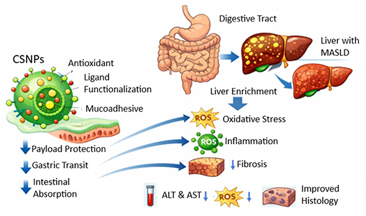 |
Figure 1 Chitosan Nanoparticles for MASLD Therapy. |
Pharmacokinetics and Molecular Mechanisms of Translational Efficacy
Validation of CS-based delivery platforms requires more than physical characterisation; the key evidence lies in in vivo data. PK and PD data bridge the gap between materials engineering and biological outcomes, demonstrating how rationally designed nanosystems have the potential to enhance the translational feasibility of low-bioavailability antioxidant compounds for MASLD.
Addressing the Pharmaceutical Gap via Pharmacokinetic Profiles
The main challenges of natural antioxidants (such as curcumin, silymarin, resveratrol) include limited solubility and/or permeability, as well as extensive first-pass metabolism.45 CSNPs have been proposed to enhance exposure through the following main pathways:
- Mucosal retention and gastrointestinal residence time. The polycationic nature of CS can interact with negatively charged intestinal mucin, thereby increasing mucosal retention and prolonging gastrointestinal residence time. In several studies, this increased retention has been reported to be associated with increased absorption and increased AUC compared to free drug suspensions.46
- Modulation of paracellular transport. By transiently modulating tight junctions (such as changes in the expression/redistribution of tight junction proteins such as claudins and occludin), CS has been reported to enhance the paracellular transport of hydrophilic drugs and macromolecules that typically have difficulty crossing the intestinal epithelium.47
- Cargo protection and absorption pathway modulation. Encapsulation in a polymer matrix can protect the cargo from enzymatic degradation in the intestinal lumen and potentially modulate absorption pathways. In certain designs (such as more hydrophobically modified CS-based systems), some absorption may involve contributions from the lymphatic pathway, potentially reducing the contribution of first-pass metabolism; however, the degree of this effect is design- and context-specific and requires adequate PK verification.43,48 The incorporation of ligands may increase hepatocyte interaction, hepatic uptake, or liver enrichment.
Dual Pharmacodynamic Mechanisms of Action
Therapeutic efficacy in MASLD/MASH models is influenced not only by drug exposure but also by modulation of relevant signalling pathways. In the literature, CSNPs and their antioxidant cargoes have been reported to influence several intersecting biological axes, particularly the oxidative stress–inflammation and gut–liver axes.
Hepatocyte-Targeted Modulation of Oxidative Stress and Lipogenesis
Functionalized particles can enhance interaction with specific receptors and, under certain conditions, potentially increase hepatocyte uptake. Released antioxidants have been reported to activate the Nrf2/Keap1 pathway and increase the expression of cytoprotective genes (such as HO-1, SOD, GSH/GPx), thereby reducing oxidative stress and lipid peroxidation.32 This reduction in oxidative stress may further be associated with suppression of NF-κB activation and reduction of pro-inflammatory cytokines (such as TNF-α, IL-6, IL-1β) in some models.49 Besides the charge contribution, free amino groups on CS and its breakdown fragments have been noted to enhance specific scavenging capabilities, potentially yielding an additive impact.50,51
Targeting the Gut-Liver Axis for Upstream Improvement
In the oral route, CS-based systems can modulate the gut compartment through mucoadhesive/mucosal interactions and effects on epithelial tight junctions, and, in some models, they also influence barrier integrity markers and gut microbiota composition. Several studies have reported that gut barrier repair and microbiota changes can reduce LPS translocation to the portal circulation, which in turn has the potential to dampen TLR4 activation on Kupffer cells and reduce inflammatory signalling in the gut-liver axis.52,53 When referring to microbiota metrics such as the Firmicutes/Bacteroidetes ratio, they should be interpreted as context-dependent compositional changes rather than a unidirectional signature, because reported shifts vary across populations, disease stages, sampling conditions, and analytical workflows.54,55
Specific Preclinical Evidence
Based on in vivo studies summarized in Table 1, a number of CSNPs formulations have been reported to be associated with improvements in biochemical and histological outcomes (such as markers of steatosis, ballooning, inflammation, and fibrogenic markers), although the magnitude of the effects varies across payloads, particle designs, doses, and disease models. Therefore, the “payload protection–improved absorption–hepatic enrichment” pattern should be viewed as a frequently occurring design framework, rather than as a universal claim for all CSNPs systems.
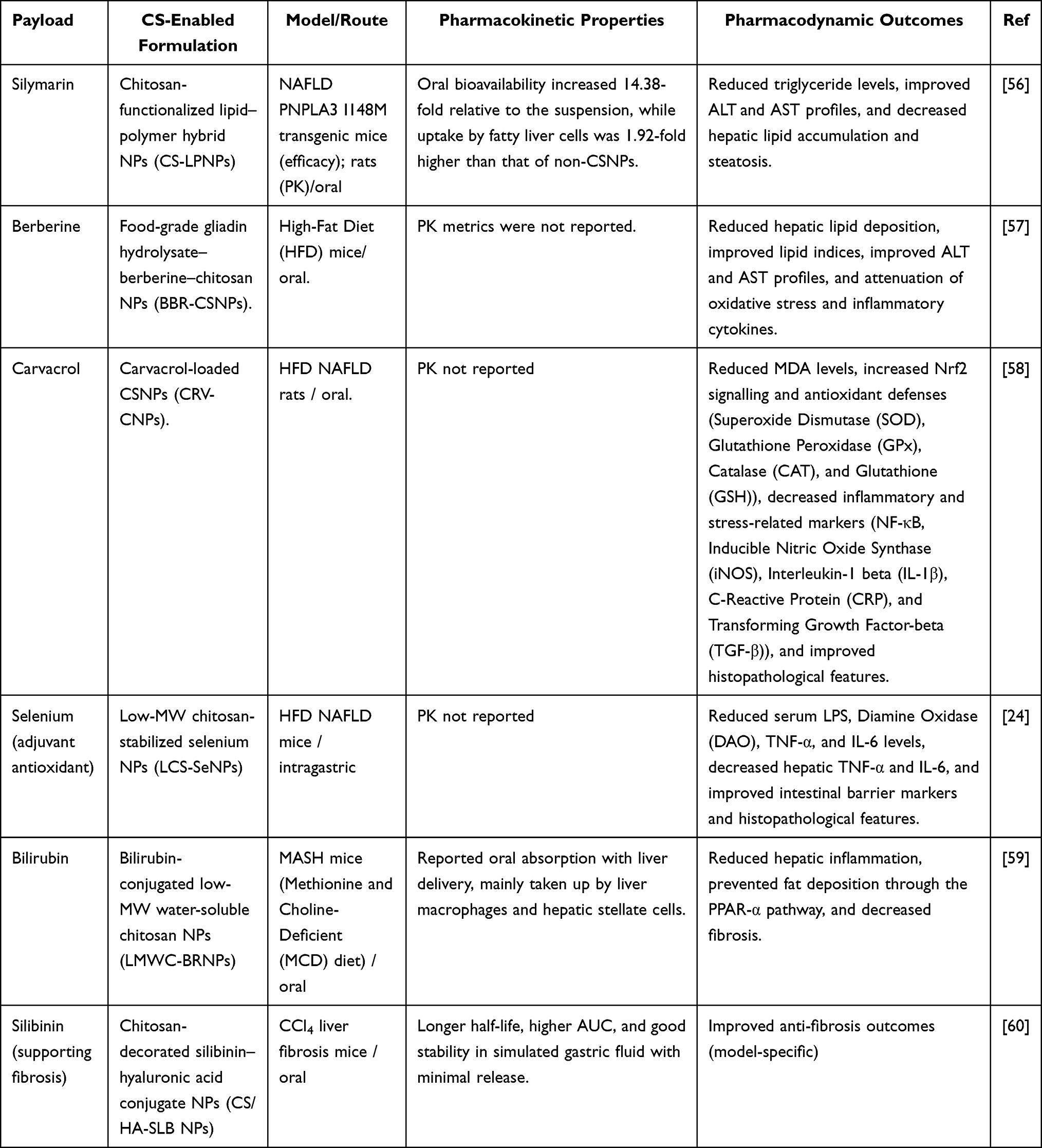 |
Table 1 Summary of Improved Pharmacokinetic and Pharmacodynamic Results of Chitosan-Based Formulations in the MASLD Model |
In general, the most consistently reported outcomes are improved liver pharmacodynamics, particularly decreased ALT/AST, decreased indicators of oxidative stress, and increased endogenous antioxidant enzymes (SOD, GPx, CAT), as well as improvements in steatosis and/or markers of inflammation–fibrosis in some models. Conversely, quantitative pharmacokinetic evidence (such as AUC or t½) is available only in a subset of studies, resulting in many reports being more robust to “biological effect” than “measured exposure”. Finally, the variation in results between studies confirms that the performance of chitosan-based systems is design- and context-specific, influenced by polymer attributes, formulation architecture, route of administration, and disease model and stage.
Conceptual Case Study
As a conceptual illustration, consider a nano-CS formulation containing curcumin and decorated with GalNAc ligands. When administered orally, these particles are expected to have improved stability (such as through crosslinking/PEC/enteric strategies, if used) and can interact with the intestinal mucosa to promote absorption. Once in the circulation, GalNAc can enhance interaction with ASGPR on hepatocytes and potentially enhance internalization. Within the cells, curcumin has been reported to activate Nrf2 and suppress NF-κB activation, thereby reducing oxidative stress and inflammation. The expected outcomes of this design are improved PK profiles and improved PD outcomes; however, a causal relationship still requires robust PK–PD data and quantitative biodistribution to support interpretation of hepatic enrichment.
As an example of preclinical support, one study reported that low-molecular-weight water-soluble chitosan nanoparticles conjugated with bilirubin (LMWCS-BRNP) exhibited oral absorption and hepatic enrichment in a mouse model of MASH, along with uptake in selected liver cell populations under the reported experimental conditions. These effects were associated with attenuation of oxidative and inflammatory stress and modulation of metabolic pathways, including PPAR-α signalling, in that model. However, these findings should be interpreted as platform-level preclinical evidence rather than direct validation of all oral CS-based targeting strategies, and translation to human MASLD still requires confirmation through reproducible quantitative biodistribution, PK–PD relationships, and repeated-dose safety studies.56
Table 2 provides a fit-for-purpose decision matrix for MASLD delivery; platform choice should follow the dominant bottleneck (solubility, gastric protection, mucus interaction, release control, or desired hepatic enrichment), rather than assuming any universally superior system. In this framework, chitosan and its derivatives are most suitable when the key barrier is mucosal retention and absorption enhancement, enabled by mucoadhesion and transient epithelial permeability effects; however, performance is design-dependent and constrained by pH sensitivity, MW/degree of deacetylation variability, batch reproducibility, and charge-related tolerability on repeat dosing.57–59
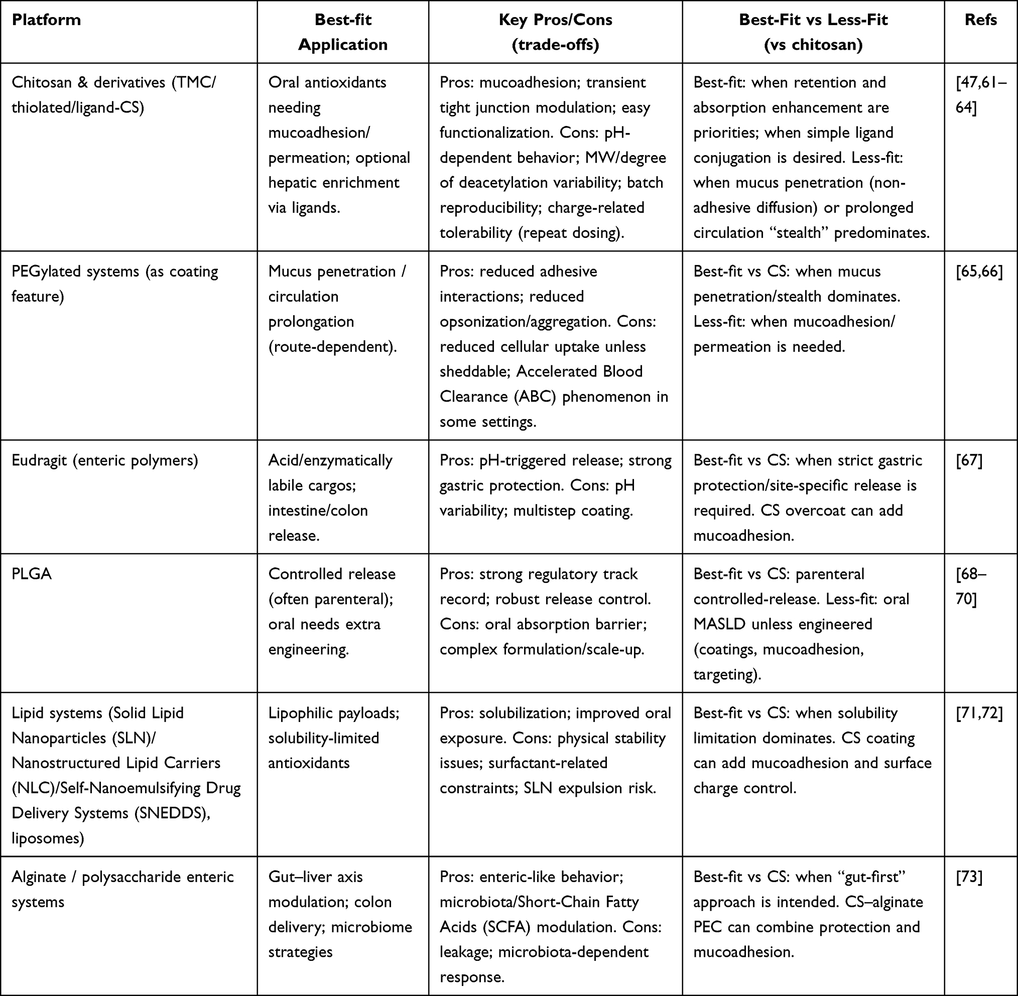 |
Table 2 Comparison Between Chitosan Platform and Other Polymers in MASLD Therapy |
In contrast, PEGylated coatings are preferable when the objective is mucus penetration and/or stealth behavior, although uptake can be reduced unless the coating is shed, and accelerated blood clearance has been reported in some repeat-dose settings.60,61 Enteric polymers (Eudragit) are the clearest choice when acid/enzymatic protection and distal intestinal/colonic release are primary requirements, at the cost of pH-dependent variability and multistep processing; chitosan can be used as an overcoat when mucoadhesion is desired.62 PLGA remains a benchmark for parenteral controlled release, with strong regulatory precedent, but oral MASLD applications typically require additional engineering to overcome absorption barriers and the associated manufacturing complexity.63–65 For highly lipophilic antioxidants, lipid systems (SLN/NLC/SNEDDS/liposomes) often address the solubility bottleneck most directly, although stability and surfactant constraints apply, and chitosan coatings can be leveraged to add mucoadhesion or tune surface charge in select designs.66,67 Finally, alginate/polysaccharide enteric systems are most relevant for gut-first strategies targeting the gut–liver axis and distal release, with trade-offs including leakage and microbiota-dependent response; CS–alginate PECs can combine protection with mucoadhesion.68 Overall, Table 2 positions chitosan as a strong option in the oral absorption/mucoadhesion niche, while clarifying when alternatives are more appropriate and where hybridization can be rational.
Cross-platform literature also shows that most MASLD nanomedicine candidates are still in preclinical trials, therefore animal model efficacy does not imply clinical performance. Thus, a translational agenda should focus on long-term toxicity, immunogenicity, PK/biodistribution characteristics, PK-PD interactions, and repeated-dose efficacy. Nanomedicine evaluation guidelines/standards are still lacking. Besides evaluation and regulatory issues, the liver’s physiological barriers to “targeting” without quantitative biodistribution data include Kupffer cell dominance of particle uptake, LSEC defenestration, and ECM remodelling in advanced disease, which can alter tissue distribution patterns and require verification before inferring “hepatoselectivity”.
Furthermore, the clinical context of MASLD itself continues to evolve—including the emergence of new therapies and the need for more precise patient stratification—so nanomedicines should be understood as part of an integrated management strategy, not a replacement for foundational non-pharmacological therapies and comorbidity control.25
With this framework, nanomedicine-based antioxidant innovations (including chitosan platforms) can be realistically placed in clinical practice, while also reviewing the most critical translational barriers and future research agendas (such as long-term safety, manufacturing scalability, and directions for developing more “clinically implementable” systems).15
Clinical Context, Challenges, and Future Perspectives
Before discussing final conclusions, it is important to place this pharmacological innovation within the broader context of MASLD clinical management and assess the challenges and future research directions. This advanced therapy is not a stand-alone solution, but rather part of an integrated and evolving treatment paradigm.
Role of Non-Pharmacological Therapies
Lifestyle modification is the cornerstone and first-line therapy for the entire spectrum of steatotic liver disease associated with metabolic dysfunction (MASLD). These non-pharmacological interventions remain the cornerstone of disease management, even with the advent of effective pharmacological therapies.69 These approaches include adopting a balanced, nutrient-rich, low-energy diet, reducing the intake of added sugars and trans fats, and engaging in regular aerobic physical activity and weight-bearing exercise. A gradual weight-loss target of 7–10% has consistently been shown to improve steatosis and liver biochemical parameters across various patient populations.70,71
In MASLD patients with renal comorbidities (such as CKD), dietary interventions need to be tailored to balance metabolic control, nitrogen/uremic load, and gut microbiota health. Several dietary approaches (such as the Mediterranean pattern or very low-protein diets in certain indications) and fiber-enhancing strategies have been reported to modulate the gut microenvironment and systemic inflammation.72,73 However, the details of the regimens should be interpreted in the context of the CKD indication and are not intended to extend the scope of this review beyond MASLD.
In this context, nanomedicines, including CS-based antioxidant systems, are positioned as promising complementary therapies. Their role becomes particularly important in high-risk cases, such as MASLD with significant fibrosis, or in patients who show a suboptimal response to lifestyle interventions and standard comorbidity management.74,75 The development of these nanomedicines aims to improve the bioavailability of therapeutic agents and potentially enhance hepatic enrichment or exposure to relevant liver compartments, without disrupting existing standard treatment protocols.
Clinical Translation Challenges
Several preclinical studies have reported that peptide-modified CSNPs exhibit good tolerability, reduced liver fibrosis markers, and altered tissue distribution under certain conditions.75 The potential of CSNPs as a dual-action platform (such as pRNA and enzyme delivery) offers efficacy beyond conventional therapies.75 However, the transition from laboratory scale to clinical application faces multidimensional challenges.
First, maintaining the integrity of the environmentally sensitive antioxidant payload is a major obstacle. CS has pH-dependent solubility and can undergo physicochemical changes during storage/in aqueous environments, potentially leading to premature release of the therapeutic payload or changes in particle characteristics. Furthermore, physical swelling due to water absorption can alter the particle size distribution, thus limiting the stability and shelf life of the formulation under certain conditions.43,76
Second, industrial-scale production requires processes that comply with Good Manufacturing Practice (GMP) standards. A major obstacle here is inter-batch variability in CS raw materials, particularly regarding molecular weight and degree of deacetylation, which directly impacts the reproducibility of nanoparticle performance. The implementation of advanced Process Analytical Technology (PAT) is often recommended to enable real-time monitoring and ensure a uniform performance profile consistent with pharmaceutical standards.18,77
Third, although CS is biodegradable into oligosaccharides that are generally considered non-toxic, its cationic nature at high doses may increase the risk of cell membrane interactions or hemolytic effects under certain conditions. Several nanoparticle biodistribution studies have also reported long-term retention in mononuclear phagocyte system organs (such as liver/spleen), which should be considered in repeat-dose designs, including the potential for macrophage activation and minor organ changes in chronic models.78–80 Therefore, optimization of particle size, surface charge, and colloidal stability remains crucial to balance efficacy and safety; however, the goal of “renal clearance” cannot be assumed solely with a target size of <200 nm and typically requires a much smaller particle design or specific surface strategies.78–80 Future developments will depend heavily on a more precise understanding of cellular mechanisms, which is beginning to be addressed through advances in single-cell technologies, enabling the identification of more specific antifibrotic targets at the cellular level.
Future Research Directions
To overcome translational challenges and optimize the potential of nanomedicine platforms, future research should focus on developing stimulus-responsive delivery systems. Systems capable of responding to specific triggers in the diseased liver microenvironment, such as decreased pH or increased levels of reactive oxygen species (ROS), could potentially enable more “intelligent” and localized payload release. This approach could strategically maximize therapeutic effects at the site of pathology while minimizing exposure to healthy cells. Similarly, integrating therapeutic and diagnostic functions into a single theranostic platform opens the possibility of monitoring tissue responses in real time. By incorporating tracers or contrast agents into nanoparticle systems, drug distribution and efficacy in the liver can be non-invasively assessed, ultimately accelerating clinical iterations toward more personalized therapies.
Synergy between metabolic modulation and targeted antioxidants is also an important research direction. Combining therapies targeting insulin resistance or lipogenesis pathways with specific antioxidants could potentially provide synergistic effects that simultaneously improve steatosis, suppress inflammation, and dampen pro-fibrotic signals. On the other hand, strengthening the gut-liver axis through synbiotic co-encapsulation strategies that combine probiotics with functional components such as prebiotics and polyphenols offers an effective method for improving microbiota health. The introduction of advanced manufacturing technologies, including microfluidics, 3D printing, and electrospinning, allows for precise structural control of nanoparticles to ensure probiotic survival in the gastrointestinal tract and enhance colonization efficiency in the gut.81,82
Systemic biomarkers for disease risk monitoring are essential for future studies, along with advances in delivery platforms. In some populations, hydration/metabolic status-based biomarkers like serum osmolality have been linked to CKD risk, but their use as risk stratification tools in MASLD patients needs further study and should be framed as evidence of association.83
Conclusion
Chitosan-based nanosystems have the potential to serve as a complementary therapy for the management of MASLD by overcoming the challenges of delivering natural antioxidants via the oral route. The reviewed preclinical studies consistently reported improved pharmacodynamic outcomes - particularly reductions in ALT/AST and indicators of oxidative stress, as well as improvements in steatosis and/or markers of inflammation/fibrosis in some models. However, quantitative PK evidence and measurable biodistribution are limited in a subset of studies, so claims of “targeting” should be framed as hepatic enrichment unless supported by quantitative tissue data, consistent PK–PD relationships, and repeated-dose safety evaluations.
Data Sharing Statement
No data was used for the research described in the article.
Funding
This study is funded by Universitas Padjadjaran through the Indonesian Endowment Fund for Education (LPDP) on behalf of the Indonesian Ministry of Higher Education, Science and Technology and managed under the EQUITY Program (Contract No. 4303/ 83/ DT.03.08/ 2025 and 3927/ UN6.RKT/HK.07.00/2025) through scheme the Equity Review Article Grant awarded to WCU Padjadjaran University (contract number No. 5692/ UN6.3.1/PT.00/2025).
Disclosure
The author(s) report no conflicts of interest in this work.
References
1. Kan C, Zhang K, Wang Y, et al. Global burden and future trends of metabolic dysfunction-associated Steatotic liver disease: 1990-2021 to 2045. Ann Hepatol. 2025;30(2):101898. doi:10.1016/j.aohep.2025.101898
2. Younossi ZM, Kalligeros M, Henry L. Epidemiology of metabolic dysfunction-associated steatotic liver disease (MASLD) and alcohol-related liver disease (ALD). Metab Target Organ Damage. 2025;2025(31):1–14. doi:10.20517/mtod.2024.57
3. Michalopoulou E, Thymis J, Lampsas S, et al. The triad of risk: linking MASLD, cardiovascular disease and type 2 diabetes; from pathophysiology to treatment. J Clin Med. 2025;14(2):1–36. doi:10.3390/jcm14020428
4. Mejía-Guzmán JE, Belmont-Hernández RA, Chávez-Tapia NC, Uribe M, Nuño-Lámbarri N. Metabolic-dysfunction-associated steatotic liver disease: molecular mechanisms, clinical implications, and emerging therapeutic strategies. Int J Mol Sci. 2025;26(7):1–42. doi:10.3390/ijms26072959
5. Cooper J, Patel P, Tristeza J, Yang A. Metabolic-associated steatotic liver disease: from molecular mechanisms to novel therapies. Int J Hepatol. 2025;2025(1):1–9. doi:10.1155/ijh/5580454
6. Chen D, Guo Z, Yao L, et al. Targeting oxidative stress-mediated regulated cell death as a vulnerability in cancer. Redox Biol. 2025;84(May):103686. doi:10.1016/j.redox.2025.103686
7. Zheng Y, Sun J, Luo Z, Li Y, Huang Y. Emerging mechanisms of lipid peroxidation in regulated cell death and its physiological implications. Cell Death Dis. 2024;15(11):1–19. doi:10.1038/s41419-024-07244-x
8. Wang Y, Fu X, Li H. Mechanisms of oxidative stress-induced sperm dysfunction. Front Endocrinol. 2025;16(February):1–15. doi:10.3389/fendo.2025.1520835
9. Zhang Z, Yang H, Han F, Guo P. Reactive oxygen species as key molecules in the pathogenesis of alcoholic fatty liver disease and nonalcoholic fatty liver disease: future perspectives. Curr Issues Mol Biol. 2025;47(6):1–27. doi:10.3390/cimb47060464
10. Acosta-Quiroga K, Rocha-Valderrama E, Zúñiga-Bustos M, et al. Gross antioxidant capacity and anti-inflammatory potential of flavonol oxidation products: a combined experimental and theoretical study. Antioxidants. 2025;14(4):1–31. doi:10.3390/antiox14040479
11. Forman HJ, Zhang H. Targeting oxidative stress in disease: promise and limitations of antioxidant therapy. Nat Rev Drug Discov. 2021;20(9):689–709. doi:10.1038/s41573-021-00233-1
12. Posey KL. Curcumin and resveratrol: nutraceuticals with so much potential for pseudoachondroplasia and other ER-stress conditions. Biomolecules. 2024;14(2):1–14. doi:10.3390/biom14020154
13. Jalouli M, Rahman MA, Biswas P, et al. Targeting natural antioxidant polyphenols to protect neuroinflammation and neurodegenerative diseases: a comprehensive review. Front Pharmacol. 2025;16(January):1–18. doi:10.3389/fphar.2025.1492517
14. Yang Y, Wang X. Nano-drug delivery systems (NDDS) in metabolic dysfunction-associated steatotic liver disease (MASLD): current status, prospects and challenges. Front Pharmacol. 2024;15(August):1–18. doi:10.3389/fphar.2024.1419384
15. Herdiana Y. Nano-enhanced diets: advancing metabolic dysfunction-related steatotic liver disease (MASLD) – a review. Diabet Metab Syndr Obes Diabet. 2025;18(December):4715–4731. doi:10.2147/DMSO.S562536
16. Herdiana Y, Husni P, Nurhasanah S, Shamsuddin S, Wathoni N. Chitosan-based nano systems for natural antioxidants in breast cancer therapy. Polymers. 2023;15(13):1–28. doi:10.3390/polym15132953
17. Gutiérrez-Ruíz SC, Cortes H, González-Torres M, et al. Optimize the parameters for the synthesis by the ionic gelation technique, purification, and freeze-drying of chitosan-sodium tripolyphosphate nanoparticles for biomedical purposes. J Biol Eng. 2024;18(1):1–16. doi:10.1186/s13036-024-00403-w
18. Herdiana Y. Bridging the gap: the role of advanced formulation strategies in the clinical translation of nanoparticle-based drug delivery systems. Int J Nanomed. 2025;20(October):13039–13053. doi:10.2147/IJN.S554821
19. Yu J, Yang C, Zhang P, Wei M, Li Y. Asialoglycoprotein receptor-targeted perfluorooctylbromide as a targeted contrast agent for evaluating the severity of carbon tetrachloride-induced acute liver damage in rats. Front Chem. 2025;13(April):1–9. doi:10.3389/fchem.2025.1475026
20. Ramírez-Cortés F, Ménová P. Hepatocyte targeting via the asialoglycoprotein receptor. RSC Med Chem. 2024;16(2):525–544. doi:10.1039/d4md00652f
21. Farid MM, Hathout RM, Fawzy M, Abou-Aisha K. Silencing of the scavenger receptor (Class B – type 1) gene using siRNA-loaded chitosan nanaoparticles in a HepG2 cell model. Colloids Surfaces B Biointerfaces. 2014;123(1 November 2014):930–937. doi:10.1016/j.colsurfb.2014.10.045
22. Wang X, Yang Y, Zhao S, Wu D, Li L, Zhao Z. Chitosan-based biomaterial delivery strategies for hepatocellular carcinoma. Front Pharmacol. 2024;15(August 2024):1–14. doi:10.3389/fphar.2024.1446030
23. Li G, Dai Z, Guo J. Therapeutic nanomaterials in NAFLD: current advances and potential applications in patients with concurrent HBV infection. Int J Nanomed. 2025;20(March):3803–3823. doi:10.2147/IJN.S510271
24. Luo Y, Peng S, Cheng J, et al. Chitosan-stabilized selenium nanoparticles alleviate high-fat diet-induced Non-Alcoholic Fatty Liver Disease (NAFLD) by modulating the gut barrier function and microbiota. J Funct Biomater. 2024;15(8):1–23. doi:10.3390/jfb15080236
25. Li F, Yuan R, Zhang J, Su B, Qi X. Advances in nanotechnology for the diagnosis and management of metabolic dysfunction-associated steatotic liver disease. Asian J Pharm Sci. 2025;20(101025):1–20. doi:10.1016/j.ajps.2025.101025
26. Biswas R, Mondal S, Ansari MA, Atanase LI. Chitosan and its derivatives as nanocarriers for drug delivery. Molecules. 2025;30(1297):1–44. doi:10.1016/S1773-2247(12)50003-1
27. Kira AY, Abdelhamid AM, Nasr M. Navigating liver targeting: fine-tuning chitosan nanocarriers through saccharide modification. J Drug Deliv Sci Technol. 2024;95(April):105644. doi:10.1016/j.jddst.2024.105644
28. Shete A, Chavan A, Potekar P, Yadav G, Shah N. Modification of physicochemical properties of chitosan to improve its pharmaceutical and agrochemical potential applications. Int J Biol Macromol. 2024;267(1):1–16. doi:10.1016/j.ijbiomac.2024.131404
29. Suryani S, Chaerunisaa AY, Joni IM, et al. The chemical modification to improve solubility of chitosan and its derivatives application, preparation method, toxicity as a nanoparticles. Nanotechnol Sci Appl. 2024;17(1):41–57. doi:10.2147/NSA.S450026
30. Adetunji JB, Salaudeen WO, Adetunji CO, Adeyomoye OI, Oyewole OA. Chapter 13 - Application of chitosan-based nanoparticles for the treatment of liver diseases. In: Adetunji CO, Danquah MK, Jeevanandam J, Hefft DI, Pan S, Khan Tabtcbn A editors. Woodhead Publishing Series in Biomaterials. Woodhead Publishing; 2025:275–287. doi:10.1016/B978-0-443-13997-0.00013-8
31. Rostami E. Progresses in targeted drug delivery systems using chitosan nanoparticles in cancer therapy: a mini-review. J Drug Deliv Sci Technol. 2020;58(101813):1–17. doi:10.1016/j.jddst.2020.101813
32. Zhang Y, Liu H, Zhen W, Jiang T, Cui J. Advancement of drugs conjugated with GalNAc in the targeted delivery to hepatocytes based on asialoglycoprotein receptor. Carbohydr Res. 2025;552(February):109426. doi:10.1016/j.carres.2025.109426
33. Liang Z, Li J, Luo S, et al. Galactose receptor-mediated hepatic targeting system: engineering of quinary cationic liposomes for resveratrol delivery against hepatic steatosis. RSC Adv. 2025;15(25):19786–19801. doi:10.1039/d5ra02554k
34. Peng M, Fang F, Wang B. Nanoparticle technologies for liver targeting and their applications in liver diseases. Front Bioeng Biotechnol. 2025;13(October):1–21. doi:10.3389/fbioe.2025.1661872
35. Cai Y, Nie T, Luo X, Wu Y, Wu J. Nanotechnology-driven biomaterials for chronic liver diseases: stage-specific strategies for advanced theranostics. Acta Biomater. 2026;212:103–123. doi:10.1016/j.actbio.2026.01.043
36. Zhu Y, Ge D, Wang J, Sun H, Li W, Ren H. Emerging nanomedicine for liver diseases treatment. J Nanobiotechnology. 2025;23(1). doi:10.1186/s12951-025-03832-x
37. Hosseini-Kharat M, Bremmell KE, Prestidge CA. Why do lipid nanoparticles target the liver? Understanding of biodistribution and liver-specific tropism. Mol Ther Methods Clin Dev. 2025;33(1):101436. doi:10.1016/j.omtm.2025.101436
38. Guadarrama-Escobar OR, Serrano-Castañeda P, Anguiano-Almazán E, et al. Chitosan nanoparticles as oral drug carriers. Int J Mol Sci. 2023;24(5):1–17. doi:10.3390/ijms24054289
39. Meng X, Zhu G, Yang YG, Sun T. Targeted delivery strategies: the interactions and applications of nanoparticles in liver diseases. Biomed Pharmacother. 2024;175(116702):1–17. doi:10.1016/j.biopha.2024.116702
40. Almeida A, Araújo M, Novoa-Carballal R, et al. Novel amphiphilic chitosan micelles as carriers for hydrophobic anticancer drugs. Mater Sci Eng C. 2020;112(March):110920. doi:10.1016/j.msec.2020.110920
41. Yilmaz T, Maldonado L, Turasan H, Kokini J. Thermodynamic mechanism of particulation of sodium alginate and chitosan polyelectrolyte complexes as a function of charge ratio and order of addition. J Food Eng. 2019;254(1):42–50. doi:10.1016/j.jfoodeng.2019.03.002
42. Abdel-Hafez SM, Hathout RM, Sammour OA. Towards better modeling of chitosan nanoparticles production: screening different factors and comparing two experimental designs. Int J Biol Macromol. 2014;64(March 2014):334–340. doi:10.1016/j.ijbiomac.2013.11.041
43. Stefanache A, Lungu II, Anton N, et al. Chitosan nanoparticle-based drug delivery systems: advances, challenges, and future perspectives. Polymers. 2025;17(1453):1–18. doi:10.3390/polym17111453
44. Dadashi H, Vandghanooni S, Karamnejad-Faragheh S, Karimian-Shaddel A, Eskandani M, Jahanban-Esfahlan R. A rapid protocol for synthesis of chitosan nanoparticles with ideal physicochemical features. Heliyon. 2024;10:1–14. doi:10.1016/j.heliyon.2024.e32228
45. Huang Q, An Z, Xin X, et al. The effectiveness of curcumin, resveratrol, and silymarin on MASLD: a systematic review and meta-analysis. Food Sci Nutr. 2024;12(12):10010–10029. doi:10.1002/fsn3.4595
46. Ahmad K, Zhang Y, Chen P, Yang X, Hou H. Chitosan interaction with stomach mucin layer to enhances gastric retention and mucoadhesive properties. Carbohydr Polym. 2024;333(1299):121926. doi:10.1016/j.carbpol.2024.121926
47. Mura P, Maestrelli F, Cirri M, Mennini N. Multiple roles of chitosan in mucosal drug delivery: an updated review. Mar Drugs. 2022;20(5):1–42. doi:10.3390/md20050335
48. Sulastri W, Yani S, Utami RW. Bahaya Tersembunyi Makanan Ultra-Proses: faktor Risiko Utama Gagal Ginjal di Indonesia: scoping Review The Hidden Dangers of Ultra-Processed Foods: a Major Risk Factor for Kidney Failure in Indonesia. J Ris Keperawatan. 2024;7(2):80–86.
49. Svobodová G, Horní M, Velecká E, Boušová I. Metabolic dysfunction-associated steatotic liver disease-induced changes in the antioxidant system: a review. Arch Toxicol. 2025;99(1):1–22. doi:10.1007/s00204-024-03889-x
50. Dua A, Kumari R, Singh M, et al. Dysfunction-Associated Steatotic Liver Disease (MASLD): the interplay of gut microbiome, insulin resistance, and diabetes. Front Med. 2019;2019(3).
51. Li Q, Mi Y, Tan W, Guo Z. International journal of biological macromolecules highly ef fi cient free radical-scavenging property of phenolic-functionalized chitosan derivatives: chemical modi fi cation and activity assessment. Int J Biol Macromol. 2020;164:4279–4288. doi:10.1016/j.ijbiomac.2020.08.250
52. Murao A, Jha A, Kato T, et al. Gut-primed neutrophils activate Kupffer cells to promote hepatic injury in mouse sepsis. Nat Commun. 2025;16(9657):1–12. doi:10.1038/s41467-025-65572-8
53. Zamyatina A, Heine H, Peri F, Sherwood E. Lipopolysaccharide recognition in the crossroads of TLR4 and inflammatory pathways. Front Immunol. 2020;11(November):1–22. doi:10.3389/fimmu.2020.585146
54. Guo Y, Zhang N, Pei D. Gut microbiota heterogeneity in non-alcoholic fatty liver disease: a narrative review of drivers, mechanisms, and clinical relevance. Front Microbiol. 2025;16(October):1–15. doi:10.3389/fmicb.2025.1645298
55. Okalin SS, Arslan N, Demiray Gürbüz E, et al. The association between gut microbiota composition and cardiometabolic parameters in healthy adults. BMC Microbiol. 2025;25(1). doi:10.1186/s12866-025-04261-4
56. Rahman AT, Choi J, Kyung W, Kang S, Lee S, Jon S. Oral delivery of chitosan-bilirubin nanoparticles alleviates hepatic inflammation and fibrosis in metabolic dysfunction-associated steatohepatitis. Biomaterials. 2026;325(123576):1–15. doi:10.1016/j.biomaterials.2025.123576
57. Ways TMM, Lau WM, Khutoryanskiy VV. Chitosan and its derivatives for application in mucoadhesive drug delivery systems. Polymers. 2018;10(3):1–37. doi:10.3390/polym10030267
58. Benediktsdóttir BE, Baldursson Ó, Másson M. Challenges in evaluation of chitosan and trimethylated chitosan (TMC) as mucosal permeation enhancers: from synthesis to in vitro application. J Control Release. 2014;173(10):18–31. doi:10.1016/j.jconrel.2013.10.022
59. Alfatama M, Choukaife H, Alkhatib H, Al Rahal O, Zin NZM. A comprehensive review of oral chitosan drug delivery systems: applications for oral insulin delivery. Nanotechnol Rev. 2024;13(1):1–30. doi:10.1515/ntrev-2023-0205
60. Pan J, Wang Y, Chen Y, et al. Emerging strategies against accelerated blood clearance phenomenon of nanocarrier drug delivery systems. J Nanobiotechnology. 2025;23(1):1–22. doi:10.1186/s12951-025-03209-0
61. Bain A, Vasdev N, Muley A, Sengupta P, Tekade RK. Mucus-penetrating PEGylated nanoshuttle for enhanced drug delivery and healthcare applications. Indian J Microbiol. 2025;65(1):3–14. doi:10.1007/s12088-024-01304-8
62. Ziebarth J, da Silva LM, Lorenzett AKP, et al. Oral delivery of liraglutide-loaded zein/eudragit-chitosan nanoparticles provides pharmacokinetic and glycemic outcomes comparable to its subcutaneous injection in rats. Pharmaceutics. 2024;16(5):1–16. doi:10.3390/pharmaceutics16050634
63. Ochi M, Wan B, Bao Q, Burgess DJ. Influence of PLGA molecular weight distribution on leuprolide release from microspheres. Int J Pharm. 2021;599(120450):1–7. doi:10.1016/j.ijpharm.2021.120450
64. Siepmann J, Siepmann F. Release mechanisms of PLGA-based drug delivery systems: a review. Int J Pharm X. 2025;10(August):100440. doi:10.1016/j.ijpx.2025.100440
65. Butreddy A, Gaddam RP, Kommineni N, Dudhipala N, Voshavar C. PLGA/PLA-based long-acting injectable depot microspheres in clinical use: production and characterization overview for protein/peptide delivery. Int J Mol Sci. 2021;22(16):1–25. doi:10.3390/ijms22168884
66. Moin A, Ram S, Boregowda SS, et al. Piperine-loaded solid lipid nanoparticles: a promising nano-phytomedicine for the treatment of non-alcoholic fatty liver disease. Front Pharmacol. 2026;16(January):1–15. doi:10.3389/fphar.2025.1646530
67. Patel P, Garala K, Singh S, Prajapati BG, Chittasupho C. Lipid-based nanoparticles in delivering bioactive compounds for improving therapeutic efficacy. Pharmaceuticals. 2024;17(3):1–24. doi:10.3390/ph17030329
68. Zhang H, Tang Y, Ge H, et al. Sodium alginate supplementation can alleviate non-alcoholic fatty liver disease in rats by restoring hepatic lipid metabolism and gut microbiota. Curr Res Food Sci. 2025;11(101199):1–16. doi:10.1016/j.crfs.2025.101199
69. Salmanizadeh F, Sabzevari S, Shafieipour S, Zahedi MJ, Sarafinejad A. Challenges and needs in the management of non-alcoholic fatty liver disease from the perspective of gastroenterology and hepatology specialists: a qualitative study. BMC Gastroenterol. 2025;25(1):1–40. doi:10.1186/s12876-025-03921-z
70. Thomas A, Thomas A. Managing nonalcoholic fatty liver disease through structured lifestyle modification interventions. Am J Lifestyle Med. 2025;20(3):463–478. doi:10.1177/15598276251346717
71. An G. Pharmacokinetics and pharmacodynamics of GalNAc-Conjugated siRNAs. J Clin Pharmacol. 2024;64(1):45–57. doi:10.1002/jcph.2337
72. Di Iorio BR, Rocchetti MT, De Angelis M, et al. Nutritional therapy modulates intestinal microbiota and reduces serum levels of total and free indoxyl sulfate and p-cresyl sulfate in chronic kidney disease (Medika study). J Clin Med. 2019;8(9):1424. doi:10.3390/jcm8091424
73. Wathanavasin W, Cheungpasitporn W, Thongprayoon C, Fülöp T. Effects of dietary fiber supplementation on modulating uremic toxins and inflammation in chronic kidney disease patients: a systematic review and meta-analysis of randomized controlled trials. Toxins. 2025;17(2):57. doi:10.3390/toxins17020057
74. Du X, Niu R, Liu X, et al. Nanomedicines in the treatment of liver fibrosis: a review. Int J Nanomed. 2025;20(August):9641–9665. doi:10.2147/IJN.S524078
75. Mostafa S, Shetab Boushehri MA, Ezzat AA, et al. Targeted delivery of anti-TGF-β(1)-siRNA using PDGFR-β peptide-modified chitosan nanoparticles for the treatment of liver fibrosis. Mol Pharm. 2025;22(11):1. doi:10.1021/acs.molpharmaceut.5c00715
76. Wardana AA, Marcellino V, Wigati LP, et al. Application of pickering emulsion-based coatings/films stabilized with chitosan nanoparticles for the preservation of fresh postharvest commodities. Front Sustain Food Syst. 2025;9(July):1–9. doi:10.3389/fsufs.2025.1646457
77. Sumer PDR, Singh LS, Debbarma B, Binan K, Lyngdoh W. Chitosan nanoparticles: revolutionizing targeted drug delivery systems. Adv Pharmacol Pharm. 2025;13(4):513–526. doi:10.13189/app.2025.130404
78. Salsabila S, Khairinisa MA, Wathoni N, et al. In vivo toxicity of chitosan-based nanoparticles: a systematic review. Artif Cells Nanomed Biotechnol. 2025;53(1):1–15. doi:10.1080/21691401.2025.2462328
79. Koirala P, Bhattarai P, Sriprablom J, Zhang R, Nirmal S, Nirmal N. Recent progress of functional nano-chitosan in pharmaceutical and biomedical applications: an updated review. Int J Biol Macromol. 2025;285(138324):1–13. doi:10.1016/j.ijbiomac.2024.138324
80. Herdiana Y, Levita J, Jiranusornkul S. Chitosan-based nanoparticles targeted delivery system: in treatment approach for dyslipidemia. Int J Nanomed. 2025;20(March):6611–6636. doi:10.2147/IJN.S517492
81. Ji C, Li D, Liang Y, Luo Y. Co-encapsulation of probiotics with functional components: design strategies, synergistic mechanisms, biomedical applications, and challenges for industrialization. J Mater Chem B. 2025;13(41):13122–13153. doi:10.1039/d5tb01747e
82. Kvakova M, Bertkova I, Stofilova J, Savidge TC. Co-encapsulated synbiotics and immobilized probiotics in human health and gut microbiota modulation. Foods. 2021;10(6):1–28. doi:10.3390/foods10061297
83. Mei L, Qin J, Liu X, Yu M. Association between serum osmolality and chronic kidney disease prevalence: evidence from NHANES 1999–2018. Sci Rep. 2025;15(1):1–13. doi:10.1038/s41598-025-08624-9
 © 2026 The Author(s). This work is published and licensed by Dove Medical Press Limited. The
full terms of this license are available at https://www.dovepress.com/terms
and incorporate the Creative Commons Attribution
- Non Commercial (unported, 4.0) License.
By accessing the work you hereby accept the Terms. Non-commercial uses of the work are permitted
without any further permission from Dove Medical Press Limited, provided the work is properly
attributed. For permission for commercial use of this work, please see paragraphs 4.2 and 5 of our Terms.
© 2026 The Author(s). This work is published and licensed by Dove Medical Press Limited. The
full terms of this license are available at https://www.dovepress.com/terms
and incorporate the Creative Commons Attribution
- Non Commercial (unported, 4.0) License.
By accessing the work you hereby accept the Terms. Non-commercial uses of the work are permitted
without any further permission from Dove Medical Press Limited, provided the work is properly
attributed. For permission for commercial use of this work, please see paragraphs 4.2 and 5 of our Terms.